sec2 scihi feature - bnl.gov
TRANSCRIPT

2 - 1 Science Highlights
Two papers were published in August, 2000, an-nouncing the determination of the crystal structure ofthe large ribosomal subunit from Haloarcula marismortuiat 2.4 Å resolution (Ban et al., 2000; Nissen et al., 2000).Most of the data used were collected at the NationalSynchrotron Light Source on beamlines X12B, X12Cand X25, which were then being run by Malcolm Capel,Robert Sweet and Lonnie Berman, respectively. Theribosome is a macromolecular complex found in all liv-ing cells that is about two thirds RNA and one-third pro-tein. It is the enzyme that catalyzes the synthesis ofproteins, and it is a programmed enzyme. The aminoacid sequence of the protein being made by a givenribosome, i.e. the covalent structure of its product, isdetermined by the nucleotide sequence of the mes-senger RNA molecule with which it is interacting.
The crystals used posed major technical chal-lenges. The molecular weight of the large subunit fromH. marismortui is 1,500,000, and since it lacks internalsymmetry, the molecular weight of the minimum unit ofunique structure that had to be determined was also1,500,000, which is several times larger than that ofthe next most complicated biological macromoleculewhose structure had been solved at atomic resolutionup to that time. The unit cells of crystals of moleculesthis big are necessarily large, in this case 210 x 300 x570 Å, and because crystals that have unit cells thatlarge diffract X-rays weakly, data collection is impracti-cal using X-ray sources less bright than synchrotronlight sources.
Adding to the difficulty was the extreme sensitivityto ionic conditions of the packing of ribosomal subunitsin these crystals. The space group of crystals of thelarge ribosomal subunit from H. marismortui that havealready formed can be altered by seemingly trivialchanges in the composition of the solvents used to sta-bilize them. The crystals do not dissolve and reform asthese space group transformations occur, and their
Feature HighlightThe Atomic Resolution Crystal Structure of the LargeRibosomal Subunit from Haloarcula marismortuiN. Ban1, P. Nissen1, J. Hansen1, P. B. Moore1,2 and T.A. Steitz1,2,3
1Department of Molecular Biophysics and Biochemistry, Yale University2 Department of Chemistry, Yale University3Howard Hughes Medical Institute
gross morphology does not change. Moreover, the dif-fraction patterns of the different crystal forms that canbe produced this way have virtually superimposablereciprocal lattices, and extend to similar resolutions.The crystal form solved has the symmetry C2221. Thevariant most commonly encountered belongs to spacegroup P21, but is invariably twinned in such a way thatits diffraction pattern has the same symmetry as that ofthe C2221 crystals (Ban et al., 1999).
Even if these crystals had not been so perverse, itwould have been a challenge to obtain phases for theirdiffraction patterns using conventional techniques. Alltechniques used to phase macromolecular diffractionpatterns require that one determine the positions of at-oms having unusual scattering properties (either largeatomic mass, or some resonance of the atom�s elec-trons with the x-rays) that have been introduced intocrystals by one means or another. For example, differ-ence Patterson methods are often used to locate heavymetal atoms in macromolecular crystals that have re-acted with heavy metal-containing compounds, andthey work well if the number of heavy atoms bound perunit cell is small. However, if the number of heavy metalatoms bound per unit cell is small in a ribosome crys-tal, the heavy atom contribution to its diffraction patternwill be too small to measure accurately, and no posi-tional information will emerge. If, on the other hand, aheavy metal compound that reacts at a large numberof sites is used so that the heavy metal differences ofinterest are easy to measure, the difference Pattersonsthat result may be too complicated to solve, and againno useful positional information will be obtained. In short,the crystallographer is caught between a rock and ahard place.
The phasing problem was solved for these crys-tals in two stages. In the first stage, only low resolutionphase information was sought (Ban et al., 1998). Clus-ter compounds that contain large numbers of metal at-

2 - 2NSLS Activity Report 2000
oms were used to make derivatives, and the resultingdifference data analyzed at resolutions so low that thecontribution each such molecule makes to the diffrac-tion pattern of the crystal in which it is bound could beconsidered equivalent to that of a single atom havingthe same number of electrons as the total number inthe cluster compound. When heavy metal compoundslike these are used, derivatization at a small number ofsites can produce measurable changes in diffractionintensities, and difference Pattersons will be soluble.Use was also made of the remarkably accurate three-dimensional reconstructions of macromolecular struc-tures that can now be generated by analysis of two-dimensional, electron microscopic images. Using mo-lecular replacement techniques, these electron densitymaps - and that is approximately what results from suchan analysis - can be used to phase the low resolutionreflections of X-ray diffraction patterns obtained fromcrystals of the same objects. The H. marismortui largesubunit image used was produced by Joachim Frankand his coworkers at the Wadsworth Institute in Albany.For another example of the use of this approach, seeCate et al., 1999.
Low resolution phases were critical for the successof the second stage of the phasing process, the objec-tive of which was to obtain high resolution phases. Con-ventional, single atom, heavy metal compounds thatbind to the ribosome at many locations were soakedinto crystals, and heavy atom differences measured.Because low resolution phases were available, the siteswhere these compounds bind to the ribosome couldbe determined approximately by difference Fouriermethods, rather than difference Patterson methods, anddifference Fouriers can be interpreted no matter howmany heavy metal sites there are. The heavy atompositions obtained this way were then refined using thehigher resolution difference data. Both isomorphous dif-ference data and anomalous difference data were usedfor this purpose.
The ribosome was imaged well enough in the elec-tron density maps computed, using the phases obtainedthis way, so that solvent-flipping and histogram-match-ing methods could be used to improve phase qualityand extend the resolution of the phase set. The firstelectron density map having a resolution high enoughso that its features could be interpreted chemically wascomputed in the middle of November, 1999. The datafor that map were measured at X25 with a MAR345imaging-plate detector. The native data set that madeit possible to extend the resolution of the structure to2.4 Å was obtained at APS the following spring [Figure1 (Ban et al., 2000, Fig. 1)].
The structure that has emerged reveals that thelarge ribosomal subunit from H. marismortui consistsof 31 proteins and two RNA molecules. The RNA forms
a monolithic matrix that has a shape similar to that ofthe whole particle [Figure 2 (Ban et al., 2000, Fig.2)].The globular bodies of the proteins are inserted intogaps and crevices in the surface of the RNA mass, andmany have irregularly structured extensions that reachinto the center of the particle through interstices in thefolded RNA. These extensions, or tails, are rich in ba-sic amino acids and interact very strongly with the RNAthat surrounds them over their entire lengths. Theirsequences are at least as highly conserved as thoseof their globular domains. Thus, random and irregularas their conformations seem to be, there is nothingaccidental about them or the interactions they makewith ribosomal RNA.
This amazingly complex structure will reassemblefrom its components in the test tube, but the process isextremely inefficient, so inefficient in fact that cells wouldnot be able to grow as fast as they do if their ribosomeshad to be assembled that way. It seems highly likely,therefore, that in vivo ribosome assembly is controlledand facilitated by non-ribosomal macromolecules. Verylittle about this aspect of cellular physiology is known.
In part because of the extensions possessed bymany ribosomal proteins, the surface area buried isenormous when the proteins are added(computationally) to the RNA matrix to which they bind.About half of the total solvent-accessible area presentin the isolated, but folded RNAs and proteins becomesconcealed in the process, which suggests that the in-teraction free energies involved are likely to be verylarge. Since most of these proteins bind to many differ-ent RNA sequences, it must be that stabilization of RNAconformation is a major function of ribosomal proteins.
Analysis of the architecture of the large ribosomalsubunit has revealed the existence of two new RNAmotifs. The first, which we call the A-minor motif, stabi-lizes RNA-RNA tertiary interactions throughout the par-ticle. Short runs of (often) conserved As in one part ofthe sequence interact with the minor groove edge ofadjacent base pairs belonging to helices formed by othersequences. The base-base and base-sugar hydrogenbonds that form hold the interacting sequences together.The second motif is one we call the kink-turn. It is asecondary structure motif consisting of two helical seg-ments separated by a short asymmetric loop. It is char-acterized by a sharp change in helix axis direction (thekink) that exposes the surfaces of bases to solvent.Kink-turns are preferred locations for RNA-protein in-teractions, and examples of kink-turns exist in manyribonucleoproteins in addition to the large ribosomalsubunit. (Manuscripts describing both motifs are inpreparation.)
Structures also have been obtained of the largeribosomal subunit in complex with two substrate ana-logues. These structures reveal that the site in the sub-

2 - 3 Science Highlights
Figure 1: Sample electron density from the 2.4 Å resolution electron density map of the large ribosomal subunit from H.marismortui. (A) A stereo view of a region where elements of domains II, III, IV, and V of 23S rRNA come together. (Contoursare at 2σ). (B) The tail region of protein L2 interacting with surrounding RNA structures. (C) Detail showing a Mg2+ ionbound between a segment of L2 and rNA belonging to domain V. (D) Detail from protein L2 showing protein side chains. (E)Helices 94-97, which form the heart of domain VI from 23S rRNA. Red contours are at 2σ. Yellow contours, which showphosphate groups, are at 6σ. (Figure reprinted with permission from Science, Ban et al., 2000)

2 - 4NSLS Activity Report 2000
unit where peptide bonds form is at the bottom of adeep cleft, where a tunnel originates that passes allthe way through the body of the particle [Figure 3 (Nis-sen et al., 2000, Fig. 11a)]. Proteins are synthesized atone end of this tunnel, pass through its length, andemerge at its far end. No portion of any ribosomal pro-tein comes close enough to the site where peptidebonds form to participate in the chemistry that occursthere. There can be no doubt that the active site of thisenzyme is entirely composed of RNA. For the benefitof non-biochemists, one hastens to add that the over-whelming majority of enzymes are composed entirelyof protein.
On the basis of the placement of nucleotides in theneighborhood of the active site, a proposal has beenadvanced for the way ribosomes catalyze peptide bondformation. Biochemical experiments done by ScottStrobel and his colleagues at Yale have shown that theRNA of the large ribosomal subunit includes a single
Figure 2: The subunit interface surface of the large ribosomal subunit. RNA isshown in gray in a space-filling-like representation that exaggerates its backbone.Proteins are shown in yellow in a continuous wire format that depicts the trajecto-ries of their backbones. The green object in the middle of the image is a peptidyltransferase transition state analog molecule bound to the active site. The entireassembly is about 250 Å across. (Figure reprinted with permission from Science,Ban et al., 2000)
adenine that acts as though its pKawere about 7.5 instead of 4 or less(Muth et al., 2000). It turns out thatthis adenine is positioned so that itcould function as a general basein the peptide bond formation re-action, and unless its pKa werearound 7, it would not be able tofunction that way. Detailed exami-nation of the interactions the ad-enine side chain makes with sur-rounding nucleotides indicates thatit is indeed protonated at pH 6.0,consistent with Strobel�s findings.The mechanism proposed for itsparticipation in peptide bond forma-tion is outlined in Figure 4 (Nissenet al., 2000, Fig. 9).
Why is the pKa of the adeninein question so high? Here too thestructure is suggestive. The ad-enine is held in place by an exten-sive network of hydrogen bonds,some of which involve a guaninethat is itself hydrogen bonded to anearby phosphate group. Thephosphate group is one of the threeleast solvent-accessible phos-phates in the entire molecule. Atneutral pH, nucleic acid phosphategroups carry net negative charges,which are normally neutralized byinteractions with metal ions andwater in the surrounding solvent.Neutralizing interactions of this kindare not possible in this case, and it
is proposed that its buried charge polarizes both theguanine side chain and the catalytic adenine with whichthe guanine interacts. This causes a build up of nega-tive charge on the adenine nitrogen atom that alters itspKa and facilitates peptide bond formation.
Both the proposal advanced for the mechanism ofpeptide bond formation and the explanation offered forthe anomalous pKa of the critical adenine are hypoth-eses that must now be tested experimentally, and it iscertain that they will be. In recent years, molecular bi-ologists have developed methods for altering the se-quences of ribosomal RNAs in vivo that make it pos-sible to produce ribosomes in which any base believedcritical for function can be changed at will. Mutant par-ticles like these provide the means for examining thechemistry of peptide bond formation with a level ofspecificity that was not previously possible.
The structure of the large ribosomal subunit is alsogoing have a big impact on our understanding of anti-

2 - 5 Science Highlights
biotics. Many antibiotics have been discovered sincethe Second World War that kill bacteria by blocking theactivity of their ribosomes, and some of them are clini-cally useful, e.g. erythromycin. Many anti-ribosomal an-tibiotics interact specifically with the large ribosomalsubunit, and most of those that do so inhibit peptidyltransferase activity. It would be very useful to under-stand how these compounds bind to the ribosome, andwhy they are effective as inhibitors. Also, unhappily,the clinical effectiveness of many of these antibioticshas become greatly reduced in recent years as bacte-rial strains have developed that are resistant to them,a phenomenon that threatens public health. In some
Figure 3: A space-filling representation of the exit tunnel region of the largeribosomal subunit. The subunit has been cut sagittally so that its tunnel isbisected and the two pieces of the subunit opened up like the pages of abook. Gray material represents buried RNA atoms, and green material isburied protein. Multi-colored atoms are all exposed to solvent. The whiteribbon represents the backbone of a nascent peptide traversing the tun-nel, and it is based on a model building exercise, not experimental data.The red dot marked �PT� marks the location of the peptidyl transferasesite. (Figure reproduced with permission from Science, Nissen et al., 2000)
instances, resistance results from alterations in ribo-some structure, and resistance mechanisms of this kindcan clearly be addressed using the crystal structuresof the large ribosomal subunit. If we are fortunate, wemay learn how to synthesize new antibiotics that areunaffected by these resistance mechanisms.
Structures have been obtained for several ribo-some-antibiotic complexes. All of the antibiotics stud-ied so far happen to bind to the large subunit close toits peptidyl transferase site, and we can now establishthe interactions they make with the ribosome in atomicdetail. Most of the antibiotics visualized to date inhibitprotein synthesis by getting in the way of the molecules
2 - 6NSLS Activity Report 2000
that must interact with the ribosome in order for proteinsynthesis to take place. This is the simplest way onecould imagine inhibiting an enzyme. What is not simpleare the interactions that hold these compounds in placein the ribosome, and the way the small differences thatexist between human ribosomes and bacterial ribo-somes in that region are �taken advantage of� by theclinically useful antibiotics to kill bacteria far more effi-ciently than the human body can. Thus, not only willthese structures contribute to the advance of science,they are going to be useful to the man on the street aswell.
References:N. Ban, P. Nissen, J. Hansen, P.B. Moore, and T.A. Stetiz,
�The complete atomic structure of the large ribosomalsubunit at 2.4 Å resolution,� Science 289, 905, 2000.
P. Nissen, N. Ban, J. Hansen, P.B. Moore, and T.A. Stetiz,�The structural basis of ribosome activity in peptide bondsynthesis,� Science 289, 920, 2000.
N. Ban, P. Nissen, J. Hansen, M. Capel, P.B. Moore, and T.A.Steitz, �Placement of protein and RNA structures into a5 Å-resolution map of the 50S ribosomal subunit,� Na-ture 400, 841, 1999.
N. Ban, B. Freeborn, P. Nissen, P. Penczek, R.A. Grassucci,R. Sweet, J. Frank, P.B. Moore, and T.A. Steitz, �A 9 Åresolution X-ray crystallographic map of the large ribo-somal subunit,� Cell 93, 1105, 1998.
J.H. Cate, M.M. Yusupov, G.Z. Yusupova, T.N. Earnest, andH.F. Noller, �X-ray crystal structures of 70S ribosomefunctional complexes,� Science 285, 2095, 1999.
G.W. Muth, L. Ortoleva-Donnelly, and S.A. Strobel, �A singleadenosine with a neutral pKa in the ribosomal peptidyltransferase center,� Science 289, 947, 2000.
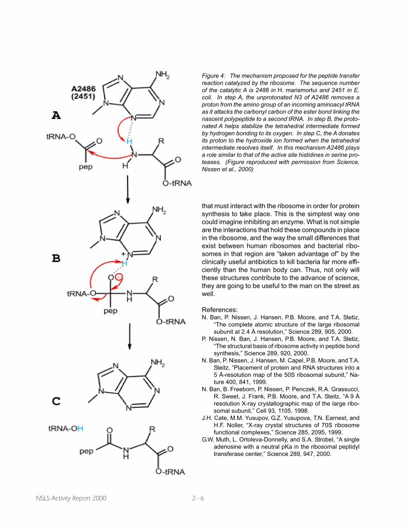
Figure 4: The mechanism proposed for the peptide transferreaction catalyzed by the ribosome. The sequence numberof the catalytic A is 2486 in H. marismortui and 2451 in E.coli. In step A, the unprotonated N3 of A2486 removes aproton from the amino group of an incoming aminoacyl tRNAas it attacks the carbonyl carbon of the ester bond linking thenascent polypeptide to a second tRNA. In step B, the proto-nated A helps stabilize the tetrahedral intermediate formedby hydrogen bonding to its oxygen. In step C, the A donatesits proton to the hydroxide ion formed when the tetrahedralintermediate resolves itself. In this mechanism A2486 playsa role similar to that of the active site histidines in serine pro-teases. (Figure reproduced with permission from Science,Nissen et al., 2000)