lia damayanti - website staff...
TRANSCRIPT
1
L ia D amayantiD epartment of H istology – Faculty of
M edicineUniversity of I ndonesia
2
IntroductionM uscle tissue
One of the four basic tissues Properties
Contractility Converting chemical energy into mechanical work
3
IntroductionThe main function of skeletal muscle
Positioning of the skeletonM ovement of the skeleton
M uscle tissue attach firmly to related boneM uscle contraction moves the skeletons
4
through skeletal muscle contraction that produce muscle tension
5
Development Derived from mesoderm Somatic mesoderm
Skeletal muscle Splanchnic mesoderm
Smooth muscle Splanchnopleuric mesoderm
Cardiac muscle
6
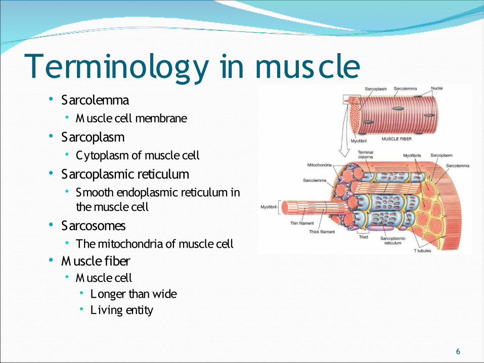
Terminology in muscle Sarcolemma
M uscle cell membrane Sarcoplasm
Cytoplasm of muscle cell Sarcoplasmic reticulum
Smooth endoplasmic reticulum in the muscle cell
Sarcosomes The mitochondria of muscle cell
M uscle fiber M uscle cell
Longer than wide Living entity
7
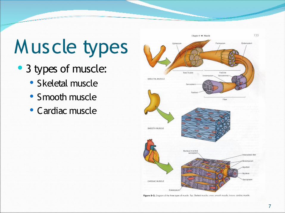
Muscle types 3 types of muscle:
Skeletal muscle Smooth muscle Cardiac muscle
8
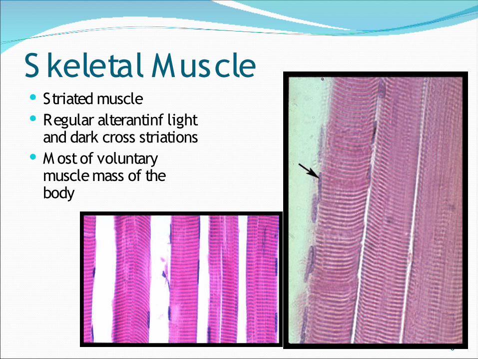
S keletal Muscle Striated muscle Regular alterantinf light
and dark cross striations M ost of voluntary
muscle mass of the body
9
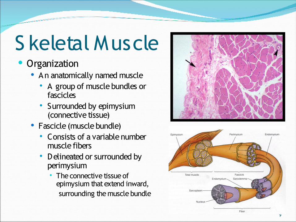
S keletal Muscle Organization
An anatomically named muscle A group of muscle bundles or
fascicles Surrounded by epimysium
(connective tissue) Fascicle (muscle bundle)
Consists of a variable number muscle fibers
Delineated or surrounded by perimysium The connective tissue of
epimysium that extend inward, surrounding the muscle bundle
10
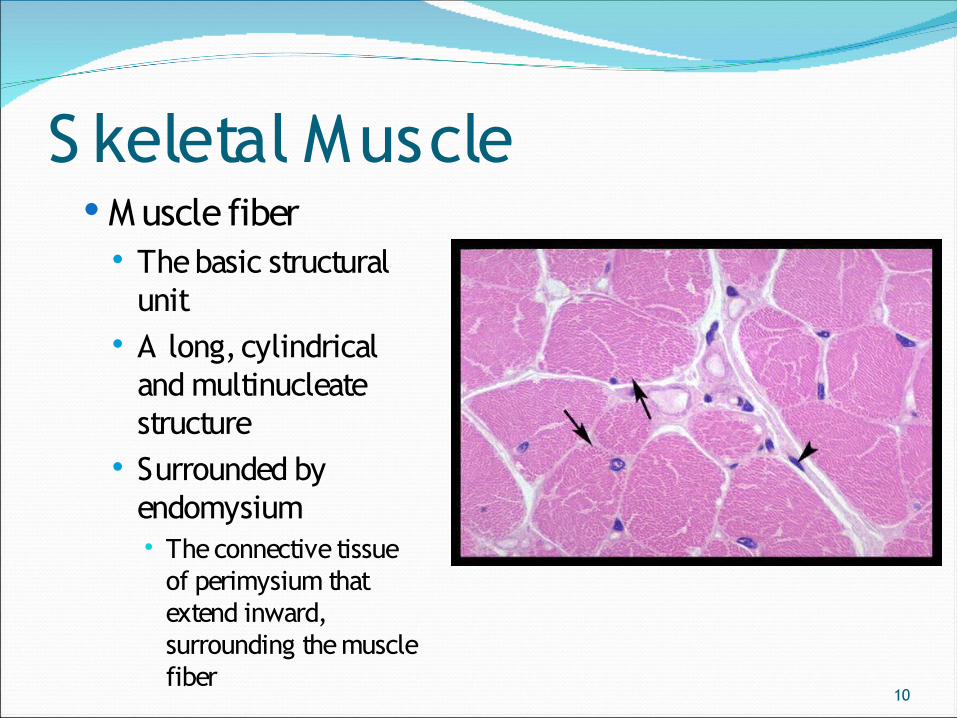
S keletal Muscle M uscle fiber
The basic structural unit
A long, cylindrical and multinucleate structure
Surrounded by endomysium The connective tissue
of perimysium that extend inward, surrounding the muscle fiber
11
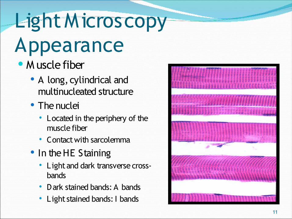
Light M icroscopy Appearance M uscle fiber
A long, cylindrical and multinucleated structure
The nuclei Located in the periphery of the
muscle fiber Contact with sarcolemma
In the HE Staining Light and dark transverse cross-
bands Dark stained bands: A bands Light stained bands: I bands
12
S keleta l M us c le U nit Organization unit
Sarcolemma Conduction of nerve impulse to the muscle fibers
Sarcoplasmic reticulum Control movement of skeletal muscle
M yofibriles Contraction of skeletal muscle
Contractile unit Sarcomere
Region of myofibril between 2 successive Z disk
13
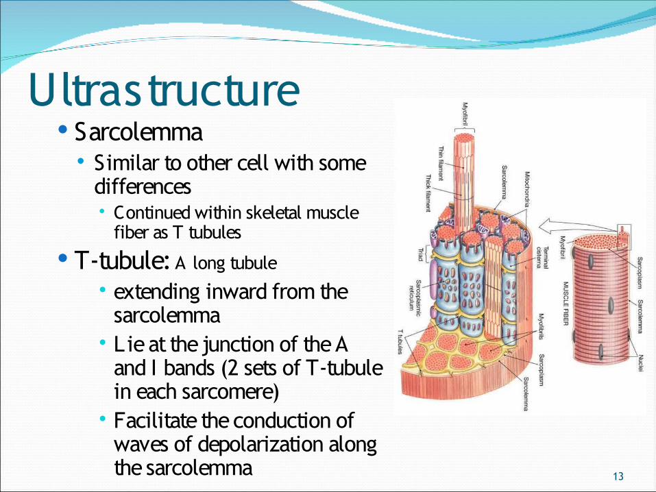
Ultrastructure Sarcolemma
Similar to other cell with some differences Continued within skeletal muscle
fiber as T tubules T-tubule: A long tubule
extending inward from the sarcolemma
Lie at the junction of the A and I bands (2 sets of T-tubule in each sarcomere)
Facilitate the conduction of waves of depolarization along the sarcolemma
14
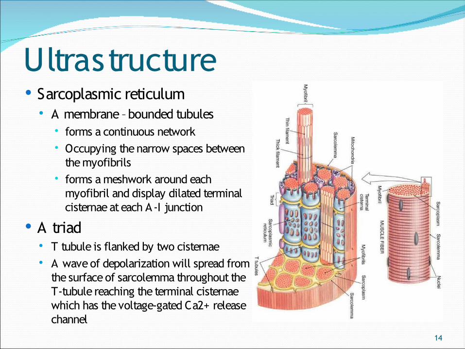
Ultrastructure Sarcoplasmic reticulum
A membrane –bounded tubules forms a continuous network Occupying the narrow spaces between
the myofibrils forms a meshwork around each
myofibril and display dilated terminal cisternae at each A-I junction
A triad T tubule is flanked by two cisternae A wave of depolarization will spread from
the surface of sarcolemma throughout the T-tubule reaching the terminal cisternae which has the voltage-gated Ca2+ release channel
15
Ultrastructure M itochondria
located just deep to the sarcoplasm numerous
M yofibril held in register with each other
by the intermediate filament desmin and vimentin the bundles of myofibril
attached to the cytoplasmic aspect of the sarcolemma by various protein including dystrophin
16
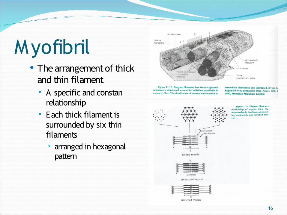
Myofibril The arrangement of thick
and thin filament A specific and constan
relationship Each thick filament is
surrounded by six thin filaments arranged in hexagonal
pattern
17
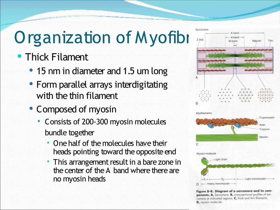
Organization of Myofibril Thick Filament
15 nm in diameter and 1.5 um long Form parallel arrays interdigitating
with the thin filament Composed of myosin
Consists of 200-300 myosin molecules bundle together One half of the molecules have their
heads pointing toward the opposite end This arrangement result in a bare zone in
the center of the A band where there are no myosin heads
18
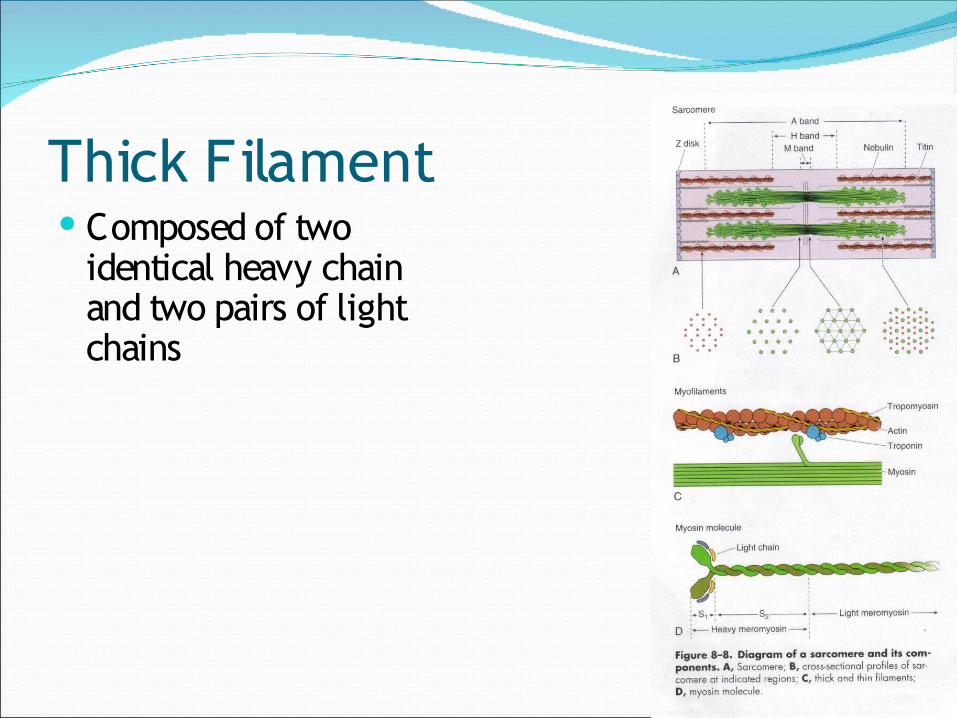
Thick Filament Composed of two
identical heavy chain and two pairs of light chains
19
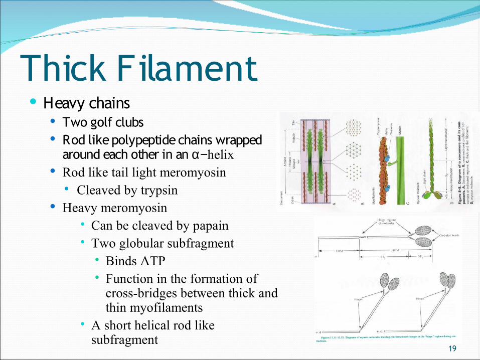
Thick Filament Heavy chains
Two golf clubs Rod like polypeptide chains wrapped
around each other in an α−helix Rod like tail light meromyosin
Cleaved by trypsin Heavy meromyosin
Can be cleaved by papain Two globular subfragment
Binds ATP Function in the formation of
cross-bridges between thick and thin myofilaments
A short helical rod like subfragment
20
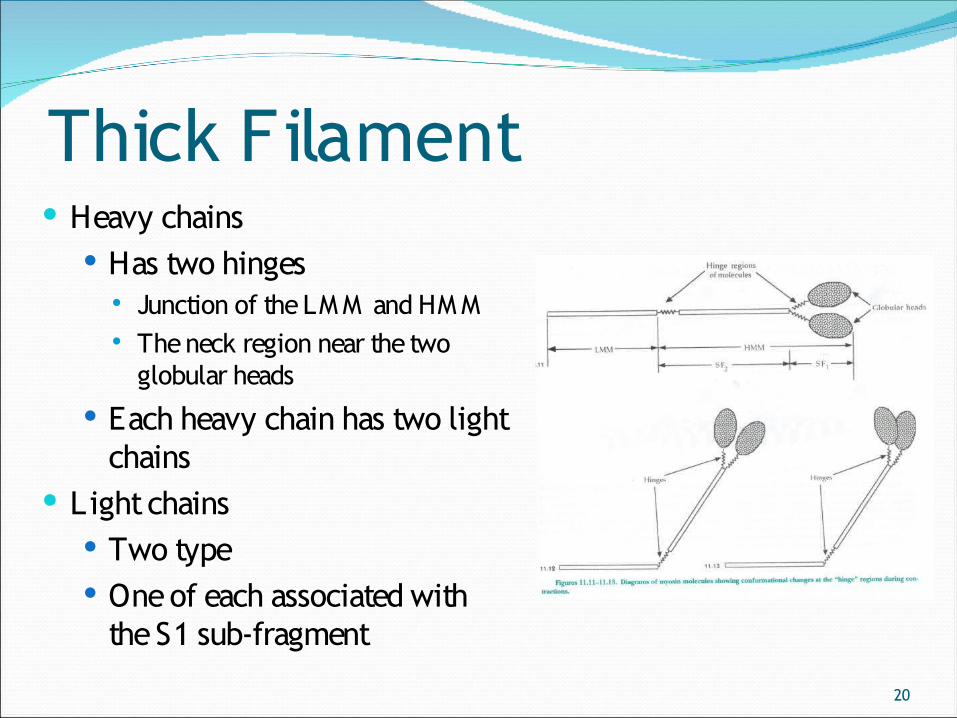
Thick Filament Heavy chains
Has two hinges Junction of the LM M and HM M The neck region near the two
globular heads
Each heavy chain has two light chains
Light chains Two type One of each associated with
the S1 sub-fragment
21
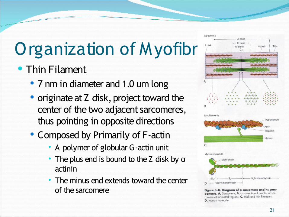
Organization of Myofibril Thin Filament
7 nm in diameter and 1.0 um long originate at Z disk, project toward the
center of the two adjacent sarcomeres, thus pointing in opposite directions
Composed by Primarily of F-actin A polymer of globular G-actin unit The plus end is bound to the Z disk by α
actinin The minus end extends toward the center
of the sarcomere
22
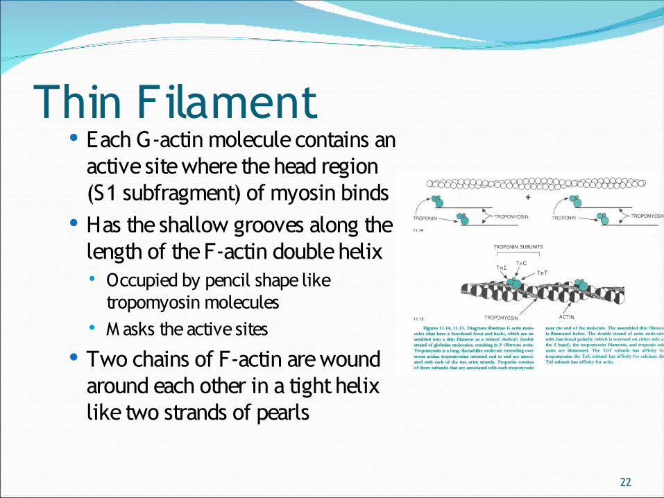
Thin Filament Each G-actin molecule contains an
active site where the head region (S1 subfragment) of myosin binds
Has the shallow grooves along the length of the F-actin double helix Occupied by pencil shape like
tropomyosin molecules M asks the active sites
Two chains of F-actin are wound around each other in a tight helix like two strands of pearls
23
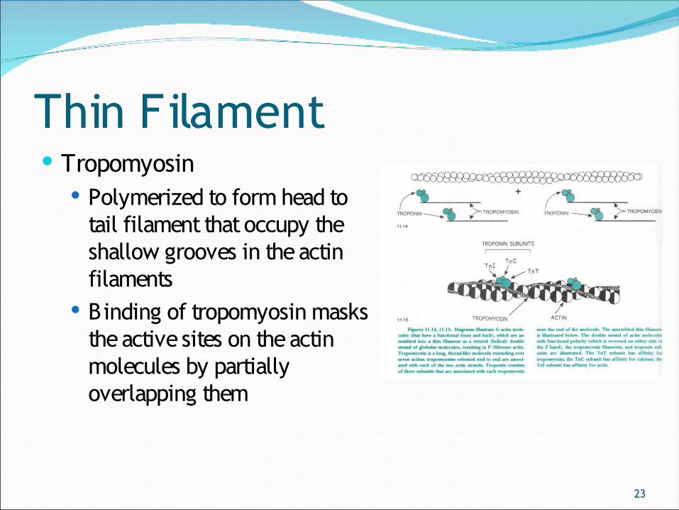
Thin Filament Tropomyosin
Polymerized to form head to tail filament that occupy the shallow grooves in the actin filaments
Binding of tropomyosin masks the active sites on the actin molecules by partially overlapping them
24
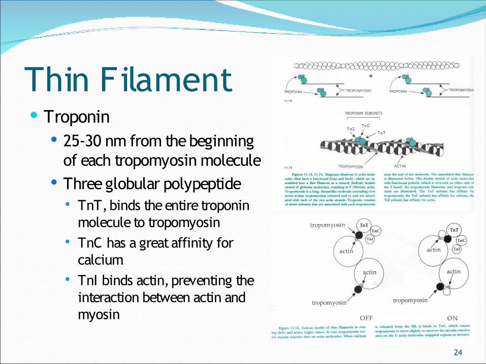
Thin Filament Troponin
25-30 nm from the beginning of each tropomyosin molecule
Three globular polypeptide TnT, binds the entire troponin
molecule to tropomyosin TnC has a great affinity for
calcium TnI binds actin, preventing the
interaction between actin and myosin
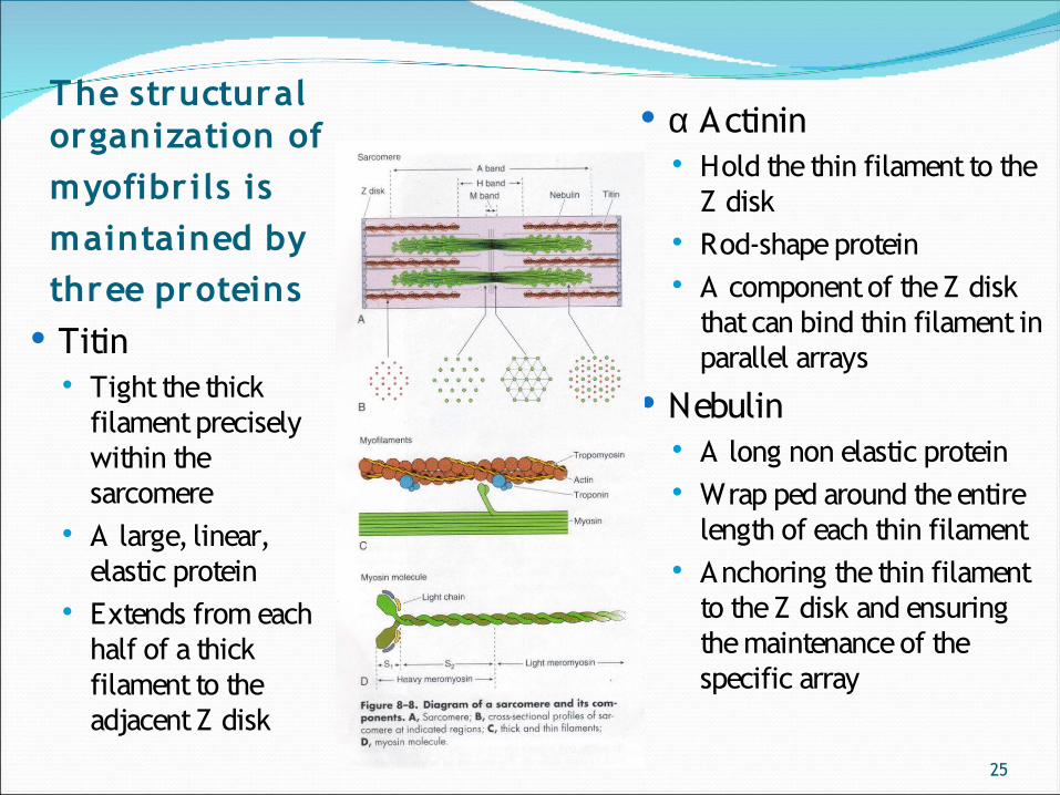
25
The structural organization of myofibrils is maintained by three proteins
Titin Tight the thick
filament precisely within the sarcomere
A large, linear, elastic protein
Extends from each half of a thick filament to the adjacent Z disk
α Actinin Hold the thin filament to the
Z disk Rod-shape protein A component of the Z disk
that can bind thin filament in parallel arrays
Nebulin A long non elastic protein Wrap ped around the entire
length of each thin filament Anchoring the thin filament
to the Z disk and ensuring the maintenance of the specific array
26
Muscle Contraction and Relaxation Contraction reduces the resting length of the muscle
fiber by an amount that is equal to the sum of all shortening that occur in all sarcomere of that particular muscle cell
The contraction process triggered by nerve impuls, obeys the all or none law in that a single muscle fiber will either contract or not contract as a result of stimulation
27
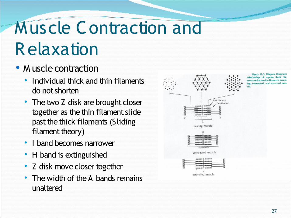
Muscle Contraction and Relaxation M uscle contraction
Individual thick and thin filaments do not shorten
The two Z disk are brought closer together as the thin filament slide past the thick filaments (Sliding filament theory)
I band becomes narrower H band is extinguished Z disk move closer together The width of the A bands remains
unaltered
28
Muscle Contraction and RelaxationM uscle Contraction
Sliding filament theory (Huxleys) Thin filament slide past the thick filament The sequences of action → Physiology lecture
29
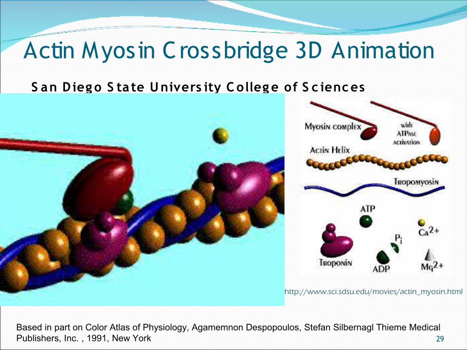
Actin Myosin Crossbridge 3D Animation S an D ieg o S ta te U nivers ity C olleg e of S c ienc es
Based in part on Color Atlas of Physiology, Agamemnon Despopoulos, Stefan Silbernagl Thieme Medical Publishers, Inc. , 1991, New York
http://www.sci.sdsu.edu/movies/actin_myosin.html
30
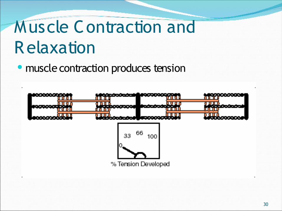
Muscle Contraction and Relaxation muscle contraction produces tension
31
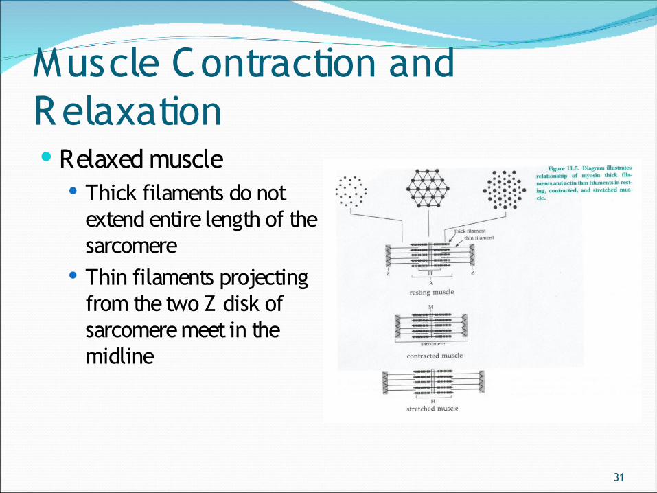
Muscle Contraction and Relaxation Relaxed muscle
Thick filaments do not extend entire length of the sarcomere
Thin filaments projecting from the two Z disk of sarcomere meet in the midline
32
Muscle Contraction and Relaxation Clinical correlation
Rigor mortis Occurs subsequently to death because the lack of ATP prevent the
dissociation of actin and myosin
Tetanus Force of contraction increases with summation of muscle twitches If action potentials continue to stimulate the muscle fiber repeatedly at
short interval (high frequency) relaxation between contractions diminished until the muscle fiber achieves a state of maximal contraction
Incomplete tetanus Complete tetanus
33
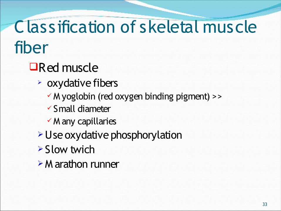
Classification of skeletal muscle fiber
Red muscle oxydative fibers
M yoglobin (red oxygen binding pigment) >> Small diameter M any capillaries
Use oxydative phosphorylationSlow twichM arathon runner
34
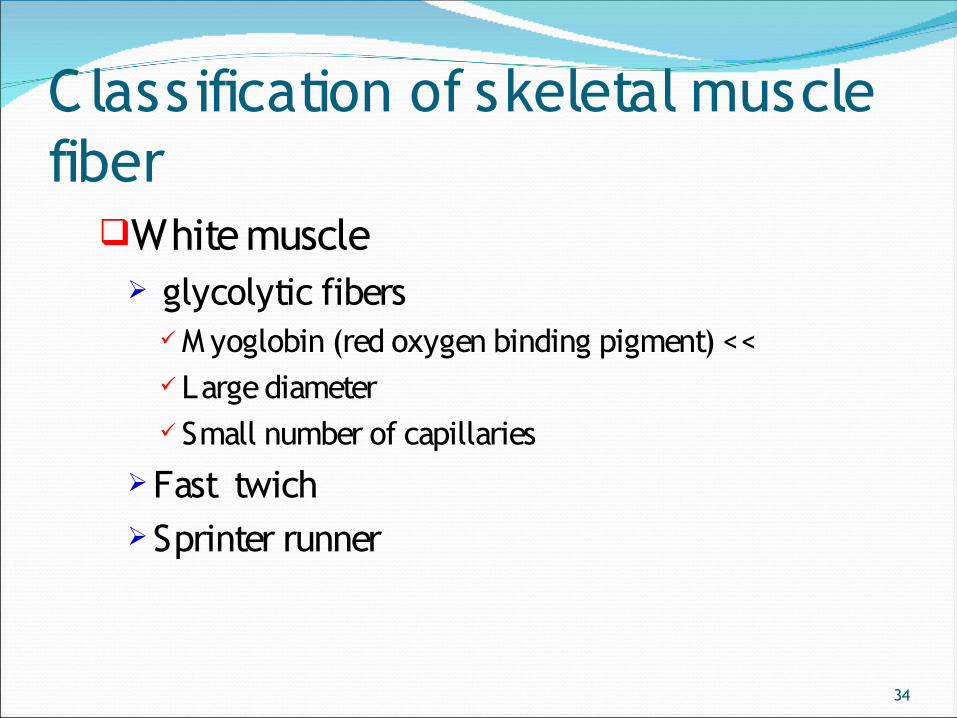
Classification of skeletal muscle fiber
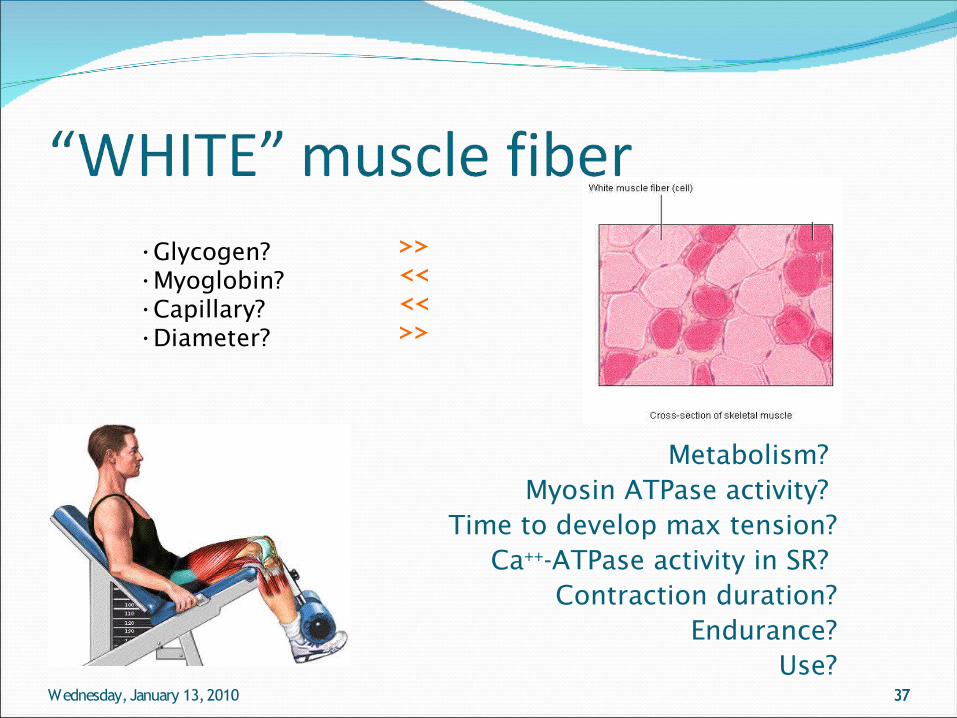
White muscle glycolytic fibers
M yoglobin (red oxygen binding pigment) << Large diameter Small number of capillaries
Fast twichSprinter runner
?aAk,ite muscle glycol
bgvbdn vvbds sscb mjhmhtttttttttttttttkmhtttttttttmttttkmmmmmmmmmmmmmmmmmmmmmmmmmmmmmmmmmmtthhhhhmbg bytic fibers M yoglobinbvbb sdc;[polo (red oxygen binding
35
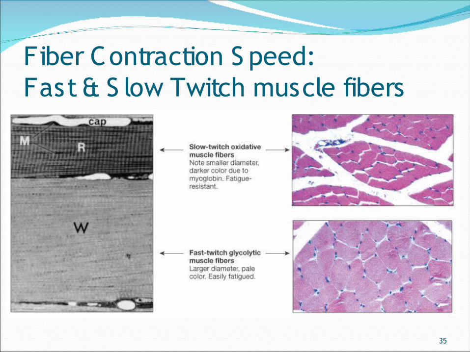
Fiber Contraction S peed: Fast & S low Twitch muscle fibers
36
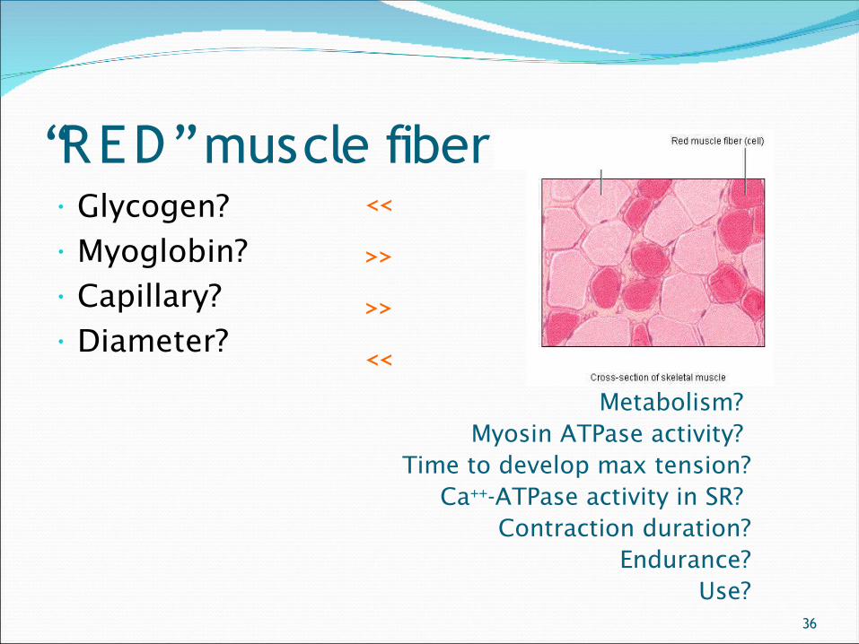
“RED” muscle fiber• Glycogen?• Myoglobin?• Capillary?• Diameter?
<<
>>
>>
<<
Metabolism? Myosin ATPase activity?
Time to develop max tension?Ca++-ATPase activity in SR?
Contraction duration?Endurance?
Use?
37Wednesday, January 13, 2010 37
•Glycogen?•Myoglobin?•Capillary?•Diameter?
>><<<<>>
Metabolism? Myosin ATPase activity?
Time to develop max tension?Ca++-ATPase activity in SR?
Contraction duration?Endurance?
Use?
38
Innervation of s keleta l mus c le Innervation of skeletal muscle by 2 nerve fiber
M otor (efferent) fiber Functions in eliciting contraction Each motor neuron and muscle fibers it controls form a motor unit
Sensory (afferent) fiber Pass to the muscle spindle
39
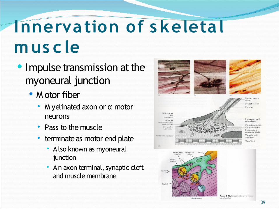
Innervation of s keleta l mus c le Impulse transmission at the
myoneural junction M otor fiber
M yelinated axon or α motor neurons
Pass to the muscle terminate as motor end plate
Also known as myoneural junction
An axon terminal, synaptic cleft and muscle membrane
40
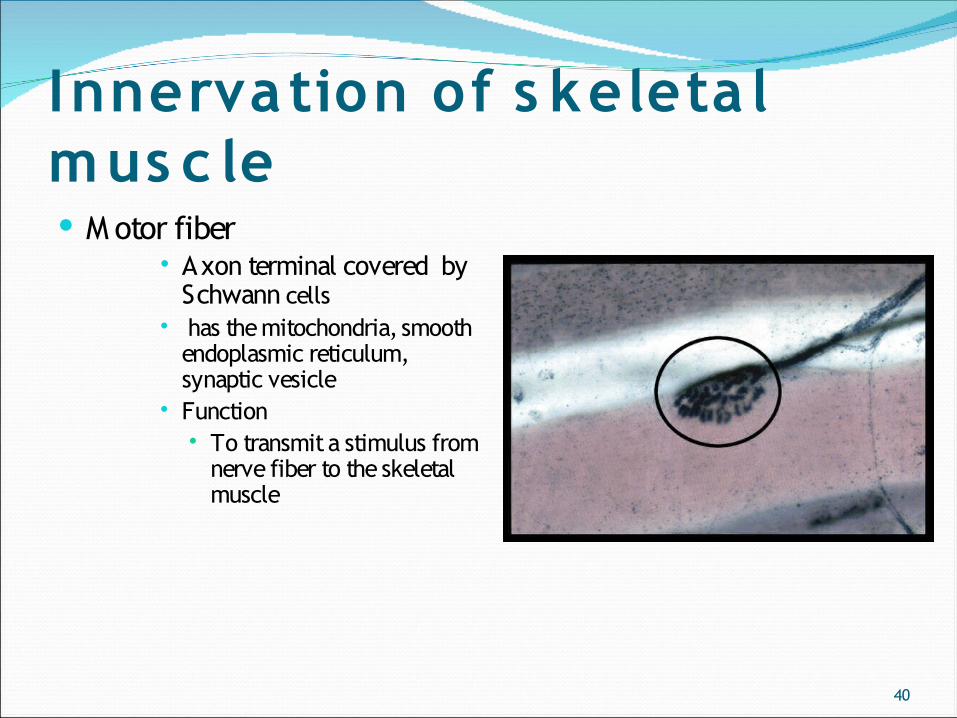
Innervation of s keleta l mus c le M otor fiber
Axon terminal covered by Schwann cells
has the mitochondria, smooth endoplasmic reticulum, synaptic vesicle
Function To transmit a stimulus from
nerve fiber to the skeletal muscle
41
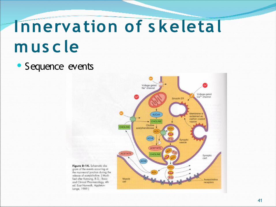
Innervation of s keleta l mus c le Sequence events
42
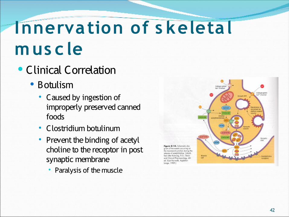
Innervation of s keleta l mus c le Clinical Correlation
Botulism Caused by ingestion of
improperly preserved canned foods
Clostridium botulinum Prevent the binding of acetyl
choline to the receptor in post synaptic membrane Paralysis of the muscle
43
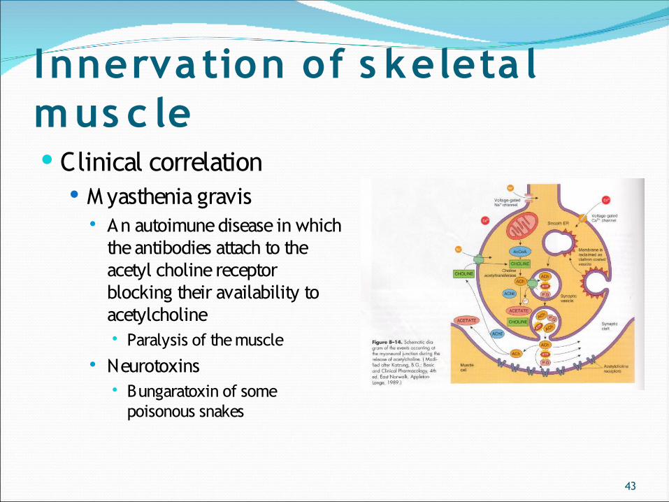
Innervation of s keleta l mus c le Clinical correlation
M yasthenia gravis An autoimune disease in which
the antibodies attach to the acetyl choline receptor blocking their availability to acetylcholine Paralysis of the muscle
Neurotoxins Bungaratoxin of some
poisonous snakes
44
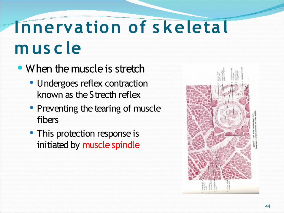
Innervation of s keleta l mus c le When the muscle is stretch
Undergoes reflex contraction known as the Strecth reflex
Preventing the tearing of muscle fibers
This protection response is initiated by muscle spindle
45
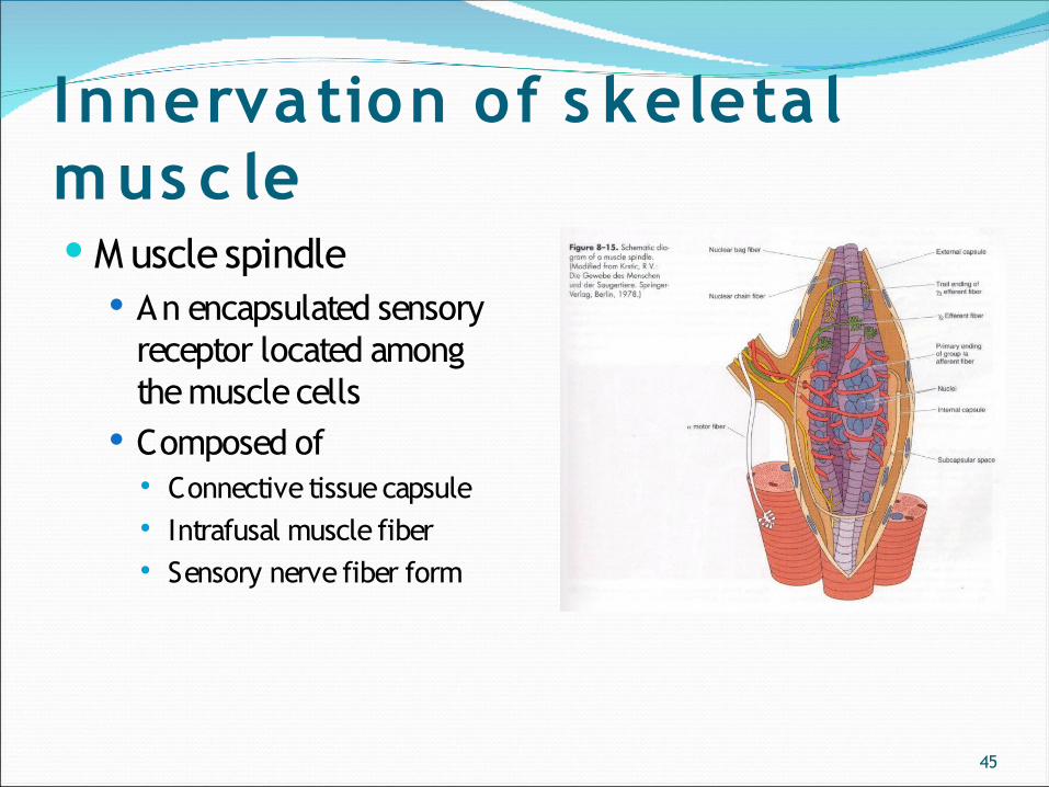
Innervation of s keleta l mus c le M uscle spindle
An encapsulated sensory receptor located among the muscle cells
Composed of Connective tissue capsule Intrafusal muscle fiber Sensory nerve fiber form
46
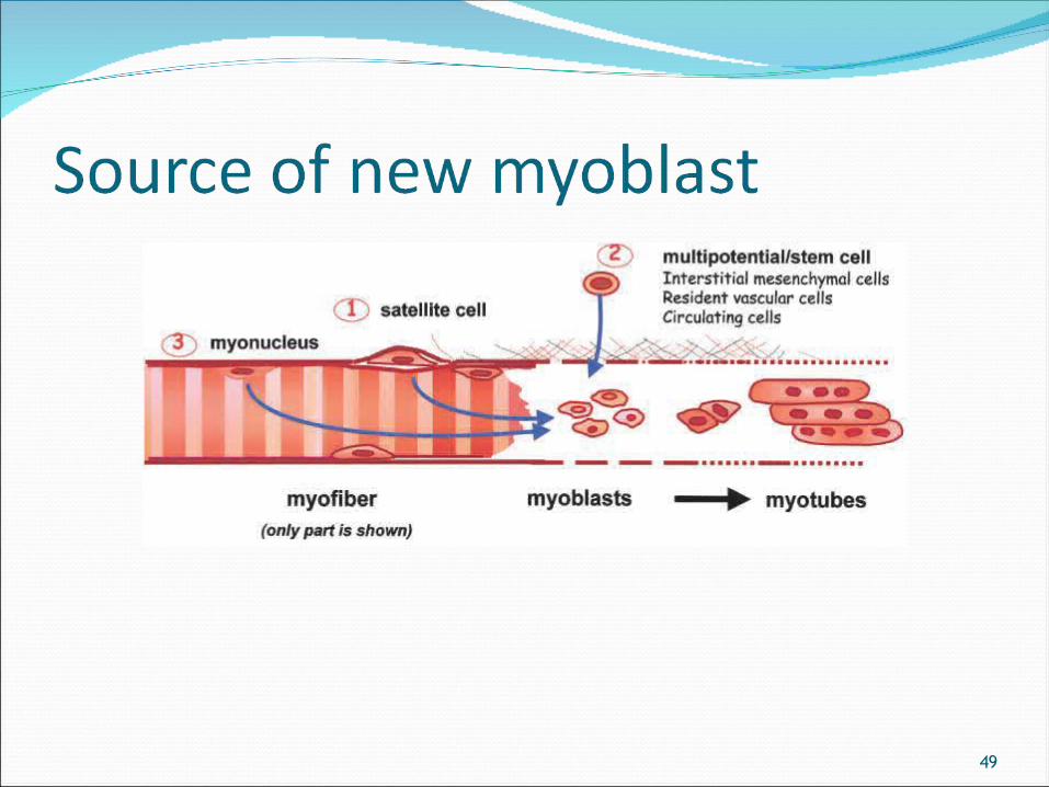
Muscle regenerationSatellite cells
Lying beneath the basement membrane next to sarcolemma
Reserve muscle precursor cells Normally quiescent Activated only in response to growth or muscle
damage
47
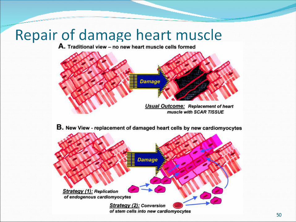
Repair/regeneration of S keletal Muscle
Traditional view Adult muscle cells→post-mitotic Regeneration is very limited Injury→repair →fibrous scar formation Satellite cells have minimal contribution especially in severe
muscle trauma Cardiac muscle do not have any satellite cells→Lack of
regeneration
48
Repair/regeneration of S keletal Muscle
New perspective Activated of:
satellite cells and other precursor cells→
M yonucleus M ultipotential cells (interstitial mesenchyme cells)
Skeletal muscle → M oderate regeneration potential Cardiac muscle → have regeneration potential
49
50
51
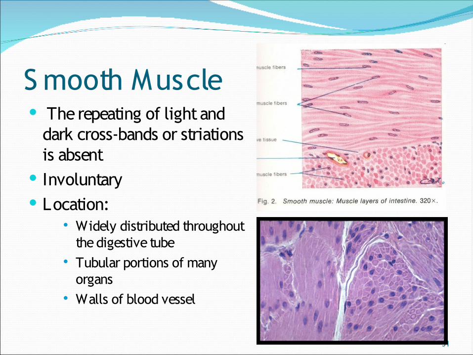
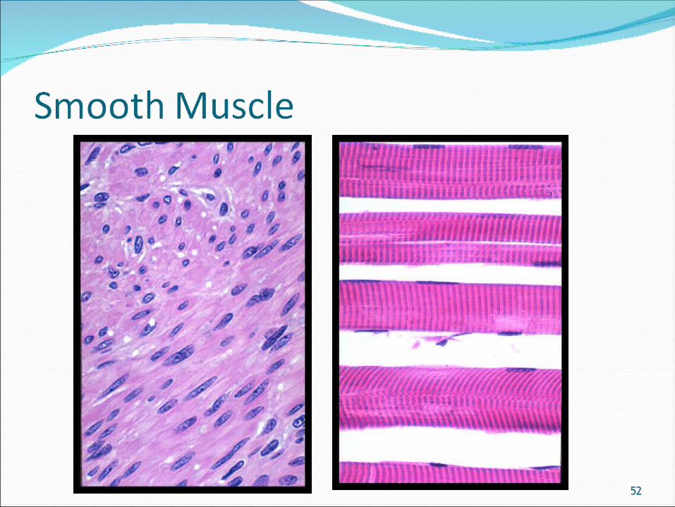
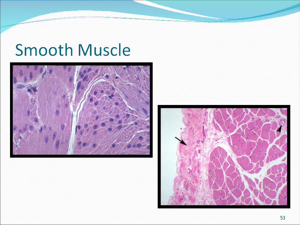
S mooth Muscle The repeating of light and
dark cross-bands or striations is absent
Involuntary Location:
Widely distributed throughout the digestive tube
Tubular portions of many organs
Walls of blood vessel
52
53

54