thoracic and abdominal trauma · 4 thoracic trauma • 25% of motor vehicle crash deaths are...
TRANSCRIPT
Thoracic and Abdominal Trauma Jami Windhorn RN BSN CPN TNCC ENPC

2
• I have nothing to disclose
• I have no conflict of interest
3
Objectives
• Identify Common Mechanisms for Thoracic Trauma
• Describe Pathophysiology of Thoracic Trauma
• Describe Nursing Assessment and Interventions for a Thoracic Trauma patient
4
Thoracic Trauma • 25% of motor vehicle crash deaths are related to thoracic trauma
• Approximately 16,000 deaths per year
• Second only to brain and spinal cord injuries as the leading cause of traumatic death
• Motor vehicle crashes and interpersonal violence are the two main causes of thoracic trauma
• Most thoracic traumas will also involve the abdominal cavity
5
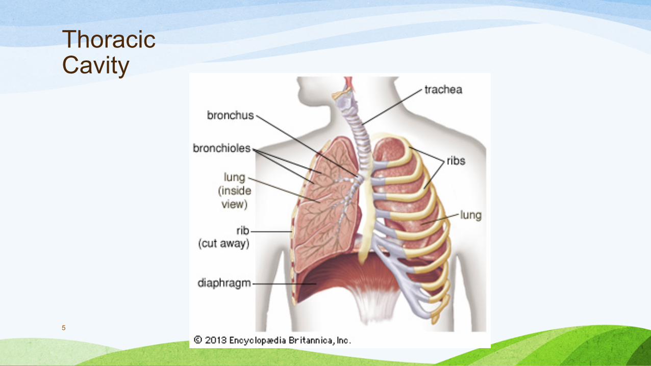
Thoracic Cavity
6
Thoracic Cavity
• Second largest hollow space of the body
• Contains the heart, lungs, diaphragm, great vessels, esophagus, ribs, vertebral column and various muscles
• Epicenter of all circulatory and oxygen flow for the body
7
Thoracic Injury • Mechanisms of Injury
• Acceleration and Deceleration forces
• First and second rib fractures can severely injure the pulmonary and cardiac tissues underneath
• Falls
• Crush Injuries
• Violence
• Motor Vehicle Crashes
8
Thoracic Injury • Pathophysiology
• Ineffective Ventilation due to disruption in the anatomical structures in the thoracic cavity
• Tears in the bronchial tree • Rib/Sternal fractures • Pain • Lung contusion • Impaled object in the chest
9
Thoracic Injury • Pathophysiology
• Ineffective Circulation • Internal or external hemorrhage due to injury to the great
vessels • Blunt trauma can lead to decrease myocardial contractility and
cardiac output • Pericardial tamponade • Air in thoracic cavity can cause venous congestion
10
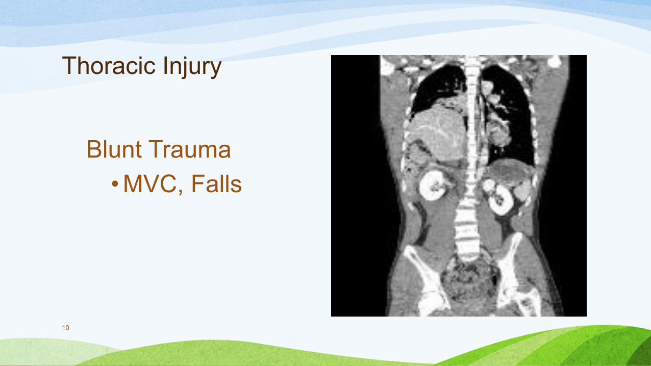
Thoracic Injury
Blunt Trauma
• MVC, Falls
11
Thoracic Injury
• Blunt Trauma • Blast Injuries: Tear blood vessels, disrupt bronchial tree,
diaphragm rupture • Crush Injuries: Body is crushed between an object and
hard surface, direct pressure to chest • Deceleration Injuries: Body hits a hard object, Body stops
but organs do not, can cause tearing of the aorta
12
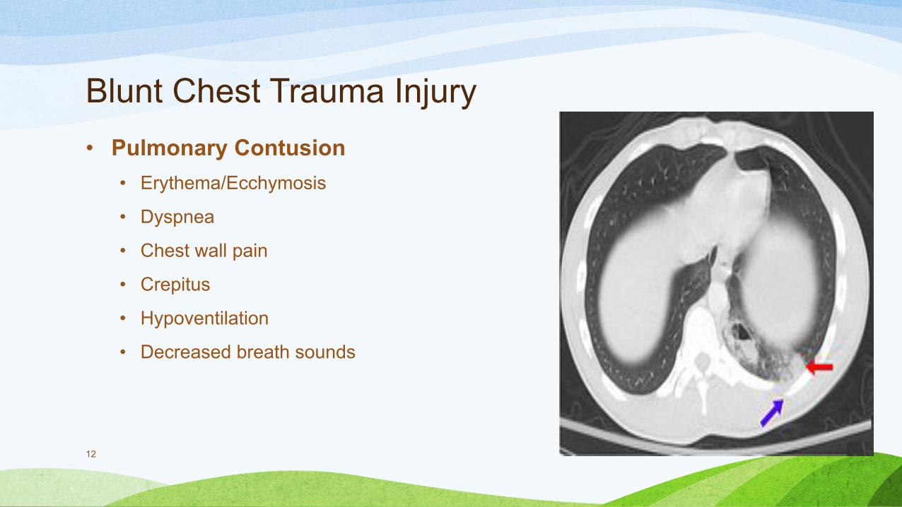
Blunt Chest Trauma Injury • Pulmonary Contusion
• Erythema/Ecchymosis
• Dyspnea
• Chest wall pain
• Crepitus
• Hypoventilation
• Decreased breath sounds
13
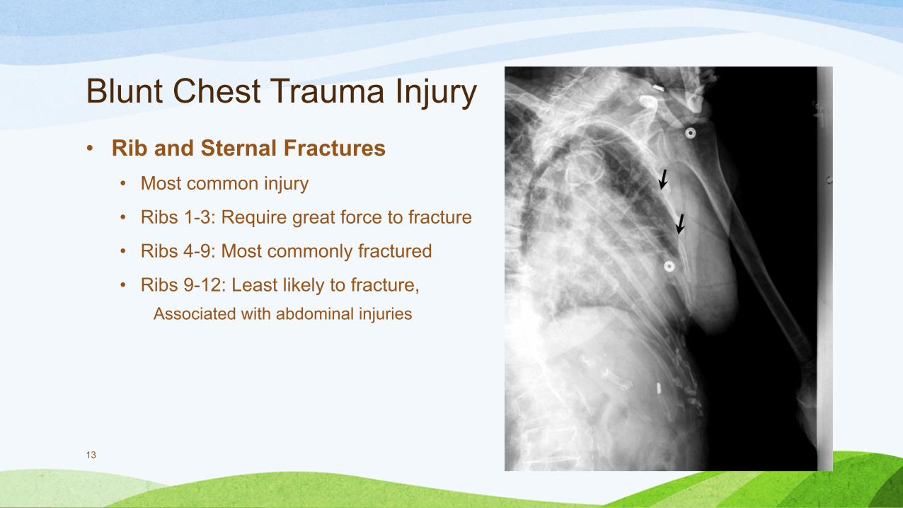
Blunt Chest Trauma Injury • Rib and Sternal Fractures
• Most common injury
• Ribs 1-3: Require great force to fracture
• Ribs 4-9: Most commonly fractured
• Ribs 9-12: Least likely to fracture, Associated with abdominal injuries
14
Blunt Chest Trauma Injury
• Rib and Sternal Fractures • Pain • Dyspnea • Chest wall bruising • Crepitus or bony deformity • Patient splints the chest for comfort
15
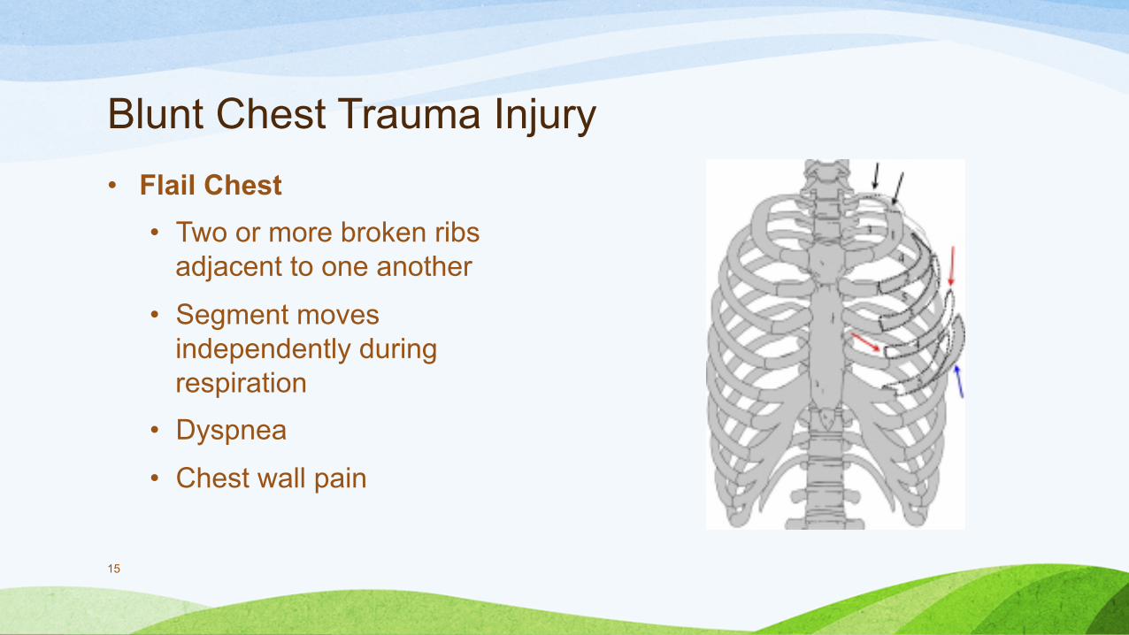
Blunt Chest Trauma Injury • Flail Chest
• Two or more broken ribs adjacent to one another
• Segment moves independently during respiration
• Dyspnea
• Chest wall pain
16
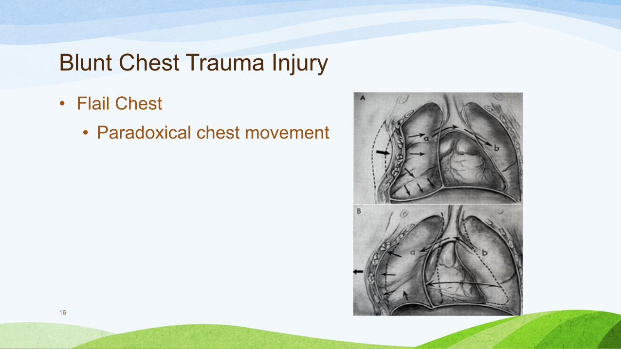
Blunt Chest Trauma Injury
• Flail Chest • Paradoxical chest movement
17
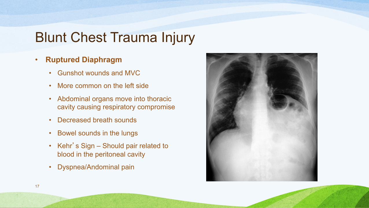
Blunt Chest Trauma Injury • Ruptured Diaphragm
• Gunshot wounds and MVC
• More common on the left side
• Abdominal organs move into thoracic cavity causing respiratory compromise
• Decreased breath sounds
• Bowel sounds in the lungs
• Kehr’s Sign – Should pair related to blood in the peritoneal cavity
• Dyspnea/Andominal pain
18
Blunt Chest Trauma Injury • Cardiac Contusion
• Bruise to the heart tissue
• MVC, falls, sports injuries
• CPR
• EKG abnormalities
• Chest Pain
• Chest wall bruising
• Irregular heart beat
• Hypotension
19
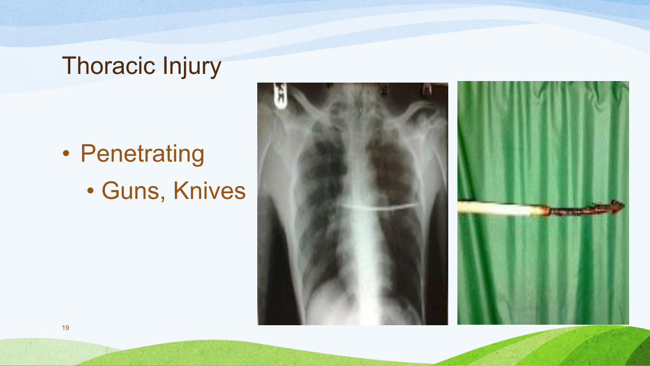
Thoracic Injury
• Penetrating • Guns, Knives
20
Thoracic Injury • Penetrating Trauma
• Low Energy: Guns, Knives and direct contact
• High Energy: High power firearms
• Damage caused by firearms increases as the distance between the gun and person decreases
• Type 1: >7 meters, soft tissue damage
• Type 2: 3-7 meters, deep fascia and internal organ damage
• Type 3: <3 meters, massive tissue destruction

21 Trauma.org
22
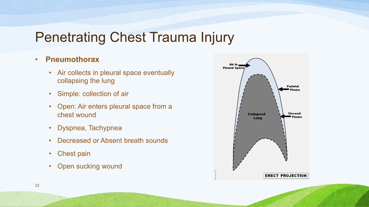
Penetrating Chest Trauma Injury • Pneumothorax
• Air collects in pleural space eventually collapsing the lung
• Simple: collection of air
• Open: Air enters pleural space from a chest wound
• Dyspnea, Tachypnea
• Decreased or Absent breath sounds
• Chest pain
• Open sucking wound
23
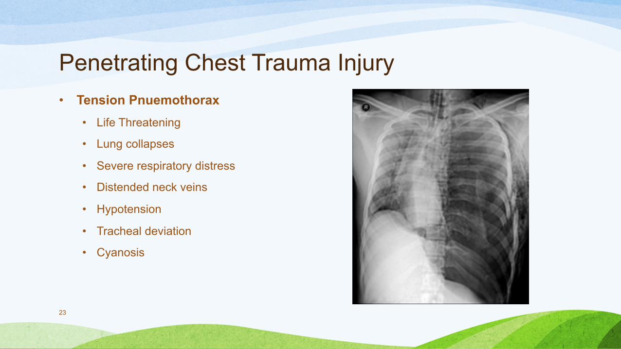
Penetrating Chest Trauma Injury • Tension Pnuemothorax
• Life Threatening
• Lung collapses
• Severe respiratory distress
• Distended neck veins
• Hypotension
• Tracheal deviation
• Cyanosis
24
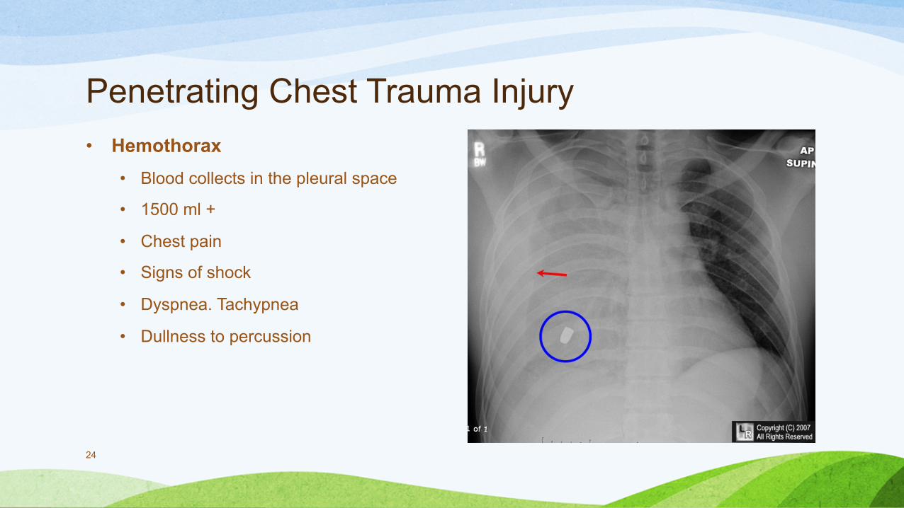
Penetrating Chest Trauma Injury • Hemothorax
• Blood collects in the pleural space
• 1500 ml +
• Chest pain
• Signs of shock
• Dyspnea. Tachypnea
• Dullness to percussion

25
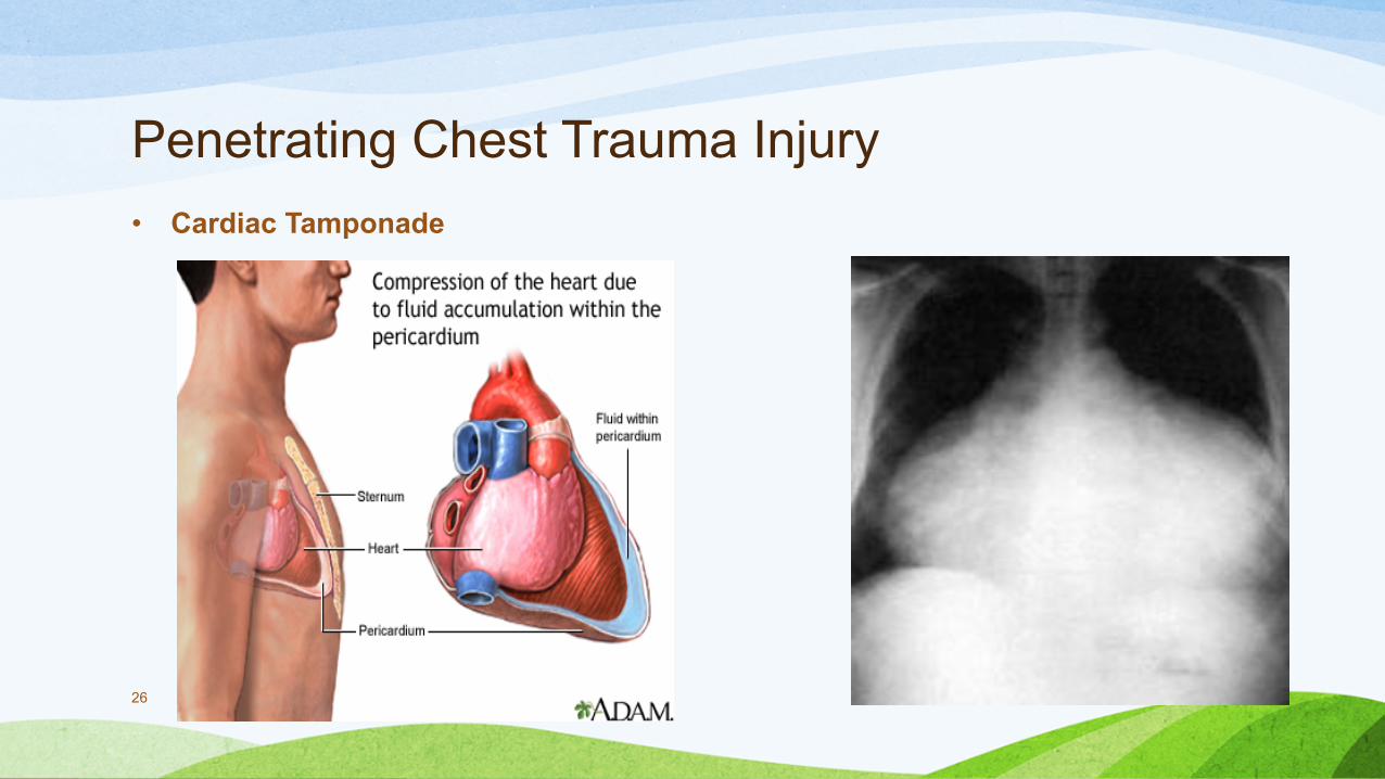
Penetrating Chest Trauma Injury • Cardiac Tamponade
• Blood collects in pericardial sac
• Decreases cardiac output
• Dyspnea
• Cyanosis
• Beck’s Triad: Distended neck veins, hypotension, muffled heart tones
• Signs of shock
26
Penetrating Chest Trauma Injury • Cardiac Tamponade
27
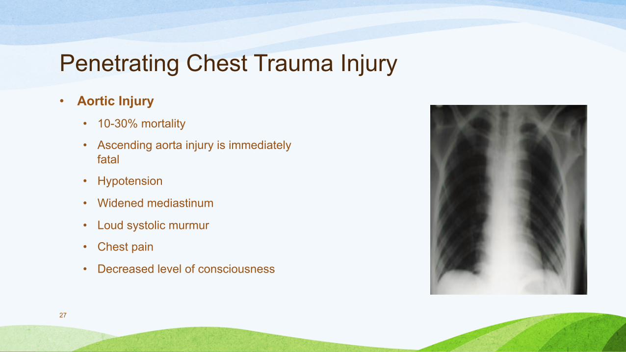
Penetrating Chest Trauma Injury • Aortic Injury
• 10-30% mortality
• Ascending aorta injury is immediately fatal
• Hypotension
• Widened mediastinum
• Loud systolic murmur
• Chest pain
• Decreased level of consciousness

28
Thoracic Injury
• Concurrent Injuries • Head • Extremities • Abdomen
29
Patient History • What was mechanism of injury?
• If MVC, what was damage to the car?
• Patient complaints?
• Vital signs?
• Previous medical history?
• Medications?
• Treatment prior to hospital?
30
Nursing Assessment • Airway
• Respiratory effort – Rate, Depth
• Symmetrical chest wall movement?
• Jugular vein distension?
• Look for chest wall injuries, bruising
• Percuss for dullness - Hemothorax
31
Nursing Assessment • Palpate
• Chest wall, clavicles and neck for crepitus, edema and pain
• Central and peripheral pulses
• Assess for tracheal deviation
• Auscultate: • Heart and lung sounds
• Listen for bowel sounds in chest
• Blood pressures in upper and lower extremities
32
Nursing Assessment • Diagnostic Procedures
• Chest X-Ray
• CT
• Bronchoscopy
• EKG
• Cardiac enzymes, CBC
• Central venous pressure
33
Nursing Assessment • Once chest tube is placed drainage must be monitored closely
• >200 ml/hour of blood from chest tube may need replaced
• FOCA for chest tube assessment • F: Fluctuation in the water seal chamber
• O: Output
• C: Color of drainage
• A: Air leak
34
Nursing Interventions • Maintain patent airway
• Oxygen
• Cover open chest wounds with sterile dressing and tape on 3 sides
• Prepare for needle thoracentesis or chest tube insertion
• 2 large bore IVs
• Pain meds
• Surgical interventions
• Stabilize impaled objects
Abdominal Trauma
36
Objectives
• Identify common mechanisms for Abdominal Trauma
• Describe Pathophysiology of Abdominal Trauma
• Describe Nursing Assessment and Interventions for an Abdominal Trauma Patient
37
Abdominal Trauma
• 3rd leading cause of traumatic death after head and chest injuries
• Blunt injuries more deadly than penetrating
• 25% require surgical intervention
• Motor vehicle crashes most common type of blunt injury
• Stab wounds and gunshots are most common penetrating injuries
38
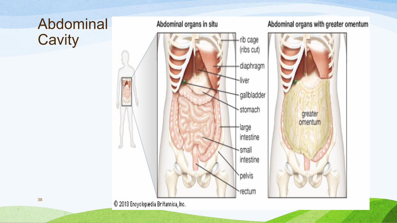
Abdominal Cavity
39
Abdominal Cavity
• Largest hollow space in the body
• Separated from the thoracic cavity by the diaphragm
• Contains digestive tract, liver, pancreas, spleen, kidneys and adrenal glands
• Entire cavity is lined with peritoneum
40
Abdominal Cavity
• Solid Organs: Liver, Pancreas, Spleen, Kidneys, Ovaries
• Hollow Organs: Stomach, Small Intestine, Appendix, Large Intestine, Gallbladder, Bladder, Uterus, Aorta, Common Bile Duct, Fallopian Tubes
41
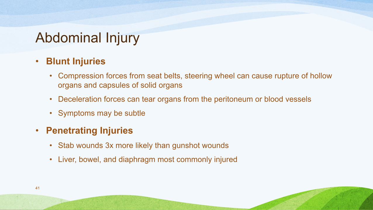
Abdominal Injury • Blunt Injuries
• Compression forces from seat belts, steering wheel can cause rupture of hollow organs and capsules of solid organs
• Deceleration forces can tear organs from the peritoneum or blood vessels
• Symptoms may be subtle
• Penetrating Injuries • Stab wounds 3x more likely than gunshot wounds
• Liver, bowel, and diaphragm most commonly injured
42
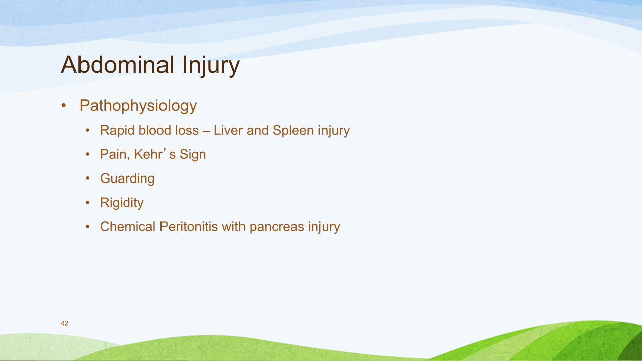
Abdominal Injury • Pathophysiology
• Rapid blood loss – Liver and Spleen injury
• Pain, Kehr’s Sign
• Guarding
• Rigidity
• Chemical Peritonitis with pancreas injury
43
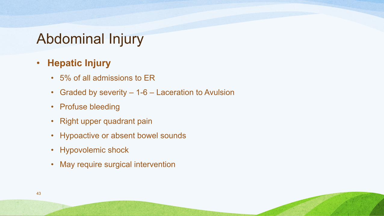
Abdominal Injury • Hepatic Injury
• 5% of all admissions to ER
• Graded by severity – 1-6 – Laceration to Avulsion
• Profuse bleeding
• Right upper quadrant pain
• Hypoactive or absent bowel sounds
• Hypovolemic shock
• May require surgical intervention
44
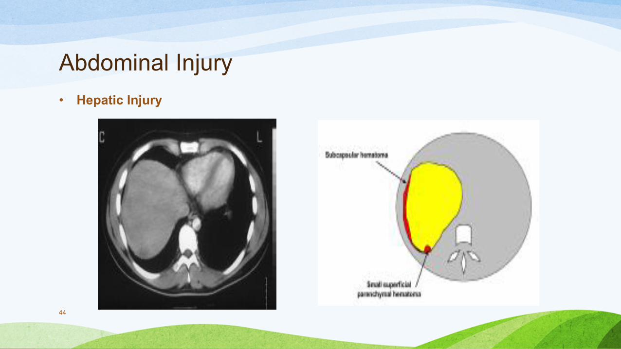
Abdominal Injury • Hepatic Injury
45
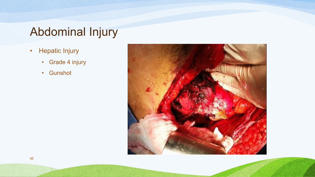
Abdominal Injury • Hepatic Injury
• Grade 4 injury
• Gunshot
46
Abdominal Injury • Splenic Injury
• Most common from blunt trauma – 49% of all blunt injuries
• Graded by severity – 1-5 – Laceration to shattered Spleen
• Signs of hypovolemic shock
• Kehr’s sign – Pain in left shoulder
• Rigidity and guarding
• Bedrest if hemodynamically stable
• May require surgical intervention
47
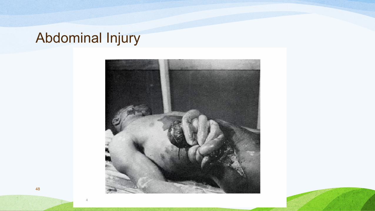
Abdominal Injury • Bowel Injuries
• Small bowel injury most common
• Blunt and penetrating trauma
• Shearing injury may cause avulsion of small bowel
• Compression may cause rupture
• Hypovolemic shock
• Bleeding from rectum
• Abdominal wall rigidity, guarding, pain
48
Abdominal Injury

49
Abdominal Injury • Esophageal Injuries
• Rare
• Associated with penetrating trauma
• Neck, shoulder, chest or abdominal pain
• Subcutaneous air in neck
• Frank blood from NG/Vomit
50
Abdominal Injury • Kidney Injuries
• Contusion from blunt trauma
• 10% of ER visit
• Suspect renal injuries with posterior rib or lumbar vertebra fracture
• Hematuria
• Flank pain
• Ecchymosis over site
• Graded by severity
51
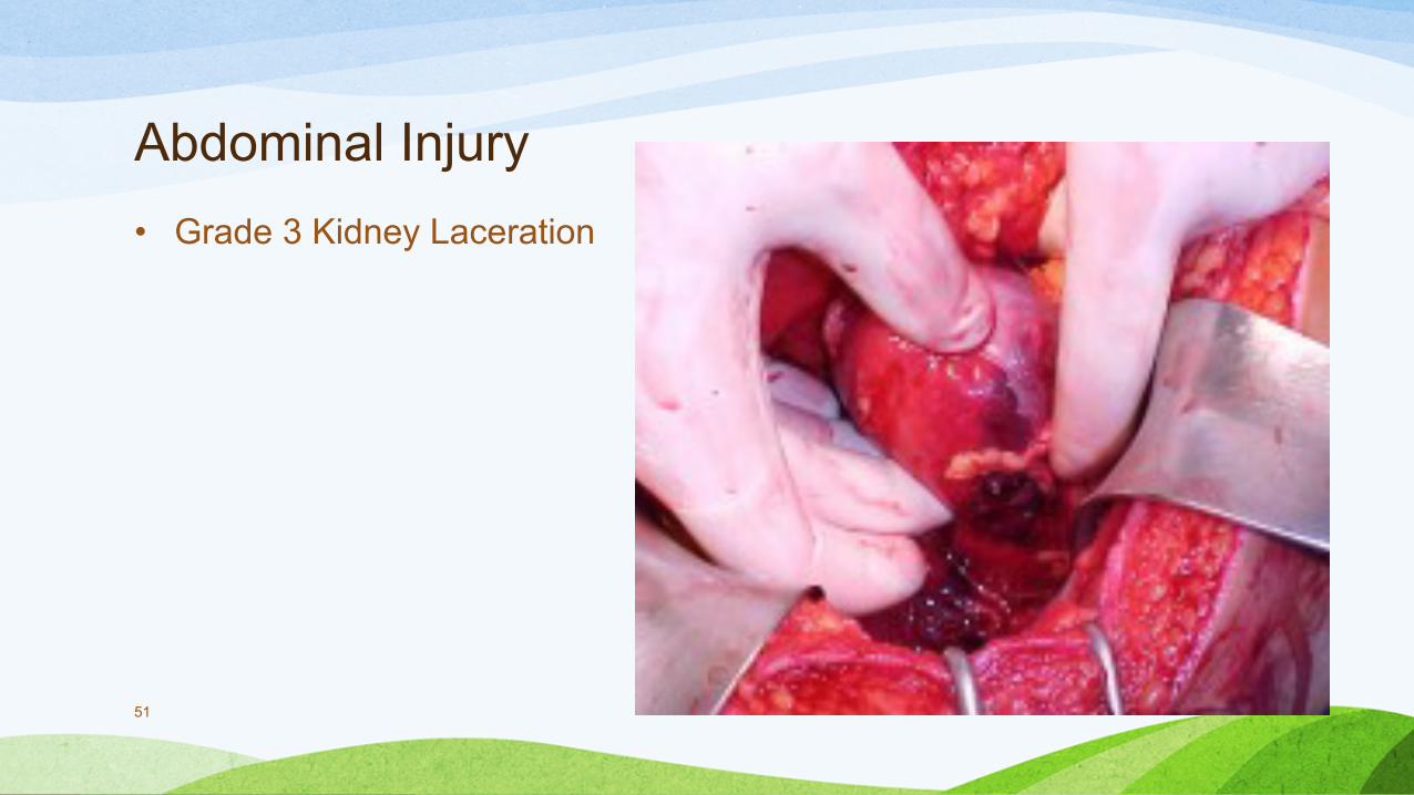
Abdominal Injury • Grade 3 Kidney Laceration
52
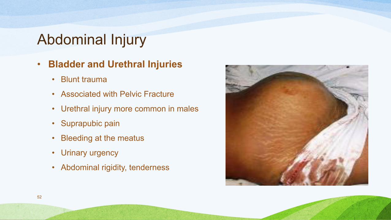
Abdominal Injury • Bladder and Urethral Injuries
• Blunt trauma
• Associated with Pelvic Fracture
• Urethral injury more common in males
• Suprapubic pain
• Bleeding at the meatus
• Urinary urgency
• Abdominal rigidity, tenderness
53
Abdominal Injury
• Concurrent Injuries • Thoracic Injuries
• Rib Fractures
• Diaphragm Injuries
• Pelvic and lower extremity injuries
54
Patient History
• What was mechanism of Injury?
• Blunt or penetrating trauma?
• Blunt – MVC? Seatbelts? Vehicle Damage? Height of Fall?
• Penetrating – Type of Weapon? Distance away from weapon? Blood Loss at scene? Pain?
55
Nursing Assessment • Airway
• Abdominal injuries
• Respiratory effort – Rate, Depth
• Symmetrical Chest Wall Movement?
• Contour of abdomen
• Bleeding Perinuem?
56
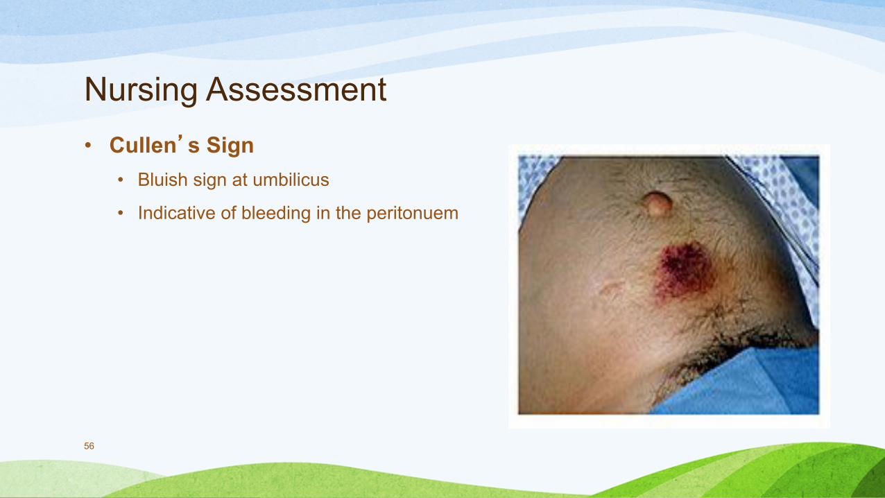
Nursing Assessment • Cullen’s Sign
• Bluish sign at umbilicus
• Indicative of bleeding in the peritonuem
57
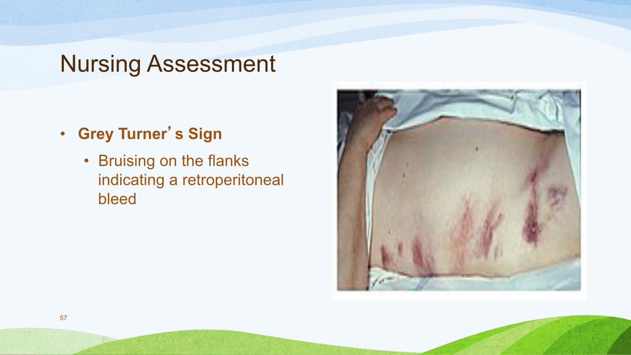
Nursing Assessment
• Grey Turner’s Sign • Bruising on the flanks
indicating a retroperitoneal bleed
58
Nursing Assessment • Auscultation
• Bowel sounds in all 4 quadrants
• Percussion • Hyperresonance – Air
• Dullness - Fluid
• Palpation • All 4 quadrants
• Pelvis for instability • Anal sphincter for tone
59
Nursing Assessment
• Diagnostic Procedures • X-Rays
• Labs – CBC, Pregnancy, Coags, UA, Stool for blood,
• CT
• FAST Exam
• Angiography
• Cystogram
60
Nursing Assessment • Focused Assessment with Sonography for Trauma (FAST) Exam
• Used to diagnose free blood in the peritoneum after blunt trauma
• Looks at 4 areas for free fluid • Perihepatic
• Perislpenic
• Pelvis • Pericardium
• 94% effective
• Test takes 4-5 minutes
61
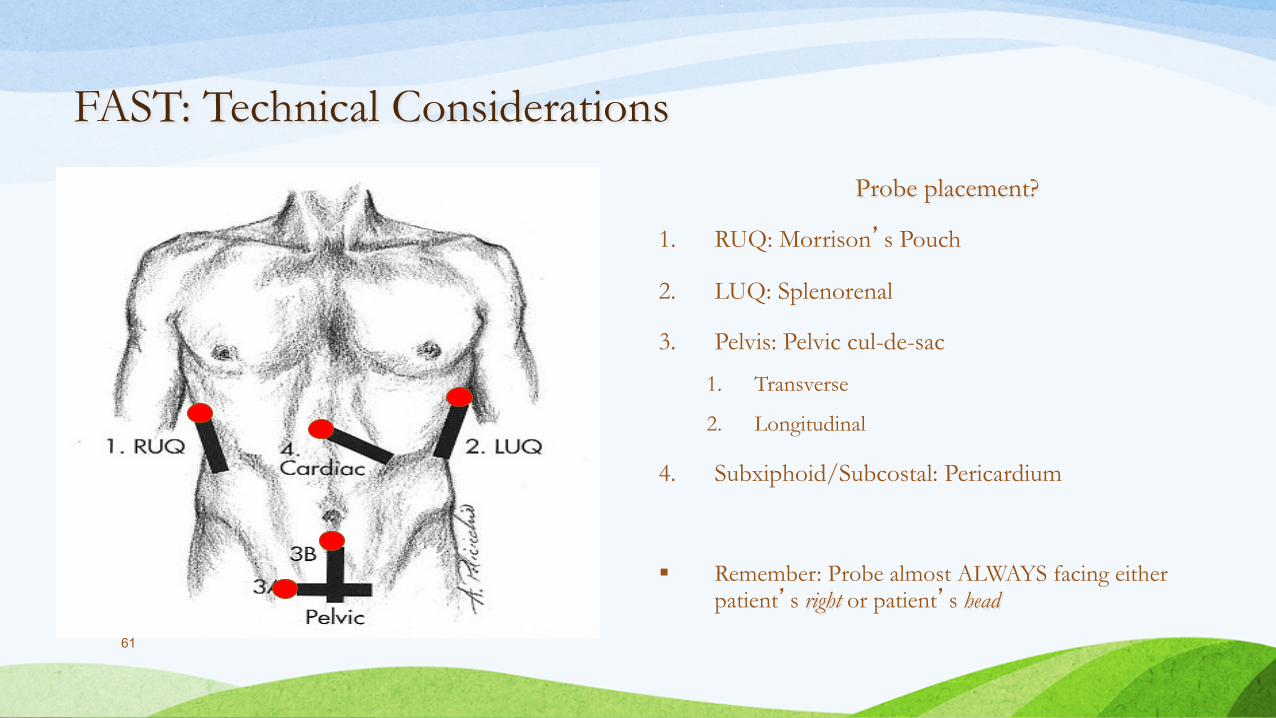
FAST: Technical Considerations
Probe placement?
1. RUQ: Morrison’s Pouch
2. LUQ: Splenorenal
3. Pelvis: Pelvic cul-de-sac
1. Transverse
2. Longitudinal
4. Subxiphoid/Subcostal: Pericardium
§ Remember: Probe almost ALWAYS facing either patient’s right or patient’s head

62
Nursing Interventions • Maintain Patent airway
• 2 large bore IVs
• IVF or Blood Volume
• Pain Meds
• Foley
• NG
• Cover open wounds

63
Nursing Interventions • Antibiotics
• Psychosocial support
• Stabilize impaled objects
• Surgical intervention
• Monitor urinary output
• Serial vital signs

Questions????