1 thoracic trauma. 2 second leading cause of trauma deaths after head injurysecond leading cause of...
TRANSCRIPT

1
Thoracic Thoracic Trauma Trauma
2
Thoracic TraumaThoracic Trauma
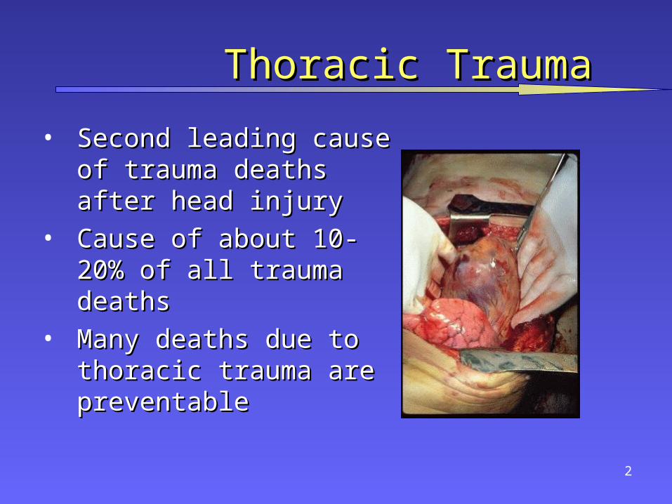
• Second leading cause of Second leading cause of trauma deaths after head trauma deaths after head injuryinjury
• Cause of about 10-20% of all Cause of about 10-20% of all trauma deathstrauma deaths
• Many deaths due to thoracic Many deaths due to thoracic trauma are preventabletrauma are preventable
3
Thoracic TraumaThoracic Trauma
• Mechanisms of InjuryMechanisms of Injury
– Blunt InjuryBlunt Injury
• DecelerationDeceleration
• CompressionCompression
– Penetrating InjuryPenetrating Injury
– BothBoth
4
Thoracic TraumaThoracic Trauma
• Anatomical InjuriesAnatomical Injuries– Thoracic Cage (Skeletal)Thoracic Cage (Skeletal)
– CardiovascularCardiovascular
– Pleural and PulmonaryPleural and Pulmonary
– MediastinalMediastinal
– DiaphragmaticDiaphragmatic
– EsophagealEsophageal
– Penetrating CardiacPenetrating Cardiac
5
Thoracic TraumaThoracic Trauma
• Often result in:Often result in:– HypoxiaHypoxia
• hypovolemiahypovolemia
• pulmonary V/P mismatchpulmonary V/P mismatch in intrathoracic pressure relationshipsin intrathoracic pressure relationships
– HypercarbiaHypercarbia in intrathoracic pressure relationshipsin intrathoracic pressure relationships level of consciousnesslevel of consciousness
– Acidosis Acidosis
• hypoperfusion of tissues (metabolic)hypoperfusion of tissues (metabolic)
6
Thoracic TraumaThoracic Trauma
• Ventilation & Respiration ReviewVentilation & Respiration Review– How & Why does ventilation (inspiration & How & Why does ventilation (inspiration &
expiration) occur? expiration) occur? • What actually happens in ventilation?What actually happens in ventilation?
• What stimulates its occurrence?What stimulates its occurrence?
• What stimulates its cessation?What stimulates its cessation?
– What happens in respiration?What happens in respiration?• How does it affect acid-base balance?How does it affect acid-base balance?
• What factors inhibit effective respiration?What factors inhibit effective respiration?
7
General PathophysiologyGeneral Pathophysiology
Impairments to cardiac outputImpairments to cardiac output
• blood lossblood loss
• increased intrapleural pressuresincreased intrapleural pressures
• blood in pericardial sacblood in pericardial sac
• myocardial valve damagemyocardial valve damage
• vascular disruptionvascular disruption
8
General PathophysiologyGeneral Pathophysiology
Impairments in ventilatory efficiencyImpairments in ventilatory efficiency
• chest excursion compromisechest excursion compromise
– painpain
– air in pleural spaceair in pleural space
– asymmetrical movementasymmetrical movement
• bleeding in pleural spacebleeding in pleural space
• ineffective diaphragm contractionineffective diaphragm contraction

9
General PathophysiologyGeneral Pathophysiology
Impairments in gas exchangeImpairments in gas exchange• atelectasisatelectasis• pulmonary contusionpulmonary contusion• respiratory tract disruptionrespiratory tract disruption

10
AssessmentAssessment
• Initial exam directed toward life threatening:Initial exam directed toward life threatening:– InjuriesInjuries
• Open pneumothoraxOpen pneumothorax
• Flail chestFlail chest
• Tension pneumothoraxTension pneumothorax
• Massive hemothoraxMassive hemothorax
• Cardiac tamponadeCardiac tamponade
– ConditionsConditions• ApneaApnea
• Respiratory DistressRespiratory Distress

11
AssessmentAssessment
• Assessment FindingsAssessment Findings– Mental Status (decreased)Mental Status (decreased)
– Pulse (absent, tachy or brady)Pulse (absent, tachy or brady)
– BP (narrow PP, hyper- or hypotension, pulsus BP (narrow PP, hyper- or hypotension, pulsus paradoxus)paradoxus)
– Ventilatory rate & effort (tachy- or bradypnea, Ventilatory rate & effort (tachy- or bradypnea, labored, retractions)labored, retractions)
– Skin (diaphoresis, pallor, cyanosis, open injury, Skin (diaphoresis, pallor, cyanosis, open injury, ecchymosis)ecchymosis)

12
AssessmentAssessment
• Assessment FindingsAssessment Findings– Neck (tracheal position, SQ emphysema, JVD, open Neck (tracheal position, SQ emphysema, JVD, open
injury)injury)
– Chest (contusions, tenderness, asymmetry, absent or Chest (contusions, tenderness, asymmetry, absent or decreased lung sounds, bowel sounds, abnormal decreased lung sounds, bowel sounds, abnormal percussion, open injury, impaled object, crepitus, percussion, open injury, impaled object, crepitus, hemoptysis)hemoptysis)
– Heart Sounds (muffled, distant, regurgitant murmur)Heart Sounds (muffled, distant, regurgitant murmur)
– Upper abdomen (contusion, open injuryUpper abdomen (contusion, open injury

13
AssessmentAssessment
• Assessment FindingsAssessment Findings– Neck (tracheal position, SQ emphysema, JVD, open Neck (tracheal position, SQ emphysema, JVD, open
injury)injury)
– Chest (contusions, tenderness, asymmetry, absent or Chest (contusions, tenderness, asymmetry, absent or decreased lung sounds, bowel sounds, abnormal decreased lung sounds, bowel sounds, abnormal percussion, open injury, impaled object, crepitus, percussion, open injury, impaled object, crepitus, hemoptysis)hemoptysis)
– Heart Sounds (muffled, distant, regurgitant murmur)Heart Sounds (muffled, distant, regurgitant murmur)
– Upper abdomen (contusion, open injuryUpper abdomen (contusion, open injury

14
Rib FracturesRib Fractures
• Most common chest wall injury from direct Most common chest wall injury from direct traumatrauma
• More common in adults than childrenMore common in adults than children• Especially common in elderlyEspecially common in elderly• Ribs form ringsRibs form rings
– Possibility of break in two placesPossibility of break in two places
• Most commonly 5th - 9th ribsMost commonly 5th - 9th ribs– Poor protectionPoor protection

15
Rib FracturesRib Fractures
• Fractures of 1st and 2nd second require high Fractures of 1st and 2nd second require high forceforce– Frequently have injury to aorta or bronchiFrequently have injury to aorta or bronchi
– Occur in 90% of patients with tracheo-bronchial Occur in 90% of patients with tracheo-bronchial rupturerupture
– May injure subclavian artery/veinMay injure subclavian artery/vein
– May result in pneumothoraxMay result in pneumothorax
• 30% will die30% will die
16
Rib FracturesRib Fractures
• Fractures of 10 to 12th ribs can cause damage to Fractures of 10 to 12th ribs can cause damage to
underlying abdominal solid organs:underlying abdominal solid organs: – LiverLiver– SpleenSpleen– KidneysKidneys

17
Rib FracturesRib Fractures
• Assessment FindingsAssessment Findings– Localized pain, tendernessLocalized pain, tenderness
– Increases on palpation or when patient:Increases on palpation or when patient:
• CoughsCoughs
• MovesMoves
• Breathes deeplyBreathes deeply
– ““Splinted” RespirationsSplinted” Respirations
– Instability in chest wall, CrepitusInstability in chest wall, Crepitus
– Deformity and discolorationDeformity and discoloration
– Associated pneumo or hemothoraxAssociated pneumo or hemothorax

18
Rib FracturesRib Fractures
• ManagementManagement– High concentration OHigh concentration O22
– Positive pressure ventilation as neededPositive pressure ventilation as needed
– Splint using pillow or swathesSplint using pillow or swathes
– Encourage pt to breath deeplyEncourage pt to breath deeply
• Helps prevent atelectasisHelps prevent atelectasis
– Analgesics for isolated traumaAnalgesics for isolated trauma
– Non-circumferential splintingNon-circumferential splinting
19
Rib FracturesRib Fractures
• ManagementManagement– Monitor elderly and COPD patients closelyMonitor elderly and COPD patients closely
• Broken ribs can cause decompensationBroken ribs can cause decompensation
• Patients will fail to breathe deeply and cough, resulting in Patients will fail to breathe deeply and cough, resulting in poor clearance of secretionspoor clearance of secretions
– Usually Non-Emergent TransportUsually Non-Emergent Transport

20
Sternal FractureSternal Fracture
• Uncommon, 5-8% in blunt chest traumaUncommon, 5-8% in blunt chest trauma• Large traumatic forceLarge traumatic force• Direct blow to front of chest byDirect blow to front of chest by
– DecelerationDeceleration
• steering wheelsteering wheel
• dashboarddashboard
– Other objectOther object
21
Sternal FractureSternal Fracture
• 25 - 45% mortality due to associated trauma:25 - 45% mortality due to associated trauma:– Disruption of thoracic aortaDisruption of thoracic aorta– Tracheal or bronchial tearTracheal or bronchial tear– Diaphragm ruptureDiaphragm rupture– Flail chestFlail chest– Myocardial traumaMyocardial trauma
• High incidence of myocardial contusion, cardiac High incidence of myocardial contusion, cardiac tamponade or pulmonary contusiontamponade or pulmonary contusion

22
Sternal FractureSternal Fracture
• Assessment FindingsAssessment Findings– Localized painLocalized pain
– Tenderness over sternumTenderness over sternum
– CrepitusCrepitus
– Tachypnea, DyspneaTachypnea, Dyspnea
– ECG changes with associated myocardial contusionECG changes with associated myocardial contusion
– Hx/Mechanism of blunt chest traumaHx/Mechanism of blunt chest trauma

23
Sternal FractureSternal Fracture
• ManagementManagement– Establish airwayEstablish airway
– High concentration oxygenHigh concentration oxygen
– Assist ventilations with BVM as neededAssist ventilations with BVM as needed
– IV NS/LRIV NS/LR
• Restrict fluids Restrict fluids
– Emergent TransportEmergent Transport• Trauma centerTrauma center

24
Fail ChestFail Chest
• Two or more adjacent ribs fractured in two or Two or more adjacent ribs fractured in two or more places producing a free floating segment more places producing a free floating segment of the chest wallof the chest wall

25
Fail ChestFail Chest
• Usually secondary to blunt traumaUsually secondary to blunt trauma– Most commonly in MVCMost commonly in MVC
– Also results fromAlso results from
• falls from heightsfalls from heights
• industrial accidentsindustrial accidents
• assaultassault
• birth traumabirth trauma
• More common in older patientsMore common in older patients

26
Fail ChestFail Chest
• Mortality rates 20-40% due to associated Mortality rates 20-40% due to associated injuriesinjuries
• Mortality increased withMortality increased with– advanced ageadvanced age
– seven or more rib fracturesseven or more rib fractures
– three or more associated injuriesthree or more associated injuries
– shockshock
– head injurieshead injuries
27
Fail ChestFail Chest
• Consequences of flail chestConsequences of flail chest– Respiratory failure due toRespiratory failure due to
• pulmonary contusionpulmonary contusion
• intrathoracic injuryintrathoracic injury
• inadequate diaphragm movementinadequate diaphragm movement
– Paradoxical movement of the chestParadoxical movement of the chest
• must be large to compromise ventilationmust be large to compromise ventilation
• Increased work of breathingIncreased work of breathing
– Pain, decreased chest expansionPain, decreased chest expansion
• leading decreased ventilationleading decreased ventilation
28
Fail ChestFail Chest
• Consequences of flail chestConsequences of flail chest– Contusion of lungContusion of lung
• decreased lung compliancedecreased lung compliance
• intra alveolar-capillary hemorrhageintra alveolar-capillary hemorrhage
– Decreased ventilationDecreased ventilation• HypercapneaHypercapnea
• HypoxiaHypoxia
29
Fail ChestFail Chest
• Assessment FindingsAssessment Findings– Chest wall contusionChest wall contusion
– Respiratory distressRespiratory distress
– Pleuritic chest painPleuritic chest pain
– Splinting of affected sideSplinting of affected side
– CrepitusCrepitus
– Tachypnea, TachycardiaTachypnea, Tachycardia
– Paradoxical movement (possible)Paradoxical movement (possible)
30
Fail ChestFail Chest
• ManagementManagement– Suspect spinal injuriesSuspect spinal injuries
– Establish airwayEstablish airway
– High concentration oxygenHigh concentration oxygen
– Assist ventilation with BVMAssist ventilation with BVM
• Treat hypoxia from underlying contusionTreat hypoxia from underlying contusion
• Promote full lung expansionPromote full lung expansion
– Consider need for intubation and PEEPConsider need for intubation and PEEP
– Mechanically stabilize chest wallMechanically stabilize chest wall
• questionable valuequestionable value
31
Fail ChestFail Chest
• ManagementManagement– IV of LR/NSIV of LR/NS
• Avoid rapid replacement in hemodynamically stable patientAvoid rapid replacement in hemodynamically stable patient
• Contused lung cannot handle fluid loadContused lung cannot handle fluid load
– Monitor EKGMonitor EKG
• Chest trauma can cause dysrhythmiasChest trauma can cause dysrhythmias
– Emergent TransportEmergent Transport• Trauma centerTrauma center
32
Simple PneumothoraxSimple Pneumothorax• IncidenceIncidence
– 10-30% in blunt chest trauma10-30% in blunt chest trauma
– almost 100% with penetrating chest traumaalmost 100% with penetrating chest trauma
– Morbidity & Mortality dependent onMorbidity & Mortality dependent on
• extent of atelectasisextent of atelectasis
• associated injuriesassociated injuries

33
Simple PneumothoraxSimple Pneumothorax• CausesCauses
– Commonly a fx rib lacerates lungCommonly a fx rib lacerates lung
– Paper bag effectPaper bag effect
– May occur spontaneously in tall, thin young males May occur spontaneously in tall, thin young males following:following:
• ExertionExertion
• CoughingCoughing
• Air TravelAir Travel
– Spontaneous may occur w/ Marfan’s syndromeSpontaneous may occur w/ Marfan’s syndrome
34
Simple PneumothoraxSimple Pneumothorax• PathophysiologyPathophysiology
– Air enters pleural space causing partial lung collapseAir enters pleural space causing partial lung collapse
• small tears self-sealsmall tears self-seal
• larger tears may progresslarger tears may progress
– Usually well-tolerated in the young & healthyUsually well-tolerated in the young & healthy
– Severe compromise can occur in the elderly or patients Severe compromise can occur in the elderly or patients with pulmonary diseasewith pulmonary disease
– Degree of distress depends on amount and speed of Degree of distress depends on amount and speed of collapsecollapse
35
Simple PneumothoraxSimple Pneumothorax• Assessment FindingsAssessment Findings
– Tachypnea, TachycardiaTachypnea, Tachycardia– Difficulty breathing or respiratory distressDifficulty breathing or respiratory distress– Pleuritic painPleuritic pain
• may be referred to shoulder or arm on affected sidemay be referred to shoulder or arm on affected side
– Decreased or absent breath soundsDecreased or absent breath sounds • not always reliablenot always reliable
– if patient standing, assess apices first if patient standing, assess apices first
– if supine, assess anteriorlyif supine, assess anteriorly
• patients with multiple ribs fractures may splint injured side by patients with multiple ribs fractures may splint injured side by not breathing deeplynot breathing deeply
36
Simple PneumothoraxSimple Pneumothorax
• ManagementManagement– Establish airwayEstablish airway– High concentration OHigh concentration O22 with NRB with NRB– Assist with BVMAssist with BVM
• decreased or rapid respirations decreased or rapid respirations • inadequate TVinadequate TV
– IV of LR/NSIV of LR/NS– Monitor for progressionMonitor for progression– Monitor ECGMonitor ECG– Usually Non-emergent transportUsually Non-emergent transport
Open PneumothoraxOpen Pneumothorax
•Hole in chest wall that allows air to enter pleural Hole in chest wall that allows air to enter pleural space. space.
•The larger the hole, the more likely air will enter The larger the hole, the more likely air will enter there than through the trachea. there than through the trachea.
•If the trauma patient does not ventilate well with an If the trauma patient does not ventilate well with an open airway, look for a holeopen airway, look for a hole
•May be subtleMay be subtle•Abrasion with deep puncturesAbrasion with deep punctures

38
Open PneumothoraxOpen Pneumothorax• PathophysiologyPathophysiology
– Result of penetrating traumaResult of penetrating trauma– Profound hypoventilation may occurProfound hypoventilation may occur– Allows communication between pleural space Allows communication between pleural space
and atmosphereand atmosphere– Prevents development of negative intrapleural Prevents development of negative intrapleural
pressurepressure– Results in ipsilateral lung collapseResults in ipsilateral lung collapse
• inability to ventilate affected lunginability to ventilate affected lung

39
Open PneumothoraxOpen Pneumothorax• PathophysiologyPathophysiology
– V/Q MismatchV/Q Mismatch• shuntingshunting• hypoventilationhypoventilation• hypoxiahypoxia• large functional dead spacelarge functional dead space
– Pressure may build within pleural spacePressure may build within pleural space– Return from Vena cava may be impairedReturn from Vena cava may be impaired

40
Open PneumothoraxOpen Pneumothorax
• Assessment FindingsAssessment Findings– Opening in the chest wallOpening in the chest wall– Sucking sound on inhalationSucking sound on inhalation– TachycardiaTachycardia– TachypneaTachypnea– Respiratory distressRespiratory distress– SQ EmphysemaSQ Emphysema– Decreased lung sounds on affected sideDecreased lung sounds on affected side

41
Open PneumothoraxOpen Pneumothorax• ManagementManagement
– Cover chest opening with occlusive dressingCover chest opening with occlusive dressing– High concentration OHigh concentration O22
– Assist with positive pressure ventilations prnAssist with positive pressure ventilations prn– Monitor for progression to tension Monitor for progression to tension
pneumothoraxpneumothorax– IV with LR/NSIV with LR/NS– Monitor ECGMonitor ECG– Emergent TransportEmergent Transport
• Trauma CenterTrauma Center
42
Tension PneumothoraxTension Pneumothorax• IncidenceIncidence
– Penetrating TraumaPenetrating Trauma– Blunt TraumaBlunt Trauma
• Morbidity/MortalityMorbidity/Mortality– Severe hypoventilationSevere hypoventilation– Immediate life-threat if not managed earlyImmediate life-threat if not managed early

43
Tension PneumothoraxTension Pneumothorax• PathophysiologyPathophysiology
– One-way valve forms in lung or chest wall One-way valve forms in lung or chest wall – Air enters pleural space, but cannot leaveAir enters pleural space, but cannot leave
• Air is trapped in pleural spaceAir is trapped in pleural space– Pressure collapses lung on affected sidePressure collapses lung on affected side– Mediastinal shift to contralateral sideMediastinal shift to contralateral side
• Reduction in cardiac outputReduction in cardiac output– Increased intrathoracic pressureIncreased intrathoracic pressure– deformed vena cava reducing preloaddeformed vena cava reducing preload
44
Tension PneumothoraxTension Pneumothorax
• Assessment Findings - Most LikelyAssessment Findings - Most Likely– Severe dyspnea Severe dyspnea extreme resp distress extreme resp distress– Restlessness, anxiety, agitationRestlessness, anxiety, agitation– Decreased/absent breath soundsDecreased/absent breath sounds– Worsening or Severe Shock / Cardiovascular collapseWorsening or Severe Shock / Cardiovascular collapse
• TachycardiaTachycardia
• Weak pulseWeak pulse
• HypotensionHypotension
• Narrow pulse pressureNarrow pulse pressure
45
Tension PneumothoraxTension Pneumothorax
• Assessment Findings - Less LikelyAssessment Findings - Less Likely– Jugular Vein DistensionJugular Vein Distension
• absent if also hypovolemicabsent if also hypovolemic– Hyperresonance to percussion Hyperresonance to percussion – Subcutaneous emphysemaSubcutaneous emphysema– Tracheal shift Tracheal shift awayaway from injured side (late) from injured side (late)– Cyanosis (late)Cyanosis (late)

46
Tension PneumothoraxTension Pneumothorax• ManagementManagement
– Recognize & Manage earlyRecognize & Manage early– Establish airwayEstablish airway– High concentration OHigh concentration O22 – Positive pressure ventilations w/BVM prnPositive pressure ventilations w/BVM prn– Needle thoracostomyNeedle thoracostomy– IV of LR/NSIV of LR/NS– Monitor ECGMonitor ECG– Emergent TransportEmergent Transport
• Consider need to intubateConsider need to intubate• Trauma Center preferredTrauma Center preferred

47
Tension PneumothoraxTension Pneumothorax
• ManagementManagement
– Needle Thoracostomy ReviewNeedle Thoracostomy Review
• Decompress with 14g (lg bore), 2-inch needleDecompress with 14g (lg bore), 2-inch needle
• Midclavicular line: 2nd intercostal spaceMidclavicular line: 2nd intercostal space
• Midaxillary line: 4-5th intercostal spaceMidaxillary line: 4-5th intercostal space
• Go over superior margin of rib to avoid blood Go over superior margin of rib to avoid blood vesselsvessels
• Be careful not to kink or bend needle or catheterBe careful not to kink or bend needle or catheter
• If available, attach a one-way valveIf available, attach a one-way valve
48
HemothoraxHemothorax
• PathophysiologyPathophysiology– Blood in the pleural spaceBlood in the pleural space– Most common result of major trauma to the Most common result of major trauma to the
chest wallchest wall– Present in 70 - 80% of penetrating and major Present in 70 - 80% of penetrating and major
non-penetrating trauma casesnon-penetrating trauma cases– Associated with pneumothoraxAssociated with pneumothorax– Rib fractures are frequent causeRib fractures are frequent cause

49
HemothoraxHemothorax
• PathophysiologyPathophysiology
– Each can hold up to 3000 cc of bloodEach can hold up to 3000 cc of blood
– Life-threatening often requiring chest tube and/or surgeryLife-threatening often requiring chest tube and/or surgery
– If assoc. with great vessel or cardiac injuryIf assoc. with great vessel or cardiac injury
• 50% die immediately50% die immediately
• 25% live five to ten minutes25% live five to ten minutes
• 25% may live 30 minutes or longer 25% may live 30 minutes or longer
– Blood loss results inBlood loss results in
• HypovolemiaHypovolemia
• Decreased ventilation of affected lungDecreased ventilation of affected lung
50
HemothoraxHemothorax
• PathophysiologyPathophysiology– Accumulation of blood in pleural spaceAccumulation of blood in pleural space
• penetrating or blunt lung injurypenetrating or blunt lung injury
• chest wall vesselschest wall vessels
• intercostal vesselsintercostal vessels
• myocardiummyocardium
– Massive hemothorax indicates great vessel or cardiac Massive hemothorax indicates great vessel or cardiac injuryinjury
– Intercostal artery can bleed 50 cc/minIntercostal artery can bleed 50 cc/min
– Results in collapse of lungResults in collapse of lung

51
HemothoraxHemothorax
• PathophysiologyPathophysiology– Accumulated blood can eventually produce a Accumulated blood can eventually produce a
tension hemothorax tension hemothorax • Shifting the mediastinum producingShifting the mediastinum producing
– ventilatory impairmentventilatory impairment– cardiovascular collapsecardiovascular collapse
52
HemothoraxHemothorax
• Assessment FindingsAssessment Findings– Tachypnea or respiratory distressTachypnea or respiratory distress– ShockShock
• Rapid, weak pulseRapid, weak pulse
• Hypotension, narrow pulse pressureHypotension, narrow pulse pressure
• Restlessness, anxietyRestlessness, anxiety
• Cool, pale, clammy skinCool, pale, clammy skin
• ThirstThirst
– Pleuritic chest painPleuritic chest pain– Decreased lung soundsDecreased lung sounds– Collapsed neck veinsCollapsed neck veins– Dullness on percussionDullness on percussion

53
HemothoraxHemothorax• ManagementManagement
– Establish airwayEstablish airway
– High concentration OHigh concentration O22
– Assist Ventilations w/BVM prnAssist Ventilations w/BVM prn
– ++ MAST in profound hypotension MAST in profound hypotension
– Needle thoracostomy if tension & unable to differentiate Needle thoracostomy if tension & unable to differentiate from Tension Pneumothoraxfrom Tension Pneumothorax
– IVs x 2 with LR/NSIVs x 2 with LR/NS
– Monitor ECGMonitor ECG
– Emergent transport to Trauma CenterEmergent transport to Trauma Center
54
Pulmonary ContusionPulmonary Contusion
• PathophysiologyPathophysiology– Blunt trauma to the chestBlunt trauma to the chest
• Rapid deceleration forces cause lung to strike chest Rapid deceleration forces cause lung to strike chest wallwall
• high energy shock wave from explosionhigh energy shock wave from explosion
• high velocity missile woundhigh velocity missile wound
• low velocity as with ice picklow velocity as with ice pick
– Most common injury from blunt thoracic traumaMost common injury from blunt thoracic trauma
• 30-75% of blunt trauma30-75% of blunt trauma
• mortality 14-20%mortality 14-20%

55
Pulmonary ContusionPulmonary Contusion
• PathophysiologyPathophysiology– Rib Fx in many but not all casesRib Fx in many but not all cases– Alveolar rupture with hemorrhage and edemaAlveolar rupture with hemorrhage and edema
• increased capillary membrane permeabilityincreased capillary membrane permeability• Large vascular shunts developLarge vascular shunts develop
– Gas exchange disturbancesGas exchange disturbances
– HypoxemiaHypoxemia
– HypercarbiaHypercarbia
56
Pulmonary ContusionPulmonary Contusion
• Assessment FindingsAssessment Findings– Tachypnea or respiratory distressTachypnea or respiratory distress– TachycardiaTachycardia– Evidence of blunt chest traumaEvidence of blunt chest trauma– Cough and/or HemoptysisCough and/or Hemoptysis– ApprehensionApprehension– CyanosisCyanosis
57
Pulmonary ContusionPulmonary Contusion
• ManagementManagement– Supportive therapySupportive therapy– Early use of positive pressure ventilation Early use of positive pressure ventilation
reduces ventilator therapy durationreduces ventilator therapy duration– Avoid aggressive crystalloid infusionAvoid aggressive crystalloid infusion– Severe cases may require ventilator therapySevere cases may require ventilator therapy– Emergent TransportEmergent Transport
• Trauma CenterTrauma Center

Cardiovascular TraumaCardiovascular Trauma
Any patient with Any patient with significant blunt or penetratingsignificant blunt or penetrating trauma to chest has heart/greattrauma to chest has heart/great
vessel injury until proven vessel injury until proven otherwise.otherwise.
59
Myocardial ContusionMyocardial Contusion
• Most common blunt injury to heartMost common blunt injury to heart• Usually due to steering wheelUsually due to steering wheel• Significant cause of morbidity and mortality in the Significant cause of morbidity and mortality in the
blunt trauma patientblunt trauma patient
60
Myocardial ContusionMyocardial Contusion
• PathophysiologyPathophysiology– Behaves like acute MIBehaves like acute MI
– Hemorrhage with edemaHemorrhage with edema
• Cellular injuryCellular injury
• vascular damage may occurvascular damage may occur
– Hemopericardium may occur from lacerated Hemopericardium may occur from lacerated epicardium or endocardiumepicardium or endocardium
– May produce arrhythmiasMay produce arrhythmias
– May cause hypotension unresponsive to fluid or May cause hypotension unresponsive to fluid or drug therapydrug therapy

61
Myocardial ContusionMyocardial Contusion• Assessment FindingsAssessment Findings
– Cardiac arrhythmias following blunt chest Cardiac arrhythmias following blunt chest traumatrauma
– Angina-like pain unresponsive to nitroglycerinAngina-like pain unresponsive to nitroglycerin– Precordial discomfort independent of Precordial discomfort independent of
respiratory movementrespiratory movement– Pericardial friction rub (late)Pericardial friction rub (late)
62
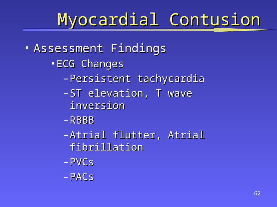
Myocardial ContusionMyocardial Contusion
• Assessment FindingsAssessment Findings• ECG ChangesECG Changes
– Persistent tachycardiaPersistent tachycardia– ST elevation, T wave inversionST elevation, T wave inversion– RBBBRBBB– Atrial flutter, Atrial fibrillationAtrial flutter, Atrial fibrillation– PVCsPVCs– PACsPACs
63
Myocardial ContusionMyocardial Contusion• ManagementManagement
– Establish airwayEstablish airway
– High concentration OHigh concentration O2 2
– IV LR/NSIV LR/NS
• Cautious fluid administration due to injured myocardiumCautious fluid administration due to injured myocardium
– ECGECG
• Standard drug therapy for arrhythmiasStandard drug therapy for arrhythmias
• 12 Lead ECG if time permits12 Lead ECG if time permits
– Consider vasopressors for hypotensionConsider vasopressors for hypotension
– Emergent TransportEmergent Transport
• Trauma CenterTrauma Center
64
Pericardial TamponadePericardial Tamponade• IncidenceIncidence
– Usually associated with penetrating traumaUsually associated with penetrating trauma– Rare in blunt traumaRare in blunt trauma– Occurs in < 2% of chest traumaOccurs in < 2% of chest trauma– GSW wounds have higher mortality than stab GSW wounds have higher mortality than stab
woundswounds– Lower mortality rate if isolated tamponadeLower mortality rate if isolated tamponade

65
Pericardial TamponadePericardial Tamponade
• PathophysiologyPathophysiology– Space normally filled with 30-50 ml of straw-Space normally filled with 30-50 ml of straw-
colored fluidcolored fluid• lubricationlubrication• lymphatic dischargelymphatic discharge• immunologic protection for the heartimmunologic protection for the heart
– Rapid accumulation of blood in the inelastic Rapid accumulation of blood in the inelastic pericardiumpericardium
66
Pericardial TamponadePericardial Tamponade• PathophysiologyPathophysiology
– Heart is compressed decreasing blood entering Heart is compressed decreasing blood entering heartheart
• Decreased diastolic expansion and fillingDecreased diastolic expansion and filling• Hindered venous return (preload)Hindered venous return (preload)
– Myocardial perfusion decreased due toMyocardial perfusion decreased due to• pressure effects on walls of heartpressure effects on walls of heart• decreased diastolic pressuresdecreased diastolic pressures
– Ischemic dysfunction may result in injuryIschemic dysfunction may result in injury– Removal of as little as 20 ml of blood may Removal of as little as 20 ml of blood may
drastically improve cardiac outputdrastically improve cardiac output
67
Pericardial TamponadePericardial Tamponade
• Signs and SymptomsSigns and Symptoms– Beck’s TriadBeck’s Triad
• Resistant hypotensionResistant hypotension• Increased central venous pressure (distended Increased central venous pressure (distended
neck/arm veins in presence of decreased neck/arm veins in presence of decreased arterial BP)arterial BP)
• Small quiet heart (decreased heart sounds)Small quiet heart (decreased heart sounds)
68
Pericardial TamponadePericardial Tamponade
• Signs and SymptomsSigns and Symptoms– Narrowing pulse pressureNarrowing pulse pressure– Pulsus paradoxicusPulsus paradoxicus
• Radial pulse becomes weak or disappears Radial pulse becomes weak or disappears when patient inhaleswhen patient inhales
• Increased intrathoracic pressure on Increased intrathoracic pressure on inhalation causes blood to be trapped in inhalation causes blood to be trapped in lungs temporarilylungs temporarily
69
Pericardial TamponadePericardial Tamponade
• ManagementManagement– Secure airwaySecure airway
– High concentration OHigh concentration O22
– PericardiocentesisPericardiocentesis
• Out of hospital, primarily reserved for cardiac arrestOut of hospital, primarily reserved for cardiac arrest
– Rapid transportRapid transport• Trauma CenterTrauma Center
– IVs of LR/NSIVs of LR/NS

70
Pericardial TamponadePericardial Tamponade
• ManagementManagement– Definite treatment is pericardiocentesis followed by Definite treatment is pericardiocentesis followed by
surgerysurgery
• Pericardial WindowPericardial Window
• Tamponade is hard to diagnosisTamponade is hard to diagnosis– Hypotension is common in chest traumaHypotension is common in chest trauma
– Heart sounds are difficult to hearHeart sounds are difficult to hear
– Bulging neck veins may be absent if hypovolemia is Bulging neck veins may be absent if hypovolemia is presentpresent
– High index of suspicion is requiredHigh index of suspicion is required
71
Traumatic Aortic DissectionTraumatic Aortic Dissection
• Caused By:Caused By:– Motor Vehicle CollisionsMotor Vehicle Collisions– Falls from heightsFalls from heights– Crushing chest traumaCrushing chest trauma– Animal KicksAnimal Kicks– Blunt chest traumaBlunt chest trauma
• 15% of all blunt trauma deaths15% of all blunt trauma deaths

72
Traumatic Aortic DissectionTraumatic Aortic Dissection
• 1 of 6 persons dying in MVC’s has aortic rupture1 of 6 persons dying in MVC’s has aortic rupture– 85% die instantaneously85% die instantaneously– 10-15% survive to hospital10-15% survive to hospital
• 1/3 die within six hours1/3 die within six hours• 1/3 die within 24 hours1/3 die within 24 hours• 1/3 survive 3 days or longer1/3 survive 3 days or longer
• MustMust have high index of suspicion have high index of suspicion
73
Traumatic Aortic DissectionTraumatic Aortic Dissection
• Separation of the aortic intima and mediaSeparation of the aortic intima and media– Tear 2° high speed deceleration at points of Tear 2° high speed deceleration at points of
relative fixationrelative fixation• Blood enters media through a small intima tearBlood enters media through a small intima tear
– Thinned layer may ruptureThinned layer may rupture• Descending aorta at the isthmus distal to left Descending aorta at the isthmus distal to left
subclavian artery most common site of rupturesubclavian artery most common site of rupture– ligamentum arteriosomligamentum arteriosom

74
Traumatic Aortic DissectionTraumatic Aortic Dissection• Assessment FindingsAssessment Findings
– Retrosternal or interscapular painRetrosternal or interscapular pain
– Pain in lower back or one legPain in lower back or one leg
– Respiratory distressRespiratory distress
– Asymmetrical arm BPsAsymmetrical arm BPs
– Upper extremity hypertension withUpper extremity hypertension with
• Decreased femoral pulses, ORDecreased femoral pulses, OR
• Absent femoral pulses Absent femoral pulses
– DysphagiaDysphagia
75
Traumatic Aortic DissectionTraumatic Aortic Dissection• ManagementManagement
– Establish airwayEstablish airway
– High concentration oxygenHigh concentration oxygen
– Maintain minimal BP in dissectionMaintain minimal BP in dissection
• IV LR/NS TKOIV LR/NS TKO
– minimize fluid administrationminimize fluid administration
• Avoid PASGAvoid PASG
– Emergent TransportEmergent Transport• Trauma CenterTrauma Center
• Vascular Surgery capabilityVascular Surgery capability

76
Traumatic AsphyxiaTraumatic Asphyxia• Name given to these patients because they looked Name given to these patients because they looked
like they had been strangled or hanged like they had been strangled or hanged
• Pathophysiology Pathophysiology – Blunt force to chest causesBlunt force to chest causes
• Increased intrathoracic pressureIncreased intrathoracic pressure• Backward flow of blood out of right heart Backward flow of blood out of right heart
into vessels of upper chest and neckinto vessels of upper chest and neck– Jugular veins engorgeJugular veins engorge– Capillaries ruptureCapillaries rupture

77
Traumatic AsphyxiaTraumatic Asphyxia
• Assessment FindingsAssessment Findings– Purplish-red discoloration of:Purplish-red discoloration of:
• Head and FaceHead and Face• NeckNeck• ShouldersShoulders
– Blood shot, protruding eyesBlood shot, protruding eyes– JVDJVD– Shock when pressure releasedShock when pressure released
78
Traumatic AsphyxiaTraumatic Asphyxia• ManagementManagement
– Airway with C-spine controlAirway with C-spine control
– Assist ventilations with high concentration OAssist ventilations with high concentration O22
– Spinal stabilizationSpinal stabilization
– IV of LRIV of LR
– Monitor EKGMonitor EKG
– ++ MAST in severely hypotensive patients MAST in severely hypotensive patients
– Rapid transportRapid transport
• Trauma CenterTrauma Center
• Consider early sodium bicarbonate in arrestConsider early sodium bicarbonate in arrest
79
Diaphragmatic RuptureDiaphragmatic Rupture
• Usually due to blunt trauma but may occur with Usually due to blunt trauma but may occur with penetrating traumapenetrating trauma
• Usually life-threateningUsually life-threatening• Likely to be associated with other severe injuriesLikely to be associated with other severe injuries

80
Diaphragmatic RuptureDiaphragmatic Rupture
• PathophysiologyPathophysiology– Compression to abdomen resulting in increased intra-Compression to abdomen resulting in increased intra-
abdominal pressureabdominal pressure
• abdominal contents rupture through diaphragm into abdominal contents rupture through diaphragm into chestchest
• bowel obstruction and strangulationbowel obstruction and strangulation
• restriction of lung expansionrestriction of lung expansion
• mediastinal shiftmediastinal shift
– 90% occur on left side due to protection of right side by 90% occur on left side due to protection of right side by liverliver

81
Diaphragmatic RuptureDiaphragmatic Rupture
• Assessment FindingsAssessment Findings– Decreased breath soundsDecreased breath sounds
• Usually unilateralUsually unilateral• Dullness to percussionDullness to percussion
– Dyspnea or Respiratory DistressDyspnea or Respiratory Distress– Scaphoid Abdomen (hollow appearance)Scaphoid Abdomen (hollow appearance)– Usually difficult to hear bowel soundsUsually difficult to hear bowel sounds
82
Diaphragmatic RuptureDiaphragmatic Rupture
• ManagementManagement– Establish airwayEstablish airway
– Assist ventilations with high concentration OAssist ventilations with high concentration O2 2
– IV of LRIV of LR
– Monitor EKGMonitor EKG
– NG tube if possibleNG tube if possible
– AvoidAvoid
• MASTMAST
• Trendelenburg positionTrendelenburg position

83
Diaphragmatic PenetrationDiaphragmatic Penetration
• Suspect intra-abdominal trauma with any Suspect intra-abdominal trauma with any injury below 4th ICSinjury below 4th ICS
• Suspect intrathoracic trauma with any Suspect intrathoracic trauma with any abdominal injury above umbilicusabdominal injury above umbilicus

84
Esophageal InjuryEsophageal Injury
• Penetrating Injury most frequent causePenetrating Injury most frequent cause
– Rare in blunt traumaRare in blunt trauma
– Can perforate spontaneouslyCan perforate spontaneously
• violent emesisviolent emesis
• carcinomacarcinoma
85
Esophageal InjuryEsophageal Injury
• Assessment FindingsAssessment Findings– Pain, local tendernessPain, local tenderness– Hoarseness, DysphagiaHoarseness, Dysphagia– Respiratory distressRespiratory distress– Resistance of neck on passive motionResistance of neck on passive motion– Mediastinal esophageal perforationMediastinal esophageal perforation
• mediastinal emphysema / mediastinal crunchmediastinal emphysema / mediastinal crunch• mediastinitismediastinitis• SQ EmphysemaSQ Emphysema• splinting of chest wallsplinting of chest wall
– ShockShock
86
Esophageal InjuryEsophageal Injury• ManagementManagement
– Establish AirwayEstablish Airway– Consider early intubation if possibleConsider early intubation if possible– IV LR/NS titrated to BP 90-100 mm HgIV LR/NS titrated to BP 90-100 mm Hg– Emergent TransportEmergent Transport
• Trauma CenterTrauma Center• Surgical capabilitySurgical capability

Tracheobronchial RuptureTracheobronchial Rupture
•Uncommon injury Uncommon injury –less than 3% of chest traumaless than 3% of chest trauma
•Occurs with penetrating or blunt chest Occurs with penetrating or blunt chest traumatrauma•High mortality rate (>30%)High mortality rate (>30%)•May involve fracture of upper 3 ribsMay involve fracture of upper 3 ribs

88
Tracheobronchial RuptureTracheobronchial Rupture• PathophysiologyPathophysiology
– Majority (80%) occur at or near carinaMajority (80%) occur at or near carina– rapid movement of air into pleural spacerapid movement of air into pleural space– Tension pneumothorax refractory to needle Tension pneumothorax refractory to needle
decompressiondecompression– continuous flow of air from needle of continuous flow of air from needle of
decompressed chestdecompressed chest

89
Tracheobronchial RuptureTracheobronchial Rupture
• Assessment FindingsAssessment Findings– Respiratory DistressRespiratory Distress
• DyspneaDyspnea
• TachypneaTachypnea
– Obvious SQ emphysemaObvious SQ emphysema
– HemoptysisHemoptysis
• Especially of bright red bloodEspecially of bright red blood
– Signs of tension pneumothorax unresponsive to needle Signs of tension pneumothorax unresponsive to needle decompressiondecompression
90
Tracheobronchial RuptureTracheobronchial Rupture
• ManagementManagement– Establish airway and ventilationsEstablish airway and ventilations– Consider early intubationConsider early intubation
• intubating right or left mainstem may be life intubating right or left mainstem may be life savingsaving
– Emergent TransportEmergent Transport• Trauma CenterTrauma Center
91
Pitfalls to AvoidPitfalls to Avoid
• Elderly do not tolerate relatively minor chest Elderly do not tolerate relatively minor chest injuriesinjuries– Anticipate progression to acute respiratory Anticipate progression to acute respiratory
insufficiencyinsufficiency• Children may sustain significant intrathoracic Children may sustain significant intrathoracic
injury w/o evidence of thoracic skeletal traumainjury w/o evidence of thoracic skeletal trauma– Maintain a high index of suspicionMaintain a high index of suspicion
92
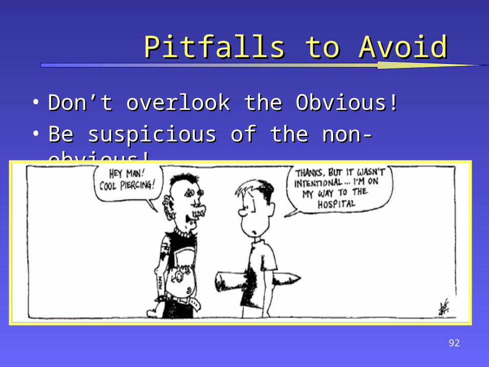
Pitfalls to AvoidPitfalls to Avoid
• Don’t overlook the Obvious!Don’t overlook the Obvious!
• Be suspicious of the non-obvious!Be suspicious of the non-obvious!

93
QUESTIONSQUESTIONS
??