thoracic trauma presentation
TRANSCRIPT
THORACIC
TRAUMA
MAZIN ERAGAT
INTRODUCTION
Thoracic trauma 25% all injury related deaths
Contributory factor in further 50% (hypoxia, hypovolaemia)
To lung and pleura low (<1%)Cardiac involvement increases by 20%!
Approx. 85% can be treated successfully without need for surgical intervention
However; important to realise that patients often have OTHER injuries (particularly to the head), which contributes to the high overall mortality
THORACIC TRAUMA
• Blunt / Penetrating / Both
• Majority may require simple procedures
(eg. Thoracostomy tube)
• Minority require urgent surgical exploration due to
bleeding
• Most life-threatening injuries can be identified in the
primary survey
• Repeated / serial examination and use of adjuncts
important, as initial normal examination does not exclude
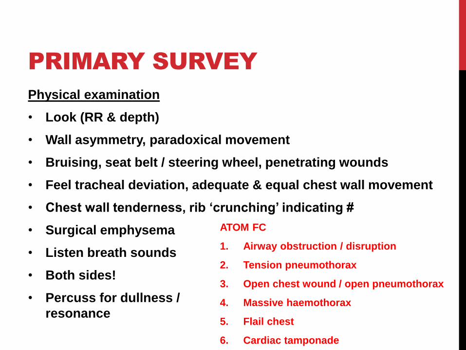
PRIMARY SURVEY
ATOM FC
1. Airway obstruction / disruption
2. Tension pneumothorax
3. Open chest wound / open pneumothorax
4. Massive haemothorax
5. Flail chest
6. Cardiac tamponade
Physical examination
• Look (RR & depth)
• Wall asymmetry, paradoxical movement
• Bruising, seat belt / steering wheel, penetrating wounds
• Feel tracheal deviation, adequate & equal chest wall movement
• Chest wall tenderness, rib ‘crunching’ indicating #
• Surgical emphysema
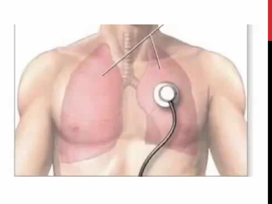
• Listen breath sounds
• Both sides!
• Percuss for dullness /
resonance
TREAT LIFE
THREATENING
INJURIES AS THEY
ARE IDENTIFIED
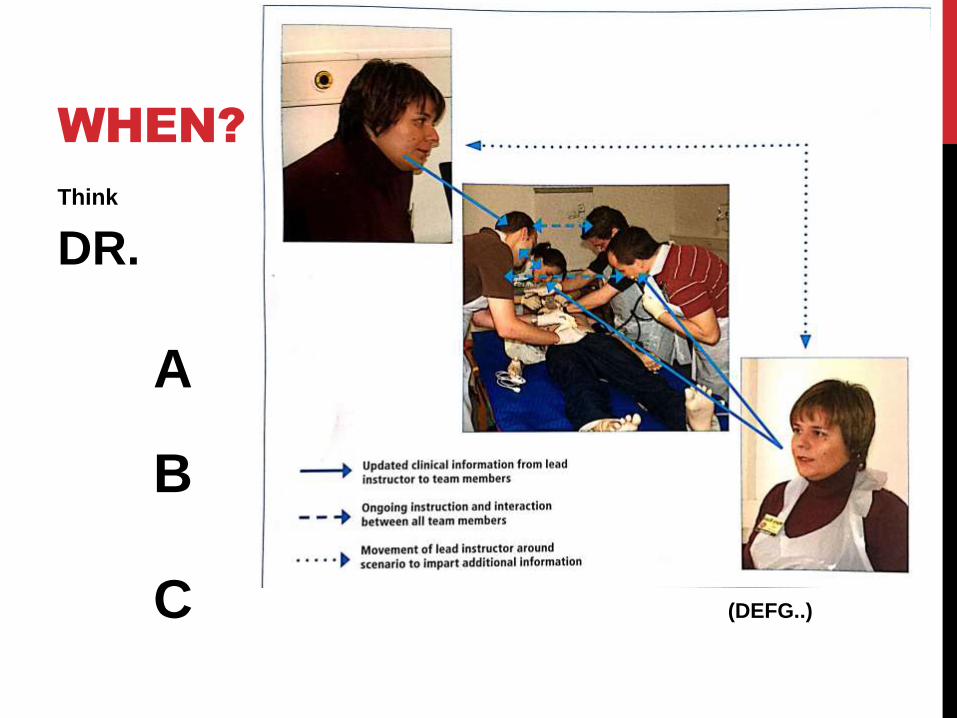
WHEN?
Think
DR.
A
B
C (DEFG..)
SECONDARY SURVEY
1. Rib # and flail chest
2. Pulmonary contusion
3. Simple pneumothorax
4. Simple haemothorax
5. Blunt aortic injury
6. Blunt myocardial injury
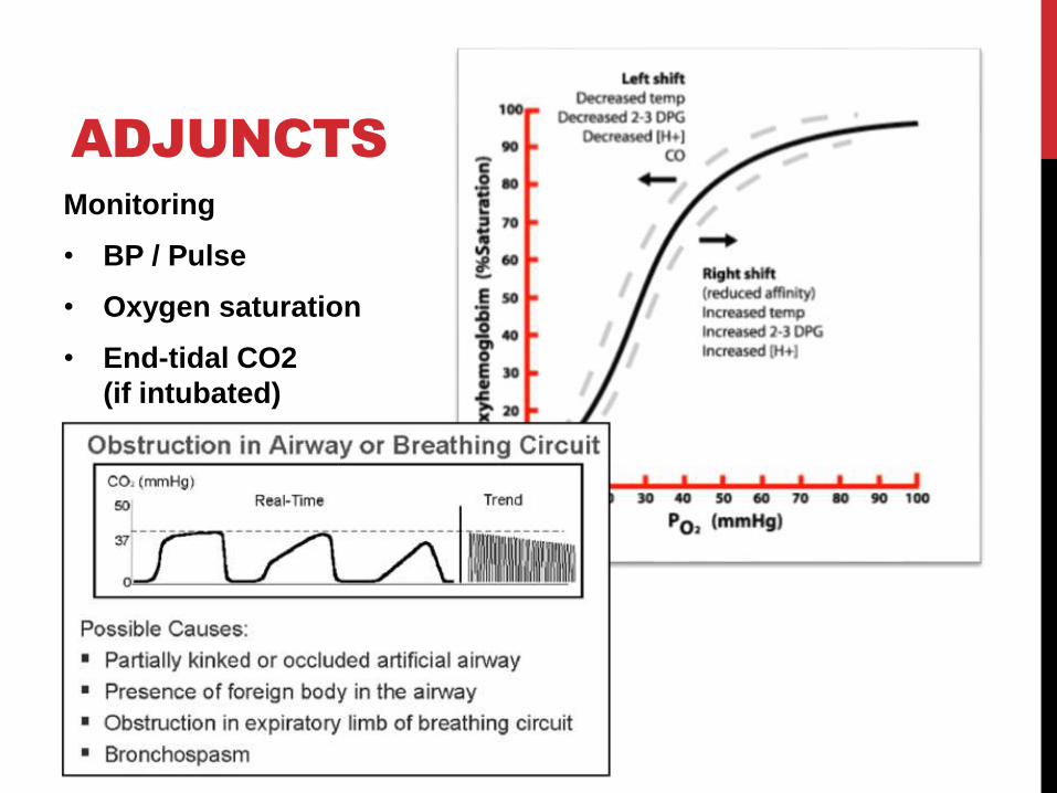
ADJUNCTS
Monitoring
• BP / Pulse
• Oxygen saturation
• End-tidal CO2
(if intubated)
ADJUNCTS
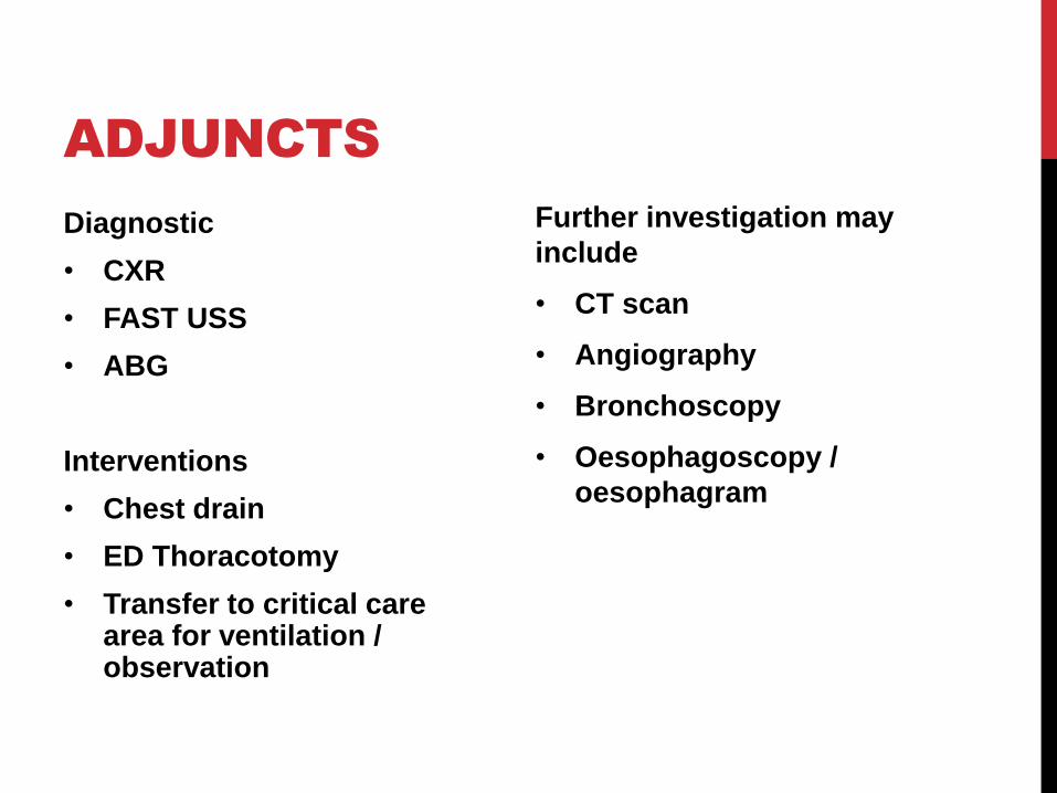
Diagnostic
• CXR
• FAST USS
• ABG
Interventions
• Chest drain
• ED Thoracotomy
• Transfer to critical care area for ventilation / observation
Further investigation may
include
• CT scan
• Angiography
• Bronchoscopy
• Oesophagoscopy /
oesophagram
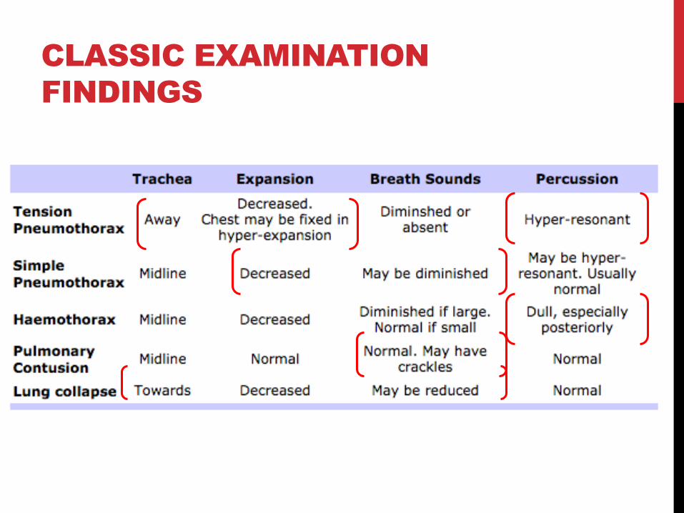
CLASSIC EXAMINATION
FINDINGS
R
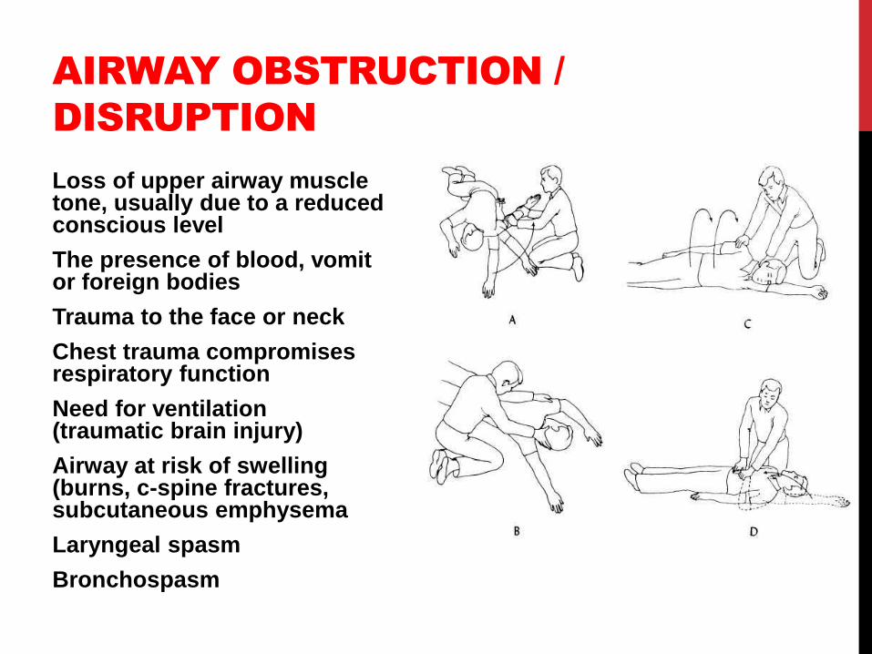
AIRWAY OBSTRUCTION /
DISRUPTION
Loss of upper airway muscle tone, usually due to a reduced conscious level
The presence of blood, vomit or foreign bodies
Trauma to the face or neck
Chest trauma compromises respiratory function
Need for ventilation (traumatic brain injury)
Airway at risk of swelling (burns, c-spine fractures, subcutaneous emphysema
Laryngeal spasm
Bronchospasm
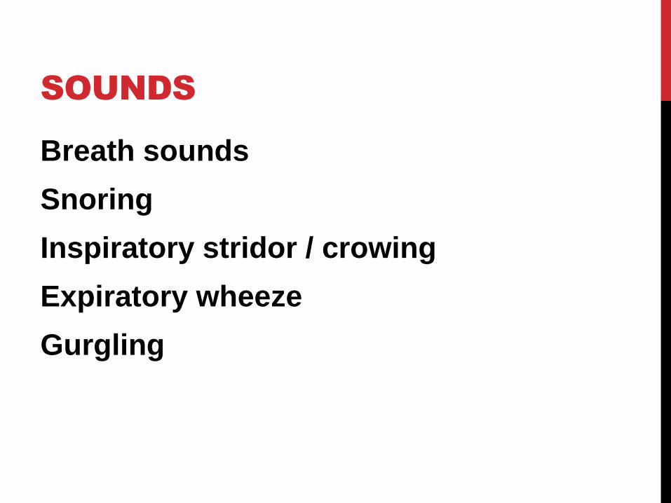
SOUNDS
Breath sounds
Snoring
Inspiratory stridor / crowing
Expiratory wheeze
Gurgling
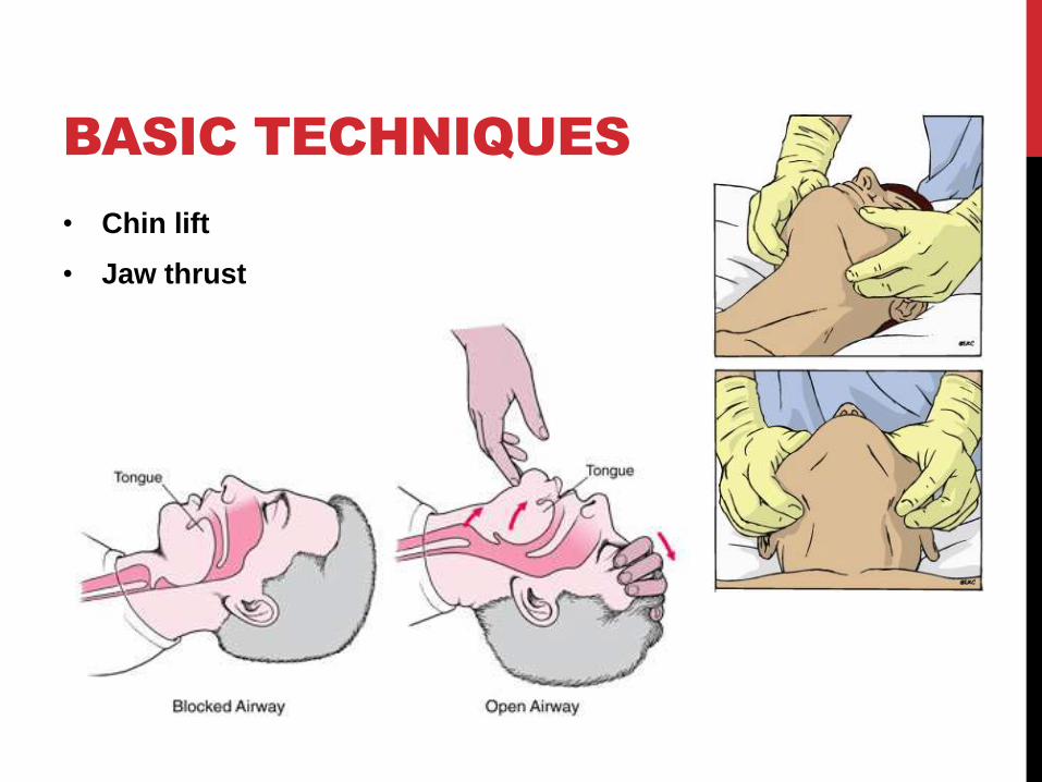
BASIC TECHNIQUES
• Chin lift
• Jaw thrust
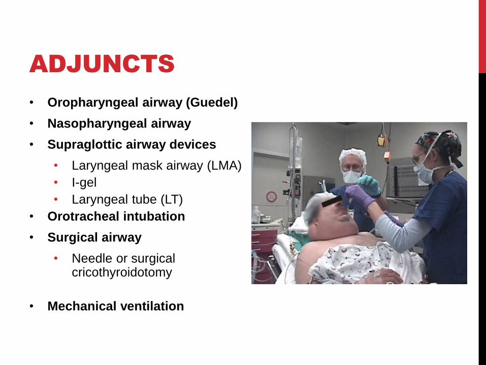
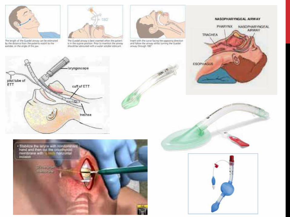
ADJUNCTS
• Oropharyngeal airway (Guedel)
• Nasopharyngeal airway
• Supraglottic airway devices
• Laryngeal mask airway (LMA)
• I-gel
• Laryngeal tube (LT)
• Orotracheal intubation
• Surgical airway
• Needle or surgical cricothyroidotomy
• Mechanical ventilation
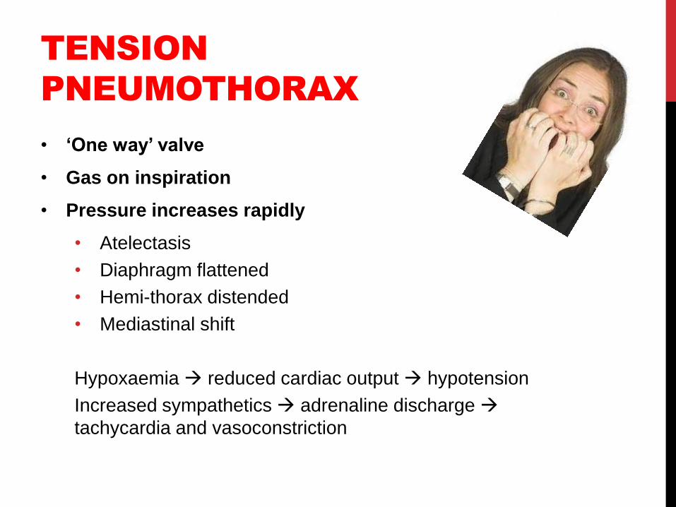
TENSION
PNEUMOTHORAX
• ‘One way’ valve
• Gas on inspiration
• Pressure increases rapidly
• Atelectasis
• Diaphragm flattened
• Hemi-thorax distended
• Mediastinal shift
Hypoxaemia reduced cardiac output hypotension
Increased sympathetics adrenaline discharge
tachycardia and vasoconstriction
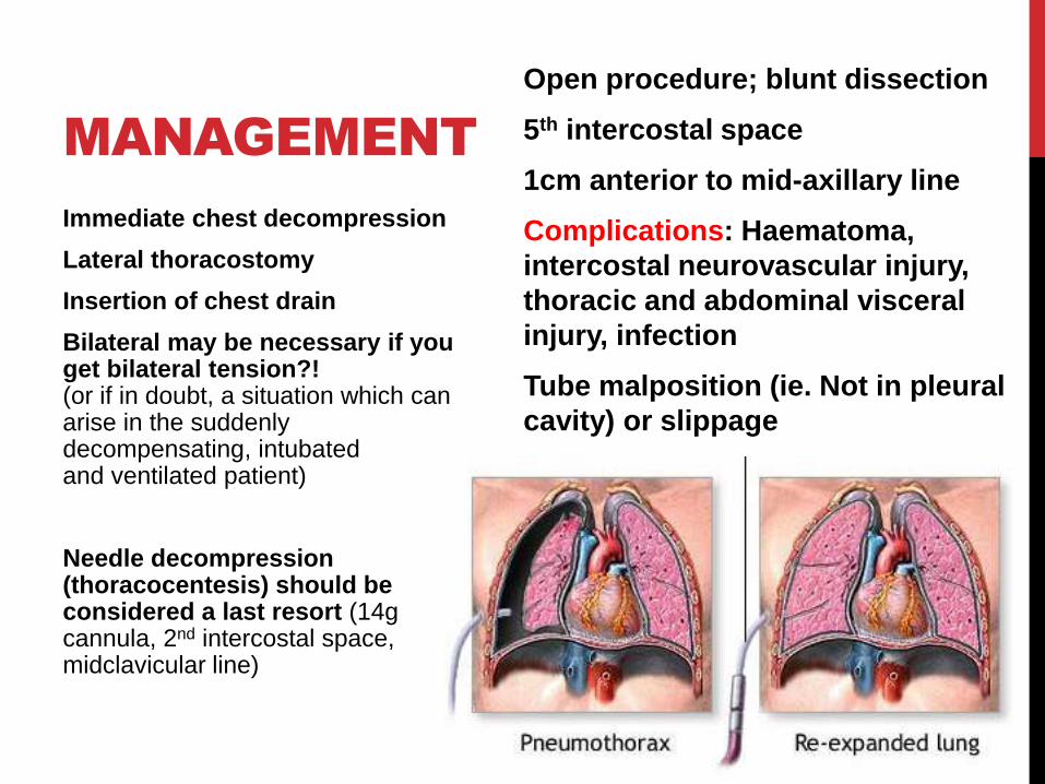
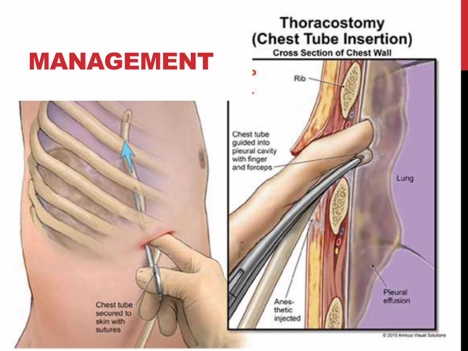
MANAGEMENT
Immediate chest decompression
Lateral thoracostomy
Insertion of chest drain
Bilateral may be necessary if you get bilateral tension?! (or if in doubt, a situation which can arise in the suddenly decompensating, intubated and ventilated patient)
Needle decompression (thoracocentesis) should be considered a last resort (14g cannula, 2nd intercostal space, midclavicular line)
Open procedure; blunt dissection
5th intercostal space
1cm anterior to mid-axillary line
Complications: Haematoma,
intercostal neurovascular injury,
thoracic and abdominal visceral
injury, infection
Tube malposition (ie. Not in pleural
cavity) or slippage
MANAGEMENT
OPEN CHEST WOUND /
OPEN PNEUMOTHORAX
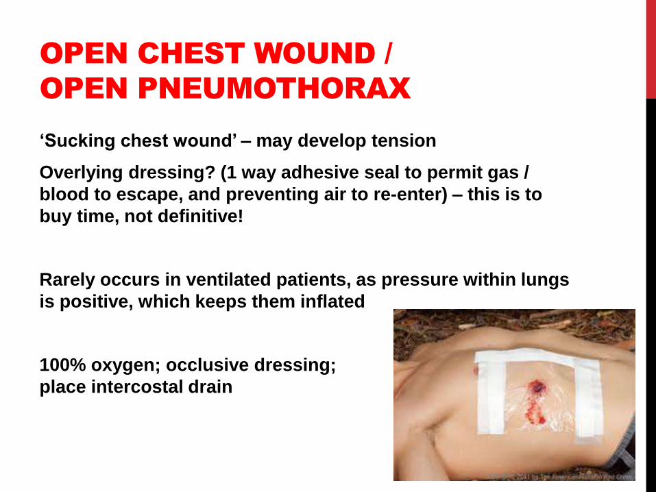
‘Sucking chest wound’ – may develop tension
Overlying dressing? (1 way adhesive seal to permit gas /
blood to escape, and preventing air to re-enter) – this is to
buy time, not definitive!
Rarely occurs in ventilated patients, as pressure within lungs
is positive, which keeps them inflated
100% oxygen; occlusive dressing;
place intercostal drain
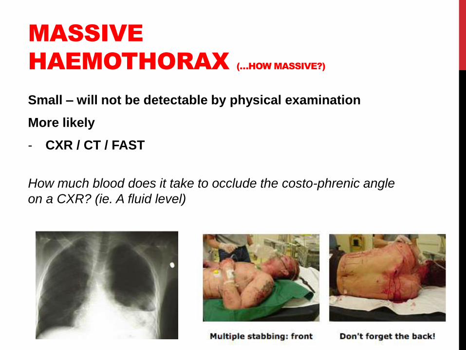
MASSIVE
HAEMOTHORAX (…HOW MASSIVE?)
Small – will not be detectable by physical examination
More likely
- CXR / CT / FAST
How much blood does it take to occlude the costo-phrenic angle
on a CXR? (ie. A fluid level)
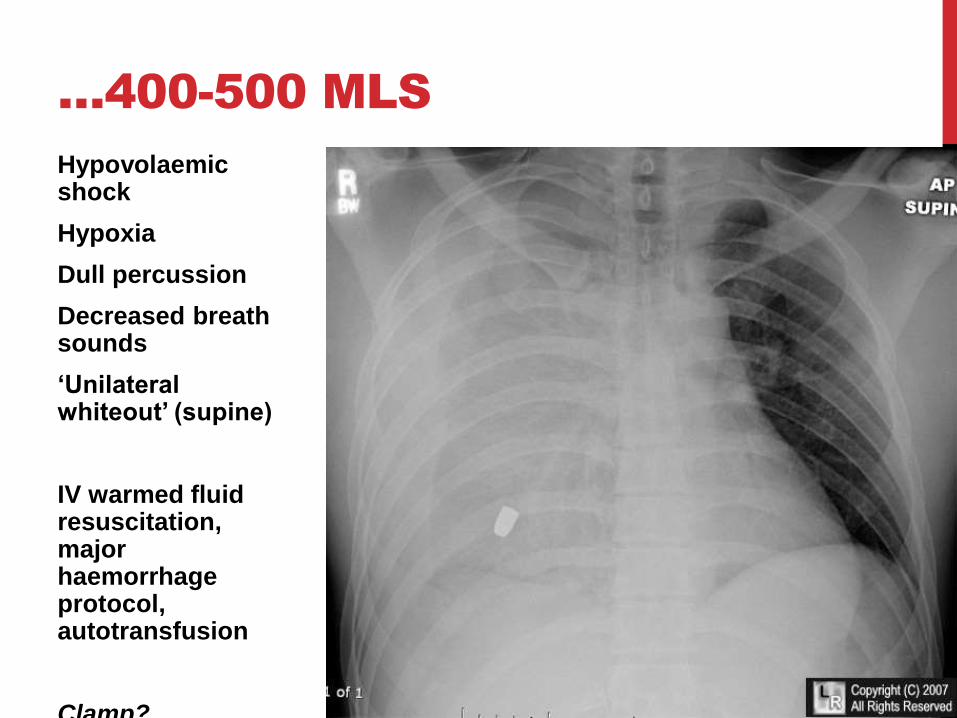
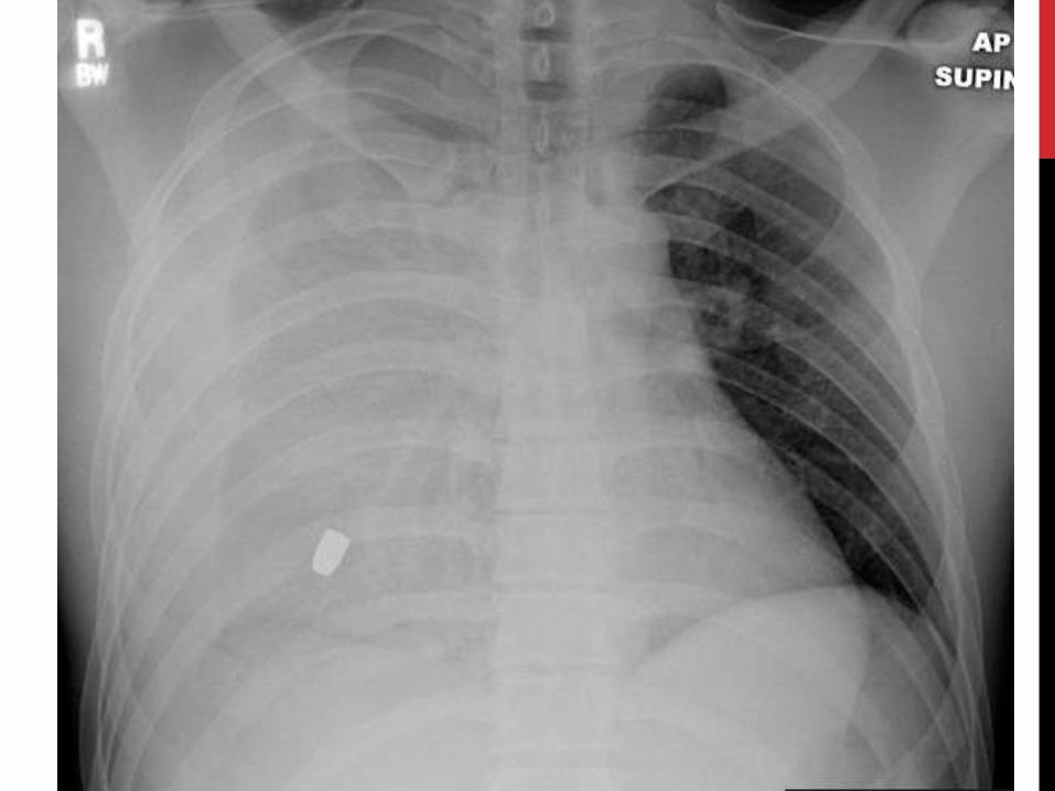
…400-500 MLS
Hypovolaemic shock
Hypoxia
Dull percussion
Decreased breath sounds
‘Unilateral whiteout’ (supine)
IV warmed fluid resuscitation, major haemorrhage protocol, autotransfusion
Clamp?
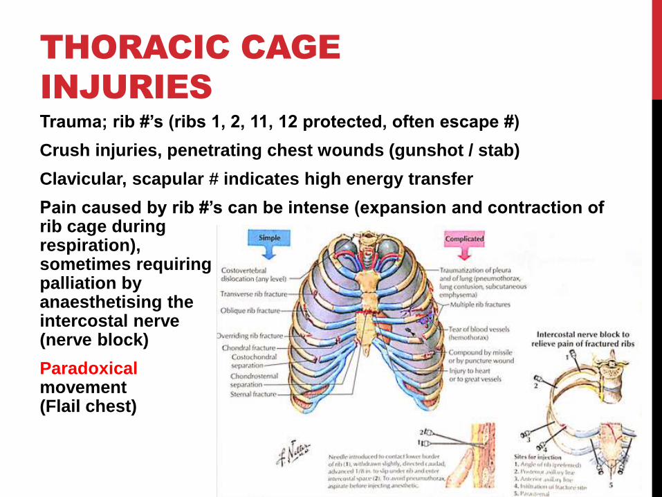
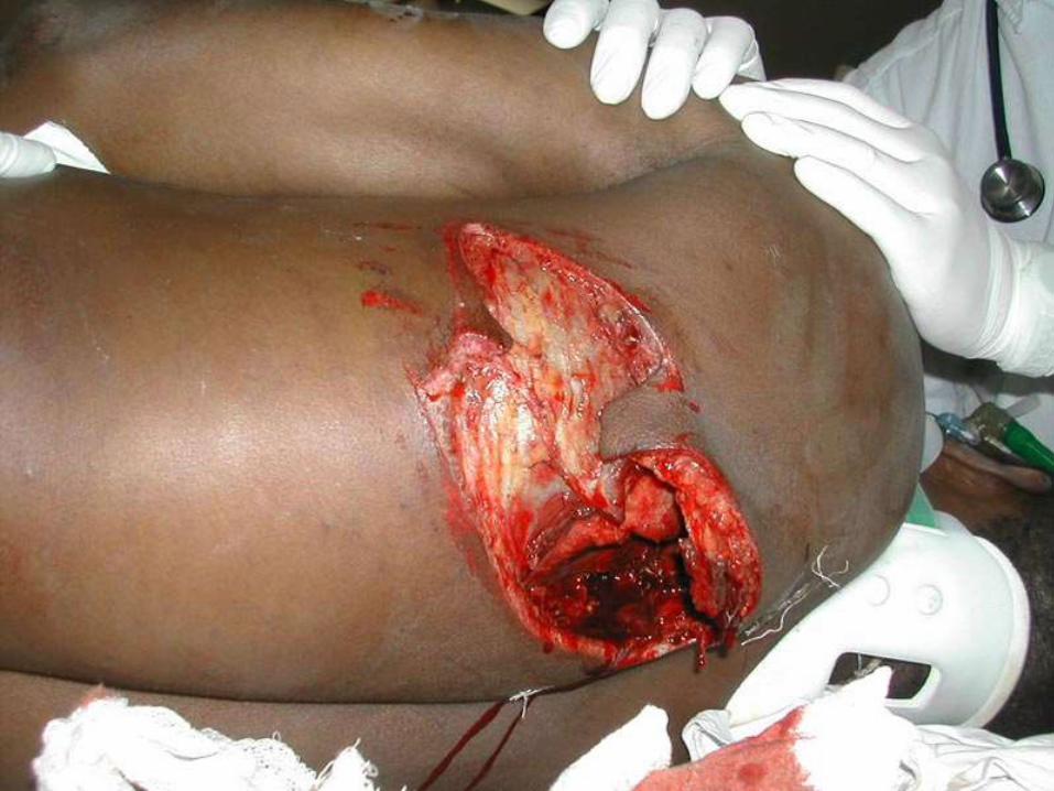
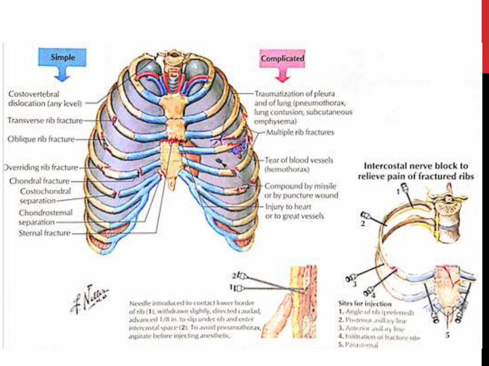
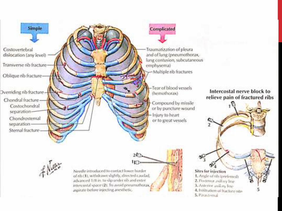
THORACIC CAGE
INJURIES
Trauma; rib #’s (ribs 1, 2, 11, 12 protected, often escape #)
Crush injuries, penetrating chest wounds (gunshot / stab)
Clavicular, scapular # indicates high energy transfer
Pain caused by rib #’s can be intense (expansion and contraction of rib cage during respiration), sometimes requiring palliation by anaesthetising the intercostal nerve (nerve block)
Paradoxicalmovement (Flail chest)
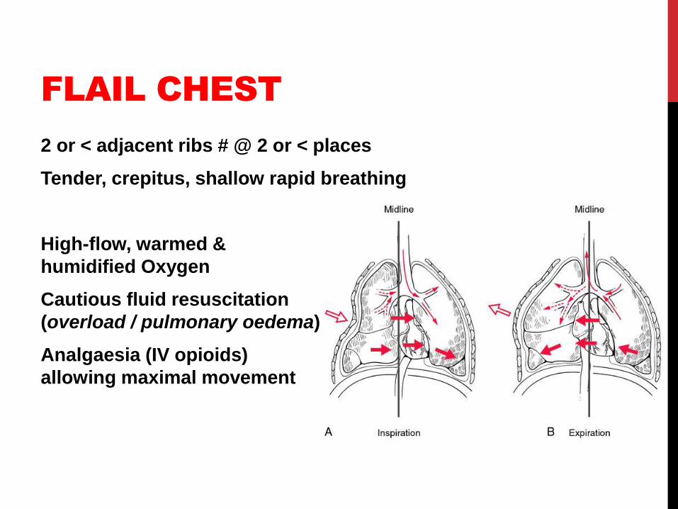
FLAIL CHEST
2 or < adjacent ribs # @ 2 or < places
Tender, crepitus, shallow rapid breathing
High-flow, warmed &
humidified Oxygen
Cautious fluid resuscitation
(overload / pulmonary oedema)
Analgaesia (IV opioids)
allowing maximal movement
INDICATIONS
For tracheal intubation and ventilation in cases of flail chest:
Oxygen
• Falling PaO2 or PaO2 <7 kPa (55 mmHg) breathing air
• PaO2 <10 kPa (75 mmHg) on high flow oxygen
Carbon dioxide
• Increasing PaCO2 or >6 kPa (45 mmHg)
Respiratory rate
• Exhaustion (RR <8 r/min)
• Tachypnoea (RR >30 r/min)
• Associated injuries compromising ventilation
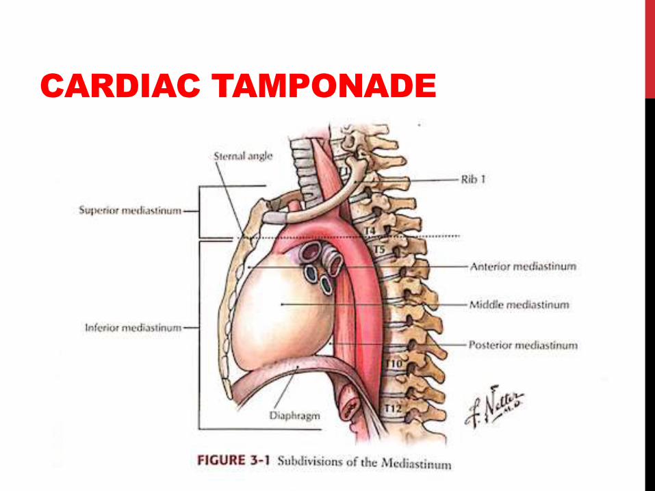
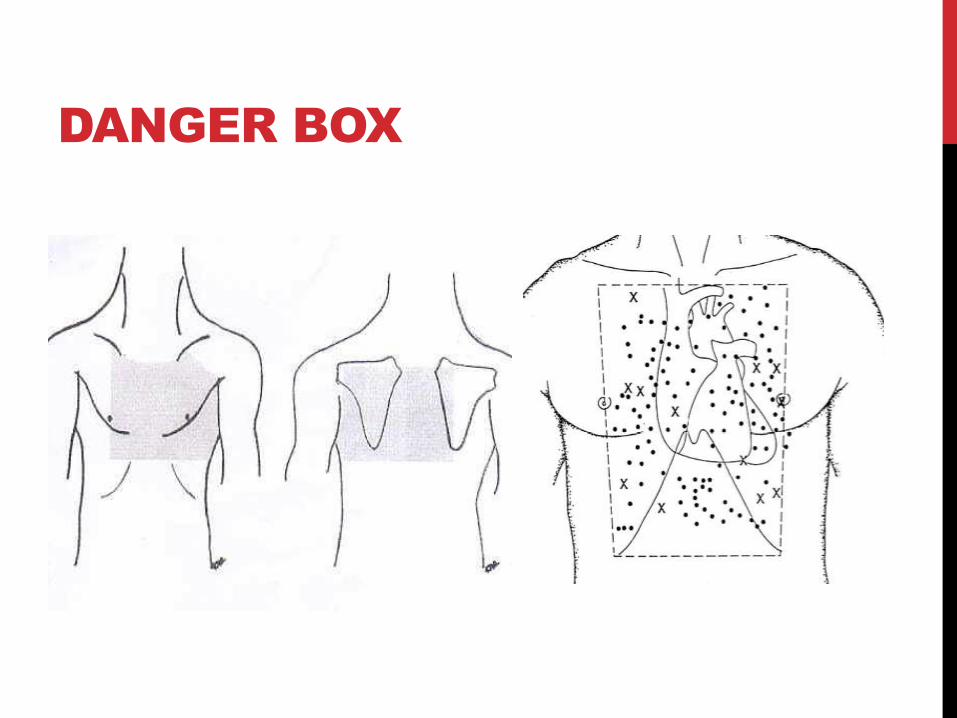
CARDIAC TAMPONADE
DANGER BOX
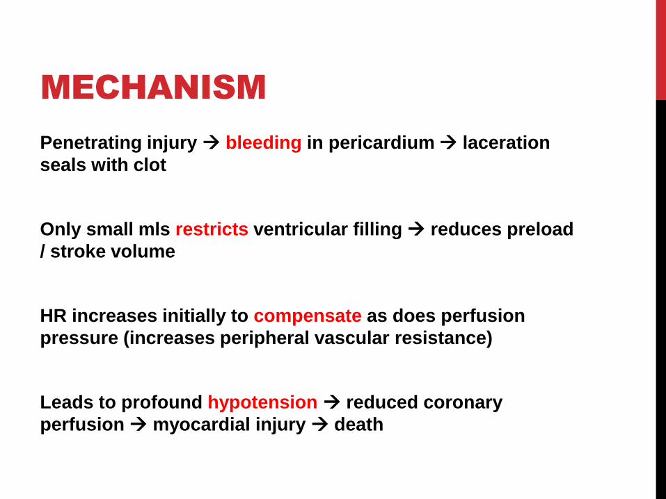
MECHANISM
Penetrating injury bleeding in pericardium laceration
seals with clot
Only small mls restricts ventricular filling reduces preload
/ stroke volume
HR increases initially to compensate as does perfusion
pressure (increases peripheral vascular resistance)
Leads to profound hypotension reduced coronary
perfusion myocardial injury death
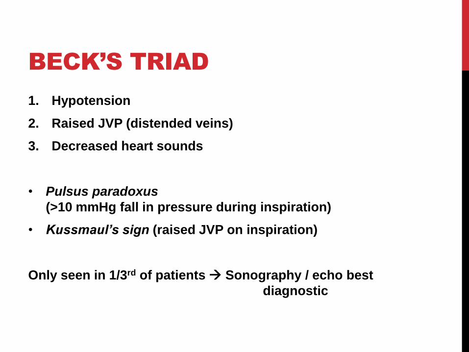
BECK’S TRIAD
1. Hypotension
2. Raised JVP (distended veins)
3. Decreased heart sounds
• Pulsus paradoxus
(>10 mmHg fall in pressure during inspiration)
• Kussmaul’s sign (raised JVP on inspiration)
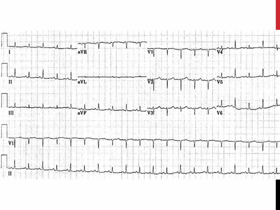
Only seen in 1/3rd of patients Sonography / echo best
diagnostic
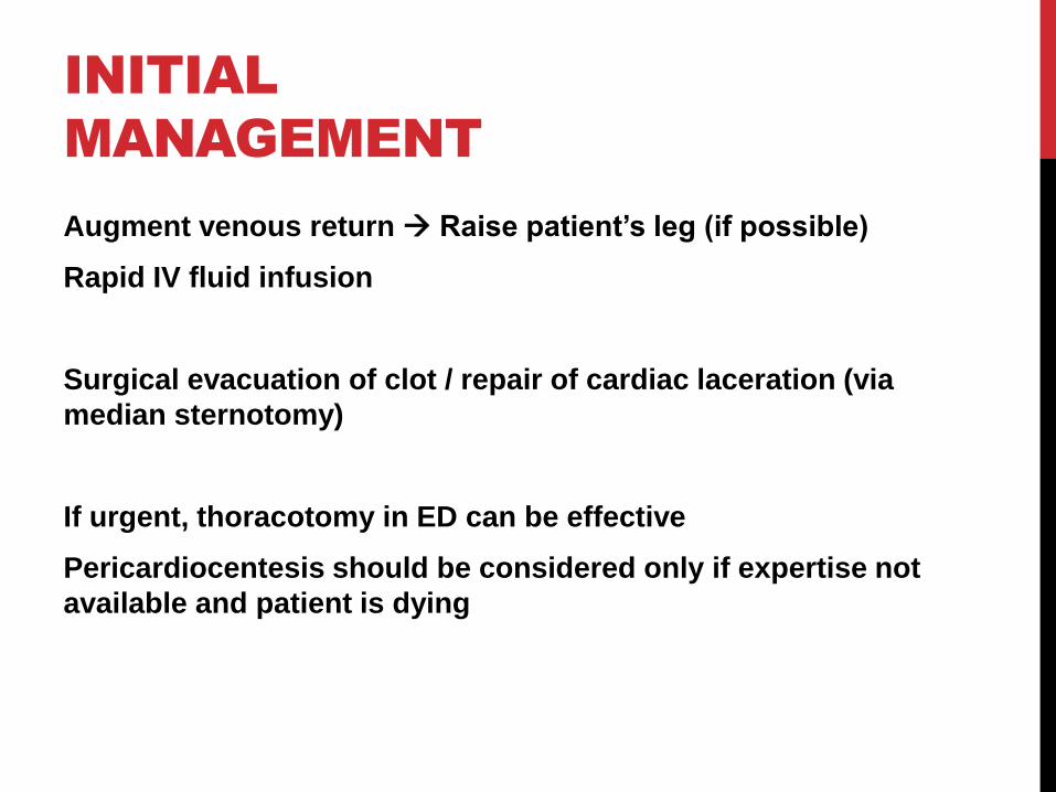
INITIAL
MANAGEMENT
Augment venous return Raise patient’s leg (if possible)
Rapid IV fluid infusion
Surgical evacuation of clot / repair of cardiac laceration (via
median sternotomy)
If urgent, thoracotomy in ED can be effective
Pericardiocentesis should be considered only if expertise not
available and patient is dying
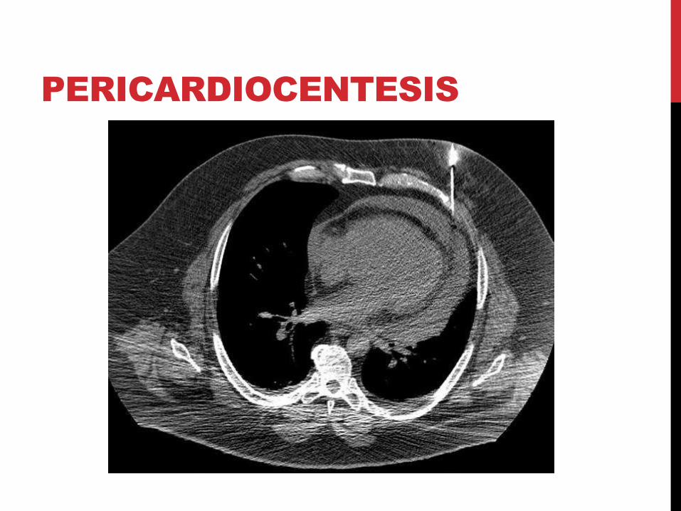
PERICARDIOCENTESIS
TRAUMATIC
ASPHYXIA
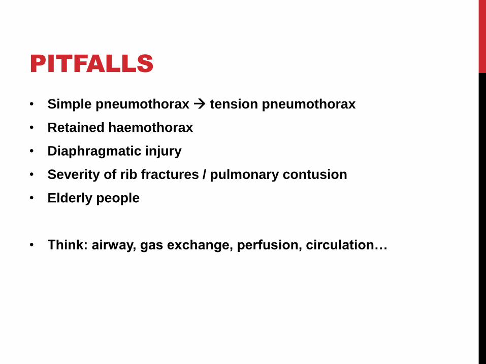
PITFALLS
• Simple pneumothorax tension pneumothorax
• Retained haemothorax
• Diaphragmatic injury
• Severity of rib fractures / pulmonary contusion
• Elderly people
• Think: airway, gas exchange, perfusion, circulation…
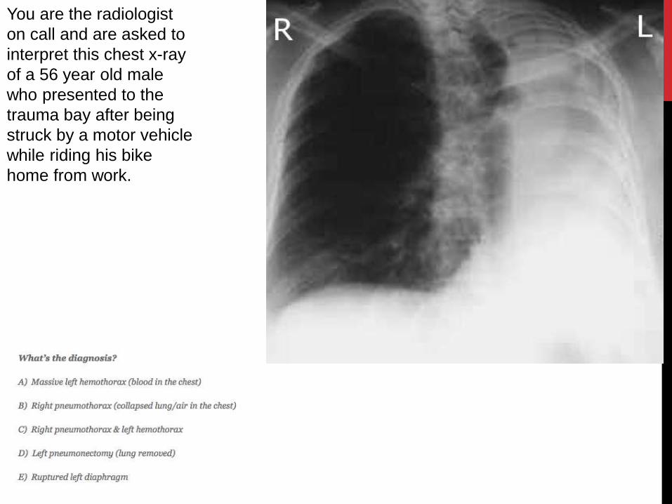
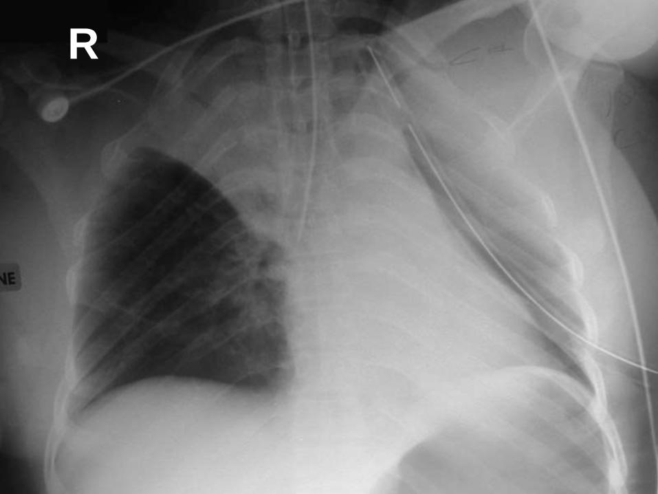
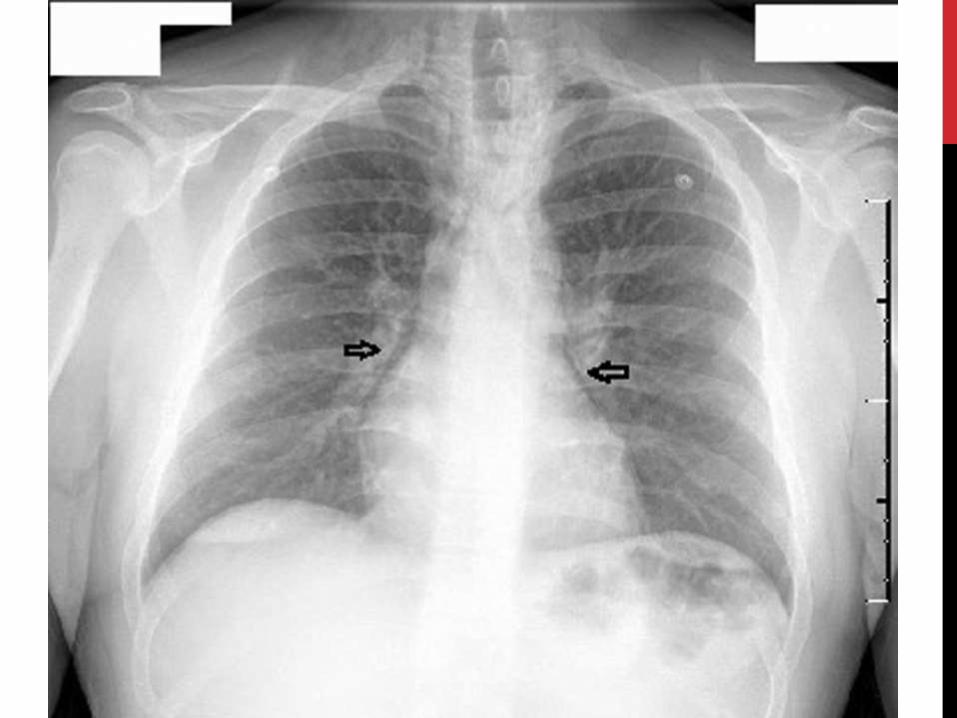
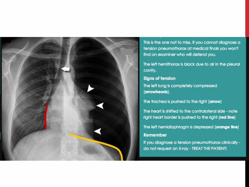
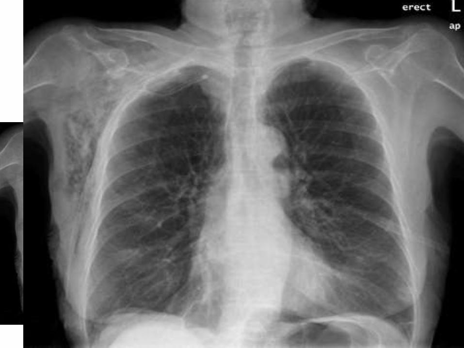
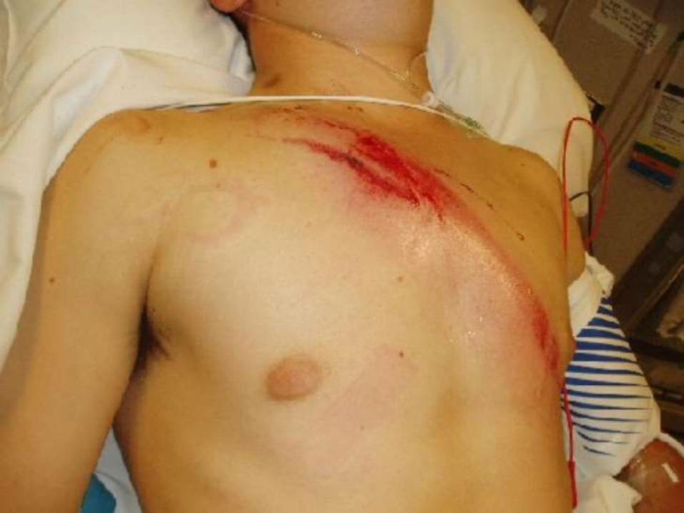
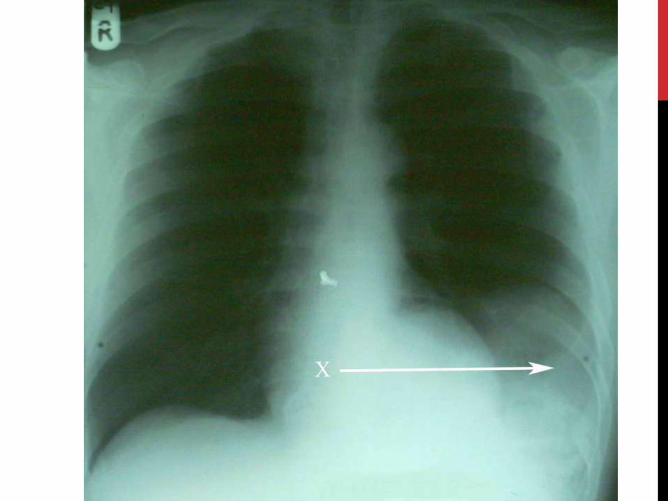
You are the radiologist
on call and are asked to
interpret this chest x-ray
of a 56 year old male
who presented to the
trauma bay after being
struck by a motor vehicle
while riding his bike
home from work.
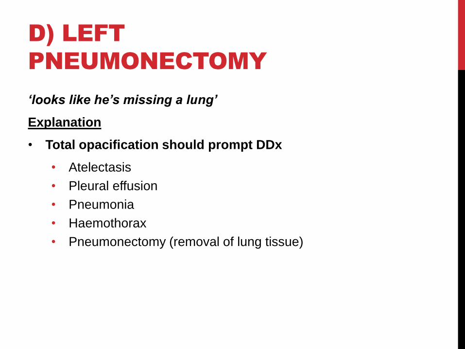
D) LEFT
PNEUMONECTOMY
‘looks like he’s missing a lung’
Explanation
• Total opacification should prompt DDx
• Atelectasis
• Pleural effusion
• Pneumonia
• Haemothorax
• Pneumonectomy (removal of lung tissue)
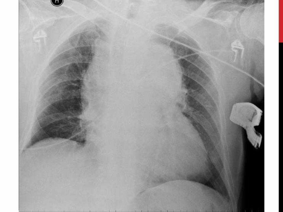
WHAT ELSE
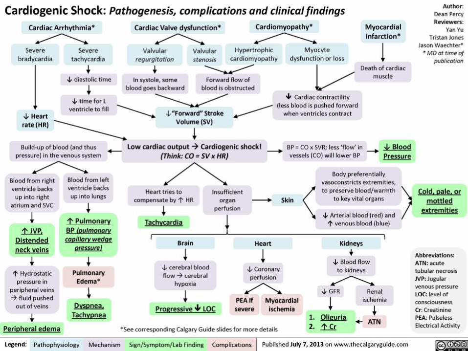
Pulmonary contusion / laceration
Blunt cardiac injury
Pneumomediastinum
Ruptured diaphragm
Traumatic disruption of aorta
Ruptured oesophagus
TOPICS
1. Cardiothoracic trauma
2. Chest wall trauma
3. Lung injuries
4. Tracheobronchial injuries
5. Diaphragmatic injuries
6. Blunt injuries to the heart
7. Trauma to great vessels
8. Oesophageal and thoracic duct injuries

QUESTIONS