imaging in thoracic trauma
TRANSCRIPT
Chest trauma
• Usually blunt, 90%
• Blunt thoracic injuries are caused by motor vehicle crashes in 63-78% of cases
• 33% of thoracic injuries require hospital admission
• Blunt chest trauma is responsible for 25% of all trauma deaths, and major contributor in another 50% of deaths
• Chest trauma is the second most common cause of death following head trauma.
EPIDEMIOLOGY OF THORACIC INJURIES IN BLUNT CHEST TRAUMA
• CHEST WALL 45%-67% • LUNG 26%-65% • HEMOTHORAX 25% • PNEUMOTHORAX 20%-30% • FLAIL CHEST 26% • HEART 5%-9% • DIAPHRAGM 7%-9% • AORTA AND GREAT VESSELS 4% • ESOPHAGUS 0,5% • MISCELLANEA 21%
North America Trauma Outcome Study-ACSCT, 1986
Devitt J et al. Can J Anaesth 1991; 38:506–510.
TRAUMA: PRIMARY SURVEY
• establishing a patent airway with cervical spine control
• adequate ventilation
• maintaining circulation (including cardiac function and intravascular volume)
• assessing the global neurologic status
• measuring the core temperature
Advanced Trauma Life Support Manual. Chicago, American College of Surgeons, 2010,
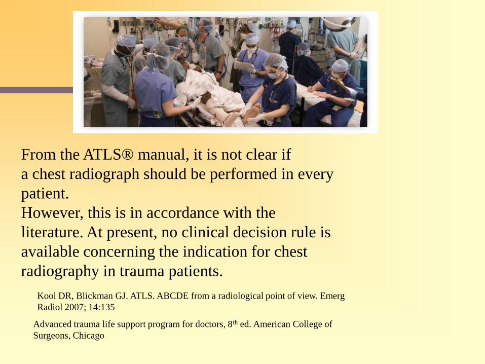
From the ATLS® manual, it is not clear if
a chest radiograph should be performed in every
patient.
However, this is in accordance with the
literature. At present, no clinical decision rule is
available concerning the indication for chest
radiography in trauma patients.
Advanced trauma life support program for doctors, 8th ed. American College of
Surgeons, Chicago
Kool DR, Blickman GJ. ATLS. ABCDE from a radiological point of view. Emerg
Radiol 2007; 14:135
Chest X-Ray in trauma?
• …is the single most valuable diagnostic study in chest trauma
• …performed early in the ATLS
• …is often done using portable x-ray equipment.
• …is taken under adverse conditions in trauma
• …It may be a suboptimal projection (supine patient!) and can be limited in penetration
• …in critically ill patients has shown low sensitivity and specificity
• …may not point out or underestimate even life-threatening lesions.
Livingston DH et al. In: Trauma. (Moore EE,
Feliciano DV et al eds.), 2008Mc Graw Hill. NY
Imaging of Thoracic Injuries In: Marincek B,
Dondelinger RF (Eds.), Emergency Radiology, 2007,
Springer, Berlin.
Other questions……..
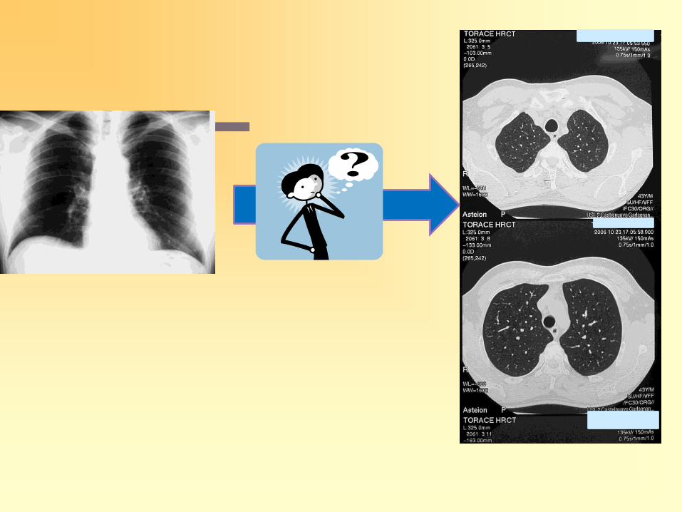
• Selective use of chest x Ray?
• Chest Xray or CT?
• Selective use of CT?
• What selective criteria for CT?
• ….. And for chest x Ray?
• Always CT?
• …..and so on……
Thoracic occult injuries
• Pneumothorax
• Hemothorax
• Pulmonary contusions
• Vascular injuries
• Cardiac injuries
• Diaphragmatic injuries
• They are injuries not suspected on the basis of clinical examination or plain radiography, but are ultimately detected with CT
Wall SD, Federle MP, Jeffrey RB, et al. AJR Am J Roentgenol
1983;141:919-21.
Tocino IM, Miller MH, Frederick PR, et al. AJR Am J Roentgenol
1984;143:987-90
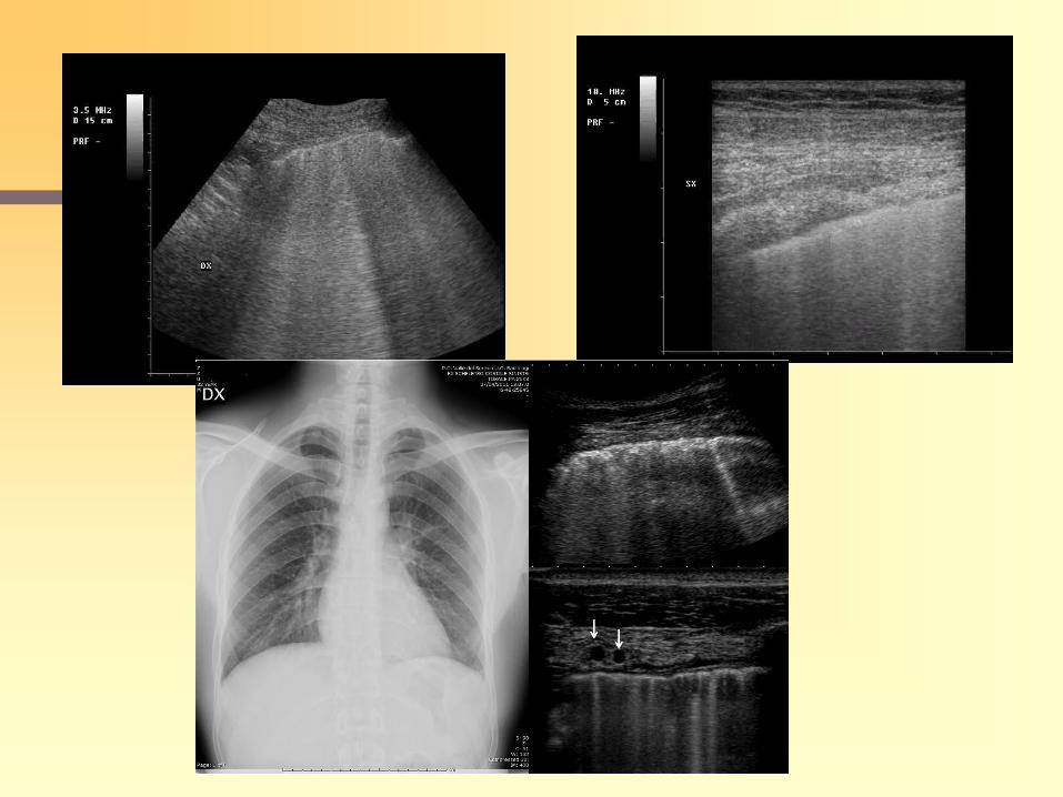
Chest sonography in trauma?
• Lung ultrasonography could be the solution between a low sensibility technique as chest Rx and the reference standard technique of CT scan for the study of trauma patients in the ED
Soldati G, Testa A et al Chest 2008
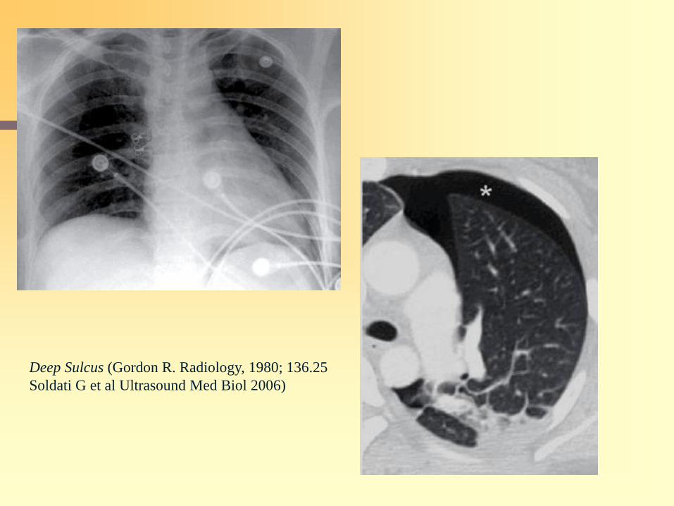
Deep Sulcus (Gordon R. Radiology, 1980; 136.25
Soldati G et al Ultrasound Med Biol 2006)
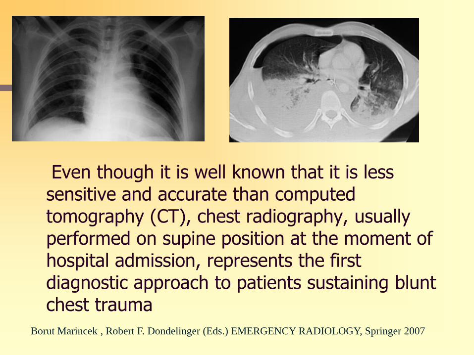
Even though it is well known that it is less sensitive and accurate than computed tomography (CT), chest radiography, usually performed on supine position at the moment of hospital admission, represents the first diagnostic approach to patients sustaining blunt chest trauma
Borut Marincek , Robert F. Dondelinger (Eds.) EMERGENCY RADIOLOGY, Springer 2007
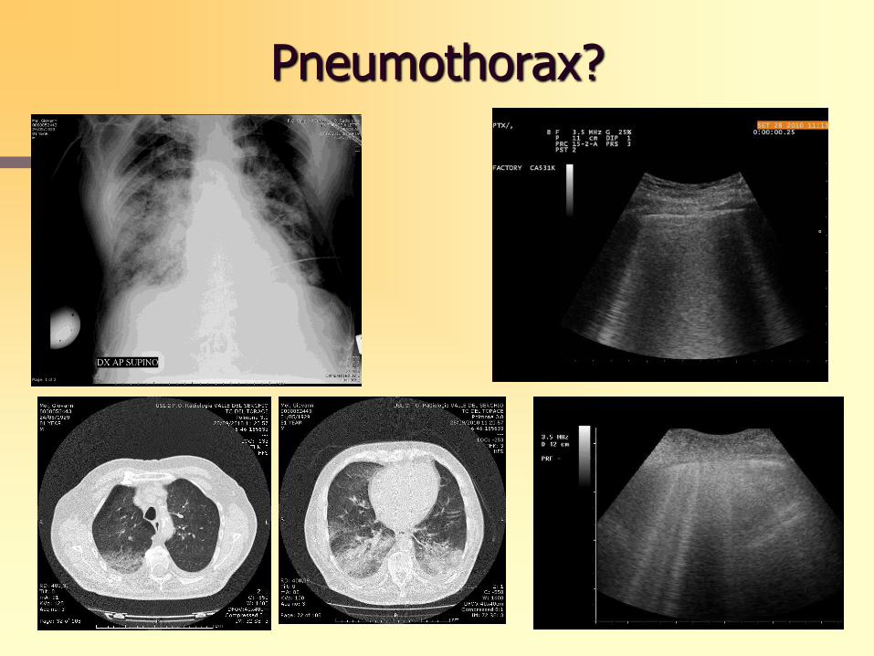
Pneumothorax?

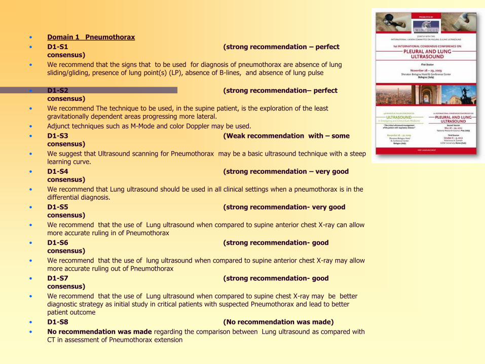
• Domain 1 Pneumothorax
• D1-S1 (strong recommendation – perfect consensus)
• We recommend that the signs that to be used for diagnosis of pneumothorax are absence of lung sliding/gliding, presence of lung point(s) (LP), absence of B-lines, and absence of lung pulse
• D1-S2 (strong recommendation– perfect consensus)
• We recommend The technique to be used, in the supine patient, is the exploration of the least gravitationally dependent areas progressing more lateral.
• Adjunct techniques such as M-Mode and color Doppler may be used.
• D1-S3 (Weak recommendation with – some consensus)
• We suggest that Ultrasound scanning for Pneumothorax may be a basic ultrasound technique with a steep learning curve.
• D1-S4 (strong recommendation – very good consensus)
• We recommend that Lung ultrasound should be used in all clinical settings when a pneumothorax is in the differential diagnosis.
• D1-S5 (strong recommendation- very good consensus)
• We recommend that the use of Lung ultrasound when compared to supine anterior chest X-ray can allow more accurate ruling in of Pneumothorax
• D1-S6 (strong recommendation- good consensus)
• We recommend that the use of lung ultrasound when compared to supine anterior chest X-ray may allow more accurate ruling out of Pneumothorax
• D1-S7 (strong recommendation- good consensus)
• We recommend that the use of Lung ultrasound when compared to supine chest X-ray may be better diagnostic strategy as initial study in critical patients with suspected Pneumothorax and lead to better patient outcome
• D1-S8 (No recommendation was made)
• No recommendation was made regarding the comparison between Lung ultrasound as compared with CT in assessment of Pneumothorax extension
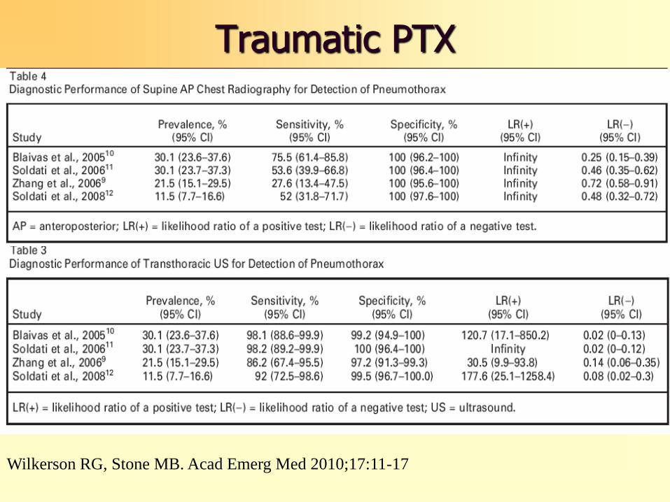
Traumatic PTX
Wilkerson RG, Stone MB. Acad Emerg Med 2010;17:11-17
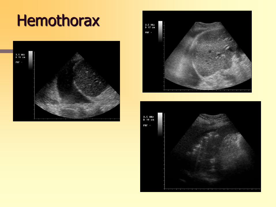
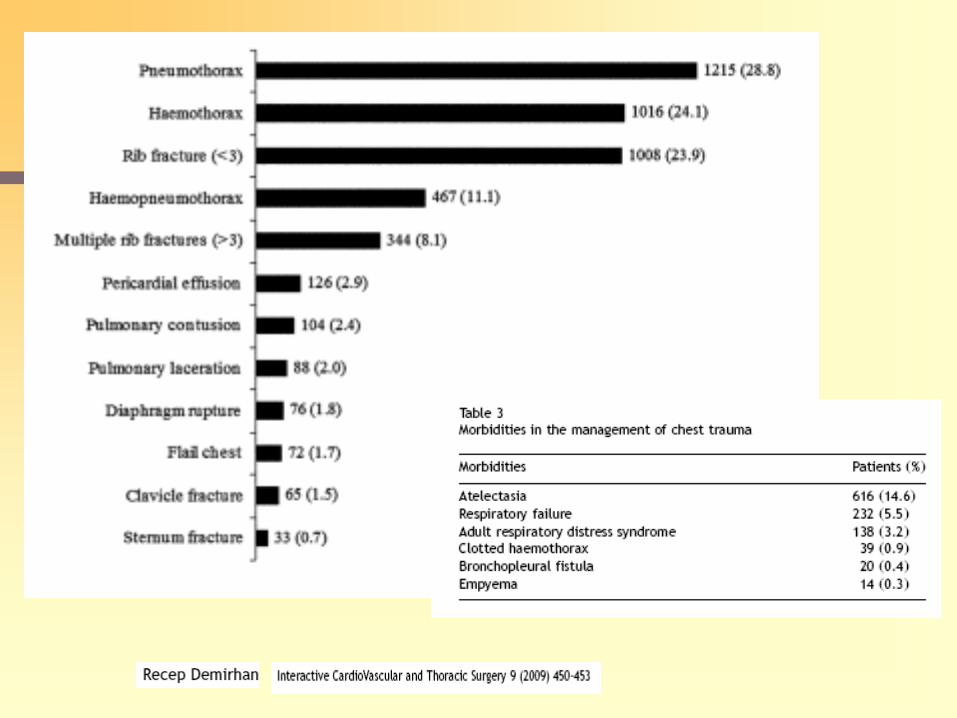
Hemothorax
one-third of patients fail to
demonstrate a lesion consistent
with this diagnosis on the initial
chest radiograph
the mean time to
opacification is 6 hours, it may take
up to 48 hours
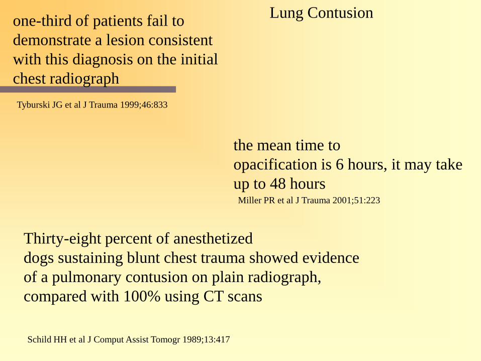
Thirty-eight percent of anesthetized
dogs sustaining blunt chest trauma showed evidence
of a pulmonary contusion on plain radiograph,
compared with 100% using CT scans
Tyburski JG et al J Trauma 1999;46:833
Miller PR et al J Trauma 2001;51:223
Schild HH et al J Comput Assist Tomogr 1989;13:417
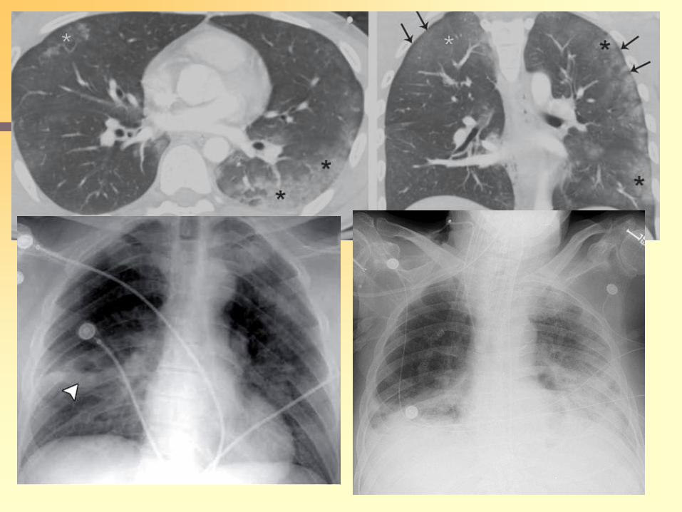
Lung Contusion
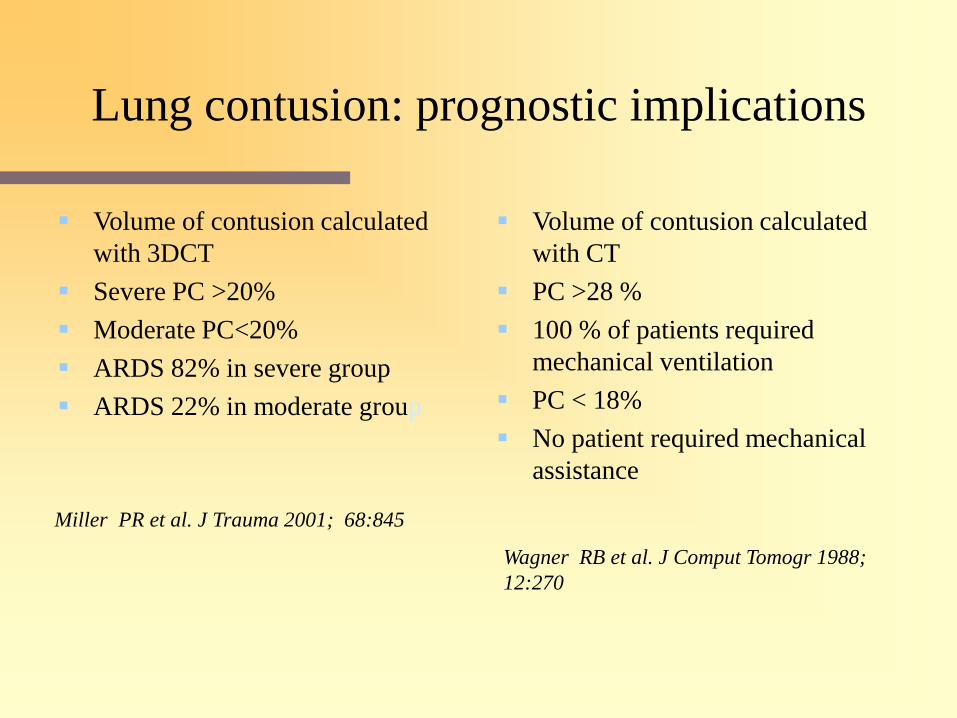
Lung contusion: prognostic implications
Volume of contusion calculated
with 3DCT
Severe PC >20%
Moderate PC<20%
ARDS 82% in severe group
ARDS 22% in moderate group
Volume of contusion calculated
with CT
PC >28 %
100 % of patients required
mechanical ventilation
PC < 18%
No patient required mechanical
assistance
Miller PR et al. J Trauma 2001; 68:845
Wagner RB et al. J Comput Tomogr 1988;
12:270
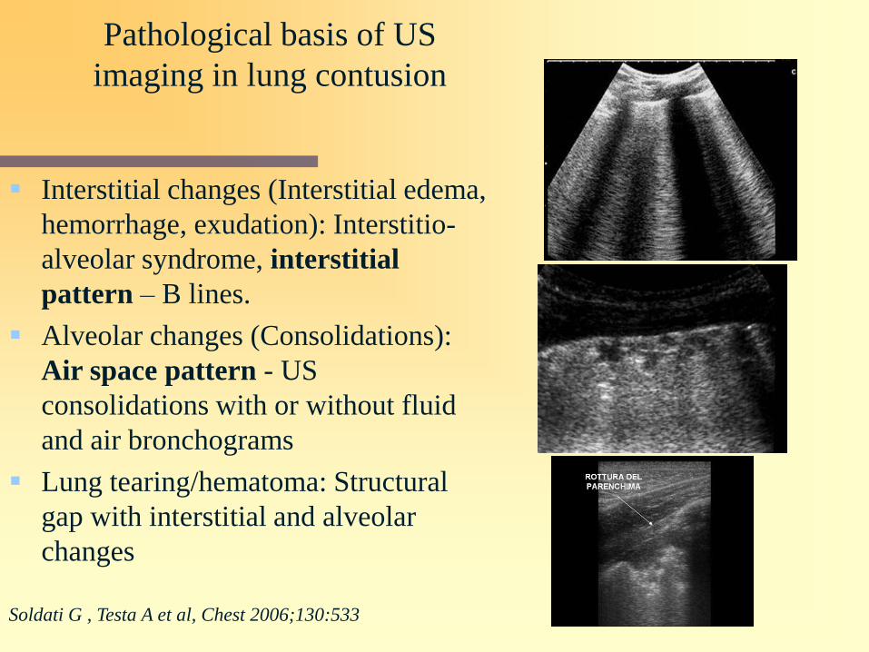
Pathological basis of US
imaging in lung contusion
Interstitial changes (Interstitial edema,
hemorrhage, exudation): Interstitio-
alveolar syndrome, interstitial
pattern – B lines.
Alveolar changes (Consolidations):
Air space pattern - US
consolidations with or without fluid
and air bronchograms
Lung tearing/hematoma: Structural
gap with interstitial and alveolar
changes
Soldati G , Testa A et al, Chest 2006;130:533
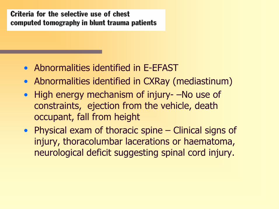
• Abnormalities identified in E-EFAST
• Abnormalities identified in CXRay (mediastinum)
• High energy mechanism of injury- –No use of constraints, ejection from the vehicle, death occupant, fall from height
• Physical exam of thoracic spine – Clinical signs of injury, thoracolumbar lacerations or haematoma, neurological deficit suggesting spinal cord injury.
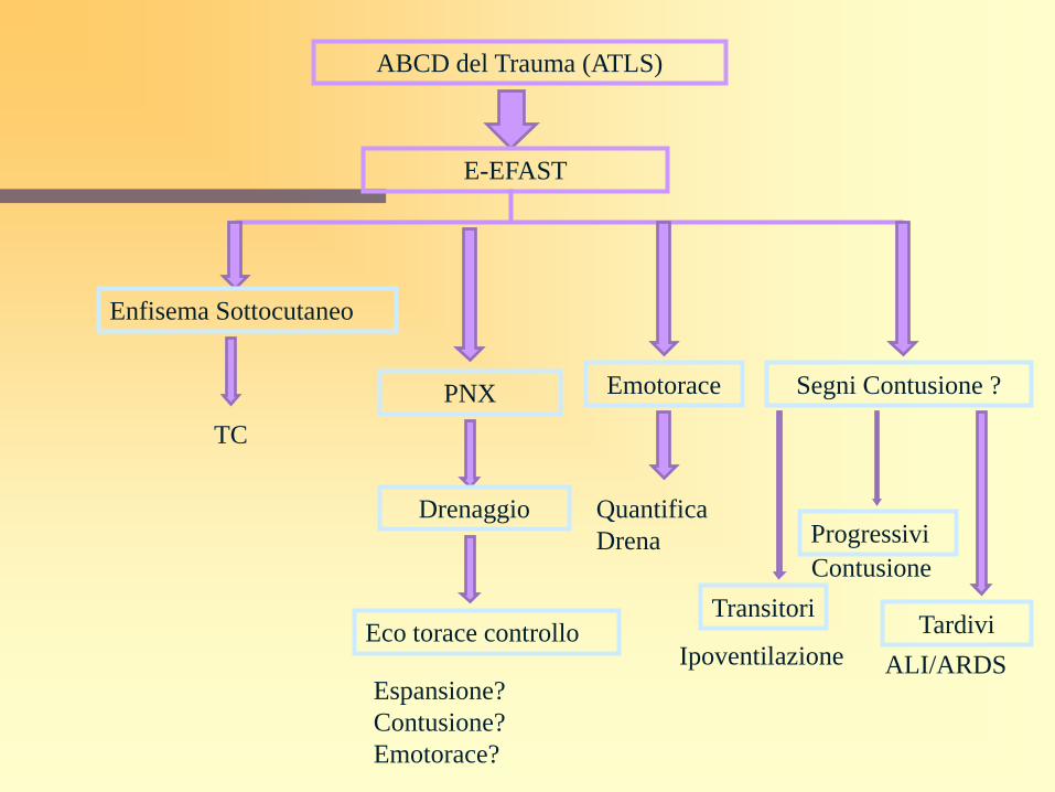
ABCD del Trauma (ATLS)
E-EFAST
Enfisema Sottocutaneo
TC
PNX
Drenaggio
Eco torace controllo
Espansione?
Contusione?
Emotorace?
Emotorace
Quantifica
Drena
Segni Contusione ?
Transitori
Progressivi
Tardivi
Ipoventilazione
Contusione
ALI/ARDS
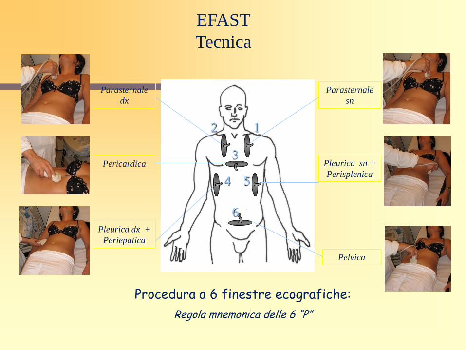
EFAST
Tecnica
Procedura a 6 finestre ecografiche:
Regola mnemonica delle 6 “P”
Parasternale
dx
Parasternale
sn
Pericardica Pleurica sn +
Perisplenica
Pleurica dx +
Periepatica
Pelvica
1 2
3
4 5
6

Thank You