tag der promotion: 30. juni 2020
TRANSCRIPT
Klinik und Poliklinik für Herz-, Thorax- und Gefäßchirurgie der Universitätsmedizin
der Johannes Gutenberg-Universität Mainz und
Klinik für Thorax-, Herz- und Gefäßchirurgie, Westpfalz-Klinikum Kaiserslautern,
Akademisches Lehrkrankenhaus der Universitätsmedizin der Johannes Gutenberg-
Universität Mainz
The perioperative use of Levosimendan as a means of optimizing the surgical
outcome in patients with severe heart insufficiency undergoing cardiac surgery.
Die perioperative Anwendung von Levosimendan zur Optimierung des chirurgischen
Ergebnisses bei Patienten mit schwerer Herzinsuffizienz, die sich einer
Herzoperation unterziehen.
Inauguraldissertation
zur Erlangung des Doktorgrades der
Medizin
der Universitätsmedizin
der Johannes Gutenberg-Universität Mainz
Vorgelegt von
Vasileios Leivaditis
aus Patras, Griechenland
Mainz, 2019

2
Tag der Promotion: 30. Juni 2020
3
Table of Contents
1. PREFACE ............................................................................................................. 18
2. ISCHEMIC HEART DISEASE ............................................................................... 19
2.1. Atherosclerosis ................................................................................................ 20
2.1.1. Endothelial dysfunction ............................................................................. 21
2.1.2. Lipid metabolism disorders ....................................................................... 23
2.1.3. Inflammation and the role of cytokines and macrophages ........................ 24
2.1.4. Vascular smooth muscle cells activation ................................................... 25
2.1.5. Extracellular matrix modification and calcification ..................................... 26
2.1.6. Platelet activation ...................................................................................... 26
2.1.7. Genetic factors of atherosclerosis ............................................................. 27
2.1.8. Rupture of the unstable plaque and arterial infarction............................... 27
2.2. Myocardial ischemia and infarction ................................................................. 28
2.3. Impact of ischemia to myocardial cellular metabolism .................................... 29
2.4. Changes in the ionic cellular equilibrium ......................................................... 30
2.5. Production of free radicals .............................................................................. 31
2.6. Cellular necrosis .............................................................................................. 32
2.7. Apoptosis ........................................................................................................ 32
3. ISCHEMIA-REPERFUSION INJURY .................................................................... 33
3.1. Myocardial Stunning ........................................................................................ 35
3.2. Myocardial Hibernation ................................................................................... 37
3.3. No-reflow Phenomenon .................................................................................. 40
3.4. Reperfusion Arrhythmias ................................................................................. 41
3.5. Mediators of lethal reperfusion injury .............................................................. 42
3.5.1. Oxygen paradox........................................................................................ 43
3.5.2. Calcium paradox ....................................................................................... 44
3.5.3. pH paradox ............................................................................................... 44
3.5.4. Inflammation ............................................................................................. 44
3.5.5. Metabolic modulation ................................................................................ 44
3.5.6. Mitochondrial permeability transition pore ................................................. 45
3.5.7. The Reperfusion Injury Salvage Kinase (RISK) pathway .......................... 46
4. APPLIED PHYSIOLOGY AND PATHOPHYSIOLOGY ......................................... 47
4.1. Basic parameters of circulatory physiology ..................................................... 48
4.1.1. Frank–Starling law .................................................................................... 48
4.1.2. Preload ..................................................................................................... 50
4
4.1.3. Afterload ................................................................................................... 51
4.1.4. Stroke volume ........................................................................................... 52
4.1.5. Cardiac output and cardiac index.............................................................. 52
4.1.6. Ejection Fraction ....................................................................................... 54
4.1.7. Pressure-Volume work .............................................................................. 54
4.2. Mechanical disorders of the myocardium ........................................................ 55
4.2.1. Systolic myocardial dysfunction ................................................................ 57
4.2.2. Diastolic myocardial dysfunction ............................................................... 57
4.3. Cardiac remodeling ......................................................................................... 59
4.3.1. Early remodeling ....................................................................................... 59
4.3.2. Late remodelling and scar formation ......................................................... 60
4.4. Activation of Renin-Angiotensin-Aldosterone-System ..................................... 60
5. CLINICAL PRESENTATIONS OF ISCHEMIC HEART DISEASE ......................... 61
5.1. Stable angina pectoris ..................................................................................... 61
5.2. Silent myocardial ischemia .............................................................................. 62
5.3 Acute coronary syndromes .............................................................................. 62
5.3.1. Unstable angina and Prinzmetal (variant) angina ..................................... 64
5.3.2. Acute myocardial infarction (NSTEMI and STEMI) ................................... 65
6. HEART FAILURE .................................................................................................. 67
6.1. Definition of heart failure ................................................................................. 67
6.2. Epidemiology of heart failure ........................................................................... 68
6.3. Defining heart failure patients regarding the ejection fraction ......................... 69
6.4. Aetiology of heart failure ................................................................................. 70
6.5. Classification of heart failure ........................................................................... 71
6.5.1. Killip classification ..................................................................................... 71
6.5.2. Forrester classification .............................................................................. 72
6.5.3. Nohria classification .................................................................................. 74
6.5.4. NYHA classification ................................................................................... 75
6.5.5. ACC/AHA classification ............................................................................. 75
6.6. Diagnosis of heart failure in the non-acute setting .......................................... 76
6.7. Pharmacological treatment of chronic heart failure ......................................... 77
6.7.1 Angiotensin-converting enzyme inhibitors (ACEI) ...................................... 78
6.7.2. Beta-blockers ............................................................................................ 78
6.7.3. Mineralocorticoid/aldosterone receptor antagonists (MRA) ...................... 78
6.7.4. Diuretics .................................................................................................... 79
6.7.5. Angiotensin receptor neprilysin inhibitor (ARNI) ....................................... 79

5
6.7.6. If-channel inhibitor ..................................................................................... 79
6.7.7. Angiotensin II type I receptor blockers (ARB) ........................................... 80
6.7.8. Combination of hydralazine and isosorbide dinitrate................................. 80
6.7.9. Digoxin and other digitalis glycosides ....................................................... 80
6.8. Cardioverter and defibrillator therapy .............................................................. 80
6.9. Cardiac resynchronization therapy (CRT) ....................................................... 81
6.10. Left ventricular assist device therapy ............................................................ 82
6.11. Heart Transplantation .................................................................................... 84
6.12. Acute heart failure ......................................................................................... 85
6.12.1. Definition of acute heart failure ............................................................... 85
6.12.2. Pathophysiology of acute heart failure .................................................... 86
6.12.3. Cardiogenic shock .................................................................................. 87
7. MYOCARDIAL DYSFUNCTION AND LOW CARDIAC OUTPUT SYNDROME
AFTER CARDIAC SURGERY ................................................................................... 88
7.1. Left ventricular systolic dysfunction ................................................................. 91
7.2. Left ventricular diastolic dysfunction ................................................................ 92
7.3. Right ventricular dysfunction ........................................................................... 93
8. CARDIOPROTECTION IN CARDIAC SURGERY ................................................. 93
8.1. Cardioplegia .................................................................................................... 93
8.1.1. Intracellular vs. extracellular type .............................................................. 95
8.1.2. Crystalloid solutions vs. blood cardioplegia .............................................. 95
8.1.3. Warm vs. cold solutions ............................................................................ 96
8.1.4. Polarized vs. depolarized arrest ................................................................ 96
8.2. Volatile Anesthetics ......................................................................................... 97
8.3. Endogenous therapeutic strategies for cardioprotection ................................. 98
8.3.1. Ischaemic preconditioning ........................................................................ 98
8.3.2. Remote ischaemic preconditioning ........................................................... 99
8.3.3. Ischaemic postconditioning ....................................................................... 99
9. RISK STRATIFICATION IN CARDIAC SURGERY ............................................. 100
9.1. EuroSCORE I & II ......................................................................................... 100
9.2. STS score ..................................................................................................... 101
10. MECHANICAL CIRCULATORY SUPPORT ...................................................... 101
10.1. Intraaortic balloon pump .............................................................................. 102
10.2. Impella® pump ............................................................................................. 103
10.3. TandemHeart® ............................................................................................ 104
10.4 Extracorporeal life support systems (ECLS) ................................................ 104
6
11. INOTROPIC AND VASOACTIVE AGENTS IN THE TREATMENT OF ACUTE
AND POSTOPERATIVE HEART FAILURE ............................................................ 106
11.1. Catecholamines .......................................................................................... 106
11.2. Phosphodiesterase inhibitors ...................................................................... 111
11.3. Vassopressors ............................................................................................ 112
12. CALCIUM SENSITIZERS – LEVOSIMENDAN ................................................. 114
12.1. Chemistry .................................................................................................... 115
12.2. Mechanism of action ................................................................................... 115
12.2.1. Positive inotropic effect ......................................................................... 116
12.2.2. Vasodilatatory effect ............................................................................. 118
12.2.3. Cardioprotective effect .......................................................................... 118
12.2.4. Phosphodiesterase III inhibitor effect .................................................... 118
12.2.5. Antiinflammatory effect ......................................................................... 119
12.2.6. Effects on the pulmonary circulation ..................................................... 119
12.2.7. Electrophysiologic effects ..................................................................... 120
12.3. Pharmacokinetics - Pharmacodynamics ..................................................... 120
12.4. Dosis ........................................................................................................... 123
12.5. Tolerance, side effects and toxicity ............................................................. 123
12.6. Combination with other agents .................................................................... 124
12.7. Importance of the application time .............................................................. 125
12.8. Most important randomized multicenter clinical trials on levosimendan ...... 126
12.8.1. Clinical trials in heart failure .................................................................. 126
12.8.2. Clinical trials in cardiac surgery............................................................. 130
13. AIM OF THIS STUDY ........................................................................................ 133
14. MATERIALS AND METHODS ........................................................................... 135
14.1. Study design and patient data collection ..................................................... 135
14.2. Inclusion and exclusion criteria ................................................................... 136
14.3. Application and dosis of Levosimendan ...................................................... 137
14.4. Ethics .......................................................................................................... 137
14.5. Statistical analysis ....................................................................................... 138
15. RESULTS .......................................................................................................... 139
15.1. Demographics ............................................................................................. 139
15.2. Clinical characteristics at baseline .............................................................. 143
15.3. Introperative surgical times ......................................................................... 158
15.4. Overall Survival ........................................................................................... 160
15.5. Postoperative parameters ........................................................................... 162
7
15.6. Laboratory haematological and biochemical parameters ............................ 174
16. DISCUSSION .................................................................................................... 190
16.1. Baseline characteristics .............................................................................. 190
16.2. Postoperative survival ................................................................................. 191
16.3. Effect on operative times and weaning from CPB ....................................... 195
16.4. Postoperative clinical parameters ............................................................... 196
16.4.1. Postoperative ejection fraction .............................................................. 196
16.4.2. Rehospitalization ................................................................................... 196
16.4.3. Postoperative angina pectoris and dyspnoea ....................................... 197
16.4.4. Duration of mechanical ventilation, ICU and total hospital stay ............ 198
16.4.5. Support with inotropic agents and IABP ................................................ 199
16.4.6. Postoperative arrhythmias .................................................................... 199
16.4.7. Postoperative renal function and need for haemodialysis ..................... 200
16.4.8. Haemoglobin and need for transfusion of blood products ..................... 203
16.5. Postoperative laboratory parameters .......................................................... 205
16.5.1. Postoperative inflammatory response ................................................... 205
16.5.2. Postoperative myocardial injury ............................................................ 206
16.5.3. Organ perfusion and metabolic balance ............................................... 207
17. LIMITATIONS OF THIS STUDY ........................................................................ 208
18. SUMMARY ........................................................................................................ 209
18.1. Summary in English .................................................................................... 209
18.2. Summary in German / Zusammenfassung .................................................. 211
19. REFERENCES .................................................................................................. 214
20. NOTE OF THANKS / DANKSAGUNG............................................................... 262
21. CURRICULUM VITAE / LEBENSLAUF ............................................................. 263
21.1. Angaben zur Person ................................................................................... 263
21.2. Arbeitserfahrung .......................................................................................... 263
21.3. Ausbildung - Weiterbildung ......................................................................... 264
21.4. Kurse - Seminare ........................................................................................ 266
21.5. Publikationen – Veröffentlichungen ............................................................. 268
21.6. Präsentationen in Kongresse ...................................................................... 271
21.7. Übrige wissentschaftliche Arbeit ................................................................. 274

8
List of Tables
Table 1. Vascular modifications in atherosclerotic disease. SMCs smooth muscle
cells. .......................................................................................................................... 21
Table 2. Mechanisms responsible for myocardial stunning and potential therapeutic
approaches. ............................................................................................................... 37
Table 3. Classification of stable angina pectoris according to the Canadian
Cardiovascular Society (CCS). .................................................................................. 62
Table 4. Classification of unstable angina. ................................................................ 64
Table 5. Definition of myocardial infarction and criteria for acute myocardial infarction.
.................................................................................................................................. 66
Table 6. Universal classification of MI. ...................................................................... 66
Table 7. Definition of heart failure with preserved (HFpEF), mid-range (HFmrEF) and
reduced ejection fraction (HFrEF). ............................................................................ 70
Table 8. Main causes of heart failure. ....................................................................... 71
Table 9. The Killip classification of heart failure. ........................................................ 72
Table 10. The NYHA classification of heart failure. ................................................... 75
Table 11. The AHA/ACC classification of heart failure. ............................................. 76
Table 12. Terms describing various indications for mechanical circulatory support. . 83
Table 13. Indications and contra-indications for heart transplantation. ...................... 85
Table 14. Factors triggering acute heart failure. ........................................................ 86
Table 15. Indications and contraindications for ECLS. ............................................ 106
Table 16. Receptor affinity and haemodynamic effects of the most common
catecholamines. ...................................................................................................... 108
Table 17. The main effects and indications of inotropic agents. .............................. 113
Table 18. Pharmacokinetic variables of levosimendan and its active metabolite OR-
1896. ....................................................................................................................... 122
Table 19. Patients’ characteristics at baseline. ........................................................ 139
Table 20. Patients’ baseline clinical characteristics prior to surgery. ....................... 145
Table 21. Intraoperative surgical times in the two cohorts. ...................................... 158
Table 22. Survival probability alongside 95% C.I. at different timepoints from
operation date by cohort. ......................................................................................... 160
Table 23. Postoperative parameters in the two groups. .......................................... 163
Table 24. Blood parameters at different timepoints in the two cohorts. ................... 177

9
List of Figures
Figure 1. a. Normal endothelial physiology induced by NO. b. Endothelial activation is
characterized by increased membrane permeability and production of ROS which
alter the function of cellular constituents, leading to phosphorylisation of transcription
factors and mitochondria, as well as protease activation (eNOS, p50/p65 nuclear
factor-kB [NF-kB] transcription factor, nicotinamide adenine dinucleotide phosphate
[NADPH])................................................................................................................... 22
Figure 2. The role of oxidized LDL cholesterol in the formation of foam cells and the
development of an atherosclerotic plaque. ................................................................ 24
Figure 3. Cellular damage in ischemia-reperfusion. .................................................. 35
Figure 4. The main factors involved in the pathogenesis of myocardial stunning. ..... 36
Figure 5. Schematic figure, summarizing different mechanisms, involved in the
development of no-reflow, and accompanying ultrastructural alterations of the
microvascular bed. .................................................................................................... 40
Figure 6. Major mediators of lethal reperfusion injury. ............................................... 43
Figure 7. An overview of the proposed mechanism of the mitochondrial permeability
transition pore and the consequences of its opening. ............................................... 45
Figure 8. Scheme demonstrating the diverse variety of agents which activate the
Reperfusion Injury Salvage Kinase (RISK) pathway in both a receptor and non-
receptor mediated manner. ....................................................................................... 47
Figure 9. Cardiac function curve illustrating the Frank–Starling law of the heart, the y-
axis often describes the stroke volume, stroke work, or cardiac output. The x-axis
often describes end-diastolic volume, right atrial pressure, or pulmonary capillary
wedge pressure. ........................................................................................................ 48
Figure 10. Myocardial sarcomere and myofilaments. A. Diagram of the sarcomere
showing approximate spatial relationships of thick and thin filaments and putative
interactions of titin with the filaments, which would give rise to radial and axial
restorati restorative forces when the sarcomere is stretched. B. Diagram of the thick
and thin filaments illustrating the decrease in lateral separation at long lengths. The
probability of crossbridge interaction increases at long lengths due to closer proximity
to actin. ...................................................................................................................... 49
Figure 11. Major factors influencing heart rate and stroke volume and consequently
cardiac output. ........................................................................................................... 53

10
Figure 12. The left ventricular pressure-volume loop from a healthy person at rest.
The pressure-volume loop is a widely applicable pathophysiological tool. ................ 55
Figure 13. Left ventricular pressure-volume loops in a healthy person (red) and for
persons with acute (blue area) or chronic, congestive (green area) cardiac failure. .. 56
Figure 14. The pathogenetic classification of acute coronary syndrome (ACS) based
on simple clinical descriptors. .................................................................................... 63
Figure 15. The Forrester classification of heart failure. ............................................. 73
Figure 16. Clinical profiles of patients with acute heart failure based on the
presence/absence of congestion and/or hypoperfusion. ........................................... 74
Figure 17. The recommended therapeutic algorithm for a patient with symptomatic
heart failure with reduced ejection fraction. ............................................................... 77
Figure 18. A schematic presentation of the pathophysiology of postoperative LCOS.
The most common causes and typical signs are presented. ..................................... 91
Figure 19. The potential cellular targets for cardioplegic arrest and their influence on
the action potential of the cell membrane. ................................................................. 95
Figure 20. Intracellular actions of β-adrenergic agonists. ........................................ 107
Figure 21. Postulated mechanisms of intracellular action of α1-adrenergic agonists.
................................................................................................................................ 110
Figure 22. Basic mechanism of action of PDIs. ....................................................... 112
Figure 23. Mechanisms of action of inotropic agents in the cardiac muscle cell...... 114
Figure 24. Structural formula of levosimendan. ....................................................... 115
Figure 25. A. Role of troponin C in the mechanism of contraction. B. Levosimendan
selectively binds to calcium saturated cardiac troponin C. ...................................... 117
Figure 26. Metabolic pathways of Levosimendan. ................................................... 121
Figure 27. Free plasma concentrations of levosimendan and OR -1896 during and
after a 24-h infusion. ................................................................................................ 122
Figure 28. Frequency of participants’ gender in the control and drug group. .......... 140
Figure 29. Histograms of age by group. .................................................................. 140
Figure 30. Histograms of weight by group. .............................................................. 141
Figure 31. Histograms of height by group. .............................................................. 141
Figure 32. Histograms of BMI by group. .................................................................. 142
Figure 33. Histograms of BSA by group. ................................................................. 142
Figure 34. Boxplot of Euroscore 1 by group. ........................................................... 145
Figure 35. Boxplot of Euroscore 2 by group. ........................................................... 146
Figure 36. Frequency of ASA score in the two cohorts. .......................................... 147

11
Figure 37. Boxplot of preoperative ejection fraction by group.................................. 148
Figure 38. Staging of preoperative dyspnoea according to NYHA classification. .... 148
Figure 39. Frequency of staging of angina according to CCS in the two cohorts. ... 149
Figure 40. Frequency of type of surgery (regular of urgent) in the two cohorts. ...... 150
Figure 41. Frequency of preoperartive acute myocardial infarction in the two cohorts.
................................................................................................................................ 150
Figure 42. Frequency of simple or combined surgery in the two cohorts. ............... 151
Figure 43. Boxplot of number of bypass-grafts by group. ........................................ 152
Figure 44. Frequency of arrhythmias prior to surgery in the two cohorts. ................ 153
Figure 45. Frequency of pulmonary hypertension prior to surgery in the two cohorts.
................................................................................................................................ 153
Figure 46. Frequency of arterial hypertension prior to surgery in the two cohorts. .. 154
Figure 47. Frequency of Diabetes mellitus prior to surgery in the two cohorts. ....... 155
Figure 48. Frequency of Hyperlipidemia prior to surgery in the two cohorts. ........... 155
Figure 49. Frequency of chronic obstructive pulmonary disease prior to surgery in the
two cohorts. ............................................................................................................. 156
Figure 50. Frequency of renal failure prior to surgery in the two cohorts. ................ 156
Figure 51. Frequency of extracardiac arteriopathy prior to surgery in the two cohorts.
................................................................................................................................ 157
Figure 52. Frequency of cerebrovascular accidents prior to surgery in the two
cohorts. ................................................................................................................... 157
Figure 53. Boxplot of Cross-Clamp-Time (min) by group. ....................................... 158
Figure 54. Boxplot of duration of surgery (min) by group. ....................................... 159
Figure 55. Boxplot of Bypass-time (min) by group. .................................................. 159
Figure 56. Overall survival from date of operation (30 days). .................................. 161
Figure 57. Overall survival from date of operation (1year). ..................................... 161
Figure 58. Overall survival from date of operation (3 years). ................................... 162
Figure 59. Histograms of post-operative ejection fraction by cohort. ....................... 164
Figure 60. Boxplot of postoperative ejection fraction by cohort. .............................. 164
Figure 61. Boxplot of ejection fraction difference (ΔEF) by cohort. .......................... 165
Figure 62. Boxplot of number of readmissions to hospital by cohort. ...................... 166
Figure 63. Frequency of every angina CCS class before and after surgery in the two
cohorts. ................................................................................................................... 167
Figure 64. Frequency of dyspnoea NYHA class before and after surgery in the two
cohorts. ................................................................................................................... 167

12
Figure 65. Boxplot of hospitalization (days) by cohort. ............................................ 168
Figure 66. Boxplot of intensive care unit (stay in days) by cohort. ........................... 169
Figure 67. Boxplot of artificial ventilation (days) by cohort. ..................................... 169
Figure 68. Boxplot of duration of patient support with inotropic drugs in the ICU (in
days) by cohort. ....................................................................................................... 170
Figure 69. Boxplot of duration of support with IABP (days) by cohort. .................... 171
Figure 70. Need for haemodialysis by cohort. ......................................................... 171
Figure 71. Frequency of postoperative arrhythmias by cohort................................. 172
Figure 72. Histograms of total units of erythrocytes (RBC) transfused by cohort. ... 173
Figure 73. Histograms of fresh frozen plasma (FFP) units transported by cohort. .. 173
Figure 74. Boxplot of total platelet (PLT) units transfused by cohort. ...................... 174
Figure 75. Histograms of haemoglobin on the 5th postoperative day in the two groups.
................................................................................................................................ 178
Figure 76. Distribution of hemoglobin values in the two cohorts. ............................. 178
Figure 77. Histograms of CRP at 5 days after surgery in the two cohorts. .............. 179
Figure 78. Distribution of CRP values in the two cohorts. ....................................... 180
Figure 79. Histograms of troponin on the 5th postoperative day in the two cohorts. 181
Figure 80. Distribution of troponin I in the two cohorts. ........................................... 181
Figure 81. Histograms of CK on the 5th postoperative day in the two cohorts. ........ 182
Figure 82. Distribution of CK in the two cohorts. ..................................................... 183
Figure 83. Histograms of CKMB on the 5th postoperative day in the two cohorts. ... 183
Figure 84. Distribution of CKMB in the two cohorts. ................................................ 184
Figure 85. Histograms of creatinine on the 2nd postoperative day in the two cohorts.
................................................................................................................................ 185
Figure 86. Distribution of creatine values in the two cohorts. .................................. 186
Figure 87. Histograms of creatinine clearance on the 2nd postoperative day in the two
cohorts. ................................................................................................................... 186
Figure 88. Distribution of Clearance creatinine in the two cohorts. .......................... 187
Figure 89. Histograms of GFR on the 3rd postoperative day in the two cohorts. ..... 187
Figure 90. Distribution of GFR in the two cohorts. ................................................... 188
Figure 91. Histograms of pH on the 2nd postoperative day in the two groups. ......... 189
Figure 92. Distribution of pH values in the two cohorts. .......................................... 189
Figure 93. Distribution of Lactic acid in the two cohorts. ......................................... 190
Figure 94. Differential effects of renal vasodilators on preglomerular (afferent
arteriole) and postglomerular (efferent arteriole) vascular resistance sections........ 202

13
List of Abbreviations
ACC American College of Cardiologists
ACEI Angiotensin-converting enzyme inhibitors
ACS Acute Coronary Syndrome
AF Atrial Fibrillation
AHA American Heart Association
AHF Acute Heart Failure
AIVR Accelerated Idioventricular Rhythm
AMI Acute Myocardial Infarction
ANT Adenine Nucleotide Translocase
APOB Apolipoprotein B
ARB Angiotensin Receptor Blockers
ATP Adenosine Triphosphate
AV Atrioventricular
BiVAD Biventricular Assist Device
BMI Body Mass Index
BNP Brain Natriuretic Peptide
BSA Body Surface Area
BTT Bridge to Transplantation
CABG Coronary Artery Bypass Grafting
CAD Coronary Artery Disease
CAM Cell Adhesion Molecules
CCS Canadian Cardiovascular Society
cGMP cyclic Guanosine Monophosphate
CI Cardiac Index

14
CK Creatine Kinase
CKMB Myocardial Creatine Kinase
CMR Cardiac Magnetic Resonance imaging
CO Cardiac Output
CPB Cardiopulmonary Bypass
CrCl Creatinine Clearance
CRP C - Reactive Protein
CRT Cardiac Resynchronization Therapy
CyP-D Cyclophilin-D
DNA Deoxyribonucleic Acid
DT Destination Therapy
ECC Extracorporeal Circulation
ECLS Extracorporeal Life Support
ECM Extracellular Matrix
EDV End Diastolic Volume
EF Ejection Fraction
eNOS endothelial Nitric Oxide Synthase
ESC European Society of Cardiology
ESV End Systolic Volume
EuroSCORE European System for Cardiac Operative Risk Evaluation
FFP Fresh Frozen Plasma
GFR Glomerular Filtration Rate
HDL High Density Lipoprotein
HF Heart Failure
HFmrEF Heart Failure with mid-range Ejection Fraction

15
HFpEF Heart Failure with preserved Ejection Fraction
HFrEF Heart Failure with reduced Ejection Fraction
HMG-CoA 3-hydroxy-3-methyl-glutaryl-coenzyme A
HR Heart Rate
IABP Intraaortic Balloon Pump
ICD Implantable cardioverter-defibrillator
IFN-g Interferon-g
IHD Ischemic Heart Disease
IL-1 Interleukin 1
IMM Inner Mitochondrial Membrane
IPC Ischemic Preconditioning
LA Left Atrium
LBBB Left Bundle Branch Block
LDL Low Density Lipoprotein
LDLR LDL Receptor
LV Left Ventricle
LVAD Left Ventricular Assist Device
LVEF Left Ventricular Ejection Fraction
MAP Mean Arterial Pressure
MCP-1 Monocyte Chemoattractant Protein-1
MCSF Macrophage Colony-stimulating Factor
MI Myocardial Infarction
MMP Matrix Metalloproteinases
mPTP mitochondrial Permeability Transition Pore
MRA Mineralocorticoid/aldosterone Receptor Antagonists
16
NO Nitric Oxide
NOS Nitric Oxide Synthase
NP Natriuretic Peptide
NSTEMI Non-ST-elevation Myocardial Infarction
NYHA New York Heart Association
OMT Optimal Medical Therapy
PCSK9 Proprotein Convertase Subtilisin/Kexin type 9
PCWP Pulmonary Capillary Wedge Pressure
PET Positron Emission Tomography
PiC Phosphate Carrier
PKC Protein Kinase C
PLT Platelets
PTCA Percutaneous Coronary Angioplasty
PTP Permeability Transition Pore
RA Right Artium
RAAS Renin-Angiotensin-Aldosetrone-System
RBBB Right Bundle Branch Block
RBC Red Blood Cells
RIC Remote Ischemic Conditioning
RISK Reperfusion Injury Salvage Kinase
ROS Reactive Oxygen Species
RRT Renal Replacement Therapy
RV Right Ventricle
RVAD Right Ventricular Assist Device
RVEF Right Ventricular Ejection Fraction

17
SIRS Systemic Inflammatory Response Syndrome
SOD Superoxide Dismutase
STEMI ST-elevation Myocardial Infarction
STS Society of Thoracic Surgeons
SV Stroke Volume
SVI Stroke Volume Index
SVR Systemic Vascular Resistance
TnC Troponin C
TNF-α Tumor Necrosis Factor alpha
TnI Troponin I
TnT Troponin T
UA Unstable Angina
VA-ECLS Venoarterial Ectracorporeal Life Support
VEGF Vascular Endothelial Growth Factor
VSMC Vascular Smooth Muscle Cell
WHO World Health Organisation

18
1. PREFACE
In recent years the number of patients with severely impaired myocardial function
has been steadily increasing. This increase is primarily attributable to the aging of the
population and improved treatment of acute myocardial infarction. Over the last
decade the frequency of surgical procedures carried out in patients with heart failure
has been constantly increasing. This group of high risk patients was considered to be
inoperable some decades ago, but they nowadays consist part of a cardiac surgeon’s
daily clinical practice. Patients with poor left ventricular function usually require
inotropic drug support immediately after cardiopulmonary bypass. Indeed, preexisting
impaired ventricular function is further compromised by variable degrees of
myocardial injury as a result of ischemia during aortic cross clamping. Therefore
physicians involved in the treatment of such patients including surgeons,
anesthesiologists and intensivists are increasingly confronted with the challenging
perioperative management of patients with heart failure.
Significant improvements have been made in treating patients with heart failure,
mainly due to a better understanding of the undelying pathophysiological
mechanisms. However, despite the steadily improved experience in the management
of these patients and the evolution of pharmacological and mechanical means to
support impaired heart function, the successful treatment and the survival of patients
with severe heart failure still remains a great challenge in cardiac surgery. This
constant increase of high risk patients as candidates for cardiac surgery has led to an
increasing use of pharmacologic support in the form of vasodilator and inotropic
therapy. Traditionally, perioperatively used inotropic agents, such as epinephrine,
dobutamine and milrinone are limited by significant increases in myocardial oxygen
consumption, proarrhythmia, or neurohormonal activation. They tend to enhance
myocardial contractility by increasing cyclic adenosine monophosphate
concentrations, which ultimately increases the myocardial concentrations of calcium.
Over the last years a new pharmacological agent, Levosimendan, has started being
used in the daily practice during an operation as well as postoperatively in the
intensive care unit, promising to ameliorate the haemodynamic condition of these
patients.
The aim of the current study is to evaluate the possible positive preoperative and
postoperative effect of Levosimendan, a new inodilator in the treatment of
19
decompensated heart failure. This new inotropic agent has already shown promising
effects in the treatment of cardiac surgical patients with high perioperative risk or
compromised left ventricular function, as well as in rescue therapy of patients with
difficult weaning from cardiopulmonary bypass. Levosimendan has a unique
mechanism of action. Briefly, levosimendan binds to the regulatory protein troponin C
(TnC) and stabilizes the Ca2+-bound conformation of TnC, thereby allowing
unopposed interaction between actin and myosin filaments and enhancing the rate
and extent of myocyte contraction.
Over the last years many patients with low ejection fraction have been admitted to
our ward and received a variety of cardiac surgical procedures. Levosimendan has
been widely used in such patients over the last years with promising postoperative
results. This study will retrospectively include such patients with preoperative low
ejection fraction who underwent heart surgery in our department. The group of
patients who were treated with levosimendan will be compared to a similar historical
group of patients with heart failure who underwent similar procedures in the past and
did not receive this medical treatment.
2. ISCHEMIC HEART DISEASE
Ischemic heart disease consists a major part of the nosological entity of
cardiovascular disease, which also includes hypertension, stroke, valvular, muscular
and congenital heart disease. About 15% of worldwide mortality is attributable to
ischemic heart disease, making it the leading cause of death globally. It is the most
important and most common contributor to the development of heart failure,
accounting for up to 50% of cases (1).
The current treatment of ischemic heart disease consists of medical pharmacological
therapy and revascularization procedures including thrombolysis, percutaneous
coronary angioplasty (PTCA) and coronary artery bypass grafting (CABG) through
open heart surgery. The main goals of these treatments are establishing reperfusion
in coronary arteries, enhancement of coronary blood flow, reduction of myocardial
oxygen consumption, avoidance of the incidence of arrhythmic disorders and in
cases of patients with acute myocardial infarction limitation of the infarct size and
loss of vital myocardium. Despite intensive pharmacological therapies and the

20
increasing use of evolving surgical and interventional procedures which are
nowadays widely available in clinical practice, the prognosis of patients with heart
failure due to ischemic heart disease remains unfortunately poor (2, 3).
2.1. Atherosclerosis
The term atherosclerosis is used to describe a chronic inflammatory condition of the
vessel wall. Atherosclerosis is the pathological basis of peripheral vascular, coronary
artery and cerebrovascular diseases, all major causes of mortality and morbidity
throughout the world. Despite the development of effective cholesterol reducing
agents and lifestyle modification initiatives, only modest decreases in rates of
atherosclerosis and its clinical manifestations have been achieved over the last
decades (4, 5).
It is considered to be a complex, chronic disease which is traditionally thought of as
the accumulation of fibrofatty deposits in the intima of medium and large muscular
arteries, a passive process and an inevitable aspect of aging. Current knowledge has
however shown that it consists a complex interplay of lipid metabolism, active cellular
interactions, inflammation, and matrix remodelling (4-6). The most appropriate
approach to the definition of atherosclerotic lesions would therefore be a dynamic
process which evolves from fatty streaks to stable or unstable plaques (4).
Scientific understanding of the pathogenesis of atherogenesis is constantly
developing. From Virchow’s observations 160 years ago we are nowadays familiar
with the endothelial response to injury as inflammatory, involved in all stages of
atherosclerosis (6). The following paragraphs are intended to give a general overview
of the main factors and pathophysiological mechanisms involed in the several phases
of atherosclerotic development. The main vascular modifications which take part in
atherosclerotic disease are summarized in Table 1.
Vascular modification
Characteristics
Intimal thickening Layers of smooth muscle cells (SMCs) and extracellular matrix. More frequent in coronary artery, carotid artery, abdominal aorta, descending aorta, and iliac artery.
Fatty streak Abundant macrophage foam cells mixed with SMCs and

21
proteoglycan-rich intima
Pathologic intimal thickening
Layers of SMCs in proteoglycan-collagen matrix aggregated near the lumen. Underlying lipid pool: acellular area rich in hyaluronan and proteoglycans with lipid infiltrates.
Fibroatheromas Acellular necrotic core (cellular debris). Necrotic core is covered by a thick fibrous cap=SMCs in proteoglycan-collagen matrix.
Vulnerable plaque Thin-cap fibroatheroma’. Type I collagen, very few/absent SMCs. Fibrous cap thickness is ≤65 µm.
Ruptured plaque Ruptured fibrous cap. Presence of luminal thrombus. Larger necrotic core and increased macrophage infiltration of the thin fibrous cap.
Table 1. Vascular modifications in atherosclerotic disease. SMCs smooth muscle cells.
(Source: Bergheanu SC, Bodde MC, Juke JW. Pathophysiology and treatment of atherosclerosis. Current view and future perspective on lipoprotein modification treatment. Neth Heart J. 2017 Apr; 25(4): 231–242.)
2.1.1. Endothelial dysfunction
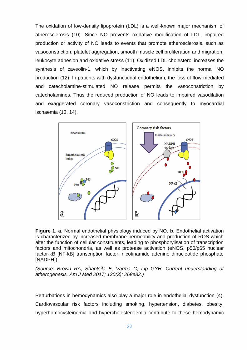
Normal endothelium helps maintain vascular homeostasis. This can be achieved by
secreting vasodilatory nitric oxide (NO) as well as vasoconstrictors such as
endothelin and angiotensin II (6). NO inhibits inflammation, proliferation and
thrombosis. NO is produced in endothelial cells from its precursor L-arginine via the
enzymatic action of endothelial NO synthase (eNOS) (7). In the vascular wall, NO
activates soluble guanylate cyclase in vascular smooth muscle cells (VSMCs),
leading to elevation of cyclic guanosine monophosphate (cGMP), activation of
cGMP-dependent protein kinase (PKG), and vasorelaxation (8).
Alterations in NO secretion and vasoconstrictive or vasodilatory response
respectively are among the earliest pathologies noted in vascular disease, often
preceeding the formation of atherosclerotic lesions (9). Conventional cardiovascular
risk factors favor a reduction in L-arginin induced nitric oxide synthesis, leading to an
increase in reactive oxygen species (ROS) and subsequent oxidative stress (Figure
1) (6). The impairment of the nitric oxide mediated vasodilation is therefore
considered to be the hallmark of endothelial dysfunction.
22
The oxidation of low-density lipoprotein (LDL) is a well-known major mechanism of
atherosclerosis (10). Since NO prevents oxidative modification of LDL, impaired
production or activity of NO leads to events that promote atherosclerosis, such as
vasoconstriction, platelet aggregation, smooth muscle cell proliferation and migration,
leukocyte adhesion and oxidative stress (11). Oxidized LDL cholesterol increases the
synthesis of caveolin-1, which by inactivating eNOS, inhibits the normal NO
production (12). In patients with dysfunctional endothelium, the loss of flow-mediated
and catecholamine-stimulated NO release permits the vasoconstriction by
catecholamines. Thus the reduced production of NO leads to impaired vasodilation
and exaggerated coronary vasoconstriction and consequently to myocardial
ischaemia (13, 14).
Figure 1. a. Normal endothelial physiology induced by NO. b. Endothelial activation is characterized by increased membrane permeability and production of ROS which alter the function of cellular constituents, leading to phosphorylisation of transcription factors and mitochondria, as well as protease activation (eNOS, p50/p65 nuclear factor-kB [NF-kB] transcription factor, nicotinamide adenine dinucleotide phosphate [NADPH]).
(Source: Brown RA, Shantsila E, Varma C, Lip GYH. Current understanding of atherogenesis. Am J Med 2017; 130(3): 268e82.)
Perturbations in hemodynamics also play a major role in endothelial dysfunction (4).
Cardiovascular risk factors including smoking, hypertension, diabetes, obesity,
hyperhomocysteinemia and hypercholesterolemia contribute to these hemodynamic

23
perturbations (6, 9, 15). The role of hemodynamics can be demonstrated by the non-
random distribution of atherosclerotic lesions, which have a strong predilection for
regions of turbulent, high pressure flow, such as the branch points of the aorta (4-6,
14). Endothelial cells are believed to alter their gene expression in response to shear
stress.
Endothelial dysfunction appears already during the early phases of atherosclerosis,
before the plaque formation can be identified through angiographic or
echocardiographic. Damage to the endothelium causes a severe disturbance to the
balance between vasoconstriction and vasodilation and initiates the processes that
promote or exacerbate atherosclerosis. Such processes include endothelial
permeability, platelet aggregation and generation of cytokines (11, 16).
2.1.2. Lipid metabolism disorders
Hyperlipidemia and abnormal lipid metabolism play a crucial role in the development
of atherosclerosis. High plasma levels of LDL are considered to be atherogenic, while
high density lipoproteins (HDL) on the contrary appear to be significant
atheroprotective (17). This is due to the function of HDLs in reverse cholesterol
transport, which brings cholesterol from the periphery to the liver for degradation.
Statins act by lowering LDL via inhibiting 3-hydroxy-3-methyl-glutaryl-coenzyme A
(HMG-CoA) reductase. HMG-CoA reductase is the rate-controlling enzyme of the
mevalonate pathway, the metabolic pathway that produces cholesterol and other
isoprenoids. Statins, diet and exercise can also lead to an increase in the plasma
levels of HDL (4, 17).
The creation of fatty streaks in the vascular wall is a result of increased circulating
lipid concentrations and lipid insudation of the intima, particularly from LDL
cholesterol. Cholesterol and phospholipids within these early accumulations are
susceptible to oxidation by enzymes such as myeloperoxidases, lipoxygenases,
NADPH oxidases, and nitric oxide synthases (4, 17). Oxidized LDLs and ROS that
result are toxic and induce endothelial dysfunction, inflammation and increased
vascular permeability (17). There is subsequent upregulation of leukocyte adhesion
molecules by the endothelium, further inciting migration of lymphocytes and
macrophages. Macrophages take up LDLs via endocytosis and then transport them
to lysosomes to be degraded, but oxidized LDLs are less susceptible to degradation.

24
Thus a macrophage transitions into a foam cell when it becomes inundated with
cholesterol faster than it can be degraded (Figure 2) (4, 17).
Figure 2. The role of oxidized LDL cholesterol in the formation of foam cells and the development of an atherosclerotic plaque.
(Source: www.mayo.edu/research/labs/atherosclerosis-lipid-genomics/research/genetics-of-atherosclerosis)
2.1.3. Inflammation and the role of cytokines and macrophages
The two mechanisms described above, endothelial dysfunction and abnormal lipid
metabolism lead to the release of many pro-inflammatory molecules. A wide variety
of inflammatory cells and cytokines are involved at all stages of atherosclerosis (4).
The upregulation of several cell adhesion molecules (CAM), such as ICAM1, VCAM1
and P-selectin leads to increased binding of inflammatory cells. Circulating
monocytes and leukocytes initially bind CAMs on the endothelial surface, but several
chemokines are additionally required for recruitment of these cells into the
subendothelial space. Among the most commonly expressed chemokines in these
atherosclerotic lesions are monocyte chemoattractant protein-1 (MCP-1),
macrophage colony-stimulating factor (MCSF) and interferon-g (IFN-g). MCP-1
activates leukocyte integrin, resulting in firm initial monocyte attachment (18). MCSF
promotes scavenger receptor protein synthesis and differentiation of monocytes into
macrophages and IFN-g promotes plaque development and foam cell formation (18).
Once monocytes enter the subendothelial space, they may mature and evolve into
macrophages. Mature macrophages are able to take up oxidized LDL and turn into
foam cells (Figure 2). Macrophages are however not just merely passive storage
“vessels” for LDL, but they also actively promote inflammation, T lymphocyte
activation, and additional macrophage migration through the secretion of cytokines

25
like interleukin 1 (IL-1), IL-6, IL-12, and MCP-1 (4, 18, 19). The IL-1 family of
cytokines upregulates CAM expression and regulates the activation of macrophages
and lymphocytes (19). IL-6 has been implicated in angiogenesis, re-vascularization,
and induction of C-reactive protein (CRP) and vascular endothelial growth factor
(VEGF) expression (18). IL-12 has been implicated in the activation of T cells (19).
Macrophages also produce matrix metalloproteinases, which can remodel the
extracellular matrix and potentially weaken plaque stability (5). The localization of
macrophages near sites of plaque rupture supports the notion that macrophages are
involved in matrix degradation and plaque instability (18). T lymphocytes comprise a
significant portion of inflammatory cells in atherosclerotic plaques. T cells are a
significant producer of IFN-g and tumor necrosis factor alpha (TNF-α). These factors
promote atherosclerosis by enhancing macrophage activation and uptake of oxidized
LDL. They can also induce macrophage apoptosis, which contributes to the necrotic
core and reduces plaque stability (19). TNF-α also upregulates CAM expression and
stimulates vascular smooth muscle cell (VSMC) migration (18).
2.1.4. Vascular smooth muscle cells activation
Activation of VSMCs offers a significant contribution to the development of
atherosclerosis. Quiescent VSMCs maintain a contractile phenotype, with expression
of smooth muscle actin alpha (ACTA2) and smooth muscle myosin heavy chain
(MYH11) (4, 5). Normally there are only few smooth muscle cells in the intima, but in
atherosclerotic plaques they can be plentiful. Activated VSMCs downregulate these
markers and enter a synthetic state, where they proliferate and produce extracellular
matrix (ECM) proteins. This increases the size of the plaque lesion, but conversely
also helps provide structural stability (5, 20). Apoptosis and necrosis of VSMCs can
contribute both to the necrotic core and also detract from the structural stability of the
plaque. Areas of rupture often show reduced VSMCs and increased macrophages.
Dying VSMCs also upregulate inflammatory factors such as IL-1, which further
promotes inflammation and endothelial dysfunction (20). Even viable VSMCs show
signs of premature aging, expressing senescence associated β-galactosidase activity
and upregulating their secretion of proinflammatory IL-6 and MCP-1 (20).

26
2.1.5. Extracellular matrix modification and calcification
The cellular components of atherosclerotic plaques interact with and actively modify
their extracellular matrix (ECM). Matrix metalloproteinases (MMP) are a family of
zinc-dependent endopeptidases that function to degrade the ECM (4, 21).
Macrophages are as already mentioned a major producer of these enzymes. Several
MMPs are purported to promote atherosclerosis, including MMP-2, MMP-8, and
MMP-12 (21). The mechanisms are unclear, but they appear to promote the
accumulation of macrophages, possibly through the liberation of matrix entrapped
growth factors and cytokines (21). MMPs have been implicated in plaque
destabilization, with MMP-1, MMP-9, MMP-12 showing localization in the fibrous cap
and shoulder of plaques. Increased levels of these enzymes have been associated
with thinning of fibrous caps (21).
Calcification and matrix remodeling in atherosclerotic plaques may have both
protective and pathological qualities. For instance, while calcification decreases
arterial elasticity and contributes to stenosis, it can also help strengthen the plaque
and reduce the risk of rupture (21, 22). Large areas of plaque calcification,
particularly in the cap area, can be associated with better stability (22). Depending on
the stage of the plaque, the benefits may outweigh the risks or vice versa. However,
extensive vascular calcification is still a marker of cardiovascular risk, possibly as a
surrogate for overall atherosclerotic disease burden (4, 22).
2.1.6. Platelet activation
Increasingly platelets are being recognized as important contributors to inflammation
and both innate and adaptive immune responses. Activated platelets interact with all
types of leucocytes, particularly monocytes, leading to upregulation of a wide range
of proinflammatory functions, such as release of proinflammatory cytokines,
production of reactive oxygen species, and endothelial adhesion (6). This is mediated
through intracellular compartments containing a-granules, lysosomes, and dense
core granules, as well as a complex membranous system allowing storage and
release of the various factors (23).
27
2.1.7. Genetic factors of atherosclerosis
The traditional clinical risk factors for atherosclerosis and cardiovascular disease,
such as gender, age, dyslipidemia, diabetes, smoking, hyperhomocysteinemia,
hypertension and family history remain relevant today. Most of these risk factors are
predictive of atherosclerosis and are often co-existent. While not traditionally
regarded as a genetic disease, an understanding of the genetic factors of
atherosclerosis is nowadays considered to be essential. When it comes to
atherosclerosis, high plasma LDL has long been identified as a clinical risk factor.
There are several monogenic conditions associated with familial
hypercholesterolemia. These include aberrations in LDL receptor (LDLR),
apolipoprotein B (ApoB), and proprotein convertase subtilisin/kexin type 9 (PCSK9),
which encodes a protein that degrades LDL receptor (24). Since LDLR interacts with
ApoB, loss of function mutations in either can disrupt this interaction and prevent the
uptake and breakdown of LDL by the liver. Conversely, gain of function mutations in
PCSK9 can lead to the excess degradation of LDLR, also resulting in increased
plasma LDL (24). Consequently, these patients have a markedly higher incidence of
cardiovascular disease.
However, the majority of people who suffer from atherosclerosis do not present a
clearly defined monogenetic abnormality. Population based genetics studies have
relied extensively on genome-wide association studies (GWAS) to help identify single
nucleotide polymorphisms (SNPs) that may serve as genetic markers for increased
cardiovascular disease risk. Nonetheless, these methods have uncovered dozens to
hundreds of potentially relevant genes, involved in processes ranging from lipid
metabolism to regulation of endothelial or smooth muscle cell phenotype (25).
Understanding the genetics of atherosclerosis progresses may let genetic factors
may find a place on risk nomograms and thus influence clinical decision making in
the future (26).
2.1.8. Rupture of the unstable plaque and arterial infarction
All the above mentioned factors and processes are often combined and gradually
contribute to the formation of an atheromatous plaque. The rupture of plaques is
considered to be the common pathophysiological substrate of acute coronary
syndromes (ACS), involving unstable angina (UA), and ST-elevation or transmural
28
(STEMI) and non-ST-elevation or non-transmural (NSTEMI) myocardial infarction.
When episodes of stable angina are associated with plaque rupture and formation of
intraplaque thrombus, then UA is associated with thrombi that project, but do not
occlude the lumen of the coronary artery, thus preserving some antegrade flow in the
artery. Several potential mechanisms of UA attacks, such as constriction of coronary
artery, intermittent change in size of thrombus and platelet disposition, have been
proposed. Acute myocardial infarction (AMI), on the other hand, occurs when total
coronary artery occlusion develops. In case of transmural (STEMI) infarction,
occlusion develops over a relatively short time frame of a few hours and persists for
at least 6-8 hours. The infarcted tissue is a structurally homogenous entity, i.e. all the
involved myocardium dies at around the same time. Non-transmural (NSTEMI)
infarcts have a different structure, built up by the coalescence of many small areas of
necrosis of very different ages. A factor in limiting the spread of necrosis and
preserving the subepicardial zone is the existence of collateral flow to the affected
artery. The development of AMI results in apoptosis and necrosis of myocardiocytes
(27).
2.2. Myocardial ischemia and infarction
Myocardial ischemia occurs because of a mismatch between coronary blood flow
and myocardial metabolic requirements. This happens when the rate of oxygen and
metabolic substrates delivery to the myocardium is insufficient to meet the myocardial
energy requirements for a given myocardial workload. Coronary atherosclerosis and
other diseases reduce the supply of oxygenated blood by obstructing the coronary
artery system. Although the obstructions may not be enough to produce myocardial
ischemia at rest, increases in myocardial oxygen demand during physical activities
can precipitate myocardial ischemia. Some patients may develop transient increases
in the degree of coronary artery obstruction as a result of platelet and thrombus
formation or through increased coronary vasomotor tone. In addition, in the presence
of other cardiac diseases, especially those that cause a pressure load on the left
ventricle, myocardial oxygen demand may outstrip the ability of normal coronary
arteries to provide oxygenated blood, resulting in myocardial ischemia or infarction.
Acute obstruction of a coronary artery may result in necrosis, the extension of which
is determined by the width of the area in danger, collateral circulation, regional
metabolic oxygen demand in the beginning of ischemia and the duration of ischemia.
29
The manifestations of ischemic heart disease have their basis in a complex
pathophysiology of multiple factors that affect myocardial oxygen supply and demand
(3). Early revascularization can rescue more of the affected myocardium, yet may
also lead to severe contractility disorders (myocardial stunning or hibernation).
2.3. Impact of ischemia to myocardial cellular metabolism
During ischemia, substantial changes occur in cardiac energy metabolism, as a
consequence of the reduced oxygen availability. Some of these metabolic changes
are beneficial and may help the heart adapt to the ischemic state. However, most of
the changes are maladaptive and contribute to the severity of the ischemic injury
leading to stunned or hibernating myocardium, cell death and ultimately to contractile
dysfunction. Dramatic changes in cardiac metabolism and contractile function also
occur during myocardial reperfusion as a consequence of the generation of oxygen
free radicals, loss of cation homeostasis, depletion of energy stores, and changes in
subcellular activities. This condition is known as ischemia/reperfusion injury.
During acute ischemia the relative substrate concentration is the prime factor defining
preference and utilization rate. Allosteric enzyme regulation and protein
phosphorylation cascades modulate the concentration effect. The expression of
metabolic genes is also dynamically regulated in response to developmental and
pathophysiological conditions, leading to long-term adjustments. Specific nuclear
receptor transcription factors and co-activators regulate the expression of these
genes (28). The prolongation of ischemia or restoration of the coronary flow,
alterations in ions and overall Ca2+ homeostasis occur, together with an oxidative
stress mediated by oxygen free radicals, which are not adequately counteracted by
the cellular antioxidant defence mechanisms. All these biochemical alterations lead
to membrane damage, mitochondrial swelling, and irreversible deterioration of
contractile function (9).
Ischaemia does not only cause changes to the cell’s glucose supply routes but also
to glycolysis pathways because of the transition from aerobic to anaerobic glycolysis.
The available cytosolic glucose is metabolized by anaerobic glycolysis and becomes
the main source of ATP. The efficiency of this process is much lower than that of
aerobic glycolysis coupled to oxidative phosphorylation. Consumption quickly
exceeds production of ATP, and the intracellular concentration of ATP decreases

30
dramatically. The degree of glycolysis inhibition is in fact directly proportional to the
severity of coronary flow restriction (30). Ischaemia also influences the metabolism of
lipids. During ischaemia the cytosolic concentrations of fatty acids, acyl-CoA and
acylcarnitine rise gradually (31, 32). The accumulation of these amphiphilic
compounds in ischemic tissues has some significant functional implications. They
dissolve readily in cell membranes and affect the functional properties of membrane
proteins. Decreased activity of Na+/K+-ATPase and the sarcoplasmic and
endoplasmic reticulum Ca2+-ATPase pumps, as well as the activation of ATP-
dependent potassium channels, reduces the inwardly rectifying potassium current
and prolongs the opening of Na+ channels, delaying their inactivation. The
accumulation of amphiphilic compounds produces a time-dependent reversible
reduction in gap-junction conductance (32, 33).
Intracellular acidosis is a severe consequence of cellular ischemia. The metabolic
modifications mentioned above lead to an increased production of protons. As a
result the buffering capacity of the cell is quickly saturated. Intracellular acidosis
interferes directly and indirectly with the optimal functioning of the cell by increasing
intracellular Na+ through the activation of Na+/H+ exchangers and by Ca2+ activation
of Na+/Ca2+ exchangers, increasing the production of free radicals; changing the
affinity of different proteins, such as enzymes and troponin C, to Ca2+; modifying
tertiary protein structures; inhibiting enzymes; and disrupting the function of
sarcoplasmic pumps and carriers. The main source of protons during ischemia
comes from the production of lactate from pyruvate by lactate dehydrogenase. The
accumulation of extracellular lactate greatly reduces the effectiveness of the
lactate/proton cotransporter, preventing the removal of protons. Additionally, the
residual metabolic activity also contributes to acidosis, as the hydrolysis of an ATP
molecule releases a proton (32, 33).
2.4. Changes in the ionic cellular equilibrium
The ionic homeostasis of a cell is highly influenced when ischaemia occurs. The two
major changes are the loss of ionic transmembrane gradients, which causes
membrane depolarization, and increased intracellular sodium ([Na+]), which is
responsible for inducing a rise in the intracellular calcium ([Ca2+]) levels, leading to
cellular oedema (34, 35).

31
Cellular depolarization occurs very rapidly after the onset of ischemia. Both the
inhibition of the Na+/K+-ATPase and the opening of ATP-dependent K+ channels play
a crucial role. Cellular depolarization is characterized by a negative outgoing current
and a decrease in the extracellular concentrations of Na+, Cl- and Ca2+, as well as an
increase in the extracellular concentration of K+. Progressive depolarization of the
cell also promotes prolonged activation of voltage-dependent sodium channels. The
accumulation of sodium in the cytosol is multifactorial. Acidosis stimulates Na+/H+
exchangers to purge cellular H+, which results in increased intracellular Na+ (34, 35).
This net movement of Na+ is accompanied by osmotic water movement. Moreover,
inhibition of the Na+/K+-ATPase due to a lack of ATP prevents the removal of excess
intracellular Na+. The high intracellular concentration of Na+ affects the function of
other membrane transporters, such as the Na+/Ca2+ antiporter, an accelerator. This
allows the extrusion of sodium from the cell at the expense of an intracellular
accumulation of Ca2+. The massive entry of calcium into the cell disrupts the
mechanisms that regulate its intracellular concentration and induces the release of
calcium from the intracellular endoplasmic reticulum stores. The lack of ATP prevents
calcium excretion into the interstitium and its sequestration in the endoplasmic
reticulum. The accumulation of cytosolic calcium induces degradation of membrane
phospholipids and cytoskeletal proteins, alters both calcium affinity and efficiency of
proteins involved in contractility, activates nitric oxide synthase (NOS) and proteases
such as calpains and caspases, promotes the production of free radicals and alters
the tertiary structure of enzymes such as xanthine dehydrogenase, which is
converted to xanthine oxidase (36, 37). Moreover, the local membrane depolarisation
causes the creation of injury current. Injury currents flowing from the depolarized
ischemic regions to normal healthy regions result in the appearance of ST segment
elevation or depression, depending upon whether the ischemic region is non-
transmural, subendocardial (ST depression) or transmural (ST elevation). The injury
current may create reentry circuits in the margins of the ischaemic and non/ischaemic
area resulting in the manifestation of ventricular arrythmias and ventricular fibrillation.
2.5. Production of free radicals
Free radical oxygen species (ROS) are highly reactive chemical compounds because
they have unpaired electrons in their electron cloud. ROS are capable of oxidizing
cellular constituents such as proteins, deoxyribonucleic acid (DNA), membrane
32
phospholipids and other adjacent biological structures. In addition to their role in
ischemia, ROS are constitutively generated during metabolic processes and have an
important role in cell signalling. Mitochondrial respiration constitutively produces a
small amount of ROS, primarily the superoxide anion O2- at complexes I and III of the
electron transport chain. The anion is rapidly converted to hydrogen peroxide (H2O2)
by metalloenzymes and superoxide dismutase (SOD). Cellular stress, particularly
oxidative stress, dramatically increases mitochondrial ROS production by disrupting
and later inhibiting oxidative phosphorylation. Moreover, the rise in mitochondrial
calcium increases ROS production and greatly decreases the antioxidant capacity of
mitochondria by decreasing the glutathione peroxidase concentration and SOD
activity (38, 39).
2.6. Cellular necrosis
Necrosis is characterised by the rapid loss of cellular homeostasis, rapid swelling as
a result of the accumulation of water and electrolytes, early plasma membrane
rupture and disruption of cellular organelles (40). Different patterns of necrosis have
been described. Coagulation necrosis, resulting from severe, persistent ischaemia, is
present usually in the central region of infarction. The coagulation necrosis results in
the arrest of muscle cells in the relaxed state and is characterised by shrinkage and
loss of nucleus. The other form, contraction band necrosis, results primarily from
severe ischaemia followed by reflow (reperfusion). It is caused by calcium ion influx
into dying cells, resulting in the arrest of cells in the contracted state and is
characterised by contracted myofibrils in contraction bands and mitochondrial
damage with calcification and vascular congestion (41).
2.7. Apoptosis
Although myocyte necrosis was thought to be the sole cause of death in myocardial
infarction for a long time, several studies provide growing evidence that apoptosis
plays an important role in the process of myocyte loss after AMI, as well as in the
process of LV remodeling and development of heart failure (42, 43). Apoptosis, a
form of programmed cell death, represents a highly regulated and energy-requiring
process by which activation of specific signalling cascades leads to cell death (44).

33
Apoptosis plays an important role in various physiological processes including
embryogenesis, normal tissue homeostasis and aging (43, 45). However, excessive
or insufficient apoptosis results in many diseases, including cancer, some infectious,
autoimmune and neurological diseases (43, 46). Several recent studies have also
demonstrated an important role of apoptosis in ischemic heart disease. Apoptosis
significantly contributes to myocyte cell death in AMI and occurs predominantly in the
peri-infarcted region (43, 47). High grade of apoptosis is present also at the subacute
phase of MI and correlates with parameters of progressive LV remodelling (48).
Moreover, patients who developed symptomatic heart failure shortly after AMI were
associated with significantly increased apoptotic rates (42). Thus apoptosis is shown
to play an important role in determining infarct size, extent of LV remodeling and
development of early symptomatic heart failure after AMI (43).
3. ISCHEMIA-REPERFUSION INJURY
The immediate restoration of coronary perfusion through the various
revascularisation procedures (thrombolysis, percutaneous coronary intervention of
coronary artery bypass surgery) is crucial regarding the amount of myocardium which
can be saved and therefore the clinical outcome of the patient. The process of
restoring blood flow to the ischemic myocardium, however, can induce further injury
to the myocardium. This phenomenon, termed myocardial reperfusion injury, can
paradoxically reduce the beneficial effects of myocardial reperfusion (49). The injury
culminates in the death of cardiac myocytes that were viable immediately before
myocardial reperfusion (50). Myocardial reperfusion injury was first postulated in
1960 by Jennings et al. in their description of the histologic features of reperfused
ischemic canine myocardium. They reported cell swelling, contracture of myofibrils,
disruption of the sarcolemma, and the appearance of intramitochondrial calcium
phosphate particles (51).
This form of myocardial injury, which by itself can induce cardiomyocyte death and
increase infarct size, may in part explain why, despite optimal myocardial reperfusion,
the rate of death after an acute myocardial infarction approaches 10%, and the
incidence of cardiac failure after an acute myocardial infarction is almost 25% (49,
52).
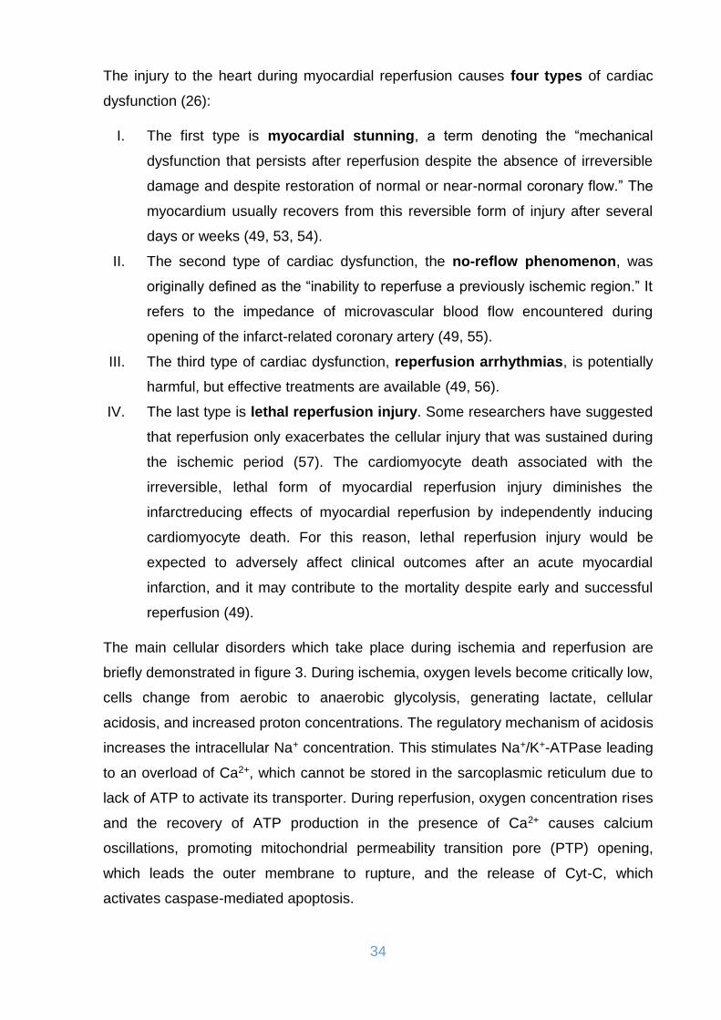
34
The injury to the heart during myocardial reperfusion causes four types of cardiac
dysfunction (26):
I. The first type is myocardial stunning, a term denoting the “mechanical
dysfunction that persists after reperfusion despite the absence of irreversible
damage and despite restoration of normal or near-normal coronary flow.” The
myocardium usually recovers from this reversible form of injury after several
days or weeks (49, 53, 54).
II. The second type of cardiac dysfunction, the no-reflow phenomenon, was
originally defined as the “inability to reperfuse a previously ischemic region.” It
refers to the impedance of microvascular blood flow encountered during
opening of the infarct-related coronary artery (49, 55).
III. The third type of cardiac dysfunction, reperfusion arrhythmias, is potentially
harmful, but effective treatments are available (49, 56).
IV. The last type is lethal reperfusion injury. Some researchers have suggested
that reperfusion only exacerbates the cellular injury that was sustained during
the ischemic period (57). The cardiomyocyte death associated with the
irreversible, lethal form of myocardial reperfusion injury diminishes the
infarctreducing effects of myocardial reperfusion by independently inducing
cardiomyocyte death. For this reason, lethal reperfusion injury would be
expected to adversely affect clinical outcomes after an acute myocardial
infarction, and it may contribute to the mortality despite early and successful
reperfusion (49).
The main cellular disorders which take place during ischemia and reperfusion are
briefly demonstrated in figure 3. During ischemia, oxygen levels become critically low,
cells change from aerobic to anaerobic glycolysis, generating lactate, cellular
acidosis, and increased proton concentrations. The regulatory mechanism of acidosis
increases the intracellular Na+ concentration. This stimulates Na+/K+-ATPase leading
to an overload of Ca2+, which cannot be stored in the sarcoplasmic reticulum due to
lack of ATP to activate its transporter. During reperfusion, oxygen concentration rises
and the recovery of ATP production in the presence of Ca2+ causes calcium
oscillations, promoting mitochondrial permeability transition pore (PTP) opening,
which leads the outer membrane to rupture, and the release of Cyt-C, which
activates caspase-mediated apoptosis.
35
Figure 3. Cellular damage in ischemia-reperfusion.
(Source: Alpuche J, Quírino L, Sánchez-Vega JT, Yap J, Pérez-Campos E, Cabrera-Fuentes HA. The Role of Platelets in Ischemic Conditioning. Cond Med. 2018 Oct;1(6):313-318.)
3.1. Myocardial Stunning
The term myocardial stunning refers to the transient myocardial dysfunction that
persists after reperfusion, despite the absence of irreversible myocardial damage
(54, 58, 59). The duration of myocardial stunning far exceeds the duration of the
previous ischemia. Transient occlusion of coronary perfusion for 15 minutes may for
example cause contraction disorders beyond 24 hours. Myocardial stunning causes
not only systolic but also diastolic myocardial dysfunction and is usually diagnosed by
the persistent contractile disorder that progressively improves over time, as long as
normal coronary flow is definitively restored (58, 59).
A number of hypotheses have been proposed to explain the pathogenesis of stunned
myocardium. The main factors and mechanisms involved and potential therapeutic
approaches are presented below in figure 4 and table 2. The most probable
mechanisms responsible for myocardial stunning are the damage caused by the
generation of oxygen-derived free radicals, disruption of calcium intracellular
homeostasis and possibly the change in the structure of contractile myofilaments as
well as decreased myofilament sensitivity to calcium. With reperfusion, production of

36
free oxygen radicals causes damage to the sarcolemma and cause a large influx of
calcium. Calcium activates endogenous proteases (calpains) that reduce the
response of myofilaments to calcium. This injury to the myofilaments most likely
leads to a decrease in sensitivity to calcium and the decrease in postischemic
contractile function, which is characteristic of stunned myocardium. In this regard, the
severity of damage to the myofilaments and possible mechanisms involved in
eliciting stunning may be determined by the intensity and/or duration of the ischemic
insult. If this large calcium discharge is prevented, myocardial stroke does not occur.
Diastolic dysfunction may be due to the disruption of calcium-induced relaxation (54,
58, 60, 61).
Figure 4. The main factors involved in the pathogenesis of myocardial stunning.
(Source: Gross GJ, Kersten JR, Warltier DC. Mechanisms of postischemic contractile dysfunction. Ann Thorac Surg. 1999 Nov;68(5):1898-904.)

37
Table 2. Mechanisms responsible for myocardial stunning and potential therapeutic approaches.
(Source: Gross GJ, Kersten JR, Warltier DC. Mechanisms of postischemic contractile dysfunction. Ann Thorac Surg. 1999 Nov;68(5):1898-904.)
3.2. Myocardial Hibernation
The term myocardial hibernation was initially used to describe chronic wall
contractility disorders in patients with chronic ischemic heart disease, without
however having experienced a myocardial infarction. This entity represents a new
state of equilibrium of the heart in response to chronic hypoperfusion, with reduced
contractility and reduced peripheral oxygen demand and consumption, so that
myocardial cells can remain viable (62, 63). After revascularization, regional
contractile function and ventricular ejection fraction are restored to normal.
The experimental counterpart is characterized by: 1) sustained perfusion-contraction
matching, 2) recovery of contractile function upon reperfusion, and 3) lack of
necrosis; frequently, there is also 4) recovery of metabolism during ongoing
ischemia, and 5) the potential for recruitment of inotropic reserve, and therefore
recovery of function upon reperfusion (64).
Through hibernation, despite the persistent hypoperfusion, a new state of cellular
energy equilibrium is achieved in which levels of concentration of lactic acid and high
38
energy phosphates remain normal. Morphologically, hibernating myocardium
displays features of dedifferentiation, with loss of cardiomyocytes and myofibrils, and
of degeneration, with increased interstitial fibrosis. Regardless of whether or not
hibernating myocardium has reduced baseline blood flow, and regardless of whether
its morphology appears more dedifferentiated or degenerative, it will benefit from
reperfusion, as will the patient’s prognosis (65). However if revascularisation takes
place in the late phase of remodelling, then the recovery potential is questionable.
The hibernating myocardium undergoes morphological changes with loss of its
sarcomeres and diminishing the size of mitochondria and the endoplasmic reticulum.
Remaining sarcomeres ingest larger amounts of glucose, resulting in glycogen
deposition in the myocardium. This kind of morphological change is called
dedifferentiation and does not seem to be as stable as originally believed and
probably reflects apoptotic death. By restoring coronary circulation, the rate of
functional and morphological recovery depends on the extent of changes in myocytes
and of interstitial fibrosis. This could explain why the reduction in coronary flow
observed in cases of hibernation is not necessarily relevant. Restoring coronary flow
with coronary artery bypass surgery (66) or percutaneous angioplasty (67) implies an
impressive improvement in contractility and increase in ejection fraction.
Improvement of myocardial function is so spectacular that coronary artery bypass
surgery is considered to be the treatment of choice in cases of patients with severe
heart failure of ischemic aetiology. Recruiting the hibernating parts of the
myocardium through reperfusion is the best option when conservative treatment fails.
The time course and extent of recovery after revascularization of viable dysfunctional
myocardium are variable. Although fibrosis is a major determinant, myocyte structural
and molecular remodeling may also play important roles. Delayed recovery of
hibernating myocardium in the absence of scar may reflect persistent reductions in
the amounts of contractile and metabolic proteins. Even in the absence of infarction,
the delayed improvement in left ventricular function after revascularization of
hibernating myocardium is associated with an incomplete reversal of the molecular
phenotype and the stimulation of new cardiomyocytes originally lost via regional
apoptosis from reversible ischemia. Although revascularization appeared to stimulate
myocyte proliferation, the persistence of small immature myocytes may have
contributed to delayed functional recovery (68). Several adjunctive treatments, such
as intracoronary stem cells (69) and growth factors (70), have been proposed as

39
methods to accelerate functional recovery following revascularization of high-risk
patients with heart failure, as well as those in whom complete revascularization is not
technically feasible. The benefits of such therapeutic strategies, particularly stem cell
therapy for patients with chronic symptomatic systolic heart failure due to ischemic
and nonischemic cardiomyopathy remain however unclear. Stem cells are a
promising therapeutic modality, however more data are required from larger blinded
trials to determine which combination of cell type and delivery mode will yield the
most benefit with avoidance of harm (71).
Stress echocardiography using dobutamine can be used to assess the existence of
viable myocardium in the ischemic area as well as the reversibility of systolic
dysfunction in patients to undergo reperfusion procedures. Improvement of
contractility is inversely proportional to the degree of myocardial fibrosis.
Administration of dobutamine increases the myocardial oxygen requirements, which
aggravates the functioning of the hibernating myocardium. Thus, the emergence of a
new area with a contrctility disorder or the reduction of the ejection fraction suggest
the presence of hibernation and predict the improvement of cardiac function after
reperfusion. Conversely, improvement in myocardial function after the administration
of dobutamine demonstrates the presence of an infract area, a positive response of
healthy areas and the absence of hibernation (72).
Advances in other cardiac imaging techniques have further contributed to knowledge
in the area, first with thallium-201 imaging, then later with Tc-99m-based tracers for
SPECT imaging and metabolic tracers used in conjunction with positron emission
tomography (PET), most commonly F-18 FDG in conjunction with blood flow imaging
with N-13 ammonia or Rb-82 Cl. More recently, cardiac magnetic resonance imaging
(CMR) has contributed further information in combination with either late gadolinium
enhancement imaging or dobutamine stress (73, 74).
Similar to myocardial stunning, the hibernating myocardium has recruitable areas
that improve their contractility after administration of positive inotropic agents. In
contrast to myocardial stunning, however, the increase in this contractility is
accompanied by an increased production of lactic acid and myocyte necrosis.
Functionality improves with afterload reduction, but the definitive and long-term
treatment is reperfusion (64-68).
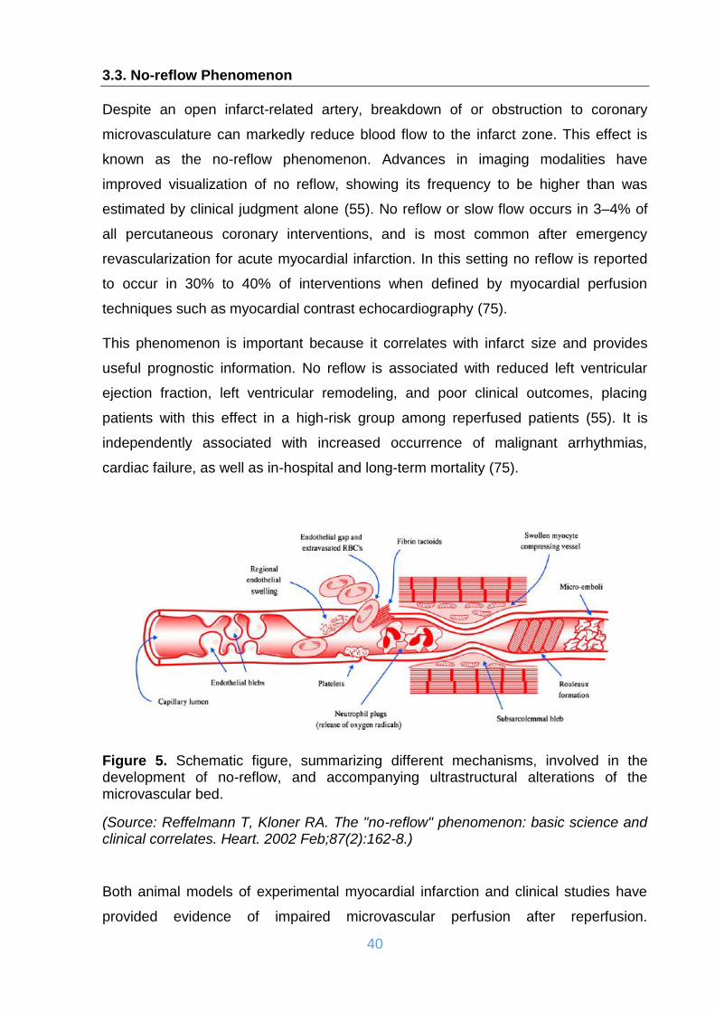
40
3.3. No-reflow Phenomenon
Despite an open infarct-related artery, breakdown of or obstruction to coronary
microvasculature can markedly reduce blood flow to the infarct zone. This effect is
known as the no-reflow phenomenon. Advances in imaging modalities have
improved visualization of no reflow, showing its frequency to be higher than was
estimated by clinical judgment alone (55). No reflow or slow flow occurs in 3–4% of
all percutaneous coronary interventions, and is most common after emergency
revascularization for acute myocardial infarction. In this setting no reflow is reported
to occur in 30% to 40% of interventions when defined by myocardial perfusion
techniques such as myocardial contrast echocardiography (75).
This phenomenon is important because it correlates with infarct size and provides
useful prognostic information. No reflow is associated with reduced left ventricular
ejection fraction, left ventricular remodeling, and poor clinical outcomes, placing
patients with this effect in a high-risk group among reperfused patients (55). It is
independently associated with increased occurrence of malignant arrhythmias,
cardiac failure, as well as in-hospital and long-term mortality (75).
Figure 5. Schematic figure, summarizing different mechanisms, involved in the development of no-reflow, and accompanying ultrastructural alterations of the microvascular bed.
(Source: Reffelmann T, Kloner RA. The "no-reflow" phenomenon: basic science and clinical correlates. Heart. 2002 Feb;87(2):162-8.)
Both animal models of experimental myocardial infarction and clinical studies have
provided evidence of impaired microvascular perfusion after reperfusion.

41
Characteristics of no-reflow found in basic science investigations, such as distinct
perfusion defects, progressive decrease of resting myocardial flow with ongoing
reperfusion and functional vascular alterations are paralleled by clinical observations
demonstrating similar features. The main pathophysiologic mechanisms involved are
summarized in figure 5 (76).
The focus of reperfusion therapy is shifting towards improved myocardial perfusion,
which could promote functional recovery of viable muscle, reduce infarct expansion,
and increase the delivery of blood-borne components, thereby accelerating the
healing process (55).
Previously the no-reflow phenomenon has been difficult to treat effectively, but recent
advances in the understanding of the pathophysiology of no reflow have led to
several novel treatment strategies. These include prophylactic use of vasodilator
therapies, mechanical devices, ischaemic postconditioning and potent platelet
inhibitors. As no reflow is a multifactorial process, a combination of these treatments
is more likely to be effective than any of these alone (75).
3.4. Reperfusion Arrhythmias
The time of reperfusion is often accompanied by ventricular arrhythmias, i.e.
reperfusion arrhythmias (77). Reperfusion arrhythmias originate as a consequence of
the complex of cellular and humoral reactions accompanying the opening of coronary
artery. The primary cause of their generation are considered to be the chemically
defined substances that are produced and accumulated in the myocardium during
reperfusion. The key role is ascribed to free oxygen radicals but of importance are
also other substances such as calcium, thrombin, platelet activating factor, inositol
triphosphate, angiotensin II and others. These chemical mediators of reperfusion
arrhythmias operate as modulators of cellular electrophysiology causing the complex
changes at the level of ion channels.
It is supposed that in the genesis of reperfusion arrhythmias, unlike ischemic
arrhythmias operate nonreentrant mechanisms such as abnormal or enhanced
automacy and triggered activity due to afterdepolarizations. As a typical reperfusion
arrhythmia is considered an early (within 6 hours after start of thrombolysis), frequent
(>30 episodes/hour) and repetitive (occurring during >3 consecutive hours)

42
accelerated idioventricular rhythm (AIVR). AIVR with such characteristics has a high
specificity and positive predictive accuracy but relative low sensitivity as a predictor of
reperfusion. Thus, in occurrence of AIVR, recanalization of infarction-related
coronary artery is very probable, but in absence of AIVR reperfusion is still not
excluded (78).
The following arrhythmias are regarded also as markers of reperfusion:
frequent premature ventricular complexes
a significant increase of episodes in nonsustained ventricular tachycardia
sinus bradycardia
high-degree atrioventricular blocks
The genesis of lethal reperfusion injury and reperfusion arrhythmias concentrates
around intracellular calcium overload for both events. This conformity in their
pathological basis leads to the conclusion that reperfusion arrhythmias and fatal
reperfusion injury should be considered to have the same underlying
pathophysiology. Reperfusion arrhythmias can therefore be seen as a marker of fatal
reperfusion injury instead of an independent entity (77). Reperfusion arrhythmias are
an important noninvasive marker of successful recanalization of infarction-related
coronary artery. However, they are also a sign of reperfusion injury and a finding
which may limit the favourable effect of reperfusion (78).
3.5. Mediators of lethal reperfusion injury
During myocardial reperfusion, the acute ischemic myocardium is subjected to
several abrupt biochemical and metabolic changes, which compound the changes
generated during the period of myocardial ischemia. These are described below and
are being summarized in figure 6.
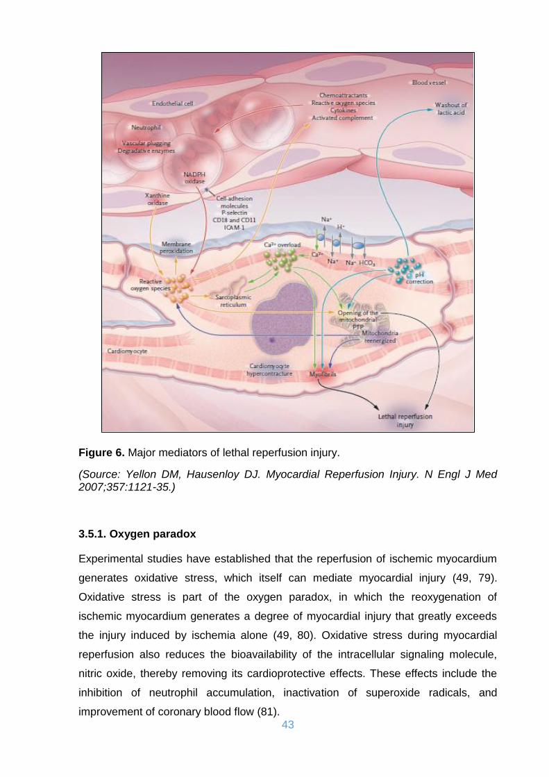
43
Figure 6. Major mediators of lethal reperfusion injury.
(Source: Yellon DM, Hausenloy DJ. Myocardial Reperfusion Injury. N Engl J Med 2007;357:1121-35.)
3.5.1. Oxygen paradox
Experimental studies have established that the reperfusion of ischemic myocardium
generates oxidative stress, which itself can mediate myocardial injury (49, 79).
Oxidative stress is part of the oxygen paradox, in which the reoxygenation of
ischemic myocardium generates a degree of myocardial injury that greatly exceeds
the injury induced by ischemia alone (49, 80). Oxidative stress during myocardial
reperfusion also reduces the bioavailability of the intracellular signaling molecule,
nitric oxide, thereby removing its cardioprotective effects. These effects include the
inhibition of neutrophil accumulation, inactivation of superoxide radicals, and
improvement of coronary blood flow (81).

44
3.5.2. Calcium paradox
At the time of myocardial reperfusion, there is an abrupt increase in intracellular Ca2+,
which is secondary to sarcolemmal-membrane damage and oxidative stress–induced
dysfunction of the sarcoplasmic reticulum. These two forms of injury overwhelm the
normal mechanisms that regulate Ca2+ in the cardiomyocyte; this phenomenon is
termed the calcium paradox (49, 82). The result is intracellular and mitochondrial
Ca2+ overload, and this excess of Ca2+ induces cardiomyocyte death by causing
hypercontracture of the heart cells and mitochondrial PTP opening (82).
3.5.3. pH paradox
The rapid restoration of physiologic pH during myocardial reperfusion, which follows
the washout of lactic acid and the activation of the sodium–hydrogen exchanger and
the sodium–bicarbonate symporter, contributes to lethal reperfusion injury. This
phenomenon is termed the pH paradox (49, 83).
3.5.4. Inflammation
After an acute myocardial infarction, the release of chemoattractants draws
neutrophils into the infarct zone during the first 6 hours of myocardial reperfusion,
and during the next 24 hours they migrate into the myocardial tissue. This process is
facilitated by cell-adhesion molecules. These neutrophils cause vascular plugging
and release degradative enzymes and reactive oxygen species (84).
3.5.5. Metabolic modulation
Several experimental and numerous clinical studies have examined the
cardioprotective potential of therapy with glucose, insulin, and potassium
administered as an adjunct to myocardial reperfusion (85). These studies have been
conducted on the premise that ischemic myocardium benefits more from
metabolizing glucose than from fatty acids (49). However recent clinical evidence
reported no cardioprotective benefit from therapy with glucose, insulin, and
potassium as an adjunct to myocardial reperfusion in patients with acute myocardial
infarction (86).
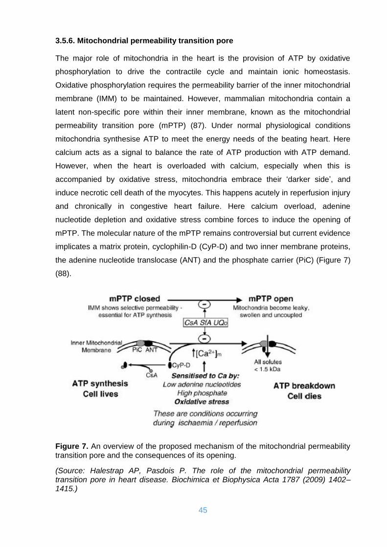
45
3.5.6. Mitochondrial permeability transition pore
The major role of mitochondria in the heart is the provision of ATP by oxidative
phosphorylation to drive the contractile cycle and maintain ionic homeostasis.
Oxidative phosphorylation requires the permeability barrier of the inner mitochondrial
membrane (IMM) to be maintained. However, mammalian mitochondria contain a
latent non-specific pore within their inner membrane, known as the mitochondrial
permeability transition pore (mPTP) (87). Under normal physiological conditions
mitochondria synthesise ATP to meet the energy needs of the beating heart. Here
calcium acts as a signal to balance the rate of ATP production with ATP demand.
However, when the heart is overloaded with calcium, especially when this is
accompanied by oxidative stress, mitochondria embrace their ‘darker side’, and
induce necrotic cell death of the myocytes. This happens acutely in reperfusion injury
and chronically in congestive heart failure. Here calcium overload, adenine
nucleotide depletion and oxidative stress combine forces to induce the opening of
mPTP. The molecular nature of the mPTP remains controversial but current evidence
implicates a matrix protein, cyclophilin-D (CyP-D) and two inner membrane proteins,
the adenine nucleotide translocase (ANT) and the phosphate carrier (PiC) (Figure 7)
(88).
Figure 7. An overview of the proposed mechanism of the mitochondrial permeability transition pore and the consequences of its opening.
(Source: Halestrap AP, Pasdois P. The role of the mitochondrial permeability transition pore in heart disease. Biochimica et Biophysica Acta 1787 (2009) 1402–1415.)

46
The mitochondrial permeability transition pore (PTP) is a nonselective channel of the
inner mitochondrial membrane. Opening the channel collapses the mitochondrial
membrane potential and uncouples oxidative phosphorylation, resulting in ATP
depletion and cell death (89). During myocardial ischemia, the mitochondrial PTP
remains closed, only to open within the first few minutes after myocardial reperfusion
in response to mitochondrial Ca2+ overload, oxidative stress, restoration of a
physiologic pH, and ATP depletion (90). Therefore, the mitochondrial PTP is a critical
determinant of lethal reperfusion injury, and as such it is an important new target for
cardioprotection (49).
3.5.7. The Reperfusion Injury Salvage Kinase (RISK) pathway
The Reperfusion Injury Salvage Kinase (RISK) Pathway, refers to a group of pro-
survival protein kinases (including Akt and Erk1/2), which confer powerful
cardioprotection. When activated specifically at the time of myocardial reperfusion,
the RISK pathway provides an amenable pharmacological target for cardioprotection
(91). In a sense, the RISK pathway mediates a form of programmed cell survival (49).
There is extensive preclinical evidence that activation of the RISK pathway by
pharmacologic agents such as insulin, erythropoietin, adipocytokines, adenosine,
volatile anesthetics, natriuretic peptides and statins, when administered specifically at
the time of myocardial reperfusion or by mechanical interventions such as ischemic
preconditioning or postconditioning reduce myocardial infarct size by up to 50% (91-
93). These treatments recruit various survival pathways providing cardioprotection.
The cardioprotection has been attributed to inhibition of mitochondrial PTP opening,
improved uptake of Ca2+ in the sarcoplasmic reticulum, and the recruitment of
antiapoptotic pathways (49, 91). The several agents which can activate the RISK
pathway are summarized in figure 8.

47
Figure 8. Scheme demonstrating the diverse variety of agents which activate the Reperfusion Injury Salvage Kinase (RISK) pathway in both a receptor and non-receptor mediated manner.
(Source: Hausenloy DJ, Yellon DM. Reperfusion injury salvage kinase signalling: taking a RISK for cardioprotection. Heart Fail Rev. 2007 Dec;12(3-4):217-34.)
4. APPLIED PHYSIOLOGY AND PATHOPHYSIOLOGY
The failure of cardiac output to meet the metabolic needs of the body mobilizes
various reversal mechanisms that result in increased intravascular volume and
vascular tone (94). These mechanisms tend to restore cardiac output to normal
levels, but they also cause pressure increases in left cavities and pulmonary
circulation. In this way, most patients with congestive heart failure have sufficient
cardiac output to meet the metabolic needs at rest, yet with increased pulmonary
pressures that cause pulmonary congestion. Lack of tolerance during exercise is
probably due to increased pulmonary pressures and not to inadequate cardiac output
(95). For this reason, the syndrome of circulatory distress due to impaired left
ventricular function is called congestive heart failure. Additionally, congestive heart
failure may also occur in patients with diastolic dysfunction whose systolic function
remains intact (96).

48
4.1. Basic parameters of circulatory physiology
4.1.1. Frank–Starling law
The Frank–Starling law of the heart (Figure 9) represents the relationship between
stroke volume and end diastolic volume. The law states that the stroke volume of the
heart increases in response to an increase in the end diastolic volume, when all other
factors remain constant. As a larger volume of blood flows into the ventricle, the
blood stretches the cardiac muscle fibers, leading to an increase in the force of
contraction. The Frank-Starling mechanism allows the cardiac output to be
synchronized with the venous return, arterial blood supply and humoral length,
without depending upon external regulation to make alterations. The Frank-Starling
relationship is an intrinsic property of myocardium by which increased length (or
ventricular volume) results in enhanced performance during the subsequent
contraction. This relationship appears to be very important in cardiac function
because increased venous return and the corresponding increase in end-diastolic
volume result in greater stroke volume during the next beat. The ventricles can thus
accommodate increased venous return by means of a more vigorous contraction that
ejects the greater volume of blood from the heart (97-101).
Figure 9. Cardiac function curve illustrating the Frank–Starling law of the heart, the y-axis often describes the stroke volume, stroke work, or cardiac output. The x-axis often describes end-diastolic volume, right atrial pressure, or pulmonary capillary wedge pressure.

49
Although the physiological significance of the Frank-Starling relationship is widely
appreciated, its cellular basis is not well understood. One hypothesis is that more
cross-bridges interact with actin at longer sarcomere lengths due to length-
dependent reductions in lateral spacing between thick and thin filaments. Due to
closer proximity to actin more crossbridges bind and thereby increase contractile
force (Figure 10). Measurements in permeabilized myocardium held at constant
length have shown that osmotic compression increases force at each [Ca2+]. Thus,
the greater Ca2+ sensitivity of force at long lengths can be achieved at short lengths
by reducing fiber diameter. Moreover, osmotic compression actually eliminates the
length dependence of Ca2+ sensitivity. This evidence suggests that lateral filament
spacing is a primary determinant of the Frank-Starling relationship. Diameter and
filament separation are likely to increase or decrease in concert, but these changes
need not be proportionate because the shape of the muscle cross section can
change with stretch or with osmotic compression (101, 102).
Figure 10. Myocardial sarcomere and myofilaments. A. Diagram of the sarcomere showing approximate spatial relationships of thick and thin filaments and putative interactions of titin with the filaments, which would give rise to radial and axial restorati restorative forces when the sarcomere is stretched. B. Diagram of the thick and thin filaments illustrating the decrease in lateral separation at long lengths. The probability of crossbridge interaction increases at long lengths due to closer proximity to actin.
(Source: Moss RL, Fitzsimons DP. Frank-Starling relationship=long on importance, short on mechanism. Circ Res. 2002 Jan 11;90(1):11-3.)
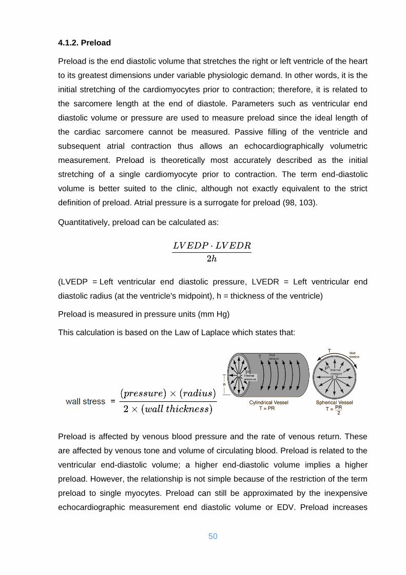
50
4.1.2. Preload
Preload is the end diastolic volume that stretches the right or left ventricle of the heart
to its greatest dimensions under variable physiologic demand. In other words, it is the
initial stretching of the cardiomyocytes prior to contraction; therefore, it is related to
the sarcomere length at the end of diastole. Parameters such as ventricular end
diastolic volume or pressure are used to measure preload since the ideal length of
the cardiac sarcomere cannot be measured. Passive filling of the ventricle and
subsequent atrial contraction thus allows an echocardiographically volumetric
measurement. Preload is theoretically most accurately described as the initial
stretching of a single cardiomyocyte prior to contraction. The term end-diastolic
volume is better suited to the clinic, although not exactly equivalent to the strict
definition of preload. Atrial pressure is a surrogate for preload (98, 103).
Quantitatively, preload can be calculated as:
(LVEDP = Left ventricular end diastolic pressure, LVEDR = Left ventricular end
diastolic radius (at the ventricle's midpoint), h = thickness of the ventricle)
Preload is measured in pressure units (mm Hg)
This calculation is based on the Law of Laplace which states that:
Preload is affected by venous blood pressure and the rate of venous return. These
are affected by venous tone and volume of circulating blood. Preload is related to the
ventricular end-diastolic volume; a higher end-diastolic volume implies a higher
preload. However, the relationship is not simple because of the restriction of the term
preload to single myocytes. Preload can still be approximated by the inexpensive
echocardiographic measurement end diastolic volume or EDV. Preload increases

51
with exercise (slightly), increasing blood volume (overtransfusion, polycythemia) and
neuroendocrine excitement (sympathetic tone) (98).
4.1.3. Afterload
Afterload is the end load against which the heart contracts to eject blood. Afterload is
readily broken into components: one factor is the aortic pressure the left ventricular
muscle must overcome to eject blood. The greater the aortic/pulmonary pressure, the
greater the afterload on the left/right ventricle, respectively. Following Laplace's law,
the tension upon the muscle fibers in the heart wall is the pressure within the
ventricle multiplied by the volume within the ventricle divided by the wall thickness
(this ratio is the other factor in setting the afterload). Afterload can also be described
as the pressure that the chambers of the heart must generate in order to eject blood
out of the heart and thus is a consequence of the aortic pressure (for the left
ventricle) and pulmonic pressure or pulmonary artery pressure (for the right
ventricle). The pressure in the ventricles must be greater than the systemic and
pulmonary pressure to open the aortic and pulmonic valves, respectively. As
afterload increases, cardiac output decreases. Cardiac imaging is a somewhat
limited modality in defining afterload because it depends on the interpretation of
volumetric data (98, 104).
Afterload is largely dependent upon aortic pressure. Systemic hypertension
increases the LV afterload because the LV must work harder to eject blood into the
aorta. This is because the aortic valve will not open until the pressure generated in
the left ventricle is higher than the elevated blood pressure in the aorta. Pulmonary
hypertension indicates a regionally applied increase in afterload dedicated to the right
side of the heart, divided and isolated from the left heart by the interventricular
septum. Aortic stenosis often increases afterload because the left ventricle must
overcome the pressure gradient caused by the calcified and stenotic aortic valve in
addition to the blood pressure in order to eject blood into the aorta. In the long-term,
the increased afterload will result in LV hypertrophy to account for the increased work
required. Aortic Regurgitation increases afterload because a percentage of the blood
that is ejected forward regurgitates back through the diseased aortic valve. Mitral
regurgitation decreases afterload. The remaining blood loaded into the LV is
optimally ejected out through the aortic valve. With an extra pathway for blood flow

52
through the mitral valve, the left ventricle does not have to work as hard to eject its
blood (98, 104).
4.1.4. Stroke volume
Stroke volume (SV) is the volume of blood pumped from the left ventricle per beat.
Stroke volume is calculated using measurements of ventricle volumes from an
echocardiogram and subtracting the end-systolic volume (ESV) from the end-
diastolic volume (EDV). The term stroke volume can apply to each of the two
ventricles of the heart, although it usually refers to the left ventricle (98, 103, 104).
Stroke volume index (SVI) is a method of relating the SV to the size of the person
Body surface area (BSA).
The normal range is between 35 and 65 ml/beat/m².
Stroke volume is dependent upon the difference between end diastolic volume and
end systolic volume. The three primary factors involved are preload, afterload and
contractility.
4.1.5. Cardiac output and cardiac index
Cardiac output (CO) is a term used in cardiac physiology that describes the volume
of blood being pumped by the heart, in particular by the left or right ventricle, per unit
time. Cardiac output is the product of the heart rate (HR) and the SV.
There are a number of clinical methods to measure cardiac output, ranging from
direct intracardiac catheterization to non-invasive measurement of the arterial pulse.
Each method has advantages and drawbacks. Relative comparison is limited by the
absence of a widely accepted "gold standard" measurement. Cardiac output can also

53
be affected significantly by the phase of respiration, as intra-thoracic pressure
changes influence diastolic filling and therefore cardiac output. This is especially
important during mechanical ventilation, in which cardiac output can vary by up to
50% across a single respiratory cycle (98, 104). Figure 11 shows the factors which
can affect CO.
Figure 11. Major factors influencing heart rate and stroke volume and consequently cardiac output.
(Source: Illustration from Anatomy & Physiology, Connexions Web site. http=//cnx.org/content/col11496/1.6)
Cardiac index (CI) is a haemodynamic parameter that relates the (CO) from left
ventricle in one minute to BSA, thus relating heart performance to the size of the
individual. The unit of measurement is litres per minute per square metre (L/min/m2)
(98, 104).
The normal range of cardiac index at rest is 2.6–4.2 L/min/m2. The cardiac index is
frequently measured and used in both intensive care medicine and cardiac intensive
care. The CI is a useful marker of how well the heart is functioning as a pump by
54
directly correlating the volume of blood pumped by the heart with an individual's body
surface area. If the CI falls acutely below 2.2 L/min/m2, the patient may be in
cardiogenic shock (98, 104).
4.1.6. Ejection Fraction
The ejection fraction (EF) of the left heart, known as the left ventricular ejection
fraction (LVEF), is calculated by dividing the stroke volume by the end-diastolic
volume. LVEF is an indicator of the effectiveness of pumping into the systemic
circulation. The EF of the right heart, or right ventricular ejection fraction (RVEF), is a
measure of the efficiency of pumping into the pulmonary circulation (98, 104).
The 2016 European Society of Cardiology Guidelines for the diagnosis and treatment
of acute and chronic heart failure subdivided heart failure into 3 categories on the
basis of LVEF (105):
normal or preserved LVEF [≥50%]
moderately reduced LVEF [in the range of 40–49%]
reduced LVEF [<40%]
4.1.7. Pressure-Volume work
The intraventricular pressure-volume loop is a time-independent representation of the
cardiac cycle, where the instantaneous intraventricular pressure and volume is
plotted (Figure 12). In diastole from B to C, the ventricle receives blood from the left
atrium. The small increase in ventricular pressure reflects passive expansion and
elastance of the myocardial wall. Pressure and volume increase with a slope that is
related to contractility. The green line represents minimal contractility. The line starts
from the ventricular dead volume that is a virtual minimal volume of blood, which can
never be ejected. A steep rise in pressure occurs from C to D, with no change in
ventricular volume (the isovolumetric contraction). At D the aortic orifice opens,
because the end-diastolic pressure in the aorta is passed. During the rapid ejection
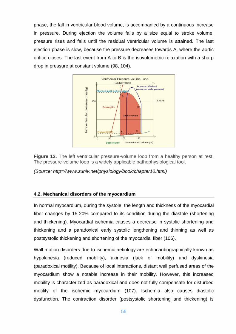
55
phase, the fall in ventricular blood volume, is accompanied by a continuous increase
in pressure. During ejection the volume falls by a size equal to stroke volume,
pressure rises and falls until the residual ventricular volume is attained. The last
ejection phase is slow, because the pressure decreases towards A, where the aortic
orifice closes. The last event from A to B is the isovolumetric relaxation with a sharp
drop in pressure at constant volume (98, 104).
Figure 12. The left ventricular pressure-volume loop from a healthy person at rest. The pressure-volume loop is a widely applicable pathophysiological tool.
(Source: http=//www.zuniv.net/physiology/book/chapter10.html)
4.2. Mechanical disorders of the myocardium
In normal myocardium, during the systole, the length and thickness of the myocardial
fiber changes by 15-20% compared to its condition during the diastole (shortening
and thickening). Myocardial ischemia causes a decrease in systolic shortening and
thickening and a paradoxical early systolic lengthening and thinning as well as
postsystolic thickening and shortening of the myocardial fiber (106).
Wall motion disorders due to ischemic aetiology are echocardiographically known as
hypokinesia (reduced mobility), akinesia (lack of mobility) and dyskinesia
(paradoxical motility). Because of local interactions, distant well perfused areas of the
myocardium show a notable increase in their mobility. However, this increased
mobility is characterized as paradoxical and does not fully compensate for disturbed
motility of the ischemic myocardium (107). Ischemia also causes diastolic
dysfunction. The contraction disorder (postsystolic shortening and thickening) is

56
accompanied by an increase in the time of isovolumic relaxation and a reduction in
the maximum ventricular filling flow. In addition, ischemia reduces the compliance of
the ventricle. This decrease is proportional to the extent and duration of ischemia and
is further reduced by coronary artery occlusion. The decrease in compliance is not
limited to the ischemic area but extends to distant and well perfused areas. A
reduction in contractility is also seen in areas not involved in the infarction due to
obstruction of the coronary blood vessels that perfuse the particular areas that have
occurred earlier and to the loss of collateral networks from the new occlusion of the
responsible vessel known as ischemia at distance (108). This effect contributes to
the abrupt increase in left ventricular end-diastolic pressure, which is often observed
after regional myocardial ischemia (109).
When ischemia or necrosis influences a large area of the myocardium a heart failure
occurs (110). Reduction of SV and fall in CO cause hypotension and tachycardia and
reduce coronary flow. Increase of enddiastolic pressures also contributes to the
reduction of coronary flow and increased wall stress causes increased O2
consumption by the myocardium (111). All of these factors interact and deteriorate
ischemia. The fall in CO causes some further impairment of peripheral tissue
perfusion. Reactive mechanisms include sympathetic stimulation and water retention
in an attempt to increase preload. These mechanisms however deteriorate the
condition due to an increase in myocardial O2 requirements and an increase in the
afterload (112). Figure 13 demonstrates the changes of the left ventricular pressure-
volume loop in patients with heart failure.
Figure 13. Left ventricular pressure-volume loops in a healthy person (red) and for persons with acute (blue area) or chronic, congestive (green area) cardiac failure.
(Source: http=//www.zuniv.net/physiology/book/chapter10.html)

57
4.2.1. Systolic myocardial dysfunction
The term systolic myocardial dysfunction defines the impairment of ejection capacity
of the LV and it can be evaluated and quantified through the EF. The induction of
myocardial ischemic damage implies the reduction of left ventricular ejection
capacity, stroke volume, cardiac output, blood pressure and dP/dt ratio, while at the
same time there is an increase in the endsystolic volume, whose rate of growth is the
most prominent mortality prognostic factor after acute coronary syndromes ACS
(113). The induction of dyskinesia entails a significant reduction in SV of the LV.
Dilatation of the ventricular area is a result of slipping of the necrotic myocytes,
resulting in thinning and extension of the infarct zone (114).
The dilation caused by the onset of the infarction, according to Laplace's law,
increases wall tension and therefore causes more dilation. The degree of ventricular
dilation depends on the extension of the infarct, the viability of the involved artery and
the activation of the renin-angiotensin-aldosterone system (RAAS) and may be
modified by inhibitors of the particular system, even in the absence of symptomatic
left ventricular dysfunction. Gradually, an increase in stiffness as a result of tissue
oedema, cellular infiltration and fibrosis of the ventricular zone is observed, resulting
in reduced dyskinesia and improved left ventricular function (115, 116).
The occurrence of symptomatic heart failure is associated with specific parameters of
the left ventricle. The reduction in compliance occurs early in small-scale infarctions.
In cases where the affected part of the myocardium exceeds 15%, there is a
reduction in the ejection fraction, while the left ventricular end-diastolic pressure and
its volume may increase. The presence of signs and symptoms of heart failure
increases with the number of areas of abnormal left ventricular mobility. An area with
an abnormal contraction of more than 25% causes the onset of heart failure, while
cardiogenic shock occurs in excess of 40% (113).
4.2.2. Diastolic myocardial dysfunction
The induction of ischemia and infarction affects the left ventricular diastolic function
as the maximal rate of decrease in left ventricular pressure (peak -dP/dt) decreases,
the time variable of the fall in ventricular pressure and the early increase in
enddiastolic pressure of the left ventricle. Gradually increases in enddiastolic volume

58
are observed while diastolic blood pressure returns to normal levels. Infarct size
determines the grade of diastolic dysfunction (117, 118).
Normal diastolic function is defined as the filling of the LV capable of achieving a
satisfactory SV without increasing left ventricular filling pressure and the pressure of
the pulmonary circulation. When EF is reduced, a larger enddiastolic volume is
required to maintain an adequate SV. Therefore the filling that the heart needs to
achieve sufficient SV depends on its systolic performance. Insufficient filling that
leads to low cardiac output mobilizes compensation mechanisms that eventually
increase pressure in pulmonary circulation. In this way diastolic dysfunction disorders
are not accompanied by a fall in CO at rest but by pulmonary congestion. In patients
with systolic dysfunction, if the increase in the enddiastolic volume is not
accompanied by a pathological increase in pulmonary circulation pressures, a
satisfactory compensation of the systolic dysfunction is achieved. If, on the other
hand, the increased enddiastolic volume leads to a pathologic increase in pulmonary
artery pressure, then the systolic dysfunction also results in diastolic dysfunction. In
this sense, systolic dysfunction is the most common cause of diastolic dysfunction
(117, 118).
Diastolic dysfunction however may also occur without the presence of systolic
dysfunction. In this case, it may be due to disturbance of the ventricular filling,
reduced ventricular compliance or external ventricular pressure. The most common
cause of primary diastolic dysfunction is the decrease in compliance. When this
occurs, significantly higher intraventricular pressures develop to fill the ventricle with
the same enddiastolic volume. If the decrease in compliance is high, ventricular filling
is achieved only with a disproportionate increase in pressure, but this also leads to
congestion. In this way reduced ventricular compliance may cause pulmonary
congestion and congestive heart failure without the coexistence of a systolic
dysfunction (117, 118).
Decreased compliance may be due to hypertrophic cardiomyopathy, ischemia or
relaxation disorders. The hypertrophy of LV is another cause of decreased
compliance. It helps to restore myocardial tension to normal levels and helps
maintain systolic function in patients with aortic valve stenosis or chronic
hypertension, but it negatively impacts diastolic dilatation and ultimately leads to
diastolic dysfunction (117, 118).

59
4.3. Cardiac remodeling
A common consequence of the MI is the appearance of changes in the size, shape
and thickness of the LV involving the entire cavity including the affected areas, but
also areas which were not directly involved in the MI. These changes are collectively
referred to as remodeling of the ventricles. Remodeling is a process which affects the
ventricular function and the prognosis of the disease. Both dilatation and hypertrophy
contribute to remodeling. Cardiac remodelling is the central mechanism of heart
failure progression in patients with IHD, occurring usually as a consequence of AMI.
Postinfarction remodelling can be divided into an early phase (within 72 hours) and a
late phase (beyond 72 hours) (119, 120).
4.3.1. Early remodeling
Early remodelling involves expansion of the infarct zone and collagen degradation,
which may result in early ventricular rupture or aneurysm formation (121). Infarct
expansion results from the degradation of the intermyocyte collagen struts by serine
proteases and from the activation of matrix metalloproteinases (MMPs) released from
neutrophils (122). Infarct expansion occurs within hours of myocyte injury, results in
wall thinning and ventricular dilatation, and causes the elevation of diastolic and
systolic wall stresses (123). Substantial changes in circulatory haemodynamics
trigger the sympathetic adrenergic system, which stimulates catecholamines,
activates the renin-angiotensin-aldosterone system (RAAS), and stimulates the
production of endothelins, and atrial and b-type natriuretic peptides (ANP and BNP).
Positive inotropic, chronotropic and also vasodilatory effects from this sympathetic
stimulation result in hyperkinesis of the noninfarcted myocardium and temporary
circulatory compensation by reduction of systemic vascular resistance and left
ventricular filling pressure (119). However, although the neurohormonal activation
initially serves an adaptive role, in later stages the responses become pathological
and contribute adversely to remodelling and ultimately to the progress of heart
failure. In addition, neurohormonal activation may precipitate further ischaemia by
increasing oxygen demand and predisposing to arrhythmias (124).
60
4.3.2. Late remodelling and scar formation
Late remodelling involves the left ventricle globally and is associated with time-
dependent dilatation, distortion of ventricular shape and hypertrophy. Hypertrophy is
an adaptive response during postinfarction remodelling that offsets increased load,
attenuates progressive dilatation, and stabilizes contractile function. It is initiated by
neurohormonal activation, myocardial stretch, activation of the RAAS, and by
paracrine/autocrine factors. Especially enhanced norepinephrine release contributes
to the hypertrophy (125).
Myocardial repair and scar formation is triggered by cytokines released from injured
myocytes. Before collagen synthesis tissue repair is initiated by the formation of a
fibrin-fibronectin matrix to which myofibroblasts become adherent (126). Deposition
of collagen occurs predominantly in the infarct zone, but also in noninfarcted
myocardium. Collagen is detectable microscopically by day 7 and its deposition then
increases dramatically, such that by 28 days, the necrotic myocytes are entirely
replaced by fibrous tissue. After the formation of a scar that equilibrates distending
and restraining forces, collagen formation is down-regulated and most fibroblasts
undergo apoptosis (122).
4.4. Activation of Renin-Angiotensin-Aldosterone-System
The circulating renin-angiotensin-aldosterone system (RAAS) plays an important role
in the maintenance of cardiovascular homeostasis. It has been demonstrated that
endogenous RAS exists in target tissues that are important in cardiovascular
regulation. Myocardial areas that have not been necrotic following myocardial
infarction show an activation of the renin-angiotensin system and cause increased
angiotensin II production. Angiotensin II leads to the production of various growth
factors, such as platelet growth factor (PGF) and transforming growth factor beta
(TGFb), which cause hypertrophy of non-infarcted myocardium and regulate the tone
and structure of the coronary vessels. Further actions of angiotensin II are the
secretion of endothelin, PAI-1 and aldosterone, which cause vasoconstriction,
decreased fibrinolysis and increased sodium retention, respectively (127, 128).
61
5. CLINICAL PRESENTATIONS OF ISCHEMIC HEART DISEASE
Ischaemic heart disease (IHD), caused as mentioned before by an imbalance
between the supply and demand of oxygen to the heart, may be symptomatic or
asymptomatic and it may have a stable or a progressive course. IHD is classified on
the basis of its symptomatology and severity (98).
5.1. Stable angina pectoris
Stable angina pectoris is the main symptom/form of IHD. The pathological substrate
for angina is almost invariably atheromatous narrowing of the coronary arteries. It is
usually considered that a coronary artery must be narrowed by at least 50-70% in
luminal diameter before coronary blood flow is inadequate to meet the metabolic
demands of the heart with exercise or stress. In this case the stenosis can be defined
as significant (129). The importance of stenosis depends also on the length and
number of stenoses. Angina pectoris results from myocardial ischaemia, which is
caused by an imbalance between myocardial oxygen requirements and oxygen
supply. Increased oxygen demand may occur due to increase in heart rate, left
ventricular wall stress or contractility. Oxygen supply, on the other hand, is
determined by coronary blood flow and coronary arterial oxygen content. The
precipitating factors causing angina due to increased myocardial oxygen
consumption include exercise, mental stress, fever, cold, tachycardia from any
cause, thyrotoxicosis, and hypoglycaemia (130). Typical angina pectoris is
substernal, across mid-thorax, anteriorly, can locate also in arms, shoulders, neck,
and interscapular region. It is characterized by a burning, heavy or squeezing feeling,
is precipitated by exertion or emotion and is promptly relieved by rest or by
nitroglycerin. The typical episode of angina pectoris usually begins gradually and
reaches its maximum intensity over a period of minutes before dissipating (129-131).
If the symptoms remain the same for several weeks and constantly occur under the
same physical or mental stress, the condition is described as “stable” angina pectoris
(95-97). Stable angina pectoris is classified by severity into 4 classes according to
the Canadian Cardiovascular Society (CCS) (Table 3) (132).
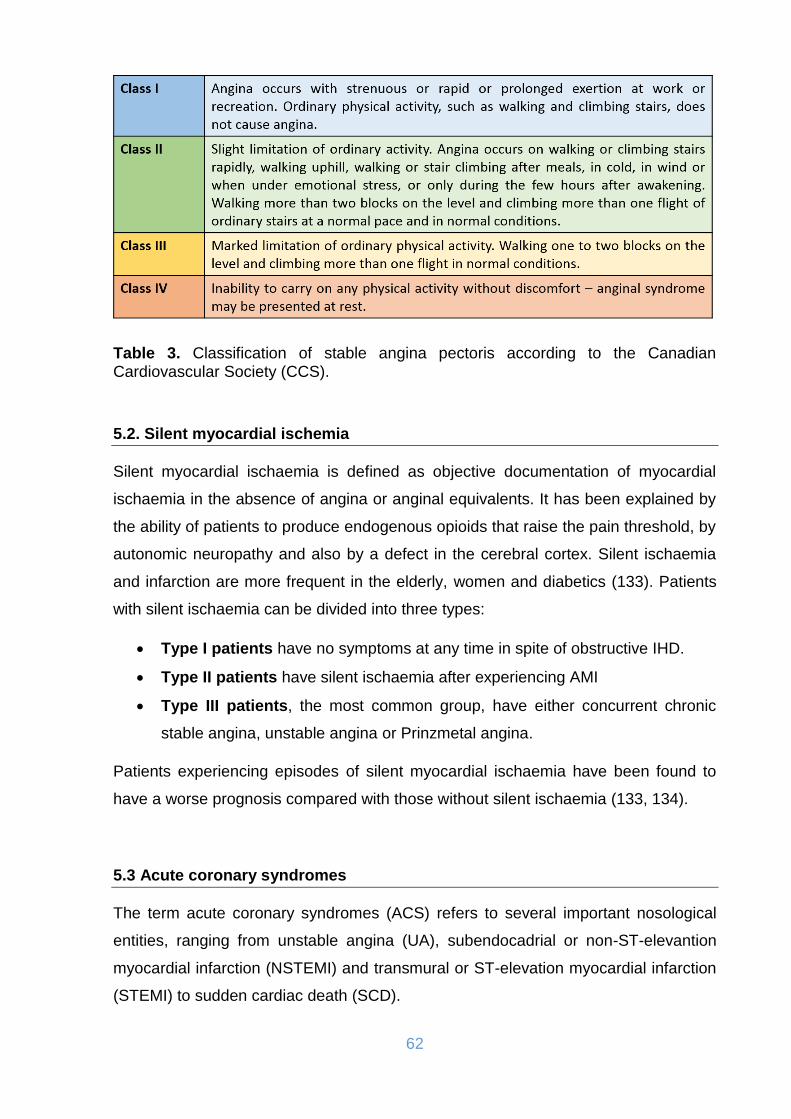
62
Table 3. Classification of stable angina pectoris according to the Canadian Cardiovascular Society (CCS).
5.2. Silent myocardial ischemia
Silent myocardial ischaemia is defined as objective documentation of myocardial
ischaemia in the absence of angina or anginal equivalents. It has been explained by
the ability of patients to produce endogenous opioids that raise the pain threshold, by
autonomic neuropathy and also by a defect in the cerebral cortex. Silent ischaemia
and infarction are more frequent in the elderly, women and diabetics (133). Patients
with silent ischaemia can be divided into three types:
Type I patients have no symptoms at any time in spite of obstructive IHD.
Type II patients have silent ischaemia after experiencing AMI
Type III patients, the most common group, have either concurrent chronic
stable angina, unstable angina or Prinzmetal angina.
Patients experiencing episodes of silent myocardial ischaemia have been found to
have a worse prognosis compared with those without silent ischaemia (133, 134).
5.3 Acute coronary syndromes
The term acute coronary syndromes (ACS) refers to several important nosological
entities, ranging from unstable angina (UA), subendocadrial or non-ST-elevantion
myocardial infarction (NSTEMI) and transmural or ST-elevation myocardial infarction
(STEMI) to sudden cardiac death (SCD).
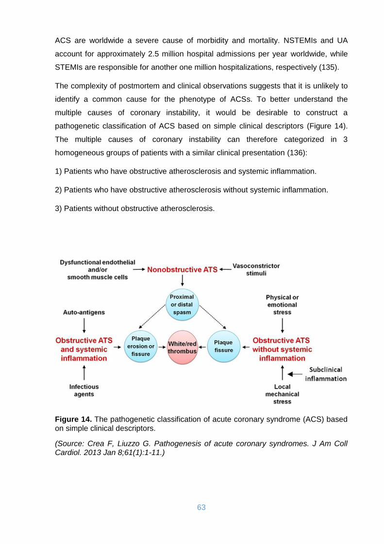
63
ACS are worldwide a severe cause of morbidity and mortality. NSTEMIs and UA
account for approximately 2.5 million hospital admissions per year worldwide, while
STEMIs are responsible for another one million hospitalizations, respectively (135).
The complexity of postmortem and clinical observations suggests that it is unlikely to
identify a common cause for the phenotype of ACSs. To better understand the
multiple causes of coronary instability, it would be desirable to construct a
pathogenetic classification of ACS based on simple clinical descriptors (Figure 14).
The multiple causes of coronary instability can therefore categorized in 3
homogeneous groups of patients with a similar clinical presentation (136):
1) Patients who have obstructive atherosclerosis and systemic inflammation.
2) Patients who have obstructive atherosclerosis without systemic inflammation.
3) Patients without obstructive atherosclerosis.
Figure 14. The pathogenetic classification of acute coronary syndrome (ACS) based on simple clinical descriptors.
(Source: Crea F, Liuzzo G. Pathogenesis of acute coronary syndromes. J Am Coll Cardiol. 2013 Jan 8;61(1):1-11.)

64
5.3.1. Unstable angina and Prinzmetal (variant) angina
The currently used definition of unstable angina pectoris depends on the presence of
one or more of the following features (98):
1) Crescendo angina (more severe, prolonged or frequent) superimposed on an
existing pattern of relatively stable, exertion-related angina pectoris.
2) Angina pectoris of new onset (usually within 1 month), which is brought on by
minimal exertion.
3) Angina pectoris at rest as well as with minimal exertion.
A classification of UA is presented in Table 4 (135).
Table 4. Classification of unstable angina.
(Source: Hamm CW, Braunwald E. A classification of unstable angina revisited. Circulation 2000;102(1):118-22.)
Prinzmetal’s (variant) angina is an unusual and uncommon form of angina secondary
to myocardial ischaemia that occurs almost exclusively at rest, is usually not
precipitated by physical exertion or emotional stress and is associated with ST-
segment elevations in electrocardiogram (136). Variant angina is demonstrated to be
due to coronary artery spasm, which narrows the coronary artery resulting in
myocardial ischaemia (104). Endothelial dysfunction, an increased platelet
aggregation together with changes in autonomic tone can trigger the vasospasms
(137, 138). Also dysfunction of K+-ATP channels may have role in variant angina
(139).

65
5.3.2. Acute myocardial infarction (NSTEMI and STEMI)
Acute myocardial infarction (AMI) represents the most critical and serious form of
IHD. Although the death rate from AMI has continuously declined over the past
decades, its development together with all complications is fatal for about one third of
the patients (140). Almost all AMIs result from coronary atherosclerosis, generally
with superimposed coronary thrombosis. During the natural evolution of
atherosclerotic plaques an abrupt transition may occur, characterized by plaque
rupture. After plaque rupture there is an exposure of substances that promote platelet
activation and aggregation, thrombin generation and ultimately thrombus formation.
The thrombus interrupts the blood flow and leads to an imbalance between oxygen
supply and demand and, if this imbalance is severe and persistent, to myocardial
necrosis. After onset of infarction, the first ultrastructural changes are noted already
within 20 minutes. The first irreversible changes are seen after about 1-2 hours from
onset of AMI. After about 6 hours of continuous occlusion the entire jeopardized area
becomes necrotic. The infarction process results in the formation of a fibrous scar
with interspersed intact muscle fibers after about 6 weeks from onset of the process
(141). The WHO criteria for diagnosis of AMI were established in 1994 and required
that at least two of three elements be present for diagnosis; these criteria have been
used for several decades for diagnosis of AMI (141). A revised definition of AMI has
been proposed by European and American cardiology societies in 2000. This was
necessary in order to achieve a better risk stratification and subsequent treatment
(142). A universal definition of myocardial infarction was finally offered in 2007 (143).
The new third universal definition of myocardial infarction was published in 2012 and
is based on troponin elevation together with ischemic symptoms, ischemic ECG
changes, and imaging evidence. MIs are classified into five types as to whether they
are spontaneous, secondary to imbalance between coronary artery blood supply and
demand, related to sudden death, or related to revascularization procedures. The
definition is based on a rise and/or fall in troponin levels occurring in a clinical setting
and is presented in table 5 (144).
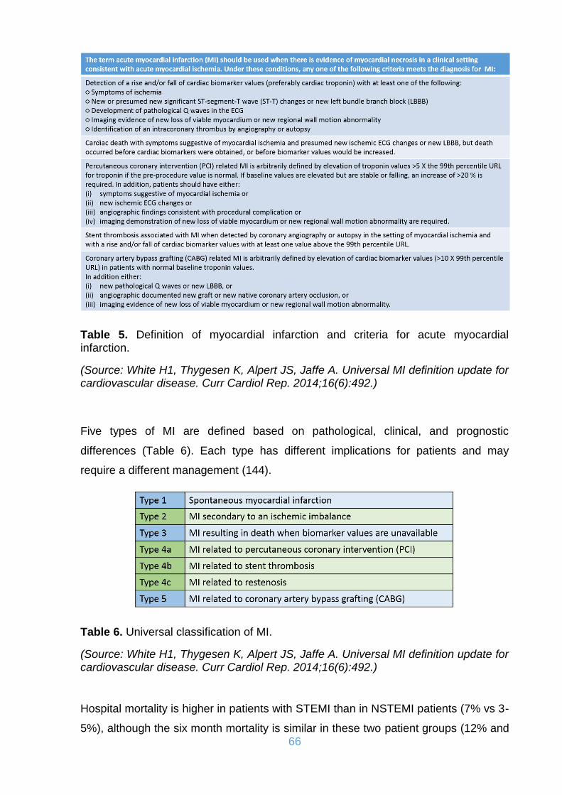
66
Table 5. Definition of myocardial infarction and criteria for acute myocardial infarction.
(Source: White H1, Thygesen K, Alpert JS, Jaffe A. Universal MI definition update for cardiovascular disease. Curr Cardiol Rep. 2014;16(6):492.)
Five types of MI are defined based on pathological, clinical, and prognostic
differences (Table 6). Each type has different implications for patients and may
require a different management (144).
Table 6. Universal classification of MI.
(Source: White H1, Thygesen K, Alpert JS, Jaffe A. Universal MI definition update for cardiovascular disease. Curr Cardiol Rep. 2014;16(6):492.)
Hospital mortality is higher in patients with STEMI than in NSTEMI patients (7% vs 3-
5%), although the six month mortality is similar in these two patient groups (12% and

67
13% respectively) (145, 146). Long-term mortality is however higher in NSTEMI
patients than in STEMI (147). This differentiation in the median and long-term
mortality is due to the different patient profiles, given that NSTEMI patients are older,
with coexisting underlying conditions, especially diabetes mellitus and renal
insufficiency (148).
As mentioned before most ACS cases are a consequence of coronary
atherosclerosis, in combination with coronary artery thrombosis. There exists
however a small percentage of cases of non-atherosclerotic coronary artery disease.
Such non-atherosclerotic causes of ACS are arteritis, trauma, dissection,
thromboembolism, congenital abnormalities, cocaine abuse and iatrogenic
complications of cardiac catheterization (149).
6. HEART FAILURE
6.1. Definition of heart failure
According to the definition given in the 2016 European guidelines heart failure (HF) is
a clinical syndrome characterized by typical symptoms (e.g. breathlessness, ankle
swelling and fatigue) that may be accompanied by signs (e.g. elevated jugular
venous pressure, pulmonary crackles and peripheral oedema) caused by a structural
and/or functional cardiac abnormality, resulting in a reduced cardiac output and/or
elevated intracardiac pressures at rest or during stress (105). Patients can however
present with a symptomatic structural or functional cardiac abnormalities before
clinical symptoms become apparent. These abnormalities are precursors of HF.
Recognition of these precursors is important because they are related to poor
outcomes, and starting treatment at the precursor stage may reduce mortality in
patients with asymptomatic systolic LV dysfunction. Demonstration of an underlying
cardiac cause is central to the diagnosis of HF. This is usually a myocardial
abnormality causing systolic and/or diastolic ventricular dysfunction. However,
abnormalities of the valves, pericardium, endocardium, heart rhythm and conduction,
or often a combination of these abnormalities can also cause HF. Identification of the
underlying cardiac problem is crucial for therapeutic reasons, as the precise
pathology determines the specific treatment used (105).

68
According to this modern definition, the fact is taken into account that approximately
half of the patients also suffer from heart failure despite having a normal systolic
function. Heart failure with impaired left ventricular function, however, is the longer
known and better characterized clinical entity.
6.2. Epidemiology of heart failure
According to the last European guidelines, the prevalence of HF is approximately 1–
2% of the adult population in developed countries, rising to ≥10% among people >70
years of age (105, 150). Among people >65 years of age presenting to primary care
with breathlessness on exertion, one in six will have unrecognized HF, mainly
patients with preserved left ventricular ejection fraction (LVEF) (105, 151). The
lifetime risk of HF at age 55 years is 33% for men and 28% for women (152). The
proportion of HF patients with normal LVEF ranges from 22 to 73%, depending on
the definition applied, the clinical setting, age and sex of the studied population,
previous myocardial infarction and the year of publication (105). Data on temporal
trends based on hospitalized patients suggest that the incidence of HF may be
decreasing, more for HF with reduced LVEF than for HF with normal LVEF (153).
These two categories of patients with HF seem to have different epidemiological and
aetiological profiles. Patients with reduced LVEF are older, more often women and
more commonly have a history of hypertension and atrial fibrillation (AF), while a
history of myocardial infarction is less common (153, 154).
Over the last 30 years, improvements in treatments and their implementation have
improved survival and reduced the hospitalization rate in patients with reduced LVEF,
although the outcome often remains unsatisfactory. The most recent European data
demonstrate that 12-month all-cause mortality rates for hospitalized and
stable/ambulatory HF patients were 17% and 7%, respectively, and the 12-month
hospitalization rates were 44% and 32%, respectively (105, 155). In patients with HF
(both hospitalized and ambulatory), most deaths are due to cardiovascular causes,
mainly sudden death and worsening HF. All-cause mortality is generally higher in
patients with reduced LVEF than those with preserved LVEF (155, 156).
Hospitalizations are often due to non-cardiovascular causes, particularly in patients
with normal LVEF (105).

69
According to the German Society of Cardiology (Deutsche Geselschaft für
Kardiologie - DGK), the prevalence in the normal population for heart failure is 2-3%.
The prevalence increases with increasing age in the 70- to 80-year-olds to more than
10% (157, 158). In the process, half of all patients die within 4 years after diagnosed
with HF, depending on etiology, age, comorbidities and individual progression of the
disease. 40% of patients admitted to hospital for heart failure die within one year or
are readmitted during this period (158). Data from the the nationwide German
database suggest that more women than men were recorded as having congestive
heart failure (66% vs. 34%). The various health insurance companies paid 2.3 times
more for patients with than without congestive heart failure. Nearly three quarters of
the cost for these patients (72%) resulted from in-patient care. Moreover, costs for
drugs were three times higher (1073 € vs. 366 €). Rising healthcare costs are
expected and will play an important economic role in the healthcare system (159).
Significant increases in heart failure are observed worldwide, especially in the
developed countries (160).
6.3. Defining heart failure patients regarding the ejection fraction
The main terminology used to describe HF is historically based on measurement of
the LVEF. HF comprises a wide range of patients, from those with normal LVEF
[typically considered as ≥50%; HF with preserved EF (HFpEF)] to those with reduced
LVEF [typically considered as <40%; HF with reduced EF (HFrEF)] (Table 7).
Patients with an LVEF in the mid-range of 40–49% represent a ‘grey area’, which we
now define as HF with moderate reduced EF (HFmrEF). Differentiation of patients
with HF based on LVEF is important due to different underlying aetiologies,
demographics, co-morbidities and response to therapies. It is mostly in patients with
HFrEF that therapies have been shown to reduce both morbidity and mortality (105,
161).
The diagnosis of HFpEF is more challenging than the diagnosis of HFrEF. Patients
with HFpEF generally do not have a dilated LV, but instead often have an increase in
LV wall thickness and/or increased left atrial (LA) size as a sign of increased filling
pressures. Most have additional ‘evidence’ of impaired LV filling or suction capacity,
also classified as diastolic dysfunction, which is generally accepted as the likely
cause of HF in these patients. However, most patients with HFrEF also have diastolic

70
dysfunction, and subtle abnormalities of systolic function have been shown in
patients with HFpEF. Patients with HFmrEF most probably have primarily mild
systolic dysfunction, but with features of diastolic dysfunction (105).
Table 7. Definition of heart failure with preserved (HFpEF), mid-range (HFmrEF) and reduced ejection fraction (HFrEF).
(Source: Ponikowski P, Voors AA, Anker SD et al. ESC Scientific Document Group. 2016 ESC Guidelines for the diagnosis and treatment of acute and chronic heart failure: The Task Force for the diagnosis and treatment of acute and chronic heart failure of the European Society of Cardiology (ESC) Developed with the special contribution of the Heart Failure Association (HFA) of the ESC. Eur Heart J. 2016 Jul 14;37(27):2129-2200.)
6.4. Aetiology of heart failure
Coronary artery disease is responsible for about 2/3 of all cases of heart failure,
followed by hypertension. Other causative factors are non-ischemic heart disease.
These include dilated cardiomyopathy (DCM), cardiomyopathies induced by alcohol,
diabetes and medications (including chemotherapy), and peripartum cardiomyopathy.
Viral, hypertensive, metabolic and valvular cardiomyopathy are also included.
Genetic factors are also discussed as triggers of cardiomyopathy (105, 162). Table 8
offers an overview of the main factors responsible for heart failure.
71
Table 8. Main causes of heart failure.
(Source: Pocket Leitlinien der DGK 2009, Hoppe 2009)
6.5. Classification of heart failure
Currently, various systems of clinical classification based on underlying clinical grade
of heart failure are used.
6.5.1. Killip classification
The oldest classification for the severity classification of the acute heart failure is the
Killip classification. This classification has prognostic significance (163, 164). It was
developed by Killip and Kimball in 1967 in patients with acute myocardial infarction
and should allow a simple clinical assessment of the hemodynamic consequences of
acute myocardial infarction with prognosis. It includes the classification according to
clinical and hemodynamic aspects into four degrees of severity, which correlate
directly with the mortality of myocardial infarction. "Killip I" indicates uncomplicated
myocardial infarction without signs of pulmonary congestion. "Killip II" is defined as
pulmonary congestion in <50% of lung sections, third heart sound, and cervical
venous stasis associated with elevated CVD. "Killip III" appears in the form of

72
pulmonary oedema with rattle noise in >50% of the lungs. Killip IV corresponds to
cardiogenic shock (163). The classification is presented in table 9.
Table 9. The Killip classification of heart failure.
(Source: Killip T, Kimball JT. Treatment of myocardial infarction in a coronary care unit. A two year experience with 250 patients. Am J Cardiol. 1967 Oct; 20(4): 457-464.)
6.5.2. Forrester classification
Another option for the classification of acute heart failure is provided by the so-called
Forrester classification (Figure 15). This was established in 1977 and originally dealt
only with patients who suffered an acute myocardial infarction. Based on clinical and
hemodynamic criteria, Forrester identified the following four Patient groups, grouped
into two pairs (165):
The first pair describes the situation "normal peripheral perfusion versus
hypoperfusion": such a distinction can be made on the one hand by typical
clinical signs such as peripheral cyanosis, flat pulse, cold wet skin,
hypotension, tachycardia and oliguria. On the other hand, measurable
hemodynamic criteria (cardiac output index <2.2 L/min/m²) are used.
The second couple discusses the issue of "pulmonary congestion versus
hypovolemia": the clinical signs of the current fluid status are both wet rales
and radiographic signs of pulmonary congestion. As a hemodynamic criterion,
differentiation via pulmonary capillary occlusion pressure (PCWP) is possible
(stasis sign at PCWP >18 mmHg).
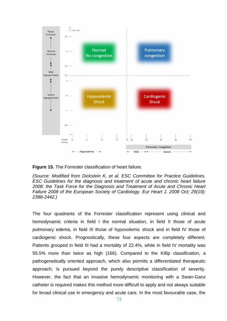
73
Figure 15. The Forrester classification of heart failure.
(Source: Modified from Dickstein K, et al. ESC Committee for Practice Guidelines. ESC Guidelines for the diagnosis and treatment of acute and chronic heart failure 2008: the Task Force for the Diagnosis and Treatment of Acute and Chronic Heart Failure 2008 of the European Society of Cardiology. Eur Heart J. 2008 Oct; 29(19): 2388-2442.)
The four quadrants of the Forrester classification represent using clinical and
hemodynamic criteria in field I the normal situation, in field II those of acute
pulmonary edema, in field III those of hypovolemic shock and in field IV those of
cardiogenic shock. Prognostically, these four aspects are completely different.
Patients grouped in field III had a mortality of 22.4%, while in field IV mortality was
55.5% more than twice as high (166). Compared to the Killip classification, a
pathogenetically oriented approach, which also permits a differentiated therapeutic
approach, is pursued beyond the purely descriptive classification of severity.
However, the fact that an invasive hemodynamic monitoring with a Swan-Ganz
catheter is required makes this method more difficult to apply and not always suitable
for broad clinical use in emergency and acute care. In the most favourable case, the

74
classification according to Killip and the Forrester classification are used in
combination for the treatment decision (165, 166).
6.5.3. Nohria classification
The Nohria classification, also known as "Clinical Severity Classification", which is
mainly used in patients with cardiomyopathies, is a classification according to the
clinical severity in patients with acute heart failure and refers exclusively to the
clinical criteria of the Forrester classification, without assuming invasive
haemodynamic monitoring (167). Patients are also divided into four groups for
peripheral perfusion and pulmonary congestion: group A - warm and dry, group B -
warm and wet, group C - cold and dry, group D - cold and wet (Figure 16). On the
one hand, the clinical diagnostic criteria include congestive criteria such as
orthopnoea, jugular vein congestion, rales, hepatojungular reflux, ascites, peripheral
oedema, and pulmonary flap tone audible to the left. On the other hand, signs of
hypoperfusion, such as low pulse amplitude, pulsus alternans, symptomatic
hypotension, cold extremities, and memory and cognitive disorders. The prognostic
significance of this classification has been proven and established (167).
Figure 16. Clinical profiles of patients with acute heart failure based on the presence/absence of congestion and/or hypoperfusion.
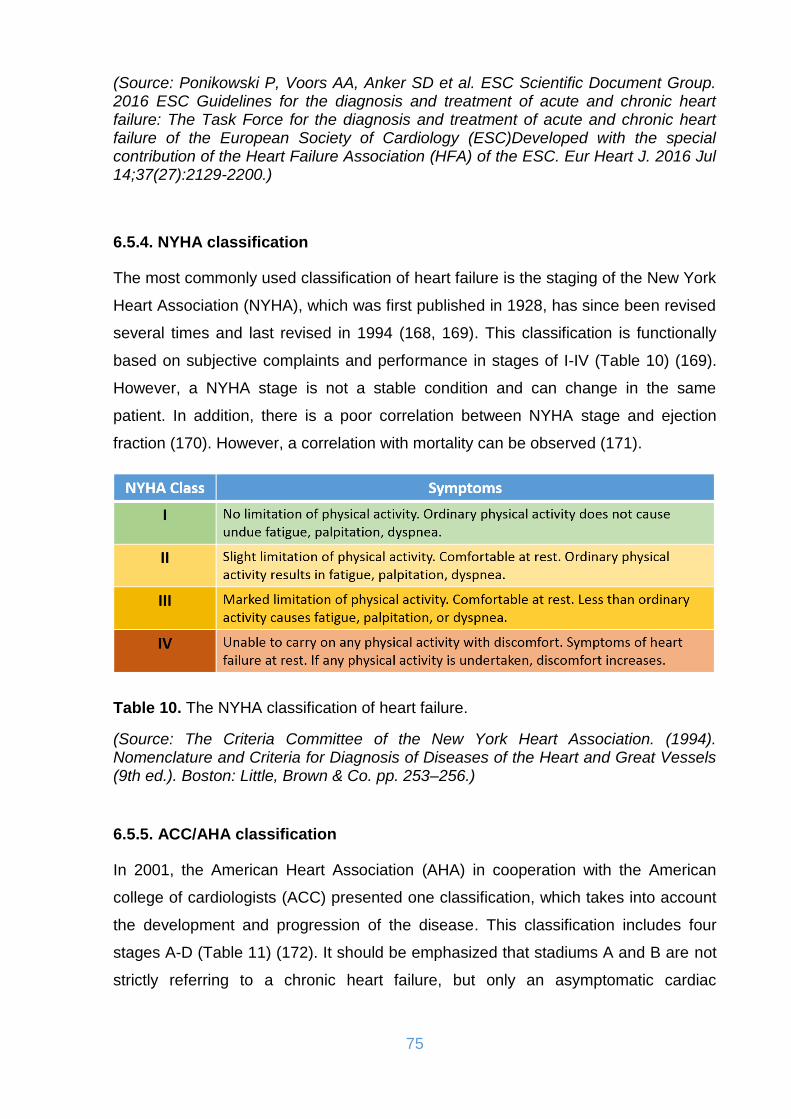
75
(Source: Ponikowski P, Voors AA, Anker SD et al. ESC Scientific Document Group. 2016 ESC Guidelines for the diagnosis and treatment of acute and chronic heart failure: The Task Force for the diagnosis and treatment of acute and chronic heart failure of the European Society of Cardiology (ESC)Developed with the special contribution of the Heart Failure Association (HFA) of the ESC. Eur Heart J. 2016 Jul 14;37(27):2129-2200.)
6.5.4. NYHA classification
The most commonly used classification of heart failure is the staging of the New York
Heart Association (NYHA), which was first published in 1928, has since been revised
several times and last revised in 1994 (168, 169). This classification is functionally
based on subjective complaints and performance in stages of I-IV (Table 10) (169).
However, a NYHA stage is not a stable condition and can change in the same
patient. In addition, there is a poor correlation between NYHA stage and ejection
fraction (170). However, a correlation with mortality can be observed (171).
Table 10. The NYHA classification of heart failure.
(Source: The Criteria Committee of the New York Heart Association. (1994). Nomenclature and Criteria for Diagnosis of Diseases of the Heart and Great Vessels (9th ed.). Boston: Little, Brown & Co. pp. 253–256.)
6.5.5. ACC/AHA classification
In 2001, the American Heart Association (AHA) in cooperation with the American
college of cardiologists (ACC) presented one classification, which takes into account
the development and progression of the disease. This classification includes four
stages A-D (Table 11) (172). It should be emphasized that stadiums A and B are not
strictly referring to a chronic heart failure, but only an asymptomatic cardiac

76
dysfunction or a pre-stage of the heart failure. Advanced disease stages are only
subsumed in stage D and clearly underrepresented.
Table 11. The AHA/ACC classification of heart failure.
(Source: Yancy CW et al. American College of Cardiology Foundation/American Heart Association Task Force on Practice Guidelines. 2013 ACCF/AHA Guideline for the Management of Heart Failure. Circulation. 2013 Oct; 128(16): e240-e327.)
At this point it should be stressed that the ACC/AHA classification is much different
than the NYHA functional classification system, in that there is no moving backwards
to prior stages. Once symptoms develop, stage C heart failure is present and stage B
will never again be achieved. In the NYHA classification, in contrast, patients can
move between class I and class IV relatively quickly, as these are all designated on
symptoms alone.
6.6. Diagnosis of heart failure in the non-acute setting
For patients presenting with symptoms or signs for the first time, non-urgently in
primary care or in a hospital outpatient clinic, the probability of HF should first be
evaluated based on the patient’s prior clinical history, presenting symptoms, physical
examination (e.g. bilateral oedema, increased jugular venous pressure, displaced
apical beat) and resting ECG. If all elements are normal, HF is highly unlikely and
other diagnoses need to be considered. If at least one element is abnormal, plasma
natriouretic peptides (NP) should be measured, if available, to identify those who
need echocardiography (an echocardiogram is indicated if the NP level is above the
exclusion threshold or if circulating NP levels cannot be assessed) (105).

77
6.7. Pharmacological treatment of chronic heart failure
No treatment has yet been shown, convincingly, to reduce morbidity or mortality in
patients with HFpEF or HFmrEF. However, since these patients are often elderly and
highly symptomatic, and often have a poor quality of life, an important aim of therapy
may be to alleviate symptoms and improve well-being (177, 178). As far as the
HFrEF patients are concerned, optimal medical therapy is crucial regarding the
quality of life and the prognosis of these patients (105).
The goals of treatment in patients with HF are to improve their clinical status,
functional capacity and quality of life, prevent hospital admission and reduce
mortality. The fact that several drugs for HF have shown detrimental effects on long-
term outcomes, despite showing beneficial effects on shorter-term surrogate
markers, has led regulatory bodies and clinical practice guidelines to seek
mortality/morbidity data for approving/recommending therapeutic interventions for
HF. However, it is now recognized that preventing HF hospitalization and improving
functional capacity are important benefits to be considered if a mortality excess is
ruled out (105, 173, 174). Figure 17 presents pharmacological treatment
recommended from the ESC.
Figure 17. The recommended therapeutic algorithm for a patient with symptomatic heart failure with reduced ejection fraction.

78
(Source: Ponikowski P, Voors AA, Anker SD et al. ESC Scientific Document Group. 2016 ESC Guidelines for the diagnosis and treatment of acute and chronic heart failure: The Task Force for the diagnosis and treatment of acute and chronic heart failure of the European Society of Cardiology (ESC) Developed with the special contribution of the Heart Failure Association (HFA) of the ESC. Eur Heart J. 2016 Jul 14;37(27):2129-2200.)
6.7.1 Angiotensin-converting enzyme inhibitors (ACEI)
ACEIs have been shown to reduce mortality and morbidity in patients with HFrEF
and are recommended unless contraindicated or not tolerated in all symptomatic
patients. ACEIs should be up-titrated to the maximum tolerated dose in order to
achieve adequate inhibition of the RAAS. ACEIs are also recommended in patients
with asymptomatic LV systolic dysfunction to reduce the risk of HF development, HF
hospitalization and death (105, 175, 176).
6.7.2. Beta-blockers
Beta-blockers reduce mortality and morbidity in symptomatic patients with HFrEF,
despite treatment with an ACEI and, in most cases, a diuretic, but have not been
tested in congested or decompensated patients (179, 180). There is consensus that
beta-blockers and ACEIs are complementary, and can be started together as soon
as the diagnosis of HFrEF is made. There is no evidence favouring the initiation of
treatment with a beta-blocker before an ACEI has been started (181). Beta-blockers
are recommended in patients with a history of myocardial infarction and
asymptomatic LV systolic dysfunction to reduce the risk of death (105).
6.7.3. Mineralocorticoid/aldosterone receptor antagonists (MRA)
MRAs block receptors that bind aldosterone and, with different degrees of affinity,
other steroid hormone (e.g. corticosteroids, androgens) receptors. Spironolactone or
eplerenone are recommended in all symptomatic patients (despite treatment with an
ACEI and a beta-blocker) with HFrEF and LVEF ≤35%, to reduce mortality and HF
hospitalization (182, 183).

79
6.7.4. Diuretics
Diuretics are recommended to reduce the signs and symptoms of congestion in
patients with HFrEF, but their effects on mortality and morbidity have not been
studied in randomaized control trials. It has been shown that in patients with chronic
HF, loop and thiazide diuretics appear to reduce the risk of death and worsening HF
and to improve exercise capacity (184). The aim of diuretic therapy is to achieve and
maintain euvolaemia with the lowest achievable dose (105).
6.7.5. Angiotensin receptor neprilysin inhibitor (ARNI)
ARNIs are a new therapeutic class of agents acting on the RAAS and the neutral
endopeptidase system. The first in class is LCZ696, which is a molecule that
combines the moieties of valsartan and sacubitril (neprilysin inhibitor) in a single
substance. By inhibiting neprilysin, the degradation of NPs, bradykinin and other
peptides is slowed. High circulating A-type natriuretic peptide (ANP) and BNP exert
physiologic effects through binding to NP receptors and the augmented generation of
cGMP, thereby enhancing diuresis, natriuresis and myocardial relaxation and anti-
remodelling. ANP and BNP also inhibit renin and aldosterone secretion. Selective
AT1-receptor blockade reduces vasoconstriction, sodium and water retention and
myocardial hypertrophy. To minimize the risk of angioedema caused by overlapping
ACE and neprilysin inhibition, the ACEI should be withheld for at least 36 h before
initiating sacubitril/valsartan. Combined treatment with an ACEI (or ARB) and
sacubitril/valsartan is contraindicated (185).
6.7.6. If-channel inhibitor
Ivabradine slows the heart rate through inhibition of the If channel in the sinus node
and therefore should only be used for patients in sinus rhythm. Ivabradine reduced
the combined endpoint of mortality or hospitalization for HF in patients with
symptomatic HFrEF or LVEF ≤35%, in sinus rhythm and with a heart rate ≥70 bpm
(186).

80
6.7.7. Angiotensin II type I receptor blockers (ARB)
ARBs are recommended only as an alternative in patients intolerant of an ACEI.
Candesartan has been shown to reduce cardiovascular mortality (187). Valsartan
showed an effect on hospitalization for HF in patients with HFrEF receiving
background ACEIs. Therefore, ARBs are indicated for the treatment of HFrEF only in
patients who cannot tolerate an ACEI because of serious side effects (188).
6.7.8. Combination of hydralazine and isosorbide dinitrate
There is no clear evidence to suggest the use of this fixed-dose combination therapy
in all patients with HFrEF. A combination of hydralazine and isosorbide dinitrate may
be considered in symptomatic patients with HFrEF who can tolerate neither ACEI nor
ARB (or they are contraindicated) to reduce mortality (105).
6.7.9. Digoxin and other digitalis glycosides
Digoxin may be considered in patients in sinus rhythm with symptomatic HFrEF to
reduce the risk of hospitalization, although its effect on top of beta-blockers has
never been tested. The effects of digoxin in patients with HFrEF and AF have not
been studied in RCTs, and recent studies have suggested potentially higher risk of
events (mortality and HF hospitalization) in patients with AF receiving digoxin (189).
In patients with symptomatic HF and AF, digoxin may be useful to slow a rapid
ventricular rate, but it is only recommended for the treatment of patients with HFrEF
and AF with rapid ventricular rate when other therapeutic options cannot be pursued.
Digitalis should always be prescribed under specialist supervision. Given its
distribution and clearance, caution should be exerted in females, in the elderly and in
patients with reduced renal function. In the latter patients, digitoxin should be
preferred (105, 190).
6.8. Cardioverter and defibrillator therapy
A high proportion of deaths among patients with HF, especially those with milder
symptoms, occur suddenly and unexpectedly. Many of these are due to electrical
81
disturbances, including ventricular arrhythmias, bradycardia and asystole, although
some are due to coronary, cerebral or aortic vascular events. Treatments that
improve or delay the progression of cardiovascular disease will reduce the annual
rate of sudden death, but they may have little effect on lifetime risk and will not treat
arrhythmic events when they occur. Implantable cardioverter-defibrillators (ICDs) are
effective in preventing bradycardia and correcting potentially lethal ventricular
arrhythmias. Some antiarrhythmic drugs might reduce the rate of tachyarrhythmias
and sudden death, but they do not reduce overall mortality and may increase it. ICDs
are widely used as primary and secondary prevention of sudden cardiac death (105).
On average, patients with IHD are at greater risk of sudden death than patients with
DCM and therefore, although the relative benefits are similar, the absolute benefit is
greater in patients with IHD (191). Patients with longer QRS durations may also
receive greater benefit from an ICD, but these patients should often receive a CRT
device (192).
6.9. Cardiac resynchronization therapy (CRT)
CRT improves cardiac performance in appropriately selected patients and improves
symptoms and well-being and reduces morbidity and mortality. Of the improvement in
quality-adjusted life-years (QALYs) with CRT among patients with moderate to
severe HF, 2/3 may be attributed to improved quality of life and 1/3 to increased
longevity (193).
Not all patients respond favourably to CRT. Several characteristics predict
improvement in morbidity and mortality, and the extent of reverse remodelling is one
of the most important mechanisms of action of CRT. Patients with ischaemic
aetiology will have less improvement in LV function due to myocardial scar tissue,
which is less likely to undergo favourable remodelling (193, 194). Conversely, women
may be more likely to respond than men, possibly due to smaller body and heart size
(195). QRS width predicts CRT response. But QRS morphology has also been
related to a beneficial response to CRT. Patients with left bundle branch block
(LBBB) morphology are more likely to respond favorably to CRT, whereas there is
less certainty about patients with non-LBBB morphology. However, patients with
LBBB morphology often have wider QRS duration, and there is a current debate
about whether QRS duration or QRS morphology is the main predictor of a beneficial

82
response to CRT. When LVEF is reduced, RV pacing may exacerbate cardiac
dyssynchrony. This can be prevented by CRT, which might improve patient outcomes
(196). Patients with HFrEF who have received a conventional pacemaker or an ICD
and subsequently develop worsening HF with a high proportion of RV pacing, despite
optimal medical therapy (OMT), may be considered for upgrading to CRT (105).
6.10. Left ventricular assist device therapy
Use of OMT, advanced pacemakers, and implantable defibrillators has changed
prognosis in HF dramatically. However, 0.5–5% of patients respond poorly to
standard therapy and develop chronic advanced HF. Prognosis in advanced HF
remains poor, with a 1-year mortality of 25–50% (197). While heart transplantation
provides excellent survival and quality of life for eligible patients, only a few can be
offered this treatment due to shortage of donor organs. More than 60% of patients
are transplanted in high-urgency status, leaving little chance for patients listed for
less urgent transplantation. Three times more patients are listed for heart
transplantation annually than are actually transplanted, and the mortality rate on the
Eurotransplant waiting list in 2013 was 21.7% (198).
Implantable left ventricular assist device (LVAD) technology has improved
considerably, and the currently used continuous flow devices may last >10 years in a
patient. LVADs have been used for decades in very advanced HF or cardiogenic
shock primarily as a bridge to transplantation (BTT). Improvements in technology,
especially the advent of smaller, durable continuous flow (CF) pumps, coupled with a
shortage of donor hearts for transplantation, have led to use of LVADs in a much
broader population of patients in the last 10 years. LVADs are being used
increasingly both as BTT and as destination therapy (DT) (Table 12).
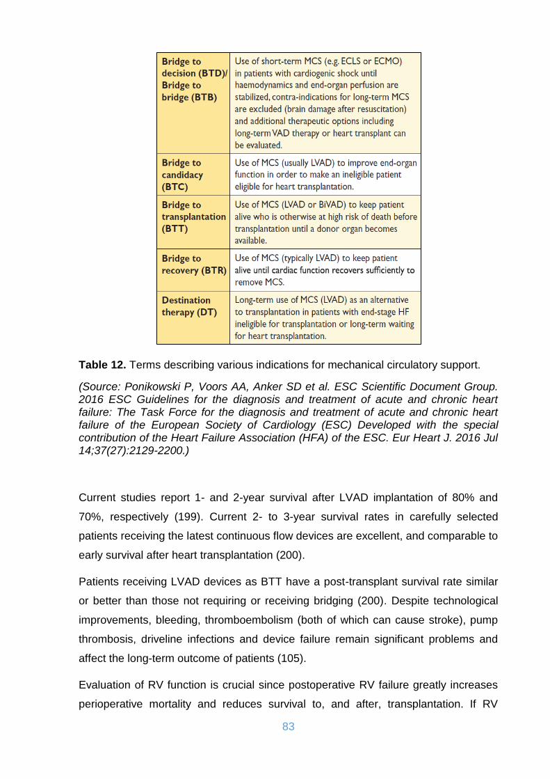
83
Table 12. Terms describing various indications for mechanical circulatory support.
(Source: Ponikowski P, Voors AA, Anker SD et al. ESC Scientific Document Group. 2016 ESC Guidelines for the diagnosis and treatment of acute and chronic heart failure: The Task Force for the diagnosis and treatment of acute and chronic heart failure of the European Society of Cardiology (ESC) Developed with the special contribution of the Heart Failure Association (HFA) of the ESC. Eur Heart J. 2016 Jul 14;37(27):2129-2200.)
Current studies report 1- and 2-year survival after LVAD implantation of 80% and
70%, respectively (199). Current 2- to 3-year survival rates in carefully selected
patients receiving the latest continuous flow devices are excellent, and comparable to
early survival after heart transplantation (200).
Patients receiving LVAD devices as BTT have a post-transplant survival rate similar
or better than those not requiring or receiving bridging (200). Despite technological
improvements, bleeding, thromboembolism (both of which can cause stroke), pump
thrombosis, driveline infections and device failure remain significant problems and
affect the long-term outcome of patients (105).
Evaluation of RV function is crucial since postoperative RV failure greatly increases
perioperative mortality and reduces survival to, and after, transplantation. If RV

84
failure is expected to be potentially reversible, temporary (days to weeks)
extracorporeal right ventricular assist device (RVAD) support using a centrifugal
pump in addition to LVAD implantation may be considered. For patients with chronic
biventricular failure or a high risk for persisting RV failure after LVAD implantation,
implantation of a biventricular assist device (BiVAD) may be necessary. Patients
requiring long-term BiVAD support must be transplant-eligible, as BiVAD therapy is
not suitable for destination therapy. The outcomes of BiVAD therapy are inferior to
those for LVAD therapy and therefore the indication for VAD therapy should be
discussed before RV function deteriorates. The implantation of a total artificial heart
with removal of the native heart should be restricted to selected patients who cannot
be treated with an LVAD (unrepairable ventricular septal defect, cardiac rupture)
(105).
6.11. Heart Transplantation
Heart transplantation is an accepted treatment for end-stage HF. Although controlled
trials have never been conducted, there is a consensus that transplantation
significantly increases survival, exercise capacity, quality of life and return to work
compared with conventional treatment. Apart from the shortage of donor hearts, the
main challenges in transplantation are the consequences of the limited effectiveness
and complications of immunosuppressive therapy in the long term. The indications
and contraindications for heart transplantation have recently been updated and are
summarized in Table 13. It needs to be considered that some contraindications are
transient and treatable. While an active infection remains a relative contraindication
to heart transplantation, patients with HIV, hepatitis, Chagas disease and
tuberculosis can be considered as suitable candidates provided certain strict
management principles are adhered to by the transplantation teams. In patients with
cancer requiring heart transplantation, a close collaboration with oncology specialists
should occur to stratify each patient according to risk of tumour recurrence (105,
201).

85
Table 13. Indications and contra-indications for heart transplantation.
(Source: Ponikowski P, Voors AA, Anker SD et al. ESC Scientific Document Group. 2016 ESC Guidelines for the diagnosis and treatment of acute and chronic heart failure: The Task Force for the diagnosis and treatment of acute and chronic heart failure of the European Society of Cardiology (ESC) Developed with the special contribution of the Heart Failure Association (HFA) of the ESC. Eur Heart J. 2016 Jul 14;37(27):2129-2200.)
6.12. Acute heart failure
6.12.1. Definition of acute heart failure
According to the 2016 ESC guidelines of heart failure acute heart failure (AHF) refers
to rapid onset or worsening of symptoms and/or signs of HF. It is a life-threatening
medical condition requiring urgent evaluation and treatment, typically leading to
urgent hospital admission. AHF may present as a first occurrence (de novo) or, more
frequently, as a consequence of acute decompensation of chronic HF, and may be
caused by primary cardiac dysfunction or precipitated by extrinsic factors, often in
patients with chronic HF. Acute myocardial dysfunction (ischaemic, inflammatory or
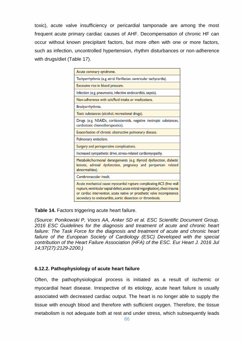
86
toxic), acute valve insufficiency or pericardial tamponade are among the most
frequent acute primary cardiac causes of AHF. Decompensation of chronic HF can
occur without known precipitant factors, but more often with one or more factors,
such as infection, uncontrolled hypertension, rhythm disturbances or non-adherence
with drugs/diet (Table 17).
Table 14. Factors triggering acute heart failure.
(Source: Ponikowski P, Voors AA, Anker SD et al. ESC Scientific Document Group. 2016 ESC Guidelines for the diagnosis and treatment of acute and chronic heart failure: The Task Force for the diagnosis and treatment of acute and chronic heart failure of the European Society of Cardiology (ESC) Developed with the special contribution of the Heart Failure Association (HFA) of the ESC. Eur Heart J. 2016 Jul 14;37(27):2129-2200.)
6.12.2. Pathophysiology of acute heart failure
Often, the pathophysiological process is initiated as a result of ischemic or
myocardial heart disease. Irrespective of its etiology, acute heart failure is usually
associated with decreased cardiac output. The heart is no longer able to supply the
tissue with enough blood and therefore with sufficient oxygen. Therefore, the tissue
metabolism is not adequate both at rest and under stress, which subsequently leads

87
to pathophysiological changes, such as a downregulation of beta-receptors,
sympathoadrenergic stimulation, altered intracellular Ca2+ homeostasis, negative
force-frequency relationship, and changes in the kidney function (202, 203).
Hypoperfusion leads to a sympathoadrenergic counterregulation (with increased
release of epinephrine, norepinephrine), activation of the vasopressor system (via
ADH) and a vascular response to induce myocardial failure. Activation of these
systems in the early phase of the disease represents a favorable compensation
mechanism for providing adequate blood pressure and sufficient organ perfusion.
Increasing and prolonged activation thereafter however causes a worsening of the
hemodynamic situation and leads to a vicious circle. These must be interrupted to
stabilize organ perfusion and circulation again. The failing heart has to fight against
the increased resistance in the left ventricular ejection tract, which is caused by an
arteriolar constriction and increase in afterload. Concomitant venous vasoconstriction
leads to an increase in both right- and left-ventricular preload with a consecutive
increase in myocardial wall sterss (204). The resulting hypoperfusion activates
proinflammatory cytokines (IL-1β, TNF-α, etc.), which, inter alia, increase capillary
permeability and increase the risk of increased fluid influx into the interstitium,
leading to further deterioration of peripheral oxygenation. In the maximum version,
this leads to interstitial pulmonary edema (205).
6.12.3. Cardiogenic shock
Cardiogenic shock is defined by end organ hypoperfusion as the result of acute heart
failure. This includes all cardiac or extracardiac diseases that lead to an immediate
dysfunction of the heart and subsequently to a shock condition (206). In most cases,
the development of cardiogenic shock is based on acute myocardial infarction. Due
to acute systolic, but also diastolic dysfunction results in a sudden reduction in
cardiac output (207). Main symptoms are marked by persistent hypotension with
systolic blood pressure values below 90 mmHg of more than 30 minutes duration and
an increased heart rate above 80/min, clinical signs of organ hypoperfusion in the
form of reduced urinary output (<0.5 ml/kg/h), conspicuously cold extremities and
mental changes such as agitation or confusion. However, even normotensive blood
pressure values and pronounced tachycardia can mask an already occurred inferior
perfusion. The normal or elevated blood pressure values result here due to an
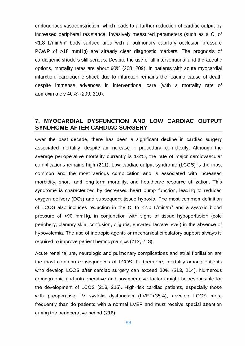
88
endogenous vasoconstriction, which leads to a further reduction of cardiac output by
increased peripheral resistance. Invasively measured parameters (such as a CI of
<1.8 L/min/m² body surface area with a pulmonary capillary occlusion pressure
PCWP of >18 mmHg) are already clear diagnostic markers. The prognosis of
cardiogenic shock is still serious. Despite the use of all interventional and therapeutic
options, mortality rates are about 60% (208, 209). In patients with acute myocardial
infarction, cardiogenic shock due to infarction remains the leading cause of death
despite immense advances in interventional care (with a mortality rate of
approximately 40%) (209, 210).
7. MYOCARDIAL DYSFUNCTION AND LOW CARDIAC OUTPUT SYNDROME AFTER CARDIAC SURGERY
Over the past decade, there has been a significant decline in cardiac surgery
associated mortality, despite an increase in procedural complexity. Although the
average perioperative mortality currently is 1-2%, the rate of major cardiovascular
complications remains high (211). Low cardiac-output syndrome (LCOS) is the most
common and the most serious complication and is associated with increased
morbidity, short- and long-term mortality, and healthcare resource utilization. This
syndrome is characterized by decreased heart pump function, leading to reduced
oxygen delivery (DO2) and subsequent tissue hypoxia. The most common definition
of LCOS also includes reduction in the CI to <2.0 L/min/m2 and a systolic blood
pressure of <90 mmHg, in conjunction with signs of tissue hypoperfusion (cold
periphery, clammy skin, confusion, oliguria, elevated lactate level) in the absence of
hypovolemia. The use of inotropic agents or mechanical circulatory support always is
required to improve patient hemodynamics (212, 213).
Acute renal failure, neurologic and pulmonary complications and atrial fibrillation are
the most common consequences of LCOS. Furthermore, mortality among patients
who develop LCOS after cardiac surgery can exceed 20% (213, 214). Numerous
demographic and intraoperative and postoperative factors might be responsible for
the development of LCOS (213, 215). High-risk cardiac patients, especially those
with preoperative LV systolic dysfunction (LVEF<35%), develop LCOS more
frequently than do patients with a normal LVEF and must receive special attention
during the perioperative period (216).

89
Myocardial dysfunction following cardiac surgery using extracorporeal circulation
(ECC) is often overlooked not only in patients with impaired preoperative myocardial
function but also in patients with normal EF (217).
Together with the reduction of the cardiac index, both the left and right ventricular
ejection fraction is reduced and even up to 35-75% compared to the preoperative
values (218). The factors contributing to the onset of cardiac dysfunction are
myocardial ischemia due to aortic cross-clamping, inadequate cardioprotection,
hypothermia and cardioplegia, surgical trauma, incomplete surgical reperfusion,
systemic inflammatory responses including complement cascade activation,
ischemia-reperfusion injury, alter at ions in signal transduction systems, as well as
the extended use of inotropic agents (219-223). The paradox is that while many of
the above-mentioned techniques are used to limit myocardial damage, they can in
themselves induce it (218).
Cardiac dysfunction after ECC can be described as a form of myocardial stunning
(53, 224). The heart undergoing surgery is subjected to reperfusion ischemia injury at
various phases of the operation resulting in Ca2+ accumulation and generation of free
ROS in the cytoplasm of the myocardial cell and the edema of these cells (225). The
generally described stunning refers to a restricted region of the myocardium (regional
stunning). The myocardial dysfunction after ECC concerns however the whole heart
(global dysfunction). The global systolic function of the heart is impaired and the
dysfunction includes both ventricles (226).
Weaning from cardiopulmonary bypass (CPB) is a crucial phase in which myocardial
dysfunction can be fully manifested and consequently inotropic support is needed.
Typically myocardial dysfunction after CPB is biphasic. Recovery of the myocardium
after the initial decline is followed by a period of further dysfunction reaching the
lowest point 3-6 hours after the end of CPB. Usually the time required for complete
recovery of the myocardium is 8-24 hours, although in patients with impaired systolic
function preoperatively this time may be longer. This knowledge is particularly
important because it determines the decision to initiate non-inotropic support as well
as its dosis and duration (219).
Ventricular dysfunction is not just about systolic function, as diastolic function is also
severely influenced. The wall compliance decreases and the volume-pressure curve
of the diastole is shifted upward, resulting in either insufficient ventricular filling or
90
sufficient ventricular filling with increased pressures that cause the reduction in
coronary driving pressure causing subendocardial ischemia (217).
After the removal of aortic clamp, the beginning of reperfusion and rewarming of the
heart in cases of hypothermic cardioplegic arrest, myocardial function gradually
improves (217). Myocardial conditions that were chronic ischemic and were
hibernating are now adequately perfused through the new grafts and may show a
spectacular improvement in their functionality, while some other areas that were
adequately perfused preoperatively through the coronary arteries may exhibit a
stunning pattern. Eventually, areas of active ischemia may coexist due to a
mechanical problem or a lack of complete reperfusion for technical reasons (217).
The total systolic function continues to improve after exiting for some time, but at
some point in the next few hours it can deteriorate again. The cause of this
phenomenon is not clear and includes mechanisms such as closure of the sternum
which can cause tamponade conditions. Other possible reasons may be an
incomplete rewarming and further hypothermia during transfer to the intensive care
unit, embolism of air or thrombus in the coronary circulation, myocardial edema due
to mechanical manipulations or due to reduced osmotic pressure or even the
continuation of reperfusion injury (217). CPB time also plays an important role. It has
been demonstrated that when the CPB time exceeds 150min the rates of myocardian
dysfunction and demand on inotropic support are higher, even in patients with normal
systolic function (227).
As mentioned before, the following pathophysiologic mechanisms of LCOS should be
highlighted:
i. Left ventricular LV systolic dysfunction
ii. Right ventricular (RV) systolic dysfunction
iii. Diastolic dysfunction, also called heart failure with preserved ejection fraction
The aforementioned mechanisms may occur isolated or in combination. Conditions
such as valvular heart disease, pulmonary hypertension, mechanical valve
dysfunction, and respiratory failure, also contribute to LCOS development (Figure 18)
(216).
91
Figure 18. A schematic presentation of the pathophysiology of postoperative LCOS. The most common causes and typical signs are presented.
(Source: Lomivorotov VV, Efremov SM, Kirov MY, Fominskiy EV, Karaskov AM. Low-Cardiac-Output Syndrome After Cardiac Surgery. J Cardiothorac Vasc Anesth. 2017 Feb;31(1):291-308.)
7.1. Left ventricular systolic dysfunction
LV function is a derivative of preload, afterload, and contractility; LV systolic
dysfunction occurs due to loss of functional myocytes or a decrease in their function.
In most cases, the loss of functional myocytes develops as a result of necrosis due to
impaired coronary circulation and ischemia/reperfusion injury or the less-understood
phenomenon of apoptosis. A loss of function of vital myocytes commonly is transient
during stunning or may be refractory to reversal with conditions such as infection;
tachycardia; cardiac valvular disease; metabolic abnormality (acidosis, hypoglycemia,
hypocalcemia); exposure to cardiac toxins; idiopathic dilated cardiomyopathy; and
genetic disorders (familial dilated cardiomyopathy, hypertrophic cardiomyopathy,
muscular dystrophies). The impairment of cardiac response to preload leads to
dramatic decreases in CO and oxygen delivery to other organs, increased left atrial
pressure and capillary wedge pressure, and cardiogenic pulmonary edema. Although
92
the LV usually works against relatively high systemic arterial pressure, the significant
afterload increase also may induce LV systolic dysfunction (216).
7.2. Left ventricular diastolic dysfunction
LCOS sometimes is associated with preserved LVEF. In such cases, the contractile
function of the myocardium is diminished, despite preserved global systolic
performance. These conditions result from the inability of the ventricular chamber to
accept an adequate volume of blood, despite normal preload, and present as
diastolic dysfunction. However, diastolic dysfunction may be accompanied by either
impaired or preserved ejection fraction. From a pathophysiologic perspective,
diastolic dysfunction is characterized by abnormal relaxation and filling of the LV
during the diastolic phase of the cardiac cycle that may be caused by the following
mechanisms: 1) severe tachycardia (upon atrial fibrillation), 2) decreased myocardial
compliance, and 3) impaired ventricular relaxation. The processes intimately involved
in the development of diastolic dysfunction, at the cardiomyocyte level, relate to
calcium removal from the cytosol and calcium homeostasis, the adequacy of cross-
bridge detachment, and intrinsic functional cytoskeletal element disorders (228).
Diastolic dysfunction is a widespread phenomenon, occurring in up to 70% of cardiac
patients postoperatively. Despite its high prevalence, diastolic dysfunction alone
often is insufficient to induce the development of acute heart failure; however, in
combination with other predisposing factors, such as atrial fibrillation, impaired
coronary perfusion, and arterial hypertension, it may lead to decompensation.
Diastolic dysfunction therefore is believed to be an early sign of myocardial ischemia
(229, 230).
The close relationship between the systolic and diastolic functions of the LV should
be acknowledged. Thus, inotropic catecholamine stimulation affects both systole and
diastole and may enhance diastolic dysfunction, whereas reduced LVEF leads to
increased end-systolic volume and prolongs the diastolic phase of the cardiac cycle
(216, 230).

93
7.3. Right ventricular dysfunction
The principal pathophysiologic mechanisms of RV dysfunction include increased RV
preload, increased RV afterload, impaired right coronary artery perfusion, and
decreased contractility (231). The specific features of RV perfusion and their
alterations during increased pulmonary artery pressure are important to understand.
Physiologically, perfusion of the right coronary artery, in contrast to the left coronary
artery, occurs during both diastole and systole. Under conditions of pulmonary
hypertension, the RV pressure increases and leads to decreased right coronary
artery perfusion, explaining why diastolic arterial pressure maintenance is highly
important for providing optimal left and right coronary blood flow (232).
In postoperative settings, RV dysfunction often develops due to a combination of
mechanisms. Thus, cardiac patients encounter many conditions associated with RV
failure. Perioperative RV ischemia and infarction are major causes of contractility
impairment. Tricuspid or pulmonic regurgitation leads to excessive volume preload,
whereas left-sided valvular disease or cardiomyopathy, pulmonary hypertension or
embolism, acute respiratory distress syndrome, and high positive-pressure ventilation
are common causes of pressure overload. Taking into account that the RV normally
provides low-pressure perfusion of the pulmonary vasculature, it is highly sensitive to
even moderate pulmonary artery pressure increases. RV failure may develop due to
pulmonary hypertension or contractile impairment associated with a rapid
progression of RV dilation, resulting in a rise in end-diastolic RV pressure. These
alterations lead to an interventricular septum shift toward the already underfilled LV
chamber, reducing LV preload and decreasing CO (233, 234).
8. CARDIOPROTECTION IN CARDIAC SURGERY
8.1. Cardioplegia
Since the beginning of cardiac surgery in the 1950s, multiple techniques have been
used to protect the heart during the surgical requirement for elective global ischemia
(and the still, relaxed, bloodless field that this provides the surgeon for repair of the
lesion). Most of these techniques have been discarded (235). The principles of
successful myocardial protection consist of a rapid and complete induction of
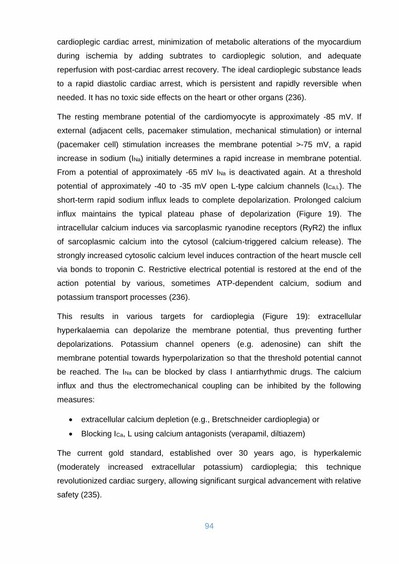
94
cardioplegic cardiac arrest, minimization of metabolic alterations of the myocardium
during ischemia by adding subtrates to cardioplegic solution, and adequate
reperfusion with post-cardiac arrest recovery. The ideal cardioplegic substance leads
to a rapid diastolic cardiac arrest, which is persistent and rapidly reversible when
needed. It has no toxic side effects on the heart or other organs (236).
The resting membrane potential of the cardiomyocyte is approximately -85 mV. If
external (adjacent cells, pacemaker stimulation, mechanical stimulation) or internal
(pacemaker cell) stimulation increases the membrane potential >-75 mV, a rapid
increase in sodium (INa) initially determines a rapid increase in membrane potential.
From a potential of approximately -65 mV INa is deactivated again. At a threshold
potential of approximately -40 to -35 mV open L-type calcium channels (ICa,L). The
short-term rapid sodium influx leads to complete depolarization. Prolonged calcium
influx maintains the typical plateau phase of depolarization (Figure 19). The
intracellular calcium induces via sarcoplasmic ryanodine receptors (RyR2) the influx
of sarcoplasmic calcium into the cytosol (calcium-triggered calcium release). The
strongly increased cytosolic calcium level induces contraction of the heart muscle cell
via bonds to troponin C. Restrictive electrical potential is restored at the end of the
action potential by various, sometimes ATP-dependent calcium, sodium and
potassium transport processes (236).
This results in various targets for cardioplegia (Figure 19): extracellular
hyperkalaemia can depolarize the membrane potential, thus preventing further
depolarizations. Potassium channel openers (e.g. adenosine) can shift the
membrane potential towards hyperpolarization so that the threshold potential cannot
be reached. The INa can be blocked by class I antiarrhythmic drugs. The calcium
influx and thus the electromechanical coupling can be inhibited by the following
measures:
extracellular calcium depletion (e.g., Bretschneider cardioplegia) or
Blocking ICa, L using calcium antagonists (verapamil, diltiazem)
The current gold standard, established over 30 years ago, is hyperkalemic
(moderately increased extracellular potassium) cardioplegia; this technique
revolutionized cardiac surgery, allowing significant surgical advancement with relative
safety (235).
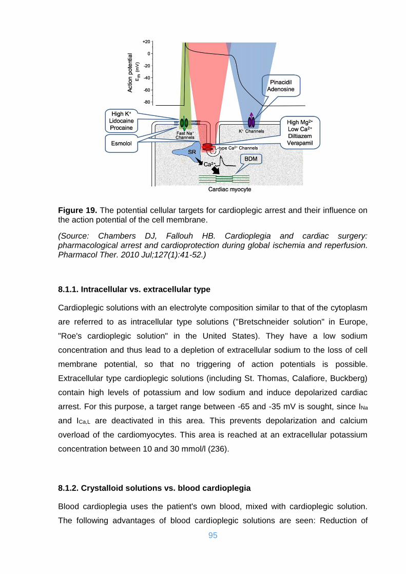
95
Figure 19. The potential cellular targets for cardioplegic arrest and their influence on the action potential of the cell membrane.
(Source: Chambers DJ, Fallouh HB. Cardioplegia and cardiac surgery: pharmacological arrest and cardioprotection during global ischemia and reperfusion. Pharmacol Ther. 2010 Jul;127(1):41-52.)
8.1.1. Intracellular vs. extracellular type
Cardioplegic solutions with an electrolyte composition similar to that of the cytoplasm
are referred to as intracellular type solutions ("Bretschneider solution" in Europe,
"Roe's cardioplegic solution" in the United States). They have a low sodium
concentration and thus lead to a depletion of extracellular sodium to the loss of cell
membrane potential, so that no triggering of action potentials is possible.
Extracellular type cardioplegic solutions (including St. Thomas, Calafiore, Buckberg)
contain high levels of potassium and low sodium and induce depolarized cardiac
arrest. For this purpose, a target range between -65 and -35 mV is sought, since INa
and ICa,L are deactivated in this area. This prevents depolarization and calcium
overload of the cardiomyocytes. This area is reached at an extracellular potassium
concentration between 10 and 30 mmol/l (236).
8.1.2. Crystalloid solutions vs. blood cardioplegia
Blood cardioplegia uses the patient's own blood, mixed with cardioplegic solution.
The following advantages of blood cardioplegic solutions are seen: Reduction of

96
myocardial ATP depletion, buffering properties and radical scavenging property. The
osmotic properties of proteinaceous blood cardioplegia reduce myocardial edema
formation. Potential disadvantages of blood cardioplegia result from the limited view
in the surgical field during the intermittent administration of cardioplegia (237).
Several randomized studies comparing blood to crystalloid cardioplegia
demonstrated a reduced rate of postoperative LCOS, improved contractility, and
lower release of myocardial necrosis parameters following the use of blood
cardioplegia. Hard endpoints such as mortality, kidney failure and heart failure were
not significantly affected (236, 238).
8.1.3. Warm vs. cold solutions
The myocardial oxygen demand, based on the heart weight (at rest at 37°C about 10
ml/100g/min), is already considerably reduced (to about 5 ml/100g/min) and due
CPB. Cardioplegic solutions reduced to approximately 10% of baseline (1
ml/100g/min). By cooling the myocardium, this reduction can be extended (reduction
by 50% for each 10°C cooling, Van't Hoff rule), so that at a temperature of the
myocardium of 10°C still an oxygen requirement of 0.3 ml/100g/min exists. The
myocardial ischemia tolerance extends reciprocally (239). From this it has long been
deduced that the cooling of the myocardium must be an indispensable part of the
myocardial protection. However, cooling causes endothelial dysfunction, myocardial
edema formation by inactivation of Na+/K+-ATPase, reduction of membrane stability
and left shift of the oxygen binding curve (240). Cold-blood cardioplegia and topical
cooling may also cause postoperative conduction disturbances and arrhythmias
(241). More recent data show no significant difference between warm and cold blood
cardioplegia in terms of clinical and laboratory outcome measures. There was a
tendency for improved results when using warm-blooded cardioplegia (236, 242,
243).
8.1.4. Polarized vs. depolarized arrest
As mentioned before almost universally cardioplegic concepts involve using a
hyperkalemic cardioplegic solution and these solutions have become the gold
standard for myocardial protection for more than 20 years. Despite the extensive and
continued research aimed at improving these basic hyperkalemic cardioplegic

97
solutions, patients undergoing surgery almost invariably experience some degree of
postoperative dysfunction. It is likely that this relates to the depolarizing nature of
hyperkalemic solutions, which results in ionic imbalance caused by continuing
transmembrane fluxes and the consequent maintenance of high energy phosphate
metabolism, even during hypothermic ischemia (244). A potentially beneficial
alternative to hyperkalemic cardioplegia is to arrest the heart in a "hyperpolarized" or
"polarized" state, which maintains the membrane potential of the arrested
myocardium at or near to the resting membrane potential. At these potentials,
transmembrane fluxes will be minimized and there should be little metabolic demand,
resulting in improved myocardial protection. Several studies have explored these
alternative concepts for myocardial protection. The use of compounds such as
adenosine or potassium channel openers, which are thought to induce
hyperpolarized arrest, have demonstrated some improved protection after
normothermic, or short periods of hypothermic, ischemia when compared to
hyperkalemic (depolarized) arrest (244). “Magnesium cardioplegia” and “esmolol
cardioplegia” has been shown to exert superior protection with comparable safety
profiles to that of hyperkalemic cardioplegia (245). However, the significant clinical
superiority of these techniques is highly debated and these alternative techniques
require further examination and investigation to challenge the traditional view that
hyperkalemic arrest is best.
8.2. Volatile Anesthetics
Volatile halogenated anesthetics are used widely worldwide for anesthetic
management of cardiac procedures. According to the CABG guidelines from the
American College of Cardiology Foundation and the American Heart Association,
volatile anesthesia can be useful in reducing the risk of perioperative myocardial
ischemia and infarction (class of recommendations IIa; Level of Evidence A) (246). A
meta-analysis of 22 studies involving 1,922 patients showed that a halogenated
anesthetic regimen was associated with improved outcomes after cardiac surgery.
Specifically, volatile anesthetics were associated with significant reductions in the
incidence of myocardial infarctions and mortality. Moreover, the need for inotropic
support also was reduced significantly in the volatile anesthetic group (216, 247).
98
8.3. Endogenous therapeutic strategies for cardioprotection
Inadequate cardioprotection during cardiac surgery, particularly in high-risk patients,
is associated with worse clinical outcomes. Therefore, novel therapeutic strategies
are required to reduce and prevent post-surgical complications. In this regard, it is
possible to ‘condition’ the heart to protect itself from the detrimental effects of acute
IRI by subjecting it to brief non-lethal episodes of ischaemia and reperfusion (248,
249). Importantly, the ‘conditioning’ stimulus may be applied either prior to [ischaemic
preconditioning (IPC)], (250) after the onset of (ischaemic perconditioning), (251) or
at the end of the index ischaemic event and at the time of reperfusion (ischaemic
postconditioning), (252) making it possible to intervene at several different time
points during cardiac surgery.
8.3.1. Ischaemic preconditioning
In 1986, Murry et al. first discovered that the heart could be ‘conditioned’ to protect
itself from MI, using brief non-lethal cycles of myocardial ischaemia and reperfusion
(250). This phenomenon, which has been termed IPC, has been shown to offer
ubiquitous cardioprotection in all animal species tested using a wide variety of
experimental in vivo and in vitro IRI models (248). IPC was the first ‘conditioning’
strategy to be applied in the clinical setting of CABG surgery. Patients randomized to
receive IPC at the time of surgery had preserved ATP levels in ventricular biopsies
(253) and less peri-operative myocardial injury as evidenced by lower serum troponin
concentrations (254). A number of clinical studies have investigated IPC in the
setting of CABG surgery, showing that IPC was associated with fewer ventricular
arrhythmias, less inotropic requirements, and a shorter intensive care unit stay (248,
255).
Early preconditioning occurs 5-15 minutes after the stimulus and lasts 1-2 hours
(256). Delayed or second window preconditioning occurs 24 hours after the stimulus
and lasts for 72 hours (257). Different mechanisms are involved in the manifestation
of the two types of preconditioning. In the early preconditioning during the stimulation
phase a number of mediators (norepinephrine, bradykinin, endothelin, opioids, and
adenosine) are released, promoting protein kinase C (PKC) movement in the cellular
membrane and mitochondria. During the ischemic phase the released adenosine
activates the PKC which causes phosphorylation of the K+-ATP channels. The result

99
of this phosphorylation is the cell membrane hyperpolarization, the shortening of
phase 2 of the action potential and ultimately the decrease in calcium influx, which
ultimately provides cardioprotection (258, 259). In delayed preconditioning, modified
gene transcription and translation leads to the synthesis of endogenous proteins
such as acute phase proteins, proto-oncogenes and various antioxidants. The
activation of PKC plays an important role by migrating to the perinuclear area where
it promotes gene expression and activates mitochondrial protein kinase (MAPK). The
acute phase proteins promote the protein synthesis while at the same time contribute
to the degradation of the due to ischemia damaged proteins. Another important
mechanism that is involved is the NO synthesis and its effect on the K+-channels
(260).
8.3.2. Remote ischaemic preconditioning
The major disadvantage of IPC as a cardioprotective strategy in patients undergoing
CABG surgery is that it requires the ‘conditioning’ stimulus to be applied directly to
the heart, which may not be practical and could actually be harmful due to the risk of
arterial thrombo-embolism from cross-clamping and declamping the aorta. The
discovery that the ‘conditioning’ stimulus could be applied to an organ or tissue away
from the heart (a phenomenon termed remote ischaemic conditioning, RIC), (261)
and the demonstration that the ‘conditioning’ stimulus could be applied non-invasively
using a standard blood pressure cuff placed on the upper or lower limb has facilitated
the translation of RIC into the clinical setting (262). The actual mechanism underlying
RIC is currently unclear but has been attributed to either a humoral or neurohormonal
pathway which links the remote organ or tissue to the heart (262, 263). Several
studies have shown some positive effects of this method, especially if combined with
volatile anesthetics (264-266). Further clinical studies are however required to
characterize the RIC stimulus and to investigate the patient population which is most
likely to benefit from this therapeutic strategy.
8.3.3. Ischaemic postconditioning
In 2003, Zhao et al. first demonstrated that the ischaemic canine heart could be
‘conditioned’ at the onset of myocardial reperfusion, by interrupting coronary reflow
with short-lived episodes of LAD occlusion and reflow (252). The mechanistic
100
pathways underlying ischaemic postconditioning (IPost) cardioprotection are complex
and some of them are similar to those utilized by IPC (267). Ischaemic
postconditioning has been applied in pediatric cardiac surgery showing some
promising benefits (268, 269). Clearly, the risk of clamping the aorta in younger
patients with relatively non-atherosclerotic aortas is not as great when compared with
adult patients undergoing CABG or valve surgery. Whether this invasive
cardioprotective strategy can impact on clinical outcomes in patients undergoing
cardiac surgery remains to be determined (248).
9. RISK STRATIFICATION IN CARDIAC SURGERY
For each intervention on patients, the risk and benefit must be weighed. This is
becoming increasingly important in the field of cardiac surgery in the context of
demographic change. Over the last decades various scoring systems have been
established which, based on known predictors of perioperative mortality, enable a
risk stratification in cardiac surgery. In the USA, the Society of Thoracic Surgeons
(STS) score has established itself. In Europe, the European System for Cardiac
Operative Risk Evaluation (EuroSCORE) and its further development EuroSCORE II
is generally used more frequently.
9.1. EuroSCORE I & II
To better estimate the perioperative mortality risk in cardiac surgery, the EuroSCORE
model was developed (270). There are an additive and a logistic version in which 17
variables that significantly influence the intraoperative and postoperative course are
included. In the case of the original additive EuroSCORE, each variable was
assigned a score of 0-3 and the individual results were added together (271, 272).
The assigned value varied depending on Significance for the operation, so that the
gender "female", for example, the value of 1 and a LVEF <30% has been assigned a
value of 3. This weighting of the individual factors allowed the perioperative mortality
risk to be estimated fairly accurately. Further development of the original idea led to
the production of the logistic EuroSCORE II, which included not only the mere
presence but also the extent of risk factors (e.g., pulmonary hypertension, graduated
to none, moderate and severe), thus making more efficient risk assessment possible,

101
especially for high-risk patients. The logistic EuroSCORE II concept was published in
2003, it is the continuation of the additive EuroSCORE of 1999 and the score was
launched at the 2011 EACTS meeting in Lisbon (272). Based on the scores obtained,
the EuroSCORE gives a percentage value that reflects the estimated perioperative
mortality. In general, the EuroSCORE II values of 0-2% are considered to be low,
those of 2-5% to medium, and those of >5% to high perioperative risks.
9.2. STS score
The STS score was published in 2007 and has been available since then as well as
the EuroSCORE as an online calculator (273). Like the EuroSCORE, the STS score
also uses preoperative risk factors to calculate, among other things, postoperative
mortality risk. However, the risk factors used here are somewhat more detailed. The
collection of these extended risk factors allows the STS score to make various other
predictions in addition to postoperative mortality, such as the probability of a
reoperation or a prolonged ventilation time. However, the prediction of these
additional endpoints only applies to patients who undergo isolated CABG surgery
(274). Despite the large number of additionally considered risk factors, both the
EuroSCORE II and the STS score show a comparably good discrimination between
high- and low-risk patients, but limited accuracy, especially in the prediction of high-
risk patients (275). Furthermore, the accuracy and discrimination of any scores vary
with respect to the different ways to intervene in different heart conditions. Above all,
the predicted mortality often exceeds the observed mortality by a factor of 2. Further
revisions of these scores in the future are probably indicated, especially since this is
based on a study population, which had been operated about several years ago and
that in the meantime, there has been a continuous improvement in both surgical
techniques and perioperative patient care (274).
10. MECHANICAL CIRCULATORY SUPPORT
For patients with either chronic or acute HF who cannot be stabilized with medical
therapy, MCS systems can be used to unload the failing ventricle and maintain
sufficient end-organ perfusion. Patients in acute cardiogenic shock are initially
102
treated with short-term assistance using extracorporeal, non-durable life support
systems so that more definitive therapy may be planned (105).
10.1. Intraaortic balloon pump
The first publication of intra-aortic balloon counter-pulsation appeared in 1962 by S.
Moulopoulos (276, 277). The device and the balloons were then developed for
commercial use between 1967 and 1969. The first clinical implant was performed at
in 1967. The patient, a 48-year-old woman, was in cardiogenic shock and
unresponsive to traditional therapy. An intraaortic balloon pump (IABP) was inserted
by a cut down on the left femoral artery. Pumping was performed for approximately 6
hours. Shock reversed and the patient was discharged. Since then it consists the
most widely used mechanical support system in cardiac infarction patients in
cardiogenic shock, for more than five decades (278-281). The main mode of action of
the IABP is the reduction of afterload, which relieves the work of the ventricle and
additionally causes an increase in coronary perfusion (280).
A balloon of about 40 ml is introduced in the Seldinger technique and implanted
percutaneously over the femoral artery. The balloon is advanced under X-ray control
or transesophageal echocardiography until the transition of the aortic arch into the
descending aorta, the tip of the balloon coming to lie about two centimeters below the
ostium of the left subclavian artery and ideally ends in the abdominal aorta above the
level of the renal arteries. The functionality of the IABP is based on the principle of
counterpulsation. The balloon is filled with helium gas during diastole, triggered by
ECG or blood pressure measurement (281, 282). On the other hand, the sudden
deflation of the balloon immediately before systole leads to a reduction in the
afterload for the left ventricle, resulting in a moderate increase in left ventricular
ejection fraction and cardiac output. In addition, left ventricular end-diastolic volume
and oxygen consumption are reduced (279, 282, 283). This procedure enjoyed a
Class 1 recommendation by 2012 and 2013 in both the European and US guidelines
(105, 284, 285). Despite these clear recommendations of the professional
associations, it was observed in various surveys and registries that only around 20-
30% of patients in cardiogenic shock were treated with IABP (286). For years, the
effectiveness and role of the IABP has been controversially discussed.

103
The IABP -Shock II trial in 2009, showed no significant reduction in 30-day mortality
in comparison to the optimal medical therapy (OMT) in patients with cardiogenic
shock in the context of myocardial infarction. In addition, there were no differences in
secondary endpoints, such as serum lactate, renal function, SAP (Simplyfied Acute
Physiology Score) II, CRP, levels of catecholamine doses, and duration of
catecholamine infusions. The evaluation of the safety endpoints, e.g. However,
severe bleeding, peripheral ischemic complications, sepsis, and insult revealed that
IABP support is considered "safe" in cardiogenic shock due to infarction (287-289). In
the current recommendations, the indication for IABP insertion in cardiogenic shock
has been downgraded to grade IIIB and the routine use of IABP in cardiogenic shock
is no longer recommended (105, 279, 286). It should be emphasized that a sensible
use of the IABP requires a measurable intrinsic performance of the left ventricle. In
the case of a severely restricted pumping function or complete arrest of the left
ventricle, other systems which ensure adequate organ perfusion and thus possibly
prevent the formation of a multi-organ failure or stabilize the patient sufficiently to
carry out diagnostic and therapeutic procedures in the sense of "bridge-to-diagnostic"
concept is considered (290).
10.2. Impella® pump
The companies Abiomed and Impella Cardiosystems offer the axial rotary pump
(Impella®) in versions with 2.5 L/min and 5 L/min pumping power. The first can be
placed percutaneously in the left ventricle at a diameter of 12 French across the
femoral artery through the aortic valve, the second must be open surgically
implanted. The blood is then aspirated from the left ventricle and delivered into the
ascending aorta. On the one hand, this improves the flow in the coronary arteries, on
the other hand, the myocardial oxygen consumption seems to be lowered (291, 292).
Impella® offers a better left ventricular relief without increase in afterload compared to
other mechanical support systems (293). In the ISAR-SHOCK 2008 study, the
Impella® 2.5 and intra-aortic balloon counterpulsation in patients with cardiogenic
shock after myocardial infarction were prospectively randomized for placement,
hemodynamic effects and mortality. In the first 30 minutes, a significantly earlier
increase in cardiac index was observed in the Impella® group. However, statistically
significant differences in disease progression and mortality were not observerd (294,
104
295). Disadvantages are the higher costs compared to the IABP, the need for
surgical implantation of the Impella® 5, the increased hemolysis rate due to the high
rotational speeds of up to 30,000 rpm and the insufficient support of the cardiac
output of the small Impella® in cardiogenic shock (296, 297).
10.3. TandemHeart®
The TandemHeart® system consists of a low-prime-volume (10 cm3) extracorporeal
centrifugal pump with flow rates of up to 5 L/min. The inflow cannula is inserted into
the femoral vein and is advanced across the interatrial septum into the left atrium; the
outflow cannula returns oxygenated blood to the femoral artery. By eliminating an
external membrane oxygenator, the foreign surface of the extracorporeal circulation
is significantly reduced. This system is therefore primarily suitable for patients with
left ventricular failure without concomitant severe pulmonary dysfunction (298).
Compared to IABP, TandemHeart® appears to provide better hemodynamic
stabilization with improved cardiac index, mean arterial pressure and reduced cardiac
work with consecutively lower cardiac oxygen consumption. However, higher rates of
complications, especially bleeding, were observed at the same time (299, 300).
10.4 Extracorporeal life support systems (ECLS)
This critical evaluation and downgrading of the indications of the IABP has created a
new impetus to reflect on the role of alternative short-term mechanical support
devices, such as veno-arterial extracorporeal life support (VA-ECLS), which can
provide partial or full circulatory support (301, 302). In addition, VA-ECLS can provide
respiratory support in patients suffering from severe, combined cardiac and
pulmonary failure. Studies have indicated beneficial effects in patients with
cardiogenic shock in acute on chronic heart failure and cardiac arrest (303).
In a VA-ECLS circuit, central venous blood is drained, relayed through an
extracorporeal pump and oxygenator and then re-infused into the arterial
compartment. A modern ECLS circuit consists of several components including
venous and arterial cannulas, tubing, a membrane oxygenator with gas blender, a
continuous-flow centrifugal pump and a heat exchanger to compensate for
extracorporeal heat loss. VA-ECLS was originally derived from (CPB) but differs in
105
several aspects (301). In cardiogenic shock, arterial and venous femoral
percutaneous cannulation via the Seldinger technique allows for rapid initiation of
VA-ECLS without need for a central approach via sternotomy. Furthermore, it can be
performed virtually everywhere inside and outside the hospital environment (304).
Consistent with Poiseuille’s law, narrower, longer cannulas result in larger resistance,
which may in turn reduce extracorporeal blood flow or necessitate pressures outside
the clinically acceptable range for a given pump speed. Drainage of central venous
blood can best be achieved through relatively large 21–25 French (Fr) multistage
cannulas. Arterial cannulas are narrower (15–19 Fr) and shorter and allow injection of
oxygenated blood retrogradely into the descending aorta. Centrifugal pumps, being
used in modern VA-ECLS circuits, are designed to provide a flow of 60–
120 ml/kg/min (range of 2–10 l/min). Maximum achievable extracorporeal blood flow
depends on many other ECLS circuit and patient characteristics, such as caval vein
diameters, volume status, thoraco-abdominal pressures, flow capacity of the
oxygenator and maximally tolerable negative and positive pressures (±300 mm Hg).
Membrane oxygenators are made up of selectively permeable membranes,
facilitating blood and gas flow to allow gas exchange (301).
The main goal of VA-ECLS in refractory cardiogenic shock is to provide immediate
circulatory and respiratory stabilisation, while accounting for sufficient cardiac
unloading. Although no randomised controlled trials exist on the effectivity of VA-
ECLS, observational studies have shown survival benefits in comparison to
conventional therapy and Class IIb and Class IIa recommendations have been
specified by European (305) and American (306) guidelines, respectively. To
maximise the potential of cardiac recovery and prevent impending multi-organ failure,
early initiation of VA-ECLS has been proposed (307). Observational evidence further
suggests that in Interagency Registry for Mechanically Assisted Circulatory Support
(INTERMACS) level 1 patients (so called ‘crash-and-burn’ patients), temporary
clinical optimisation with VA-ECLS prior to left ventricular assist device (LVAD)
support improves outcome as compared to direct implantation of a permanent LVAD
(308). The main indications and contraindication regarding the application of ECLS
are summarized in table 15.
VA-ECLS is an increasingly used cardiac and circulatory support modality, which can
provide immediate stabilisation in patients with otherwise refractory cardiogenic
shock. In the absence of randomised trials, observational studies have suggested a

106
reduction in mortality as compared to conventional treatment. VA-ECLS should be
taken into consideration in well-selected cases. Management of VA-ECLS is complex
and requires constant support of trained personnel. For this reason, the application of
this support technique should be confined to centres with sufficient experience,
ongoing exposure and a close and well-organised multidisciplinary team (301).
Table 15. Indications and contraindications for ECLS.
(Source: Modified from C. L. Meuwese, et al. Extracorporeal life support in cardiogenic shock: indications and management in current practice. Neth Heart J. 2018 Feb; 26(2): 58–66.)
11. INOTROPIC AND VASOACTIVE AGENTS IN THE TREATMENT OF ACUTE AND POSTOPERATIVE HEART FAILURE
The ideal positive inotropic agent should be able to increase the contractility of both
ventricles without causing a significant increase of the heart rate, preload, afterload
and metabolic requirements of the myocardium. It should not affect diastolic function
and not reduce coronary perfusion pressure and thus blood supply to the heart (309).
It should also have a rapid onset of action, a short half-life and a short time of
titration. Unfortunately, not only is there no ideal inotropic agent but also commonly
used agents are always accompanied by restrictions on their use (309).
11.1. Catecholamines
Catecholamines are the most widely used agents in clinical practice and exert their
positive inotropic activity through the stimulation of β-adrenergic receptors.
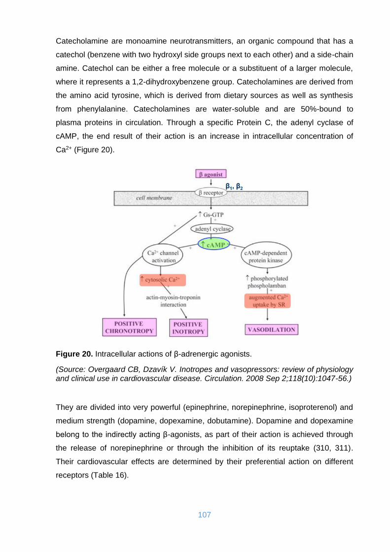
107
Catecholamine are monoamine neurotransmitters, an organic compound that has a
catechol (benzene with two hydroxyl side groups next to each other) and a side-chain
amine. Catechol can be either a free molecule or a substituent of a larger molecule,
where it represents a 1,2-dihydroxybenzene group. Catecholamines are derived from
the amino acid tyrosine, which is derived from dietary sources as well as synthesis
from phenylalanine. Catecholamines are water-soluble and are 50%-bound to
plasma proteins in circulation. Through a specific Protein C, the adenyl cyclase of
cAMP, the end result of their action is an increase in intracellular concentration of
Ca2+ (Figure 20).
Figure 20. Intracellular actions of β-adrenergic agonists.
(Source: Overgaard CB, Dzavík V. Inotropes and vasopressors: review of physiology and clinical use in cardiovascular disease. Circulation. 2008 Sep 2;118(10):1047-56.)
They are divided into very powerful (epinephrine, norepinephrine, isoproterenol) and
medium strength (dopamine, dopexamine, dobutamine). Dopamine and dopexamine
belong to the indirectly acting β-agonists, as part of their action is achieved through
the release of norepinephrine or through the inhibition of its reuptake (310, 311).
Their cardiovascular effects are determined by their preferential action on different
receptors (Table 16).
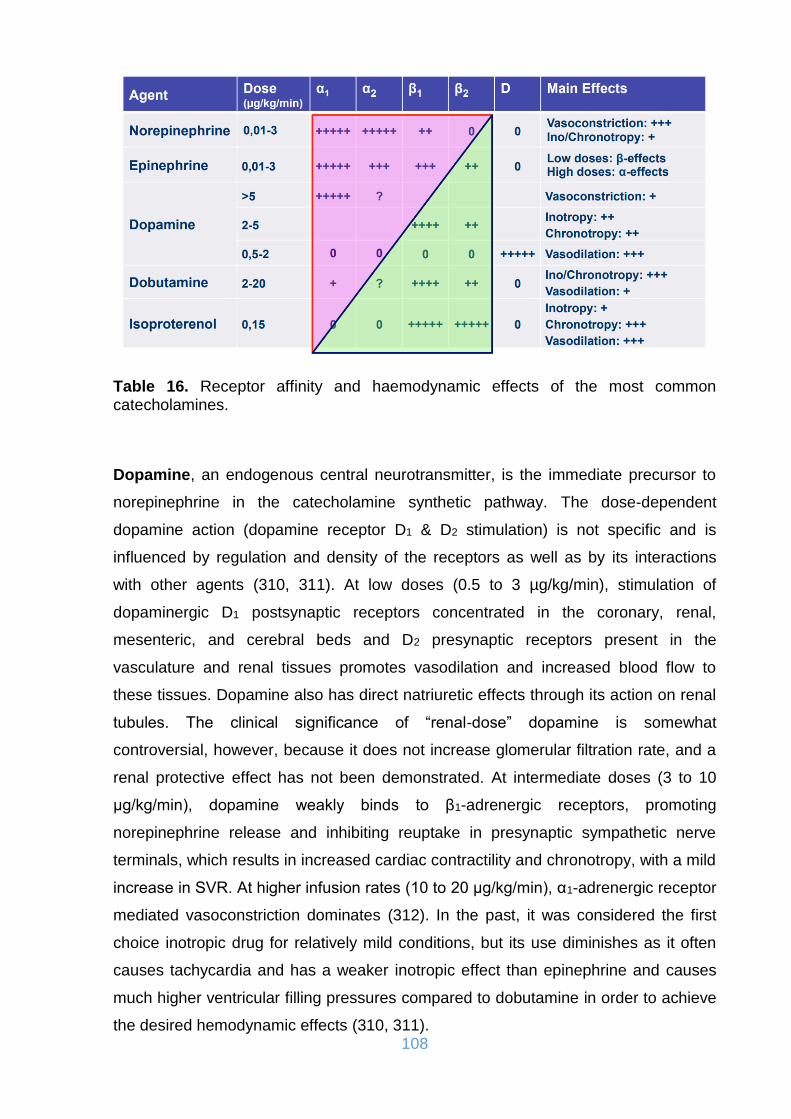
108
Table 16. Receptor affinity and haemodynamic effects of the most common catecholamines.
Dopamine, an endogenous central neurotransmitter, is the immediate precursor to
norepinephrine in the catecholamine synthetic pathway. The dose-dependent
dopamine action (dopamine receptor D1 & D2 stimulation) is not specific and is
influenced by regulation and density of the receptors as well as by its interactions
with other agents (310, 311). At low doses (0.5 to 3 µg/kg/min), stimulation of
dopaminergic D1 postsynaptic receptors concentrated in the coronary, renal,
mesenteric, and cerebral beds and D2 presynaptic receptors present in the
vasculature and renal tissues promotes vasodilation and increased blood flow to
these tissues. Dopamine also has direct natriuretic effects through its action on renal
tubules. The clinical significance of “renal-dose” dopamine is somewhat
controversial, however, because it does not increase glomerular filtration rate, and a
renal protective effect has not been demonstrated. At intermediate doses (3 to 10
µg/kg/min), dopamine weakly binds to β1-adrenergic receptors, promoting
norepinephrine release and inhibiting reuptake in presynaptic sympathetic nerve
terminals, which results in increased cardiac contractility and chronotropy, with a mild
increase in SVR. At higher infusion rates (10 to 20 µg/kg/min), α1-adrenergic receptor
mediated vasoconstriction dominates (312). In the past, it was considered the first
choice inotropic drug for relatively mild conditions, but its use diminishes as it often
causes tachycardia and has a weaker inotropic effect than epinephrine and causes
much higher ventricular filling pressures compared to dobutamine in order to achieve
the desired hemodynamic effects (310, 311).
109
Dopexamine (a synthetic dopamine analogue) other than its dopaminergic activity
exhibits greater affinity for β2 than β1 adrenergic receptors, resulting in increased
visceral and renal blood flow. But in the management of cardiac surgery patients it
seems to cause frequent and serious tachycardias, which is of course not desirable
(313).
Dobutamine is a synthetic catecholamine with a strong affinity for both β1- and β2-
receptors, which it binds to at a 3:1 ratio. With its cardiac β1-stimulatory effects,
dobutamine is a potent inotrope, with weaker chronotropic activity. Vascular smooth
muscle binding results in combined β1-adrenergic agonism and antagonism, as well
as β2-stimulation, such that the net vascular effect is often mild vasodilation,
particularly at lower doses (<5 µg/kg/min). Doses up to 15 µg/kg/min increase cardiac
contractility without greatly affecting peripheral resistance, likely owing to the
counterbalancing effects of α1-mediated vasoconstriction and β2-mediated
vasodilation. Vasoconstriction progressively dominates at higher infusion rates.
Despite its mild chronotropic effects at low to medium doses, dobutamine significantly
increases myocardial oxygen consumption. Tolerance can develop after just a few
days of therapy, due to downregulation of adrenergic receptors and malignant
ventricular arrhythmias can be observed at any dose. In cardiac surgery patients,
dobutamine increases coronary flow and oxygen supply to the myocardium
compared to dopamine, which appears to adversely affect the metabolic balance.
These beneficial effects are unfortunately eliminated by the induction of tachycardia,
which is also one of the determinants of the increase in oxygen consumption by the
myocardium (312, 314).
Norepinephrine, the major endogenous neurotransmitter liberated by postganglionic
adrenergic nerves, is a potent α1-adrenergic receptor agonist with modest β-agonist
activity, which renders it a powerful vasoconstrictor with less potent direct inotropic
properties (Figure 21). Norepinephrine primarily increases systolic, diastolic, and
pulse pressure and has a minimal net impact on CO. Furthermore, this agent has
minimal chronotropic effects, which makes it attractive for use in settings in which
heart rate stimulation may be undesirable. Coronary flow is increased owing to
elevated diastolic blood pressure and indirect stimulation of cardiomyocytes, which
release local vasodilators. Prolonged norepinephrine infusion can have a direct toxic
effect on cardiac myocytes by inducing apoptosis via protein kinase A activation and

110
increased cytosolic Ca2+ influx. The administration of norepinephrine is highly useful
in conditions with low peripheral resistances (312, 315).
Figure 21. Postulated mechanisms of intracellular action of α1-adrenergic agonists.
(Source: Overgaard CB, Dzavík V. Inotropes and vasopressors: review of physiology and clinical use in cardiovascular disease. Circulation. 2008 Sep 2;118(10):1047-56.)
Epinephrine is an endogenous catecholamine with high affinity for β1-, β2-, and α1-
receptors present in cardiac and vascular smooth muscle. β-adrenergic effects are
more pronounced at low doses and α1-adrenergic effects at higher doses. Coronary
blood flow is enhanced through an increased relative duration of diastole at higher
heart rates and through stimulation of myocytes to release local vasodilators, which
largely counterbalance direct α1-mediated coronary vasoconstriction. Arterial and
venous pulmonary pressures are increased through direct pulmonary
vasoconstriction and increased pulmonary blood flow. High and prolonged doses can
cause direct cardiac toxicity through damage to arterial walls, which causes focal
regions of myocardial contraction band necrosis, and through direct stimulation of
myocyte apoptosis (312, 316).
The combination of norepinephrine and dobutamine is currently the most favored
inotropic treatment and seems to be superior to traditional therapy with dopamine.

111
Epinephrine is not recommended as the first-line therapy. When considering global
hemodynamic effects, epinephrine is as effective as norepinephrine-dobutamine.
Nevertheless, epinephrine is associated with a transient lactic acidosis, higher heart
rate and arrhythmia, and inadequate gastric mucosa perfusion. Thus, the
combination norepinephrine-dobutamine appears to be a more reliable and safer
strategy (317, 318).
Isoproterenol is a potent, nonselective, synthetic β-adrenergic agonist with very low
affinity for α-adrenergic receptors. It has powerful chronotropic and inotropic
properties, with potent systemic and mild pulmonary vasodilatory effects. Its
stimulatory impact on stroke volume is counterbalanced by a β2-mediated drop in
systemic vascular resistance (SVR), which results in a net neutral impact on CO
(312). Its use is limited in patients with CAD due to the often caused tachycardias, is
however still used in the treatment of bradyarrhythmias and atrioventricular blocks.
Phenylephrine presents a potent synthetic α-adrenergic activity and virtually no
affinity for β-adrenergic receptors. It can be primarily used as a rapid bolus for
immediate correction of sudden severe hypotension. It can for example be used to
raise mean arterial pressure (MAP) in patients with severe hypotension and
concomitant aortic stenosis, to correct hypotension caused by the simultaneous
ingestion of sildenafil and nitrates, to decrease the outflow tract gradient in patients
with obstructive hypertrophic cardiomyopathy, and to correct vagally mediated
hypotension during percutaneous diagnostic or therapeutic procedures. This agent
has virtually no direct heart rate effects, although it has the potential to induce
significant baroreceptor-mediated reflex rate responses after rapid alterations in
MAP.
11.2. Phosphodiesterase inhibitors
Phosphodiesterase 3 is an intracellular enzyme associated with the sarcoplasmic
reticulum in cardiac myocytes and vascular smooth muscle that breaks down cAMP
into AMP. Phosphodiesterase inhibitors (PDIs) increase the level of cAMP by
inhibiting its breakdown within the cell, which leads to increased myocardial
contractility (Figure 22). These agents are potent inotropes and vasodilators and also
improve diastolic relaxation (lusitropy), thus reducing preload, afterload, and SVR
(312). Unlike sympathomimetic amines, PDE III inhibitors produce no tolerance and

112
possess the distinct advantage of directly decreasing pulmonary vascular resistance
(319). Milrinone is the PDI most commonly used for cardiovascular indications. In its
parenteral form, it has a longer half-life (2-4 hours) than many other inotropic
medications. This drug is particularly useful if adrenergic receptors are
downregulated or desensitized in the setting of chronic HF, or after chronic β-agonist
administration. Amrinone is used less often because of important side effects, which
include doserelated thrombocytopenia (312). Low-dose oral enoximone has recently
been evaluated in several small clinical trials, especially its effects during co-
administration with beta-blockers (320). Another drug, vesnarinone, a mixed PDE
inhibitor and ion-channel modifier that has modest, dose-dependent, positive
inotropic activity, but minimal negative chronotropic activity, has improved
haemodynamics and quality of life in small trials (321). However, in the largest
outcome study, the VEST trial with 3833 patients with severe heart failure, a dose-
dependent increase in mortality was identified (322).
Figure 22. Basic mechanism of action of PDIs.
(Source: Overgaard CB, Dzavík V. Inotropes and vasopressors: review of physiology and clinical use in cardiovascular disease. Circulation. 2008 Sep 2;118(10):1047-56.)
11.3. Vassopressors
Isolated in 1951 (323), the nonapeptide vasopressin or “antidiuretic hormone” is
stored primarily in granules in the posterior pituitary gland and is released after
increased plasma osmolality or hypotension, as well as pain, nausea, and hypoxia.

113
Vasopressin is synthesized to a lesser degree by the heart in response to elevated
cardiac wall stress and by the adrenal gland in response to increased catecholamine
secretion (324). It exerts its circulatory effects through V1 (V1a in vascular smooth
muscle, V1b in the pituitary gland) and V2 receptors (renal collecting duct system). V1a
stimulation mediates constriction of vascular smooth muscle, whereas V2 receptors
mediate water reabsorption by enhancing renal collecting duct permeability.
Vasopressin causes less direct coronary and cerebral vasoconstriction than
catecholamines and has a neutral or inhibitory impact on CO, depending on its dose-
dependent increase in SVR and the reflexive increase in vagal tone. A vasopressin-
modulated increase in vascular sensitivity to norepinephrine further augments its
pressor effects. The agent may also directly influence mechanisms involved in the
pathogenesis of vasodilation, through inhibition of ATP-activated potassium
channels, attenuation of nitric oxide production, and reversal of adrenergic receptor
downregulation. The pressor effects of vasopressin are relatively preserved during
hypoxic and acidotic conditions, which commonly develop during shock of any origin
(312).
Table 17 summarizes the main effects and the indications of the most commonly
used pharmacologic agents including Levosimendan which will be thoroughly
described on the following section.
Table 17. The main effects and indications of inotropic agents.

114
12. CALCIUM SENSITIZERS – LEVOSIMENDAN
β-adrenergic receptors, phosphodiesterase inhibitors and digoxin have been
extensively used as inotropic agents to improve hemodynamic parameters in patients
with heart failure. Due to their ‘retrograde’ mechanism of action interfering with
stimulation-contraction conjugation they do not lack side effects which include the
accumulation of calcium in the cytoplasm that could potentially cause cardiac
arrhythmias and myocardial damage and eventually lead to cell death (325, 326).
These agents also consume large amounts of energy for the required intracellular
calcium metabolism. Both β-agonists and phosphodiesterase inhibitors cause a
double and a threefold increase of heat and work to increase the intensity of the
contraction through an expensive and uneconomic for the body procedure (327).
An alternative way of increasing contractile intensity is the ‘antegrade’ mechanism in
which the binding of troponin C (TnC) to calcium or myofilament response for the
existing number of calcium ion-TnC junctions is altered. This mechanism provides the
desired hemodynamic effect without increasing energy consumption and avoiding the
accumulation of calcium and its adverse effects. Calcium sensitizers are a recently
developed class of inotropic agents, levosimendan being the most well-known (325,
328). The ‘antegrade’ mechanism of action of Levosimendan is compared to other
inotropic agents in figure 23.
Figure 23. Mechanisms of action of inotropic agents in the cardiac muscle cell.
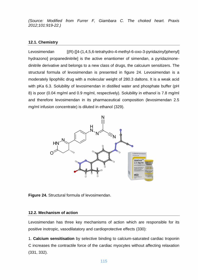
115
(Source: Modified from Furrer F, Giambara C. The choked heart. Praxis 2012;101:919-22.)
12.1. Chemistry
Levosimendan [(R)-[[4-(1,4,5,6-tetrahydro-4-methyl-6-oxo-3-pyridazinyl)phenyl]
hydrazono] propanedinitrile] is the active enantiomer of simendan, a pyridazinone-
dinitrile derivative and belongs to a new class of drugs, the calcuium sensitizers. The
structural formula of levosimendan is presented in figure 24. Levosimendan is a
moderately lipophilic drug with a molecular weight of 280.3 daltons. It is a weak acid
with pKa 6.3. Solubility of levosimendan in distilled water and phosphate buffer (pH
8) is poor (0.04 mg/ml and 0.9 mg/ml, respectively). Solubility in ethanol is 7.8 mg/ml
and therefore levosimendan in its pharmaceutical composition (levosimendan 2.5
mg/ml infusion concentrate) is diluted in ethanol (329).
Figure 24. Structural formula of levosimendan.
12.2. Mechanism of action
Levosimendan has three key mechanisms of action which are responsible for its
positive inotropic, vasodilatatory and cardioprotective effects (330):
1. Calcium sensitisation by selective binding to calcium-saturated cardiac troponin
C increases the contractile force of the cardiac myocytes without affecting relaxation
(331, 332).

116
2. Opening of KATP channels in vascular smooth muscle cells elicits both arterial
and venous vasodilation as well as improvement in coronary artery circulation (333,
334).
3. Opening of KATP channels in the mitochondria of cardiomyocytes achieves a
cardioprotective effect in situations when the heart is subjected to ischaemic events
(335, 336).
Through calcium sensitisation, levosimendan improves cardiac contractility in the
failing heart without affecting muscle electrophysiology (337). Through the opening of
KATP channels in vascular smooth muscle cells, levosimendan improves oxygen
supply to the myocardium (338). Because levosimendan augments myofibril
contractions by increasing calcium sensitivity rather than by increasing intracellular
calcium, it is not associated with increased myocardial oxygen demand, ischaemia,
or tolerance, conditions sometimes incurred with agents traditionally used to treat
decompensated heart failure. In brief, the mechanism of action for levosimendan
involves three clinically relevant features that are specific to the cardiovascular
system; levosimendan acts on the contractile apparatus of the myocardial cells, on
the vascular smooth muscle cells and on the mitochondria of the cardiomyocytes via
independent, but complementary, mechanisms (338, 339).
12.2.1. Positive inotropic effect
The heart muscle consists of cardiac myocytes that show a striated subcellular
structure: each cell contains myofibrils with actin and myosin filaments, which form
the contractile apparatus. The actin filaments are associated with the regulatory
proteins tropomyosin and troponin, which is complex of three smaller proteins (TnC,
TnI, and TnT) (Figure 25A). When intracellular Ca2+ concentration increases, TnC
becomes Ca2+-saturated, which triggers the contraction. When calcium is removed
from the cytosol, TnC, now Ca2+-free, allows the sarcomere relaxation.
Levosimendan selectively binds to Ca2+-saturated cardiac TnC (Figure 25B) (340).
By binding to TnC and stabilising the TnC-Ca2+ complex, levosimendan enhances the
sensitivity of the myofilament and facilitates the actin-myosin cross-bridge formation
(340). The calcium sensitisation effect of levosimendan has been shown in many in
vitro models from skinned fibres to isolated hearts (333, 337, 341, 342).
Levosimendan has positive inotropic effects in normal and heart failure models (343,

117
344). The formation of the TnC-Ca2+-levosimendan complex is calcium-dependent
and calcium sensitivity is enhanced only when intracellular calcium concentration is
elevated (337). As a result of this unique property, levosimendan increases
contractile force during systole when intracellular calcium concentration is increased.
Importantly, levosimendan does not impair relaxation during diastole when
intracellular calcium concentration is decreased or even improves relaxation (345).
Levosimendan has been shown to increase contractility considerably with only a
modest increase in intracellular calcium, even in ventricular muscle strips from end-
stage failing human hearts. This finding is significant in relation to clinical effect in
that levosimendan does not increase energy consumption and the risk of
proarrhythmic events is low (346, 347). Other agents shown to improve cardiac
output, such as milrinone, have different mechanisms of action from levosimendan
(340). In fact, milrinone increases cardiac contractility, but it does so by increasing
intracellular calcium concentrations, thereby increasing energy consumption and the
potential for arrhythmia (348).
Calcium sensitisation with levosimendan offers increased cardiac contractility:
• Without increasing intracellular calcium.
• Without increasing oxygen consumption.
• Without affecting cardiac rhythm and relaxation (342, 346, 347, 349).
Figure 25. A. Role of troponin C in the mechanism of contraction. B. Levosimendan selectively binds to calcium saturated cardiac troponin C.
(Source: Product monograph Simdax®, 2018 Orion Corporation, Espoo, Finland.)
118
12.2.2. Vasodilatatory effect
Vasodilation with levosimendan results from the opening of KATP channels; it reduces
preload and afterload, and improves oxygen supply to the myocardium. Vasodilation
with levosimendan has been demonstrated in both arterial and venous vascular
beds, and in the coronary arteries (333, 350, 351). Opening of KATP channels has
also been observed in ventricular myocytes - an effect that may help to protect
ischaemic myocardium. The opening of KATP channels by levosimendan has been
both electrophysiologically and pharmacologically demonstrated in arterial and
venous preparations and in coronary arteries (333, 350). It has also been shown that
the venodilatory effect of levosimendan on the noradrenaline-constricted human
portal vein or serotonin-constricted human saphenous vein is also mediated by the
opening of KATP channels. In addition, some pharmacological findings indicate that
levosimendan may open the calcium-dependent potassium channels in arteries and
veins as well as voltage dependent potassium channels in coronary arteries (352). In
light of the above-mentioned studies, it seems that levosimendan may preferentially
stimulate KATP channels in small resistance vessels. In large conductance vessels the
vasodilatation appears to be mediated mainly through opening of voltage- as well as
calcium-dependent potassium channels (353).
12.2.3. Cardioprotective effect
By opening mitochondrial adenosine triphosphate-dependent potassium (mitoKATP)
channels, levosimendan protects the heart against ischaemia-reperfusion injury (335,
336, 338). The fact that levosimendan can prevent or limit myocyte apoptosis via the
activation of mitoKATP channels provides a potential mechanism whereby this agent
might protect cardiac myocytes during episodes of acute heart failure as well as in
chronic heart failure situation (335, 354, 355).
12.2.4. Phosphodiesterase III inhibitor effect
Levosimendan and OR-1896 have also shown the potential to cause a moderate
increase in intracellular calcium concentration via phosphodiesterase inhibition. In
particular, the phosphodiesterase III inhibition with an increase in intracellular cAMP
concentration is in the foreground (356). However, this fact does not affect the
119
positive inotropic effects of levosimendan in the normal therapeutic concentrations,
as this effect occurs only at high doses of the drug (357).
12.2.5. Antiinflammatory effect
In previous studies, heart failure was simultaneously associated with elevated
plasma levels of pro-inflammatory cytokines such as TNF-α, IL-6 and IL-1β, and
increased oxidative stress, which in turn increased the rate of apoptosis of the heart
muscle and endothelial cells responsible (358). Several studies have also shown an
association between the elevation of TNF-α and IL-6 plasma levels, with an
acceleration of disease progression and increased mortality in heart failure patients
(359). At the same time, subsequent studies indicate that levosimendan has anti-
inflammatory and anti-apoptotic properties by reducing the circulation of
proinflammatory cytokines and soluble inducers of apoptosis (360, 361).
12.2.6. Effects on the pulmonary circulation
Effects of levosimendan on pulmonary circulation have been studied in an animal
model after pulmonary arterial vasculitis and were found to cause pulmonary
vasodilation in reversible at least pulmonary hypertension (362). In another study, a
significant reduction in pulmonary vascular resistance in healthy animals was
observed after administration of levosimendan (363), whereas in rat model reduction
of proinflammatory factors (IL-1β, inflammatory macrophage protein 2), nitric oxide
and metalloproteinases 2 and 9 following administration of the agent to intubated
animals (364). Research interest has been developed around the action of
levosimendan in the lungs, in shock conditions. Differences in acid-base balance,
electrolytes and muscle enzymes following congestion of the femoral artery have
been studied in rabbits treated with levosimendan and iloprost. After stopping the
ischemia, levosimendan not only had better results than prostaglandin but also in
pulmonary tissue preparations showed less tissue damage from oxidative products
such as free ROS (365).

120
12.2.7. Electrophysiologic effects
The limitation of inotropic support due to the susceptibility to lethal arrhythmias is
known. Levosimendan promotes atrioventricular conduction and the recovery of
myocardial excitability (366). The prolongation of the monophasic potential is mild,
with the result that the risk of ventricular arrhythmias is minimal as long as the QT
interval in the electrocardiogram is not prolonged. In another study, prolongation of
energy potential during ischemia through K+-ATP channel activation reduces
ventricular arrhythmogenesis and late repolarization (367).
Adding levosimendan to the category of drugs that increase myocardial contractility is
a milestone, as it does not affect calcium levels. For this purpose, 10 patients were
electrophysiologically studied without suffering from cardiac disease and a large
amount of the agent was administered. Concerning effects on rhythm generation, the
drug increased the sinus incidence significantly (from 62 to 71 beats). The venous
recovery time was reduced by 160 to 280 ms for a 600-400 ms pacing cycle). Also,
AH interval did not show a change in sinus rhythm or atrial pacing. The same was
observed for HV interval. The refractory period in the atrioventricular junction was
reduced by 40-63 ms, at vaginal myocardium by 22 to 33 ms, while in the ventricle by
5 to 9 ms on average. The drug increased the duration of energy potential by 9 to 17
ms to 50% and by 5 to 15 ms to 90% of repolarization on average. The QT interval
during endogenous rhythm and/or atrial pacing did not change, although it decreased
mildly when corrected for sinus incidence (368). The results indicate that
levosimendan in brief intravenous prospers the rate of formation and conduction in
cardiac slow conduction tissue, enhances the excitability recovery and slow
ventricular repolarisation. The actions in the ventricular myocardium are of minor
importance and therefore the risk of causing ventricular arrhythmias is not expected
to be great (368).
12.3. Pharmacokinetics - Pharmacodynamics
Levosimendan is extensively metabolised before excretion into urine and faeces. The
main pathway is conjugation with glutathione to form inactive metabolites. The minor
pathway (approximately 6% of the total levosimendan dose) is reduction in the
intestine to an intermediate metabolite (OR-1855), which is further acetylated to the
active metabolite, OR-1896 (369). Levosimendan is excreted as conjugates via the

121
urine and faeces and only traces of unchanged levosimendan are found in
experimental animals and in man (370, 371). Levosimendan metabolism is illustrated
in figure 26. The metabolite OR-1896 has been shown to have haemodynamic and
pharmacologic properties similar to those of the parent drug in preclinical models.
Preclinical findings suggest that cytochrome P450 (CYP) enzymes do not play any
role in the metabolism of levosimendan or its metabolites OR-1855 and OR-1896
(341, 343, 353, 369, 372).
Figure 26. Metabolic pathways of Levosimendan.
(Source: Product monograph Simdax®, 2018 Orion Corporation, Espoo, Finland.)
The terminal elimination half-life (t1/2el) of levosimendan is about 1 hour both in
healthy volunteers and in patients with heart failure (Table 18) and it rapidly
disappears from the circulation after the infusion is stopped. Levosimendan is highly
bound to plasma proteins (97-98%) (371). Plasma concentrations of levosimendan
increase dose-proportionally (373). The mean elimination half-life values for the
levosimendan metabolites OR-1855 and OR-1896 are approximately 80 hours and
their plasma protein binding is about 40% (Table 18) (373, 374). The time curves of
the concentrations of levosimendan and the metabolite OR-1896 are shown in figure
27.

122
Table 18. Pharmacokinetic variables of levosimendan and its active metabolite OR-1896.
t1/2el = terminal elimination half-life, CLtot = total clearance, VC = volume of distribution based on area under the curve (AUC), na = not assessed.
(Source: Product monograph Simdax®, 2018 Orion Corporation, Espoo, Finland.)
The activity of the enzyme responsible for the acetylation, the N-acetyltransferase, is
known to differ considerably in man. Most Caucasian populations in Europe and
North America have 40% to 70% slow acetylators, whereas most Asian populations
have only 10% to 30% slow acetylators (375). The acetylator status of a patient
affects the pharmacokinetics of levosimendan metabolites, but not that of the parent
drug. In rapid acetylators, the OR-1896 levels were significantly higher and OR-1855
significantly lower; in slow acetylators the opposite was seen. However, the effects
on heart rate, blood pressure, pulmonary capillary wedge pressure and cardiac
output were similar in the two acetylator types. These findings could be explained
either by assuming that both metabolites are active in man or by the fact that the
differences in OR-1896 levels seen in the study were too small to produce different
haemodynamic responses (376).
Figure 27. Free plasma concentrations of levosimendan and OR -1896 during and after a 24-h infusion.

123
(Source: Kivikko M, Antila S, Eha J, et al. Pharmacokinetics of levosimendan and its metabolites during and after a 24-hour continuous infusion in patients with severe heart failure. Int J Clin Pharmacol Ther. 2002;40(10):465-71.)
In patients undergoing cardiac surgery, the formation of the metabolites OR-1855
and OR-1896 was delayed compared to patients with chronic heart failure. In chronic
heart failure, the peak concentrations of the metabolites were seen 2-4 days after
starting the infusion, compared to 6 days in patients undergoing cardiac surgery
(374, 377). The reason is not fully known, but may be related to initiation of therapy
following a fasting state and the use of broad-spectrum antibiotics. These conditions
reduce populations of intestinal bacteria involved in the acetylation of levosimendan,
leading to reduced/delayed formation of metabolites OR-1855 and OR-1896. The
steady state plasma concentrations of the parent drug were somewhat lower in
cardiac surgery patients than in chronic heart failure with the AUC 14% lower with
similar dosing (377).
12.4. Dosis
Levosimendan therapy can be recommended to begin with a 10-minute bolus of 6 to
24 μg/kg/min, followed by continuous infusion of 0.05 to 0.2 μg/kg/min over 24 hours
(378). The initial rapid dosing is optional and in many studies have been omitted for
better tolerability (379). If patients present hypotension (systolic blood pressure <90
mmHg), either the bolus dose can be dispensed with or it is possible to additionally
administer low-dose norepinephrine (380). In addition, if signs of volume depletion
appear, it makes sense to consider cautious fluid administration under adequate
monitoring. In most cases, patients show an improvement in hemodynamic function
over the next 24 hours, as evidenced by an increase in urinary output and a
significant reduction in PCWP (Pulmonary Capillary Wedge Pressure) (381).
12.5. Tolerance, side effects and toxicity
Levosimendan is commonly well tolerated by patients with moderate to severe heart
failure, with an overall prevalence of adverse effects up to 17-29%, comparable to
placebo (17-20%). Most adverse effects are based on its property as a vasodilator
and are dose-related. They include headache (5% of patients), hypotension (5% of
124
patients), dizziness (1-10% of patients) and nausea (1-10% of patients). Prior to the
application of levosimendan, any pre-existing arrhythmia should be controlled and
evaluated because the agent may increase ventricular frequency. Arrhythmias that
have been observed are varied, with ventricular rhythm disorders seen at 1.3%, sinus
tachycardia at 2.4%, atrial fibrillation at 1.4%, and ventricular tachycardia at 1%. The
adverse clinical effects may be even more extreme in cases where it is not possible
to increase the SV or in cases that have not effective high filling pressures or present
significantly reduced filling pressures. Thus, it is not recommended to administer the
agent in cases with blood pressure below 85 mmHg (382-384).
Levosimendan may reduce hematocrit levels and hemoglobin concentration due to a
rise in circulating blood volume due to redistribution of fluid in the vessels and
secondary to venous and arterial vasodilation. It can also lower potassium levels in
the blood due to stimulation of the sympathetic system, which causes the intracellular
transport of the ion. Paradoxically, myocardial ischaemia has been reported up to 2%
of the patients due to excessive peripheral vasodilation (326).
Given the particular pharmacokinetics with the long half-life of the drug, it is clear
from the evidence of adverse events that 83% of these occurred during treatment
and a further 17% afterwards. Most of these are observed within the first 3 days after
the agent’s administration is started.
There is no particular risk of human toxicity with regard to the short-term
administration of levosimendan and the proposed dosages. In experimental animals,
it is not teratogenic but causes a decrease in the degree of osteoporosis in rat
embryos. At early stages of pregnancy, the factor reduced the number of Corpus
luteum, zygote implantation and neonates per pregnancy. In experimental animals, it
is excreted in breast milk (385).
12.6. Combination with other agents
Various studies have shown that the combination of levosimendan and dobutamine is
safe and effective in patients with severe heart failure. An increase in the
hemodynamic effect of dobutamine was even observed (386). Although concomitant
beta-blocker therapy reduced the inotropic and vasodilatory properties of
dobutamine, no loss of efficacy was reported with levosimendan in such a situation

125
(387). Levosimendan may also be used in combination with norepinephrine in
patients with low initial systolic blood pressure to maintain adequate organ and tissue
perfusion (387, 388).
12.7. Importance of the application time
Currently levosimendan is used at different timepoints namely preoperatively,
intraoperatively and postoperatively. It is higly important to choose the right time to
administer the drug, especially in terms of cost and benefit compared to conventional
inotropic drugs. Thus, the question arises as to the optimal timing of levosimendan
administration, taking into account the clinical condition of the patient. Tasouli et al.
examined patients with a LVEF <35% and a NYHA Stage III-IV undergoing cardiac
surgery and Levosimendan was administered at a dosis of 0.1 μg/kg/min for 24 to 48
hours as part of a randomized study, either intraoperatively or postoperatively in the
intensive care unit. During intraoperative levosimendan therapy, a significantly
shorter ICU stay as well as total hospital stay was observed. The levels of the
inflammatory parameter C-reactive protein (CRP) of intraoperative levosimendan-
treated patients also showed significantly lower values than in the postoperatively
treated patients. No significant results were obtained in terms of ventilation duration,
maximum CRP, maximum creatinine and hemofiltration. In particular, the shortening
of hospital stay and the trend towards a better clinical outcome are convincing
arguments for the intraoperative or preoperative use of levosimendan in the clinical
practice (379). Tritapepe et al. conducted a prospective, randomized, double-blind
study comparing the outcome of patients undergoing elective CABG surgery and
receiving levosimendan immediately prior to surgery with a placebo-administered
control group. The results showed in the study group in contrast to the control group
a significantly shorter ventilation time and a better CI with lower TnI values with
significantly lower inotropic support (389). Another study dealt even more precisely
with the administration time point. They retrospectively divided a small study
population into 3 groups, one group receiving levosimendan for anesthesia induction,
one during weaning from the heart-lung machine and one group receiving the drug
postoperatively. The results clearly showed a better postoperative course of groups 1
and 2. All inotropic drugs could be discontinued in these groups within 24 h and only
one IABP implantation was necessary. In the third group, however, there was a
prolonged catecholamine requirement as well as an increased indication for the IABP

126
system. It was also shown that Group 1 even achieved a slightly better overall
outcome compared to Group 2 (390). Another study carried out in 2014 was able to
confirm that prophylactic preoperative administration 12 hours before the start of the
operation showed significantly better effects, even compared to administration at
anesthesia induction, and that preoperative and intraoperative administration of
levosimendan is superior to administration on exit from the heart-lung machine.
Therefore, the authors suggest a correlation with the efficacy of the effective
metabolites of levosimendan, as the effect of the metabolites is by far superior when
administered 12 hours preoperatively (391). Overall, levosimendan appears to be
suitable for pharmacological preconditioning due to its cardioprotective and anti-
ischemic properties to reduce postoperative myocardial stupefaction. It seems that
early preoperative administration has a slightly better effect than administration on
anesthesia induction or only on leaving the heart-lung machine.
12.8. Most important randomized multicenter clinical trials on levosimendan
12.8.1. Clinical trials in heart failure
LIDO (The Levosimendan versus Dobutamine Study)
The 2002 LIDO study compared dobutamine with levosimendan in 203 patients with
severe heart failure and symptoms of low-output syndrome in a double-blind,
randomized study in 26 centers in 11 European countries. 103 patients were
randomized into the levosimendan group. The dobutamine group included 100
patients. The levosimendan group under continuous hemodynamic monitoring
received a loading dose of levosimendan of 24 μg/kg for 10 min as an intravenous
infusion followed by a continuous intravenous infusion of 0.1-0.2 μg/kg/min over 24
hours. Dobutamine was administered without an initial dose at a continuous dose of
5 μg/kg/min for 24 hours. The primary endpoint defined was the occurrence or
evidence of haemodynamic improvement. This was achieved in 28% (29 patients) of
levosimendan and in 15% (15 patients) of the dobutamine group. In another
endpoint, mortality was assessed within the first 31 days or 180 days after
administration of levosimendan. In both cases, a significantly lower mortality rate was

127
demonstrated in the levosimendan group (7.81% vs. 17% and 26% vs. 38%,
respectively). With regard to the hospitalization rate, this was comparatively lower in
the levosimendan group (133 days vs. 157 days). In a sub-analysis of the LIDO
study, it was observed that the use of beta-blockers did not adversely affect the
haemodynamically beneficial effects of levosimendan (387).
RUSSLAN (The Randomized Study on Safety and Effectiveness of
Levosimendan in Patients with Left Ventricular Failure Due to an Acute
Myocardial Infarct)
In 2002, Moiseyev et al. in the double-blind, placebo-controlled, randomized and
parallel-group design RUSSLAN study, examined the safety and efficacy of
levosimendan in various doses in 504 patients with left heart failure after acute
myocardial infarction. The levosimendan group received a bolus of 6-24 μg/kg for 10
min, followed by a levosimendan infusion of 4 hours in 4 increasing doses (0.1-0.4
μg/kg/min). Invasive hemodynamic parameters were not collected. The primary
endpoint was defined as the occurrence of complications such as significant
hypotension or myocardial ischemia. In the five treatment groups, no significant
differences in the primary endpoint were observed. At the selected maximum dose
with a bolus of 24 μg/kg body weight and subsequent continuous infusion of 0.4
μg/kg/min, there was a more frequent occurrence of hypotension and myocardial
ischemia compared to the highest dose placebo group. The secondary endpoints
included mortality risk, worsening heart failure, symptom control of heart failure, and
long-term mortality. The levosimendan group had a lower mortality risk (11.7% vs.
19.6%), a reduction that persisted after 6 months. A dose-dependency was not found
in the mortality. The number of patients whose heart failure continued to deteriorate
was also lower than in the placebo group (384).
REVIVE (Randomized EValuation of Intravenous LeVosimendan Efficacy I und
II)
REVIVE I was designed as a pilot study, the results of which were mainly intended
for the conception of REVIVE II. It included 100 patients with acutely decompensated
chronic heart failure (mean ejection fraction 20%). Levosimendan was compared to

128
placebo. The primary endpoint was defined here as a mixed endpoint that classified
the clinical condition of the included patients by 3 categories (improved condition,
unaltered condition and worsened condition) after 24 hours and after 5 days. No
statistically significant superiority of levosimendan versus placebo was observed
(392).
The subsequent prospective, pivotal, pivotal study, pivotal study REVIVE II examined
the administration of levosimendan versus placebo to 600 patients with acute
decompensated chronic heart failure (mean EF around 23%) who were hospitalized
and despite receiving intravenous diuretics continued to present dyspnea at rest. All
patients were treated with intravenous diuretics prior to admission, and about a
quarter received intravenous vasodilators or positive inotropics. The treated group
received a 10 minute initial dose of 6-12 μg/kg levosimendan followed by a
continuous infusion of 0.05-0.2 μg/kg/min for up to 24 hours in addition to standard
therapy. The outcome was largely consistent with REVIVE I and was also a mixed
clinical endpoint, evaluating the clinical benefit that included both the judgment of the
treating physician and that of the patient. An additional measurement time 6 hours
after start of infusion was also assessed. The result for the primary endpoint was
statistically significant (p=0.015) in favor of the calcium sensitizer. With
levosimendan, more patients improved in their clinical symptomatology than with
standard therapy alone (19.4% vs. 14.6%, respectively). In addition, less worsening
of symptoms was reported in patients on levosimendan (19.4% vs 27.2%, p<0.015).
As long as the secondary endpoints are concerned, a statistically significant
reduction in the plasma BNP concentration was observed after 24 hours under
levosimendan therapy. In addition, a shorter length of stay in the clinic of 7 vs. 8.9
days was observed. Finally, fewer patients on levosimendan needed additional
intervention therapy. Overall mortality was numerically higher in the levosimendan
group over the first 90 days than in the placebo group (15% versus 12%). In a post-
hoc analysis it was shown that a systolic blood pressure <100 mmHg or a diastolic
blood pressure <60 mmHg before starting therapy increased the mortality risk (380).
129
CASINO study (The Calcium Sensitizer or Inotrope or None in Low-Output
Heart Failure Study)
In the double-blind, randomized CASINO study, Zairis et al. in 2004 examined the
treatment of congestive heart failure with low cardiac output (NYHA stage IV, EF
<35%) with levosimendan vs. Dobutamine or placebo. Originally, this study was
designed for 600 subjects. The primary endpoint was established as a combination of
death or rehospitalization due to worsening of heart failure. Mortality was evaluated
at one, six and twelve months, respectively. Due to a survival benefit in the
levosimendan group, which resulted in an interim analysis of the data, the study was
discontinued early after 6 months after 299 patients. At one month, differences in
mortality were found, being 6.1% for levosimendan, 8.2% for placebo and 12.8% for
dobutamine (p=0.04 for dobutamine vs. placebo). At six months mortality was 15.3%
for levosimendan, 24.7% for placebo and 39.6% for dobutamine (p=0.0001 for
levosimendan vs. dobutamine, p=0.04 for levosimendan vs. placebo, p=0.04 for
placebo vs. dobutamine). Here it could be demonstrated that levosimendan improves
the prognosis in patients with low cardiac output syndrome (393).
SURVIVE (Survival of Patients with Acute Heart Failure in Need of Intravenous
Inotropic Support)
In the double-blind, randomized SURVIVE trial, Mebazaa et al. in 2007 compared
levosimendan to the standard inotropic drug dobutamine in a group of 1327 patients
hospitalized with acutely decompensated severe heart failure and in need of
intravenous inotropic support due to insufficient response to intravenous diuretics
and / or vasodilators. Levosimendan was administered as an initial dose of 12 µg/kg
for 10 minutes followed by a continuous infusion of 0.05-0.2 μg/kg/min for a
maximum of 24 hours; Dobutamine was given at a dosage of at least 5 μg/kg/min for
24 hours; however, the infusion could be continued as long as it was clinically
indicated. Overall mortality was defined as the primary endpoint after 180 days.
There was no statistically significant difference between levosimendan and
dobutamine (26.1% and 27.9%, hazard ratio 0.91, 95% CI: 0.74 to 1.13, p=0.401).
The survival advantage with levosimendan-treated patients could not be confirmed.
At the secondary endpoints, as in REVIVE, a statistically significant reduction in the
plasma BNP concentration was observed after 24 hours under calcium sensitizers.
130
The other secondary endpoints (including 24-hour symptom scores, 180-day
hospitalization shortening) did not show statistically significant differences compared
to dobutamine (394). In a follow-up work published in 2009, Mebazaa et al. reported
a significantly lower mortality rate on day 5 of the levosimendan group, in which the
patients were on therapy with beta-blockers (395). Similar relationships have already
been observed in the LIDO study.
BELIEF
In the BELIEF study published in Brazil in 2008, 115 patients with severe chronic
heart failure of various origins (ischemic, idiopathic, Chagas disease, etc.) who
needed intravenous therapy despite optimal standard oral therapy were examined
and followed-up for one year. Levosimendan was given over a period of 24 hours
with or without bolus administration. The therapeutic goal was defined as clinical
stabilization with subsequent discharge from inpatient hospital care, without the need
for additional inotropic or vasoactive support. This was achieved in 79% of patients
and not achieved in 21%. The bolus dosis did not seem to play an important role as a
significant drop in blood pressure occurred less frequently in only 20% of patients
who received only continuous infusion. In the multivariable analysis performed, low
blood pressure, which occurred before or during levosimendan therapy, was an
independent factor in the need for longer hospitalization with additional required
measures. It was discussed here whether the administration of a bolus necessarily
makes sense, since the therapeutic effective level of levosimendan is built up after 3-
4 hours without a bolus and thus a lower blood pressure drop can be circumvented at
lower blood pressure values (396).
12.8.2. Clinical trials in cardiac surgery
CHEETAH study
In 2017 a multicenter, randomized, double-blind, placebo-controlled trial was
published in New England Journal of Medicine, involving patients in whom
perioperative hemodynamic support was indicated after cardiac surgery, according to
prespecified criteria. The study was performed in 14 centers in Italy, Russia and
131
Brazil. Patients were randomly assigned to receive levosimendan (in a continuous
infusion at a dose of 0.025 to 0.2 μg/kg/min) or placebo, for up to 48 hours or until
discharge from the ICU, in addition to standard care. The primary outcome was 30-
day mortality. The trial was stopped for futility after 506 patients were enrolled. A total
of 248 patients were assigned to receive levosimendan and 258 to receive placebo.
There was no significant difference in 30-day mortality between the levosimendan
group and the placebo group (32 patients [12.9%] and 33 patients [12.8%],
respectively; absolute risk difference, 0.1 percentage points; 95% confidence interval
[CI], −5.7 to 5.9; p=0.97). There were no significant differences between the
levosimendan group and the placebo group in the durations of mechanical ventilation
(median, 19 hours and 21 hours, respectively; median difference, −2 hours; 95% CI,
−5 to 1; p=0.48), ICU stay (median, 72 hours and 84 hours, respectively; median
difference, −12 hours; 95% CI, −21 to 2; p=0.09), and hospital stay (median, 14 days
and 14 days, respectively; median difference, 0 days; 95% CI, −1 to 2; p=0.39).
There was no significant difference between the levosimendan group and the
placebo group in rates of hypotension or cardiac arrhythmias. Some positive trends
in renal function were noted. Concluding, in patients who required perioperative
hemodynamic support after cardiac surgery, low-dose levosimendan in addition to
standard care did not result in lower 30-day mortality than placebo (397).
LEVO-CTS study
This multicenter, randomized, placebo-controlled, phase 3 trial was published in
2017. The efficacy and safety of levosimendan in patients with a left ventricular
ejection fraction of 35% or less who were undergoing cardiac surgery with the use of
cardiopulmonary bypass were here evaluated. Patients were randomly assigned to
receive either intravenous levosimendan (at a dose of 0.2 μg/kg/min for 1 hour,
followed by a dose of 0.1 μg/kg/min for 23 hours) or placebo, with the infusion started
before surgery. The two primary end points were a four-component composite of
death through day 30, renal-replacement therapy through day 30, perioperative
myocardial infarction through day 5, or use of a mechanical cardiac assist device
through day 5; and a two-component composite of death through day 30 or use of a
mechanical cardiac assist device through day 5. A total of 882 patients underwent
randomization, 849 of whom received levosimendan or placebo and were included in
the modified intention-to-treat population. The four-component primary end point

132
occurred in 105 of 428 patients (24.5%) assigned to receive levosimendan and in
103 of 421 (24.5%) assigned to receive placebo (adjusted odds ratio, 1.00; 99%
confidence interval [CI], 0.66 to 1.54; p=0.98). The two-component primary end point
occurred in 56 patients (13.1%) assigned to receive levosimendan and in 48 (11.4%)
assigned to receive placebo (adjusted odds ratio, 1.18; 96% CI, 0.76 to 1.82;
p=0.45). The rate of adverse events did not differ significantly between the two
groups. Prophylactic levosimendan did not result in a rate of the short-term
composite end point of death, renal-replacement therapy, perioperative myocardial
infarction, or use of a mechanical cardiac assist device that was lower than the rate
with placebo among patients with a reduced left ventricular ejection fraction who
were undergoing cardiac surgery with the use of cardiopulmonary bypass. However,
levosimendan group had statistically significantly less LCOS events (18% vs. 26%,
p=0.007) and needed less inotropic support (55% vs. 63%, p=0.02), and cardiac
index improved more (2.86±0.61 vs. 2.68±0.65 L/min/m2; p<0.001) in levosimendan
treated patients (398).
LICORN study
The aim of this study was to assess the ability of preoperative levosimendan to
prevent postoperative low cardiac output syndrome. Is consists of a randomized,
double-blind, placebo-controlled trial conducted in 13 French cardiac surgical
centers. Patients with a left ventricular ejection fraction less than or equal to 40% and
scheduled for isolated or combined coronary artery bypass grafting with
cardiopulmonary bypass were enrolled from June 2013 until May 2015 and followed
during 6 months (last follow-up, November 30, 2015). Patients were assigned to a
24-hour infusion of levosimendan 0.1 μg/kg/min (n=167) or placebo (n=168) initiated
after anesthetic induction. Composite end point reflecting low cardiac output
syndrome with need for a catecholamine infusion 48 hours after study drug initiation,
need for a left ventricular mechanical assist device or failure to wean from it at 96
hours after study drug initiation when the device was inserted preoperatively, or need
for renal replacement therapy at any time postoperatively. It was hypothesized that
levosimendan would reduce the incidence of this composite end point by 15%in
comparison with placebo. Among 336 randomized patients (mean age 68 years; 16%
women), 333 completed the trial. The primary end point occurred in 87 patients
(52%) in the levosimendan group and 101 patients (61%) in the placebo group
133
(absolute risk difference taking into account center effect, −7% [95% CI, −17% to
3%]; p=0.15). Predefined subgroup analyses found no interaction with ejection
fraction less than 30%, type of surgery, and preoperative use of β-blockers, intra-
aortic balloon pump, or catecholamines. The prevalence of hypotension (57% vs
48%), atrial fibrillation (50% vs. 40%), and other adverse events did not significantly
differ between levosimendan and placebo. To sum up, among patients with low
ejection fraction who were undergoing coronary artery bypass grafting with
cardiopulmonary bypass, levosimendan compared with placebo did not result in a
significant difference in the composite end point of prolonged catecholamine infusion,
use of left ventricular mechanical assist device, or renal replacement therapy. These
findings do not support the use of levosimendan for this indication (399).
13. AIM OF THIS STUDY
In recent years, a new concept has started to emerge in the pharmacological
treatment of acute heart failure, cardiogenic shock and postoperative low cardiac
output. The focus shifted from the sole stimulation and increase in contractility, such
as that achieved by catecholamines, to greater relief of the ventricle, which seems to
be achievable through the administration of a relatively new group of substances, the
calcium sensitizers. In single case reports, case series and several studies, the
efficacy of the most prominent representative of the group of calcium sensitizers,
levosimendan, has already been demonstrated in heart failure patients and cardiac
surgery patients. In the present work, the use of levosimendan is retrospectively
analyzed in cardiac surgery patients of Westpfalz-Klinikum in Kaiserslautern to
investigate different parameters of an unselected patient material and to gain
experience for the use of levosimendan in cardiac surgery. The aim of the study was
the evaluation of a standardized perioperative treatment concept using the calcium
sensitizer levosimendan in patients with highly impaired left ventricular function. The
following postoperative parameters were be compared between the two groups in
order to evaluate the effect of levosimendan application:
• Short term survival in hospital (10 days and 30 days).
• Long term survival and follow up in 6 months, 1 year, 2 years and 3 years.
• Rehospitalization and number of postoperative readmissions to the hospital
related to heart failure.

134
• Postoperative physical ability, and symptomatology (Classification according
to NYHA and CCS).
• Preoperatitive and postoperative systolic heart function - ejection fraction.
• Kidney function, postoperative renal failure, need for renal replacement
therapy.
• Cardiopulmonary Bypass parameters (total bypass time, aortic cross-clamp
time).
• Invasive ventilation time, weaning from ventilator.
• Perioperative need for inotropic support.
• Need for mechanical support with IABP.
• Frequency of postoperative arrhythmias.
• Postoperative need for transfusion of blood products (RBCs, FFPs and PLTs).
The following laboratory values including the preoperative value and the values of the
first five postoperative days were also be compared between the two groups:
• Hemoglobin
• C-reactive protein (CRP)
• Troponin I (Unltrasensitive)
• Creatine Kinase (CK)
• Myocardial creatine kinase (CK-MB)
• Creatinin
• Creatinin clearance
• Glomerular filtration rate (GFR)
• pH
• Lactic acid
The influence of the modified therapeutic concept on extracorporeal circulation
parameters, clinical and laboratory parameters and mortality was thoroughly
evaluated. The necessary data were collected as mentioned before retrospectively.

135
14. MATERIALS AND METHODS
14.1. Study design and patient data collection
The present study concerns a retrospective data analysis. Data were collected from
314 consecutive patients treated in the period from 24.12.2008 to 22.03.2017 in the
department of cardiothoracic and vascular surgery of the Westpfalz-Klinikum Hospital
in Kaiserslautern, regarding cases of patients with pre-existing preoperative heart
failure undergoing cardiac surgery. 184 patients were given a preoperative and
perioperative additive therapy with the calcium sensitizer levosimendan and they
have been compared to a historical control group of 130 patients with similar
characteristics who received conventional treatment.
The definitions of chronic heart failure, acute heart failure or cardiogenic shock
mentioned above were used. All patients were postoperatively treated in the
intensive care unit and were later transferred to the intermediate care unit and finally
to the normal ward. Patients who were treated with levosimendan were
comprehensively informed about the application of the substance and informed about
the effects, side effects and overall concept. The data collection took place via the
central digital archive of the Westpfalz-Klinikum hospital in Kaiserslautern (Orbis®,
HYDMedia®).
The documentation of the primary data spanned the period of the clinical stay and
ended with discharge, transfer to another hospital, or death of the patient. The
laboratory parameters were extracted from the standard software programme used in
our hospital (Ixserv®). The follow-up data were collected either through our archive or
through direct contact with the patients, their relatives or their general practitioner /
family doctor. The last update of the patients’ actual condition was carried out on
31.01.2019. To record the patients, these were numbered in chronological order. A
selection of the patient collective was not done. All patients were treated according to
the treatment regimen of the current guidelines of the European Society of
Cardiology (ESC) and European Association of Cardiothoracic Surgery (EACTS);
Exceptional case decisions were made individually.
136
14.2. Inclusion and exclusion criteria
Inclusion criteria:
The study exclusively recruited patients with a diagnosis of heart failure who met the
following criteria:
Patients undergoing cardiac surgery with the assist of the extracorporeal
circulation.
Terminal heart failure (NYHA class III-IV, stage D ACC / AHA 2005).
Clinical signs of decompensation with indication for therapy with the positive
inotropic substance levosimendan.
Preoperative left ventricular ejection fraction (LVEF) of ≤30% in TTE, TEE or
MRI.
Minimum age: 18 years.
Exclusion criteria:
Following the known precautions for the use of levosimendan and the exclusion of
interference factors for the validity of the recorded measurements, patients with the
following criteria were excluded from the study:
Preoperative left ventricular ejection fraction (LVEF) of >30% in TTE, TEE or
MRI.
Patients who were conventionally (medical therapy) or interventionaly (PCI,
TAVI etc.) treated.
Patients with acute infective endocarditis.
Severe acute or chronic liver disease.
Systemic inflammatory syndrome (SIRS) or sepsis within the last 2 weeks
prior to surgery.
Patients who did not tolerate the administration of levosimendan due to
excessive vasodilatatory effects.
Patients who have not reached the age of 18, who are not consenting or
consenting or who are participating in another study.

137
14.3. Application and dosis of Levosimendan
The product Simdax® of Orion Pharma was used with the active ingredient
levosimendan. Each milliliter of the concentrate contains 2.5 mg of the substance.
The basis for solution for infusion is one vial containing 5 ml of the active substance.
12.5 mg of the active ingredient were mixed with 50 ml of 0.9% NaCl solution to give
an infusion solution containing 0.25 mg/ml and weight applied over a period of 24
hours. A bolus dose was dispensed with in this patient collective. All patients
received levosimendan at a dose of 0.1 μg/kg/min as far as the hemodynamic
situation allowed the administration in this dosis. The substance was infused via both
central venous and peripheral venous accesses. In patients with mildly to moderately
impaired renal or hepatic function, appropriate monitoring was performed. The
administration was performed in the intensive care unit under monitoring of the vital
parameters. The blood pressure was invasively monitored in all patients through
catheterization of the radial artery. Special care was taken regarding the
intravascular volume status of the patients providing them the appropriate infusion
therapy. When vasodilatatory effects were seen, patients were treated with low-dose
norepinephrine. The administration was interrupted in patients with persistent
hypotension due to excessive vasodilatatory effects despite the additional support
with intravenous fluids and norepinephrine. These patients were excluded from the
study.
14.4. Ethics
Due to the project of the retrospective identification and use of patient data, the
review of the study by the Ethics Commission at the state medical association
(Landesärztekammer) of Rheinland-Paltinate was required. Following a successful
presentation of the study protocol, a positive ethics vote was achieved on 16.06.2015
taking into account state hospital act (Landeskrankenhausgesetz §36 and §37) and
the good clinical practice for trials on medicine products in the european community
(ICH-GCP). In compliance with the listed requirements, the ethics committee
confirmed that there are no ethics concerns and professional concerns.
138
14.5. Statistical analysis
In this retrospective cohort study, demographics, survival, pre and postoperative
clinical data alongside laboratory parameters obtained at different timepoints were
collected for 314 patients who underwent heart surgery. Results have been reported
by Control and Levosimendan group to facilitate comparisons. Categorical variables
have been summarized using frequencies and percentages whereas continuous
variables via median, interquartile range (IQR) and mean in case of an underlying
normal distribution. Histograms and bar charts have been used to display the
distribution of continuous and categorical variables respectively.
Depending on the nature of the variables involved, the appropriate statistical test was
applied to examine if there was a statistically significant difference in the two groups.
The chi-squared test was used to assess the association of categorical variables.
The Kruskal-Wallis test was used to evaluate the association of an ordinal and a
categorical variable. The distribution of a continuous variables was inspected using
either a histogram or a Shapiro-Wilk test to assess deviations from normality.
Depending on the distribution of a continuous variable in each cohort, either the t-test
or the Mann-Whitney test was used to assess for differences. Significance level was
set to p<0.05 for all statistical tests.
The Kaplan-Meier method was used to estimate the probability of survival after heart
surgery for the two patient groups. Survival estimates at different timepoints were
presented alongside 95% confidence intervals. The log-rank test was used to assess
whether there was a significant difference in the survival functions of the two groups.
Statistical analysis was conducted using STATA13.

139
15. RESULTS
15.1. Demographics
Despite the retrospective character of the study, the groups of patients are
homogeneous with regard to the general demographic data. The demographic,
biological and clinical characteristics at baseline are presented in Tables 19 and 20.
Control
(N: 130)
Levosimendan
(N: 184)
P value
Gender (N, %) 0.551
Males 106 (81.54) 145 (78.80)
Females 24 (18.46) 39 (21.20)
Age (years)
Median (IQR) 71 (63,76) 69 (63, 76.50) 0.703
Weight (kg)
Median (IQR) 82.15 (75,88) 83.99, 85 (76,90) 0.087
Height (cm)
Median (IQR) 174.5 (168, 178) 175 (168,178) 0.874
Body Mass Index (BMI)
Median (IQR) 26.8 (25.40,28.70) 27.6 (25.5, 30.38) 0.072
Body surface area (BSA)
Median (IQR) 1.99 (1.85, 2.06) 2.01 (1.91, 2.10) 0.096
Table 19. Patients’ characteristics at baseline.
Distribution of gender was similar in the control and levosimendan group with males
forming the majority, 81.54% and 78.80% respectively (p=0.551 from chi-squared
test) (Figure 28).
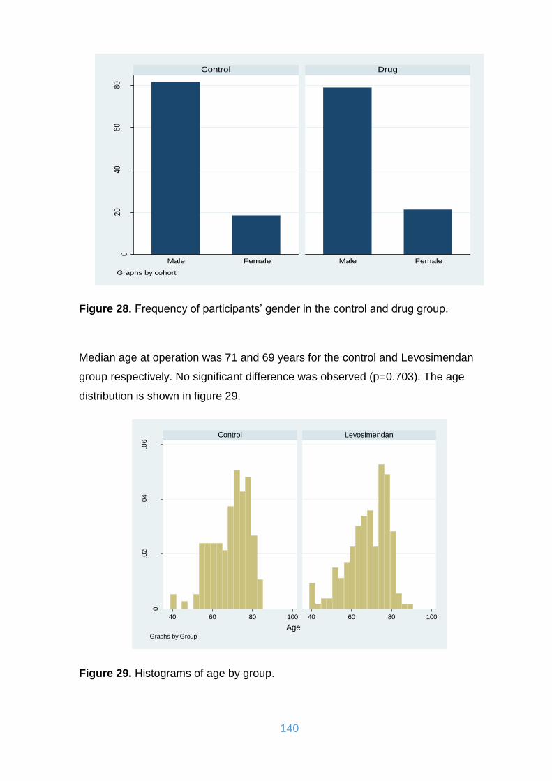
140
Figure 28. Frequency of participants’ gender in the control and drug group.
Median age at operation was 71 and 69 years for the control and Levosimendan
group respectively. No significant difference was observed (p=0.703). The age
distribution is shown in figure 29.
Figure 29. Histograms of age by group.
020
4060
80
Male Female Male Female
Control Drugpe
rcen
t
Graphs by cohort
0
.02
.04
.06
40 60 80 100 40 60 80 100
Control Levosimendan
Density
AgeGraphs by Group

141
The Mann-Witney test showed no difference in the distribution of weight (p=0.087),
height (p=0.874), body mass index (BMI) (p=0.072) and body surface area (BSA)
(p=0.096) in the two groups. The distribution of the above parameters is shown in the
following grafics (Figures 30-33).
Figure 30. Histograms of weight by group.
Figure 31. Histograms of height by group.
0
.01
.02
.03
.04
50 100 150 50 100 150
Control Levosimendan
Density
WeightGraphs by Group
0
.02
.04
.06
.08
150 160 170 180 190 150 160 170 180 190
Control Levosimendan
Density
HeightGraphs by Group

142
Figure 32. Histograms of BMI by group.
Figure 33. Histograms of BSA by group.
0
.05
.1.1
5
20 30 40 50 20 30 40 50
Control Levosimendan
Density
BMIGraphs by Group
01
23
1.5 2 2.5 3 1.5 2 2.5 3
Control Levosimendan
Density
BSAGraphs by Group

143
15.2. Clinical characteristics at baseline
The collected data regarding the preoperative clinical characteristics of the patients
who took part in the study are presented in table 20.
Control
(N: 130)
Levosimendan
(N: 184)
P value
Euroscore 1
Median (IQR) 15.72 (8.07,
27.90)
22.80 (11.59,
42.52)
<0.001
Euroscore 2
Median (IQR) 5.43 (3.41, 10.46) 8.82 (5.09, 17.10) <0.001
ASA score (N, %)
2 1 (0.77) 0 (0.00) 0.048
3 26 (20.00) 31 (16.85)
4 89 (68.46) 116 (63.04)
5 14 (10.77) 37 (20.11)
Type of surgery (N, %)
Elective/regular surgery 96 (73.85) 133 (72.28) 0.759
Emergency surgery 34 (26.15) 51 (27.72)
Number of bypass grafts (N, %)
0 15 (11.54) 21 (11.41) 0.324
1 5 (3.85) 9 (4.89)
2 12 (9.23) 28 (15.22)
3 42 (32.31) 57 (30.98)
4 43 (33.08) 49 (26.63)
5 12 (9.23) 20 (10.87)
6 1 (0.77) 0 (0.00)
Simple/Combination surgery (N, %)
Simple surgery 95 (73.08) 118 (64.13) 0.095
Combination surgery 35 (26.92) 66 (35.87)
Acute myocardial infarction (AMI) (N, %)
No 94 (72.31) 137 (74.46) 0.671
Yes 36 (27.69) 47 (25.54)
Arterial hypertension (N, %)

144
No 9 (6.92) 8 (4.35) 0.321
Yes 121 (93.08) 176 (95.65)
Pulmonary hypertension (N, %)
No 119 (91.54) 128 (69.57) <0.001
Yes 11 (8.46) 56 (30.43)
Hyperlipidemia (N, %)
No 35 (26.92) 37 (20.11) 0.157
Yes 95 (73.08) 147 (79.89)
Diabetes mellitus (N, %)
No 96 (73.85) 127 (69.02) 0.353
Yes 34 (26.15) 57 (30.98)
Arrhythmias (N, %)
No 105 (80.77) 128 (69.57) 0.025
Yes 25 (19.23) 56 (30.43)
Renal failure (N, %)
No 112 (86.15) 137 (74.46) 0.012
Yes 18 (13.85) 47 (25.54)
Chronic obstructive pulmonary disease (COPD) (N, %)
No 109 (83.85) 141 (76.63) 0.118
Yes 21 (16.15) 43 (23.37)
Extracardiac arteriopathy (N, %)
No 108 (83.08) 126 (68.48) 0.003
Yes 22 (16.92) 58 (31.52)
Cerebrovascular accidents (N, %)
No 124 (95.38) 159 (86.41) 0.009
Yes 6 (4.62) 25 (13.59)
Dyspnoea classification (NYHA) (N, %)
1 0 (0.00) 0 (0.00) 0.032
2 7 (5.38) 8 (4.35)
3 66 (50.76) 72 (31.13)
4 57 (43.85) 104 (56.52)
Classification of angina (CCS) (N, %)
0 3 (2.31) 3 (1.63) 0.124
1 6 (4.62) 10 (5.43)
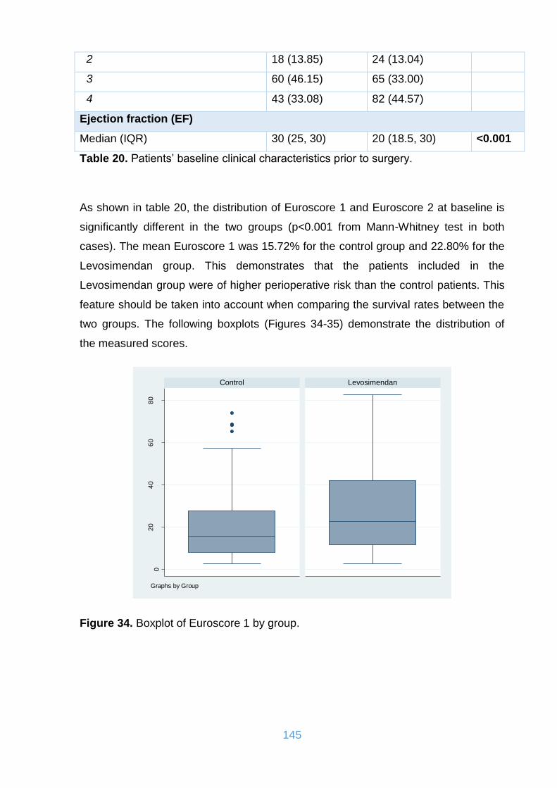
145
2 18 (13.85) 24 (13.04)
3 60 (46.15) 65 (33.00)
4 43 (33.08) 82 (44.57)
Ejection fraction (EF)
Median (IQR) 30 (25, 30) 20 (18.5, 30) <0.001
Table 20. Patients’ baseline clinical characteristics prior to surgery.
As shown in table 20, the distribution of Euroscore 1 and Euroscore 2 at baseline is
significantly different in the two groups (p<0.001 from Mann-Whitney test in both
cases). The mean Euroscore 1 was 15.72% for the control group and 22.80% for the
Levosimendan group. This demonstrates that the patients included in the
Levosimendan group were of higher perioperative risk than the control patients. This
feature should be taken into account when comparing the survival rates between the
two groups. The following boxplots (Figures 34-35) demonstrate the distribution of
the measured scores.
Figure 34. Boxplot of Euroscore 1 by group.
02
04
06
08
0
Control Levosimendan
Euro
score
1
Graphs by Group
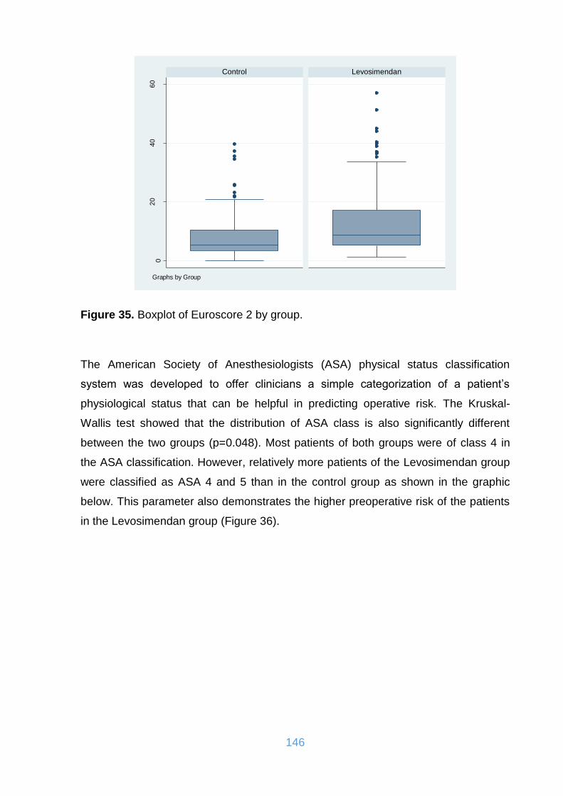
146
Figure 35. Boxplot of Euroscore 2 by group.
The American Society of Anesthesiologists (ASA) physical status classification
system was developed to offer clinicians a simple categorization of a patient’s
physiological status that can be helpful in predicting operative risk. The Kruskal-
Wallis test showed that the distribution of ASA class is also significantly different
between the two groups (p=0.048). Most patients of both groups were of class 4 in
the ASA classification. However, relatively more patients of the Levosimendan group
were classified as ASA 4 and 5 than in the control group as shown in the graphic
below. This parameter also demonstrates the higher preoperative risk of the patients
in the Levosimendan group (Figure 36).
02
04
06
0
Control Levosimendan
Euro
score
2
Graphs by Group
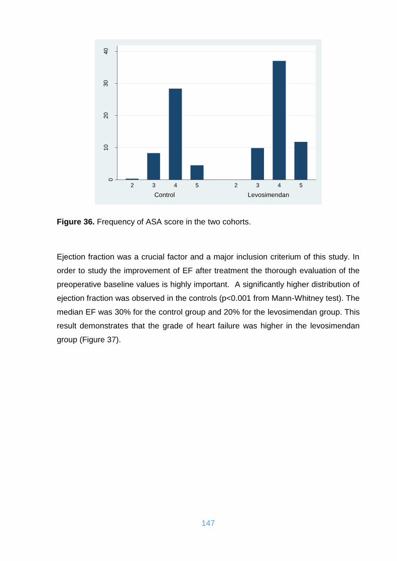
147
Figure 36. Frequency of ASA score in the two cohorts.
Ejection fraction was a crucial factor and a major inclusion criterium of this study. In
order to study the improvement of EF after treatment the thorough evaluation of the
preoperative baseline values is highly important. A significantly higher distribution of
ejection fraction was observed in the controls (p<0.001 from Mann-Whitney test). The
median EF was 30% for the control group and 20% for the levosimendan group. This
result demonstrates that the grade of heart failure was higher in the levosimendan
group (Figure 37).
010
20
30
40
perc
ent
Control Levosimendan
2 3 4 5 2 3 4 5
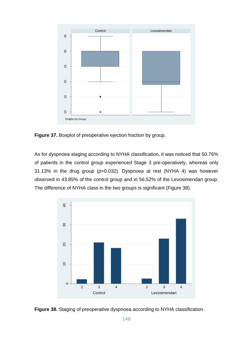
148
Figure 37. Boxplot of preoperative ejection fraction by group.
As for dyspnoea staging according to NYHA classification, it was noticed that 50.76%
of patients in the control group experienced Stage 3 pre-operatively, whereas only
31.13% in the drug group (p=0.032). Dyspnoea at rest (NYHA 4) was however
observed in 43.85% of the control group and in 56.52% of the Levosimendan group.
The difference of NYHA class in the two groups is significant (Figure 38).
Figure 38. Staging of preoperative dyspnoea according to NYHA classification.
10
15
20
25
30
35
Control Levosimendan
Eje
ction f
raction (
pre
opera
tively
)
Graphs by Group
010
20
30
40
perc
ent
Control Levosimendan
2 3 4 2 3 4
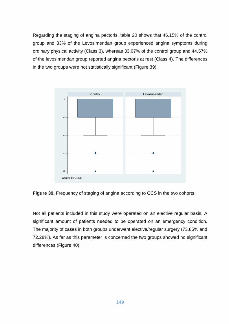
149
Regarding the staging of angina pectoris, table 20 shows that 46.15% of the control
group and 33% of the Levosimendan group experienced angina symptoms during
ordinary physical activity (Class 3), whereas 33.07% of the control group and 44.57%
of the levosimendan group reported angina pectoris at rest (Class 4). The differences
in the two groups were not statistically significant (Figure 39).
Figure 39. Frequency of staging of angina according to CCS in the two cohorts.
Not all patients included in this study were operated on an elective regular basis. A
significant amount of patients needed to be operated on an emergency condition.
The majority of cases in both groups underwent elective/regular surgery (73.85% and
72.28%). As far as this parameter is concerned the two groups showed no significant
differences (Figure 40).
01
23
4
Control Levosimendan
Sta
gin
g o
f angin
a a
gain
st
CC
S
Graphs by Group

150
Figure 40. Frequency of type of surgery (regular of urgent) in the two cohorts.
The next figure presents the percentage of the patients who needed to be operated
under acute myocardial infarction. 27.69% of the control group patients and 25.54%
of the levosimendan group patients were presented with an acute myocardial
infarction. There was no significant difference between the groups as far as this
parameter is concerned (p=0.759) (Figure 41).
Figure 41. Frequency of preoperartive acute myocardial infarction in the two cohorts.
010
20
30
40
perc
ent
Control Levosimendan
Elective/regular surgery Emergency surgery Elective/regular surgery Emergency surgery
010
20
30
40
perc
ent
Control Levosimendan
No Yes No Yes

151
In a significant amount of patients more than one surgical procedures needed to be
carried out (Bypassgrafting, aortic or mitral valve replacement or repair, surgery on
the aorta or aoric root etc.) However no significant difference was observed between
the two groups (p=0.095). 26.92% of the control group patients and 35.85% of the
levosimendan group patients underwent a combined surgery (Figure 42).
Figure 42. Frequency of simple or combined surgery in the two cohorts.
As far as the number of bypass grafts received by the patients is concerned, there
was no significant difference between control and levosimendan group (p=0.324).
The number of grafts are demonstrated in table 20 and figure 43.
010
20
30
40
perc
ent
Control Levosimendan
Simple surgery Combination surgery Simple surgery Combination surgery

152
Figure 43. Boxplot of number of bypass-grafts by group.
The following data demonstrate the rest clinical features which characterize the
comorbidity of the patients included in the study.
Occurrence of arrhythmias (Figure 44) before surgery was significantly different in the
two groups, 19.23% in the controls and 30.43% in the drug group (p=0.030 from chi-
squared test).
01
23
45
6
Control Levosimendan
Num
ber
of
bypass g
raft
s
Graphs by Group

153
Figure 44. Frequency of arrhythmias prior to surgery in the two cohorts.
Similarly, the appearance of pulmonary hypertension before surgery (Figure 45) was
significantly different in the two groups, 8.46% in the controls and 30.43% in the drug
group (p<0.001 from chi-squared test).
Figure 45. Frequency of pulmonary hypertension prior to surgery in the two cohorts.
010
20
30
40
perc
ent
Control Levosimendan
No Yes No Yes
010
20
30
40
perc
ent
Control Levosimendan
No Yes No Yes
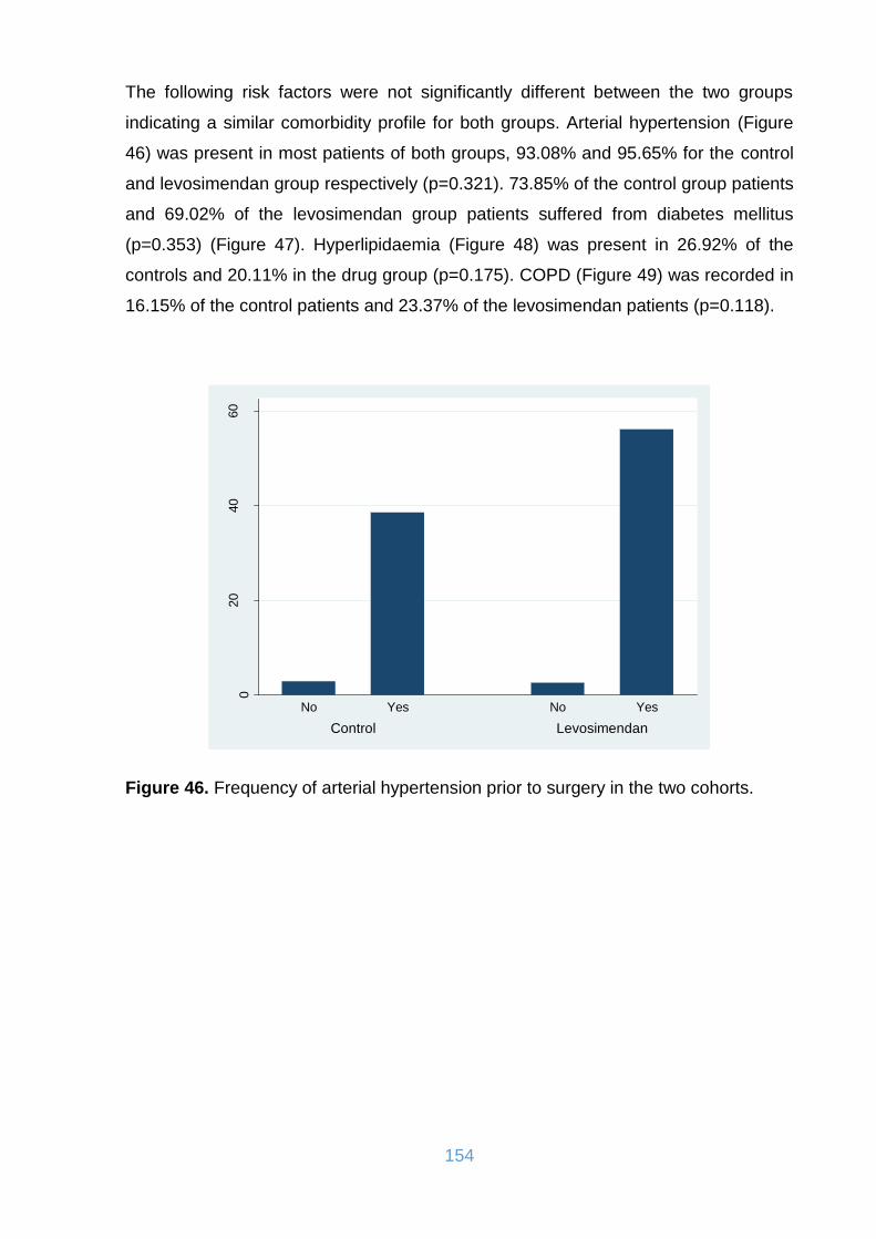
154
The following risk factors were not significantly different between the two groups
indicating a similar comorbidity profile for both groups. Arterial hypertension (Figure
46) was present in most patients of both groups, 93.08% and 95.65% for the control
and levosimendan group respectively (p=0.321). 73.85% of the control group patients
and 69.02% of the levosimendan group patients suffered from diabetes mellitus
(p=0.353) (Figure 47). Hyperlipidaemia (Figure 48) was present in 26.92% of the
controls and 20.11% in the drug group (p=0.175). COPD (Figure 49) was recorded in
16.15% of the control patients and 23.37% of the levosimendan patients (p=0.118).
Figure 46. Frequency of arterial hypertension prior to surgery in the two cohorts.
020
40
60
perc
ent
Control Levosimendan
No Yes No Yes
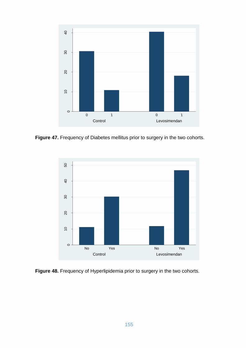
155
Figure 47. Frequency of Diabetes mellitus prior to surgery in the two cohorts.
Figure 48. Frequency of Hyperlipidemia prior to surgery in the two cohorts.
010
20
30
40
perc
ent
Control Levosimendan
0 1 0 1
010
20
30
40
50
perc
ent
Control Levosimendan
No Yes No Yes

156
Figure 49. Frequency of chronic obstructive pulmonary disease prior to surgery in the two cohorts.
Finally, some significant differences were observed in the last 3 clinical parameters.
13.85% of control cases had renal failure versus 25.54% in the drug group (p=0.012)
(Figure 50), 16.28% had extracardiac arteriopathy versus 31.52% in the
levosimendan group (p=0.003) (Figure 51) and 4.62% had a cerebrovascular
accident versus 13.59% in the drug group (p=0.009) (Figure 52).
Figure 50. Frequency of renal failure prior to surgery in the two cohorts.
010
20
30
40
50
perc
ent
Control Levosimendan
No Yes No Yes
010
20
30
40
perc
ent
Control Levosimendan
No Yes No Yes

157
Figure 51. Frequency of extracardiac arteriopathy prior to surgery in the two cohorts.
Figure 52. Frequency of cerebrovascular accidents prior to surgery in the two cohorts.
010
20
30
40
perc
ent
Control Levosimendan
No Yes No Yes
010
20
30
40
50
perc
ent
Control Levosimendan
No Yes No Yes

158
15.3. Introperative surgical times
The intraoperative data concerning the surgical times are shown below in table 21.
Control
(N:130)
Levosimendan
(N: 184)
p value
Duration of surgery (min)
Median (IQR) 218.00
(177.50, 262.75)
227.50
(184.00, 283.75)
0.220
Bypass-Time (min)
Median (IQR) 113.00
(91.00, 154.00)
111.00
(82.00, 153.25)
0.313
Cross-Clamp-Time (min)
Median (IQR) 78.00
(56.75, 103.25)
71.00
(54.00, 92.00)
0.041
Table 21. Intraoperative surgical times in the two cohorts.
Distribution of cross-clamp time (Figure 53) was significantly different in the two
groups where median was 78 minutes in the control group and 71 minutes in the
drug group, suggesting a trend towards longer cross-clamp time in the controls
(p=0.041).
Figure 53. Boxplot of Cross-Clamp-Time (min) by group.
05
01
00
15
02
00
25
0
Control Levosimendan
Cro
ss-C
lam
p-T
ime (
min
)
Graphs by Group
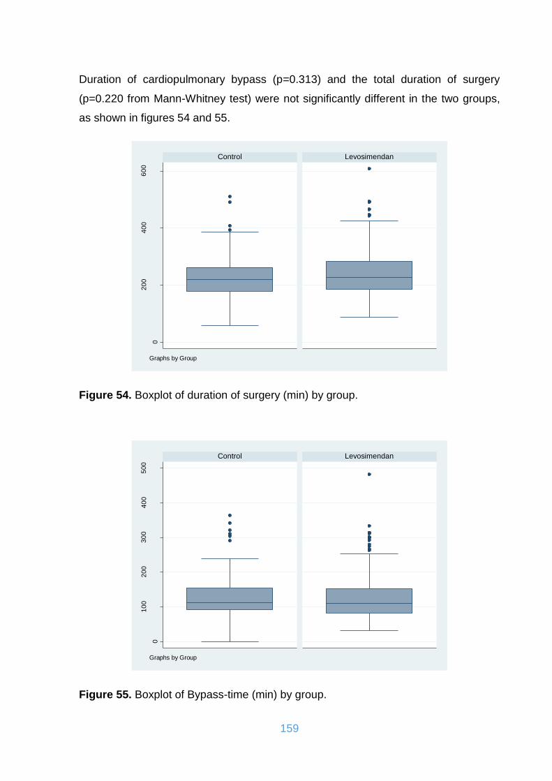
159
Duration of cardiopulmonary bypass (p=0.313) and the total duration of surgery
(p=0.220 from Mann-Whitney test) were not significantly different in the two groups,
as shown in figures 54 and 55.
Figure 54. Boxplot of duration of surgery (min) by group.
Figure 55. Boxplot of Bypass-time (min) by group.
0
20
04
00
60
0
Control Levosimendan
Dura
tion o
f surg
ery
(m
in)
Graphs by Group
0
10
02
00
30
04
00
50
0
Control Levosimendan
Bypass-t
ime (
min
)
Graphs by Group
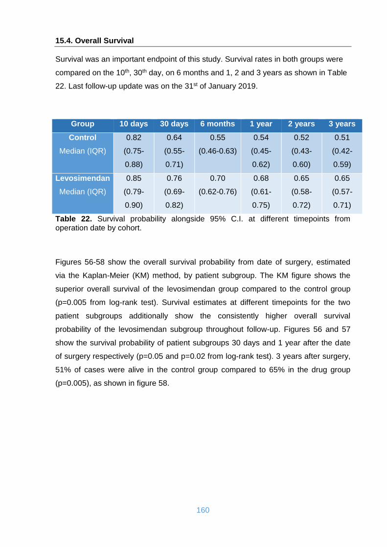
160
15.4. Overall Survival
Survival was an important endpoint of this study. Survival rates in both groups were
compared on the 10th, 30th day, on 6 months and 1, 2 and 3 years as shown in Table
22. Last follow-up update was on the 31st of January 2019.
Group 10 days 30 days 6 months 1 year 2 years 3 years
Control
Median (IQR)
0.82
(0.75-
0.88)
0.64
(0.55-
0.71)
0.55
(0.46-0.63)
0.54
(0.45-
0.62)
0.52
(0.43-
0.60)
0.51
(0.42-
0.59)
Levosimendan
Median (IQR)
0.85
(0.79-
0.90)
0.76
(0.69-
0.82)
0.70
(0.62-0.76)
0.68
(0.61-
0.75)
0.65
(0.58-
0.72)
0.65
(0.57-
0.71)
Table 22. Survival probability alongside 95% C.I. at different timepoints from operation date by cohort.
Figures 56-58 show the overall survival probability from date of surgery, estimated
via the Kaplan-Meier (KM) method, by patient subgroup. The KM figure shows the
superior overall survival of the levosimendan group compared to the control group
(p=0.005 from log-rank test). Survival estimates at different timepoints for the two
patient subgroups additionally show the consistently higher overall survival
probability of the levosimendan subgroup throughout follow-up. Figures 56 and 57
show the survival probability of patient subgroups 30 days and 1 year after the date
of surgery respectively (p=0.05 and p=0.02 from log-rank test). 3 years after surgery,
51% of cases were alive in the control group compared to 65% in the drug group
(p=0.005), as shown in figure 58.

161
Figure 56. Overall survival from date of operation (30 days).
Figure 57. Overall survival from date of operation (1year).
0
.1
.2
.3
.4
.5
.6
.7
.8
.9
1O
vera
ll Su
rviv
al
184 173 160 151 145 140 140Group = Levosimendan129 119 110 102 94 89 84Group = Control
Number at risk
0 5 10 15 20 25 30Days
Group = Control Group = Levosimendan
Kaplan-Meier survival estimates
0
.1
.2
.3
.4
.5
.6
.7
.8
.9
1
Ove
rall
Sur
viva
l
184 135 130 128 126 126 126 0Group = Levosimendan129 75 73 71 70 70 70 0Group = Control
Number at risk
0 2 4 6 8 10 12 14Months
Group = Control Group = Levosimendan
Kaplan-Meier survival estimates
p=0.05 from log-rank
test
p=0.02 from log-rank
test
test test

162
Figure 58. Overall survival from date of operation (3 years).
15.5. Postoperative parameters
Table 23 presents the frequency and percentage of the post-operative parameters in
the two groups. A statistically significant difference between the two cohorts was
observed for all variables except for the total platelet units transfused, support with
IABP, duration of patient support with inotropic drugs in the ICU and hospitalization
days. This is highly indicative of the positive effect of levosimendan versus the no
treatment option before surgery.
Control (N: 130)
Levosimendan (N: 184)
P value
Number of readmissions to hospital (N, %)
0 2 (2.74) 74 (54.41) <0.001
1 25 (34.25) 36 (26.47)
2 30 (41.10) 16 (11.76)
3 8 (10.96) 5 (3.68)
4 5 (6.85) 2 (1.47)
5 3 (4.11) 1 (0.74)
6 0 (0.00) 2 (1.47)
Number of readmissions to hospital
Median (IQR) 2 (1, 2) 0 (0, 1) <0.001
Dyspnoea staging (NYHA) (N, %)
0 0 (0) 19 (10.33) <0.001
1 14 (10.77) 76 (41.30)
2 48 (36.92) 34 (18.48)
3 22 (16.92) 11 (5.98)
0
.1
.2
.3
.4
.5
.6
.7
.8
.9
1O
vera
ll Su
rviv
al
184 126 113 81 50 37 26 16 2 0 0 0Group = Levosimendan129 70 67 66 63 59 58 55 53 21 3 0Group = Control
Number at risk
0 12 24 36 48 60 72 84 96 108 120 132Months
Group = Control Group = Levosimendan
Kaplan-Meier survival estimates
p=0.005 from log-rank
test

163
4 46 (35.38) 44 (23.91)
Staging of angina (CCS) (N, %)
0 6 (4.62) 66 (35.87) <0.001
1 45 (34.62) 67 (36.41)
2 30 (23.08) 9 (4.89)
3 19 (14.62) 7 (3.80)
4 30 (23.08) 35 (19.02)
Postoperative Ejection fraction (EF)
Median (IQR) 30.00(25.00, 30.00)
30.00(25.00, 39.50)
0.025
Need for hemodialysis (N, %)
No 92 (70.77) 163 (88.59) <0.001
Yes 38 (29.23) 21 (11.41)
Artificial ventilation (days)
Median (IQR) 2 (2, 4) 2 (1, 4) 0.007
Support with IABP (days)
Median (IQR) 1 (0, 5.5) 2 (0, 4) 0.810
Units of red blood cells (RBC) transfused
Median (IQR) 8.00 (4.00, 12.00) 4.00 (2.00, 11.50) <0.001
Fresh frozen plasma (FFP) units transfused
Median (IQR) 8.00 (4.00, 12.00) 4.00 (0.00, 8.00) <0.001
Total platelet (PLT) units transfused
Median (IQR) 0.00 (0.00, 2.00) 0.00 (0.00, 2.00) 0.179
Duration of support with inotropic drugs (days)
Median (IQR) 4 (3, 6) 4 (2, 6) 0.076
Hospitalization days
Median (IQR) 15 (13, 21) 16 (12.50, 26.50) 0.405
Postoperative arrhythmias (N, %)
No 61 (46.92) 109 (59.24) 0.031
Yes 69 (53.08) 75 (40.76)
Ejection fraction difference (ΔEF)
Median (IQR) 0 (0, 5) 9 (2.50, 9.50) <0.001
Intensive care unit (ICU) stay (days)
Median (IQR) 7 (4, 9) 6 (4, 10) 0.664
Table 23. Postoperative parameters in the two groups.
A significantly higher distribution of ejection fraction (Figures 59 & 60) was
observed in the drug group (p=0.025 from Mann-Whitney test). Taking into
consideration the fact that the preoperative ejection fractions of the levosimendan
group patients was significantly lower than in the control group patients, this makes
the improvement of the postoperative EF even more significant.
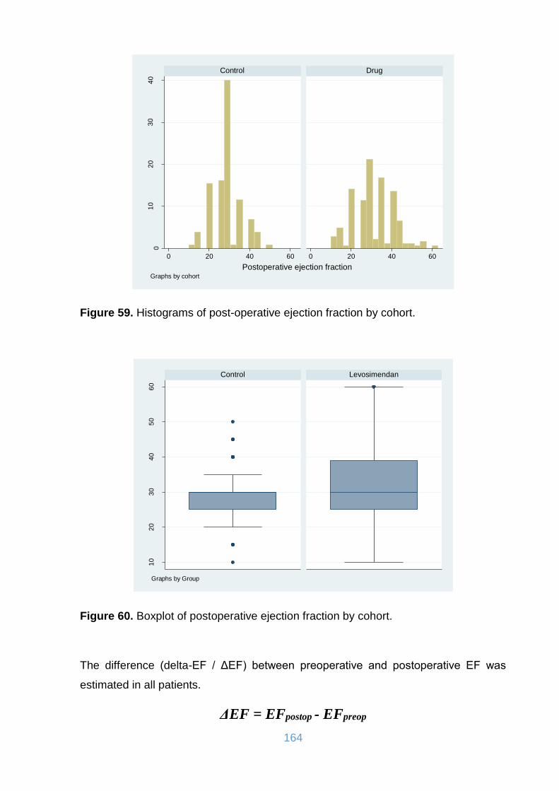
164
Figure 59. Histograms of post-operative ejection fraction by cohort.
Figure 60. Boxplot of postoperative ejection fraction by cohort.
The difference (delta-EF / ΔEF) between preoperative and postoperative EF was
estimated in all patients.
ΔEF = EFpostop - EFpreop
01
02
03
04
0
0 20 40 60 0 20 40 60
Control Drug
Pe
rcen
t
Postoperative ejection fractionGraphs by cohort
10
20
30
40
50
60
Control Levosimendan
Posto
pera
tive e
jection f
raction
Graphs by Group
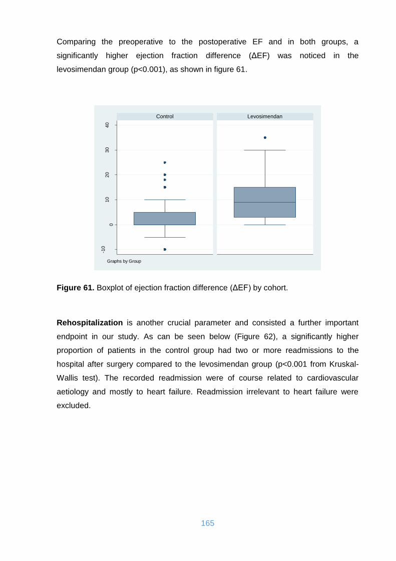
165
Comparing the preoperative to the postoperative EF and in both groups, a
significantly higher ejection fraction difference (ΔEF) was noticed in the
levosimendan group (p<0.001), as shown in figure 61.
Figure 61. Boxplot of ejection fraction difference (ΔEF) by cohort.
Rehospitalization is another crucial parameter and consisted a further important
endpoint in our study. As can be seen below (Figure 62), a significantly higher
proportion of patients in the control group had two or more readmissions to the
hospital after surgery compared to the levosimendan group (p<0.001 from Kruskal-
Wallis test). The recorded readmission were of course related to cardiovascular
aetiology and mostly to heart failure. Readmission irrelevant to heart failure were
excluded.
-10
01
02
03
04
0
Control Levosimendan
Extr
usio
n f
raction d
iffe
rence
Graphs by Group
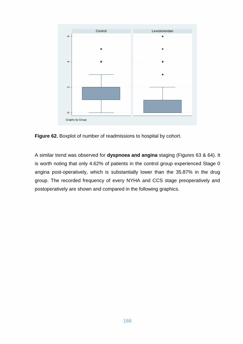
166
Figure 62. Boxplot of number of readmissions to hospital by cohort.
A similar trend was observed for dyspnoea and angina staging (Figures 63 & 64). It
is worth noting that only 4.62% of patients in the control group experienced Stage 0
angina post-operatively, which is substantially lower than the 35.87% in the drug
group. The recorded frequency of every NYHA and CCS stage preoperatively and
postoperatively are shown and compared in the following graphics.
02
46
Control Levosimendan
Num
ber
of
readm
issio
ns t
o h
ospital
Graphs by Group
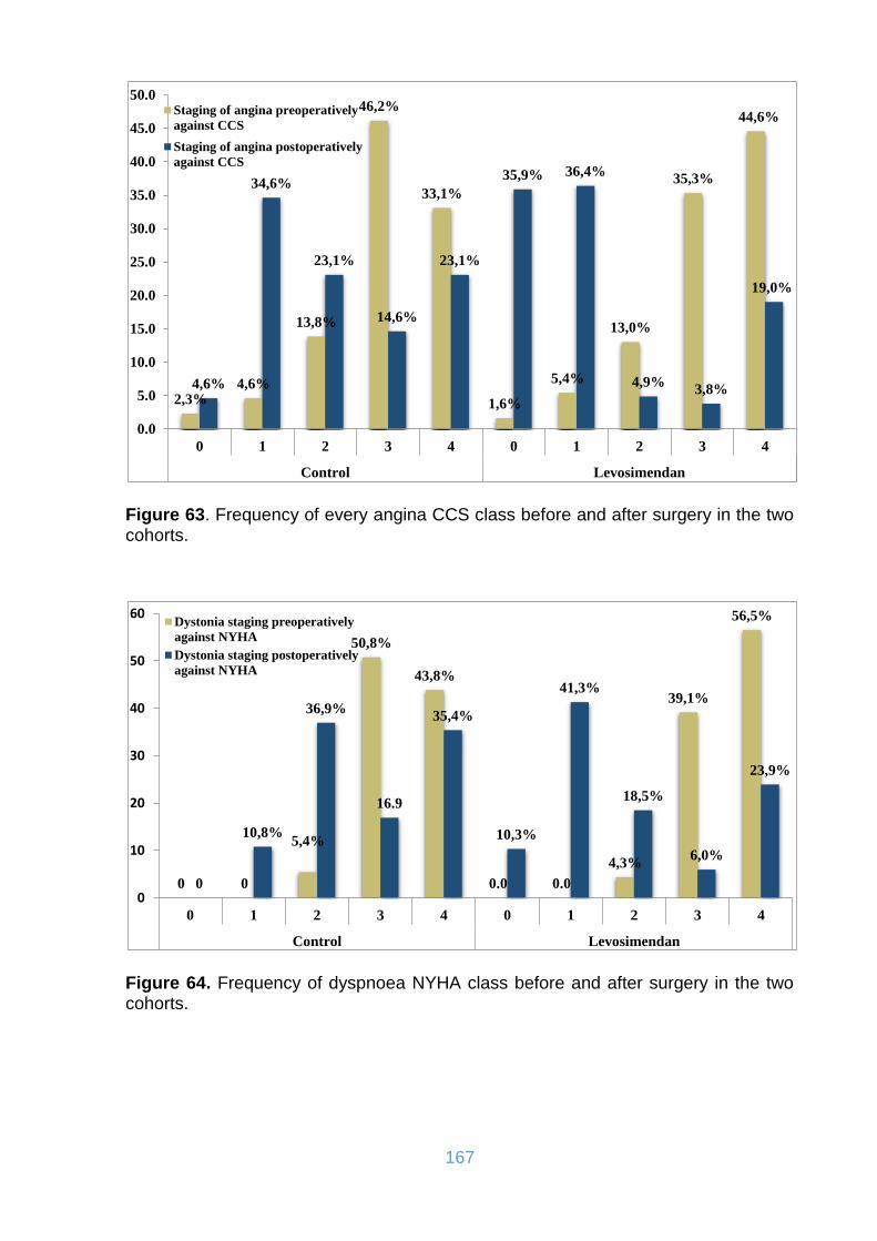
167
Figure 63. Frequency of every angina CCS class before and after surgery in the two cohorts.
Figure 64. Frequency of dyspnoea NYHA class before and after surgery in the two cohorts.
2,3%4,6%
13,8%
46,2%
33,1%
1,6%
5,4%
13,0%
35,3%
44,6%
4,6%
34,6%
23,1%
14,6%
23,1%
35,9% 36,4%
4,9%3,8%
19,0%
0.0
5.0
10.0
15.0
20.0
25.0
30.0
35.0
40.0
45.0
50.0
0 1 2 3 4 0 1 2 3 4
Control Levosimendan
Staging of angina preoperatively
against CCS
Staging of angina postoperatively
against CCS
0 0
5,4%
50,8%
43,8%
0.0 0.0
4,3%
39,1%
56,5%
0
10,8%
36,9%
16.9
35,4%
10,3%
41,3%
18,5%
6,0%
23,9%
0
10
20
30
40
50
60
0 1 2 3 4 0 1 2 3 4
Control Levosimendan
Dystonia staging preoperatively
against NYHA
Dystonia staging postoperatively
against NYHA
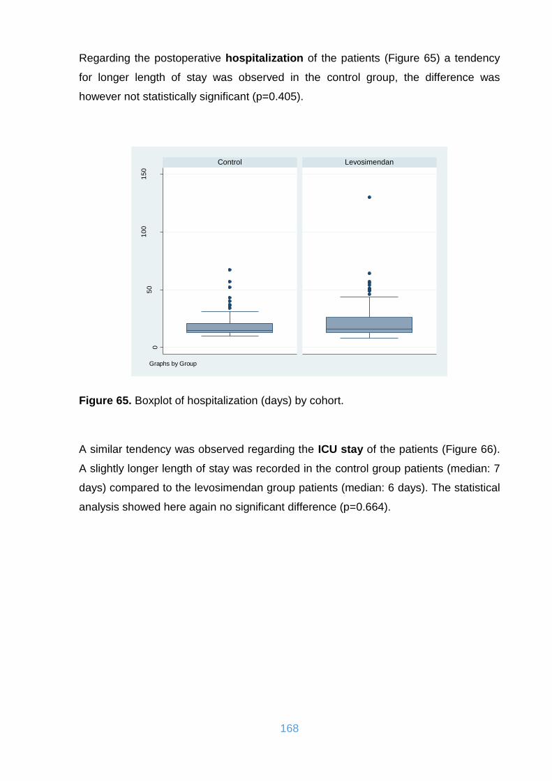
168
Regarding the postoperative hospitalization of the patients (Figure 65) a tendency
for longer length of stay was observed in the control group, the difference was
however not statistically significant (p=0.405).
Figure 65. Boxplot of hospitalization (days) by cohort.
A similar tendency was observed regarding the ICU stay of the patients (Figure 66).
A slightly longer length of stay was recorded in the control group patients (median: 7
days) compared to the levosimendan group patients (median: 6 days). The statistical
analysis showed here again no significant difference (p=0.664).
05
01
00
15
0
Control Levosimendan
Hospitaliz
ation (
days)
Graphs by Group

169
Figure 66. Boxplot of intensive care unit (stay in days) by cohort.
The patients of the levosimendan group appeared easier to wean from the ventilator
and were sooner extubated compared to the control group patients (p=0.07). This
finding may imply a possible lung protective effect of the drug (Figure 67).
Figure 67. Boxplot of artificial ventilation (days) by cohort.
02
04
06
08
01
00
Control Levosimendan
Inte
nsiv
e c
are
unit(s
tay in d
ays)
Graphs by Group
02
04
06
08
0
Control Levosimendan
Art
ific
ial aera
tion (
days)
Graphs by Group

170
The need of inotropic support and administration of catecholamines was less in the
levosimendan group (Figure 68), the difference appeared however to be not
statisticall significant (p=0.076).
Figure 68. Boxplot of duration of patient support with inotropic drugs in the ICU (in days) by cohort.
A similar trend was observed regarding the need of mechanical circulatory support
and the use of the IABP (Figure 69), where also no significant difference was
observed between the two groups (p=0.810).
05
10
15
20
25
Control Levosimendan
Dura
tion o
f patient
support
with inotr
opic
dru
gs in I
CU
(days)
Graphs by Group
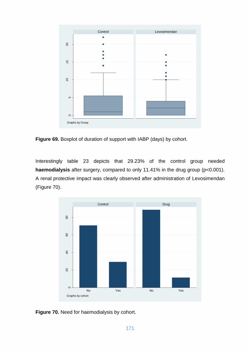
171
Figure 69. Boxplot of duration of support with IABP (days) by cohort.
Interestingly table 23 depicts that 29.23% of the control group needed
haemodialysis after surgery, compared to only 11.41% in the drug group (p<0.001).
A renal protective impact was clearly observed after administration of Levosimendan
(Figure 70).
Figure 70. Need for haemodialysis by cohort.
05
10
15
20
Control Levosimendan
Support
with I
AB
P (
days)
Graphs by Group
02
04
06
08
0
No Yes No Yes
Control Drug
perc
ent
Graphs by cohort

172
Postoperative arrhythmias are very frequent after cardiac surgery and were recorded
in patients from both groups (Figure 71). The frequency of such incidents was
however significantly higher in the control group (53%) compared to the group
treated with levosimendan (40.76%) (p=0,031).
Figure 71. Frequency of postoperative arrhythmias by cohort.
A significantly higher quantity of erythrocytes (Figure 72) and fresh frozen plasma
(Figure 73) units was required after surgery by the control group compared to the
levosimendan group (p<0.001 for both parameters). No difference was observed
regarding the need for platelet transfusion (p=0.179) (Figure 74).
010
20
30
40
perc
ent
Control Levosimendan
No Yes No Yes

173
Figure 72. Histograms of total units of erythrocytes (RBC) transfused by cohort.
Figure 73. Histograms of fresh frozen plasma (FFP) units transported by cohort.
02
04
06
0
0 20 40 60 80 0 20 40 60 80
Control Drug
Pe
rcen
t
Total units of red blood cells transfusedGraphs by cohort
02
04
06
0
0 20 40 60 80 0 20 40 60 80
Control Drug
Pe
rcen
t
Total fresh frozen plasma units transportedGraphs by cohort
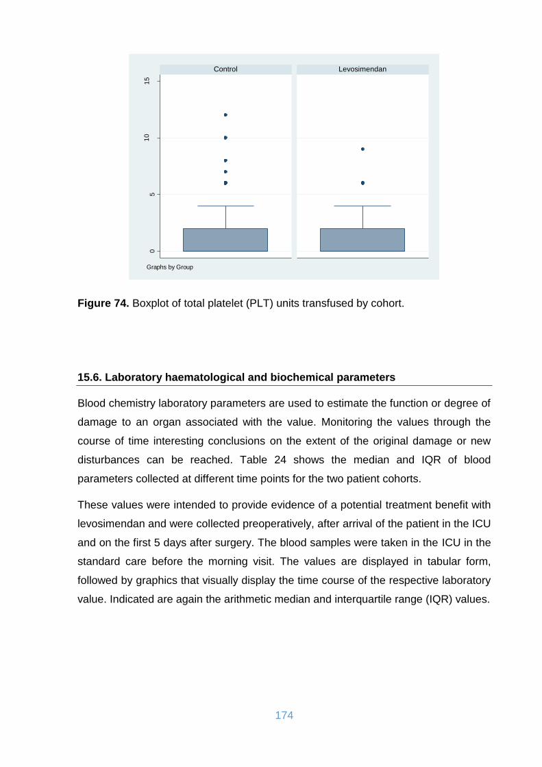
174
Figure 74. Boxplot of total platelet (PLT) units transfused by cohort.
15.6. Laboratory haematological and biochemical parameters
Blood chemistry laboratory parameters are used to estimate the function or degree of
damage to an organ associated with the value. Monitoring the values through the
course of time interesting conclusions on the extent of the original damage or new
disturbances can be reached. Table 24 shows the median and IQR of blood
parameters collected at different time points for the two patient cohorts.
These values were intended to provide evidence of a potential treatment benefit with
levosimendan and were collected preoperatively, after arrival of the patient in the ICU
and on the first 5 days after surgery. The blood samples were taken in the ICU in the
standard care before the morning visit. The values are displayed in tabular form,
followed by graphics that visually display the time course of the respective laboratory
value. Indicated are again the arithmetic median and interquartile range (IQR) values.
05
10
15
Control Levosimendan
Tota
l pla
tele
t units t
ransfu
sed
Graphs by Group

175
Control
(N: 130)
Levosimendan
(N: 184)
P value
Hemoglobin (g/dl) ; median (IQR)
0 day prior to surgery 14.30 (12.50, 15.40) 13.40 (11.65, 14.95) 0.003
1 day of surgery 9.30 (8.55, 10.00) 9.80 (9.90, 10.70) <0.001
2 1st postop. day 9.50 (8.6, 10.50) 9.8 (8.90, 10.80) 0.047
3 2nd postop. day 9.60 (8.90, 10.40) 9.6 (8.90, 10.40) 0.956
4 3rd postop. day 9.70 (9, 10.40) 9.5 (8.80, 10.30) 0.315
5 4th postop. day 10 (9.20, 10.80) 9.70 (8.90,10.70) 0.082
6 5th postop. day 10.40 (9.40, 11.30) 9.90 (9.10, 10.90) 0.028
C Reactive Protein (CRP) (mg/l) ; median (IQR)
0 day prior to surgery 6.80 (3.60, 17.32) 9.34 (3.98, 22.33) 0.158
1 day of surgery 68.70 (34.70, 88.10) 40.30 (22.73, 72.73) <0.001
2 1st postop. day 114.20 (78.40, 156.30) 106.13 (56.10, 186.30) 0.555
3 2nd postop. day 199.30 (159.10,
243.79)
194.29 (151.78, 239.86) 0.422
4 3rd postop. day 189.95 (147.20,
273.90)
171.67 (133.90, 227.84) 0.047
5 4th postop. day 148.80 (109.90, 236) 130.40 (90.30, 172.99) <0.001
6 5th postop. day 120.40 (76, 192.70) 89.65 (55.50, 131.20) <0.001
Troponin I (TnI) (ng/ml); median (IQR)
0 day prior to surgery 0.07 (0.03, 0.28) 0.08 (0.04, 0.52) 0.028
1 day of surgery 18.74 (8.31, 50) 9.90 (4.05, 24.75) <0.001
2 1st postop. day 18.47 (7.17, 50) 9.74 (4.88, 28.83) 0.001
3 2nd postop. day 11.82 (5.43, 38.14) 5.62 (2.26, 17.64) <0.001
4 3rd postop. day 8.94 (3.56, 26.94) 3.09 (1.13, 10.68) <0.001
5 4th postop. day 5.13 (2.12, 17.61) 1.88 (0.73, 6.01) <0.001
6 5th postop. day 3.96 (1.32, 10.35) 0.94 (0.36, 3.51) <0.001
Creatine Kinase (CK) (U/l); median (IQR)
0 day prior to surgery 93.50 (57, 147) 75.50 (48.50, 156) 0.114
1 day of surgery 796 (453, 1530.50) 503.50 (288.25,
1010.25)
<0.001
2 1st postop. day 1131 (597, 2026) 728.50 (374.50,
1741.50)
0.005

176
3 2nd postop. day 1109.5 (565, 2725) 738.50 (309, 2116.50) 0.014
4 3rd postop. day 852.50 (387, 1990) 464.5 (166.5, 1314) <0.001
5 4th postop. day 561 (193, 1293) 266 (99, 792.50) <0.001
6 5th postop. day 307.50 (102, 884) 135.50 (61, 389.50) <0.001
Myocardial CK (CKMB) (U/l); median (IQR)
0 day prior to surgery 17 (13,22) 21 (17,28) <0.001
1 day of surgery 78 (40.50, 202.50) 41 (27.25, 77.50) <0.001
2 1st postop. day 65 (40,148) 40.5 (24.5, 74.5) <0.001
3 2nd postop. day 44 (29, 90) 30.5 (19,58) <0.001
4 3rd postop. day 34 (21,71) 22 (14,39) <0.001
5 4th postop. day 27 (18,46) 18 (12,28) <0.001
6 5th postop. day 22 (16,36) 13 (9,21) <0.001
Creatinine (mg/dl); median (IQR)
0 day prior to surgery 1.05 (0.84, 1.30) 1.10 (0.92,1.47) 0.050
1 day of surgery 1.32 (0.99, 1.74) 1.11 (0.86, 1.52) 0.002
2 1st postop. day 1.45 (1.06, 1.94) 1.24 (0.89, 1.75) 0.005
3 2nd postop. day 1.65 (1.06, 2.78) 1.15 (0.81, 2) <0.001
4 3rd postop. day 1.91 (1.13, 3.38) 1.22 (0.80, 2.07) <0.001
5 4th postop. day 1.83 (1.14, 3.47) 1.20 (0.78, 2.19) <0.001
6 5th postop. day 1.89 (1.16, 3.25) 1.14 (0.80, 1.87) <0.001
Creatinine clearance(ml/min); median (IQR)
0 day prior to surgery 72.59 (54.59, 91.31) 71.59 (47.96, 100.54) 0.506
1 day of surgery 56.23 (43.30, 76.45) 70.23 (47.42, 102.23) 0.001
2 1st postop. day 49.83 (40.33, 69.79) 63.80 (44.38, 97.12) 0.002
3 2nd postop. day 43.33 (26.47, 73.29) 64.64 (35.60, 106.67) <0.001
4 3rd postop. day 38.92 (20.74, 65.81) 65.13 (36.50, 102.78) <0.001
5 4th postop. day 42.30 (19.41, 69.19) 63.98 (32.23, 107.84) <0.001
6 5th postop. day 39.79 (20.92, 66.35) 69.13 (36.99, 104.59) <0.001
Glomerular filtration rate (GFR) (ml/min); median (IQR)
0 day prior to surgery 68.40 (55, 82.30) 63.29 (46, 82.98) 0.061
1 day of surgery 52.20 (38.90, 72) 64.95 (43.73, 86.32) 0.002
2 1st postop. day 47.6 (33.20, 66.70) 57.45 (38.30, 83.58) 0.004
3 2nd postop. day 42 (20.80, 66.40) 59.29 (32.52, 90.57) <0.001
4 3rd postop. day 33.30 (15.50, 63.20) 58.90 (29.80, 88) <0.001
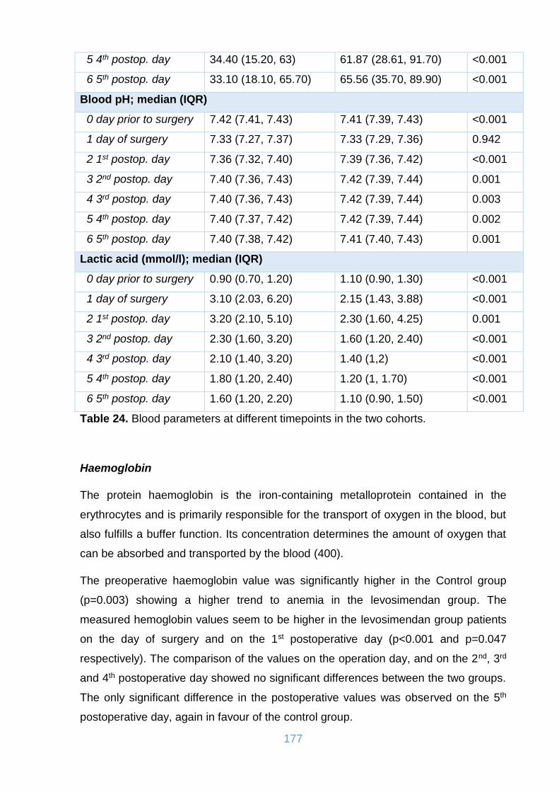
177
5 4th postop. day 34.40 (15.20, 63) 61.87 (28.61, 91.70) <0.001
6 5th postop. day 33.10 (18.10, 65.70) 65.56 (35.70, 89.90) <0.001
Blood pH; median (IQR)
0 day prior to surgery 7.42 (7.41, 7.43) 7.41 (7.39, 7.43) <0.001
1 day of surgery 7.33 (7.27, 7.37) 7.33 (7.29, 7.36) 0.942
2 1st postop. day 7.36 (7.32, 7.40) 7.39 (7.36, 7.42) <0.001
3 2nd postop. day 7.40 (7.36, 7.43) 7.42 (7.39, 7.44) 0.001
4 3rd postop. day 7.40 (7.36, 7.43) 7.42 (7.39, 7.44) 0.003
5 4th postop. day 7.40 (7.37, 7.42) 7.42 (7.39, 7.44) 0.002
6 5th postop. day 7.40 (7.38, 7.42) 7.41 (7.40, 7.43) 0.001
Lactic acid (mmol/l); median (IQR)
0 day prior to surgery 0.90 (0.70, 1.20) 1.10 (0.90, 1.30) <0.001
1 day of surgery 3.10 (2.03, 6.20) 2.15 (1.43, 3.88) <0.001
2 1st postop. day 3.20 (2.10, 5.10) 2.30 (1.60, 4.25) 0.001
3 2nd postop. day 2.30 (1.60, 3.20) 1.60 (1.20, 2.40) <0.001
4 3rd postop. day 2.10 (1.40, 3.20) 1.40 (1,2) <0.001
5 4th postop. day 1.80 (1.20, 2.40) 1.20 (1, 1.70) <0.001
6 5th postop. day 1.60 (1.20, 2.20) 1.10 (0.90, 1.50) <0.001
Table 24. Blood parameters at different timepoints in the two cohorts.
Haemoglobin
The protein haemoglobin is the iron-containing metalloprotein contained in the
erythrocytes and is primarily responsible for the transport of oxygen in the blood, but
also fulfills a buffer function. Its concentration determines the amount of oxygen that
can be absorbed and transported by the blood (400).
The preoperative haemoglobin value was significantly higher in the Control group
(p=0.003) showing a higher trend to anemia in the levosimendan group. The
measured hemoglobin values seem to be higher in the levosimendan group patients
on the day of surgery and on the 1st postoperative day (p<0.001 and p=0.047
respectively). The comparison of the values on the operation day, and on the 2nd, 3rd
and 4th postoperative day showed no significant differences between the two groups.
The only significant difference in the postoperative values was observed on the 5th
postoperative day, again in favour of the control group.

178
Figure 75. Histograms of haemoglobin on the 5th postoperative day in the two groups.
Figure 76. Distribution of hemoglobin values in the two cohorts.
C Reactive Protein (CRP)
C-reactive protein (CRP) is an annular (ring-shaped), pentameric protein found in
blood plasma, who’s circulating concentrations rise in response to inflammation and
is therefore mainly used as an inflammation marker. It is an acute-phase protein of
05
10
15
20
5 10 15 5 10 15
Control Drug
Pe
rcen
t
HemoglobinGraphs by cohort
0 dayprior tosurgery
1 daysurgery
1 day postsurgery
2 dayspost
surgery
3 dayspost
surgery
4 dayspost
surgery
5 dayspost
surgery
Control 14.3 9.3 9.5 9.6 9.7 10 10.4
Levosimendan 13.4 9.8 9.8 9.6 9.5 9.7 9.9
0
2
4
6
8
10
12
14
16
Haemoglobin

179
hepatic origin that increases following interleukin-6 secretion by macrophages and T
cells. Its physiological role is to bind to lysophosphatidylcholine expressed on the
surface of dead or dying cells (and some types of bacteria) in order to activate the
complement system via C1q. Myocardial infarction in humans provokes an acute
phase response, and C-reactive protein (CRP), the classical acute phase plasma
protein, is deposited together with complement within the infarct. CRP and
complement activation are major mediators of ischemic myocardial injury (402, 403).
CRP differed significantly from three days onwards post-surgery. This implies a lower
grade of inflammatory response of the patients who were treated with levosimendan.
Figure 77. Histograms of CRP at 5 days after surgery in the two cohorts.
01
02
03
0
0 200 400 600 0 200 400 600
Control Drug
Pe
rcen
t
C-Reactive ProteinGraphs by cohort

180
Figure 78. Distribution of CRP values in the two cohorts.
Troponin I
Troponin I is considered to be a specific marker of cardiac damage and, for the
diagnosis of acute myocardial infarction. For more than 15 years cardiac troponin I
(cTnI) has been known as a reliable marker of cardiac muscle tissue injury. It is
considered to be more sensitive and significantly more specific in diagnosis of the
myocardial infarction than the "golden marker" of last decades – CK-MB, as well as
total creatine kinase, myoglobin and lactate dehydrogenase isoenzymes. Troponin I
is not entirely specific for myocardial damage secondary to infarction. Other causes
of raised Troponin I include chronic renal failure, heart failure, subarachnoid
haemorrhage and pulmonary embolus (401).
Troponin I levels differed significantly between the two groups across all time points.
Indicatively, median of troponin I was 18.47 and 9.90 on the 1st postoperative day in
the control and levosimendan groups respectively. This finding supports the
hypothetical cardioprotective effect of levosimendan showing less myocardial
damage to the patients treated with the drug.
1 dayprior tosurgery
1 daysurgery
1 day postsurgery
2 dayspost
surgery
3 dayspost
surgery
4 dayspost
surgery
5 dayspost
surgery
Control 6.8 68.7 114.2 199.3 189.95 148.8 120.4
Levosimendan 9.34 40.3 106.13 194.29 171.67 130.4 89.65
0
50
100
150
200
250
CRP

181
Figure 79. Histograms of troponin on the 5th postoperative day in the two cohorts.
Figure 80. Distribution of troponin I in the two cohorts.
Creatine Kinase (CK) and Myocardial Creatinine Kinase (CK-MB)
The enzyme creatine kinase (CK) is used to diagnose heart and skeletal muscle
damage. By changing the concentration in the blood can be concluded on the extent
of the damage done. A subtype of creatine kinase (CKMB) allows more specific
conclusions about damage to the heart. An increasing proportion of CKMB indicates
02
04
06
08
0
0 20 40 60 0 20 40 60
Control Drug
Pe
rcen
t
TroponinGraphs by cohort
1 dayprior tosurgery
1 daysurgery
1 day postsurgery
2 dayspost
surgery
3 dayspost
surgery
4 dayspost
surgery
5 dayspost
surgery
Control 0.07 18.74 18.47 11.82 8.94 5.13 3.96
Levosimendan 0.08 9.9 9.74 5.62 3.09 1.88 0.94
02468
101214161820
Troponin I
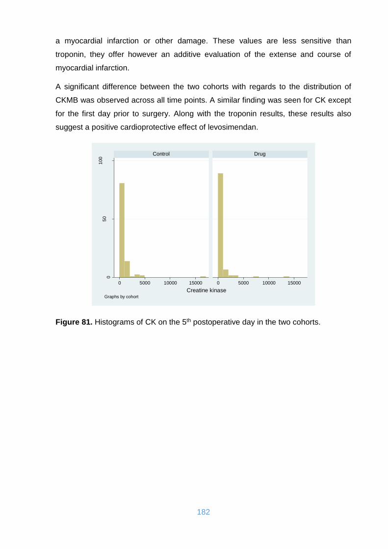
182
a myocardial infarction or other damage. These values are less sensitive than
troponin, they offer however an additive evaluation of the extense and course of
myocardial infarction.
A significant difference between the two cohorts with regards to the distribution of
CKMB was observed across all time points. A similar finding was seen for CK except
for the first day prior to surgery. Along with the troponin results, these results also
suggest a positive cardioprotective effect of levosimendan.
Figure 81. Histograms of CK on the 5th postoperative day in the two cohorts.
05
01
00
0 5000 10000 15000 0 5000 10000 15000
Control Drug
Pe
rcen
t
Creatine kinaseGraphs by cohort
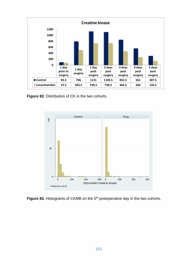
183
Figure 82. Distribution of CK in the two cohorts.
Figure 83. Histograms of CKMB on the 5th postoperative day in the two cohorts.
1 dayprior tosurgery
1 daysurgery
1 daypost
surgery
2 dayspost
surgery
3 dayspost
surgery
4 dayspost
surgery
5 dayspost
surgery
Control 93.5 796 1131 1109.5 852.5 561 307.5
Levosimendan 75.5 503.5 728.5 738.5 464.5 266 135.5
0
200
400
600
800
1000
1200
Creatine kinase
05
01
00
0 200 400 600 0 200 400 600
Control Drug
Pe
rcen
t
Myocardial Creatine kinaseGraphs by cohort
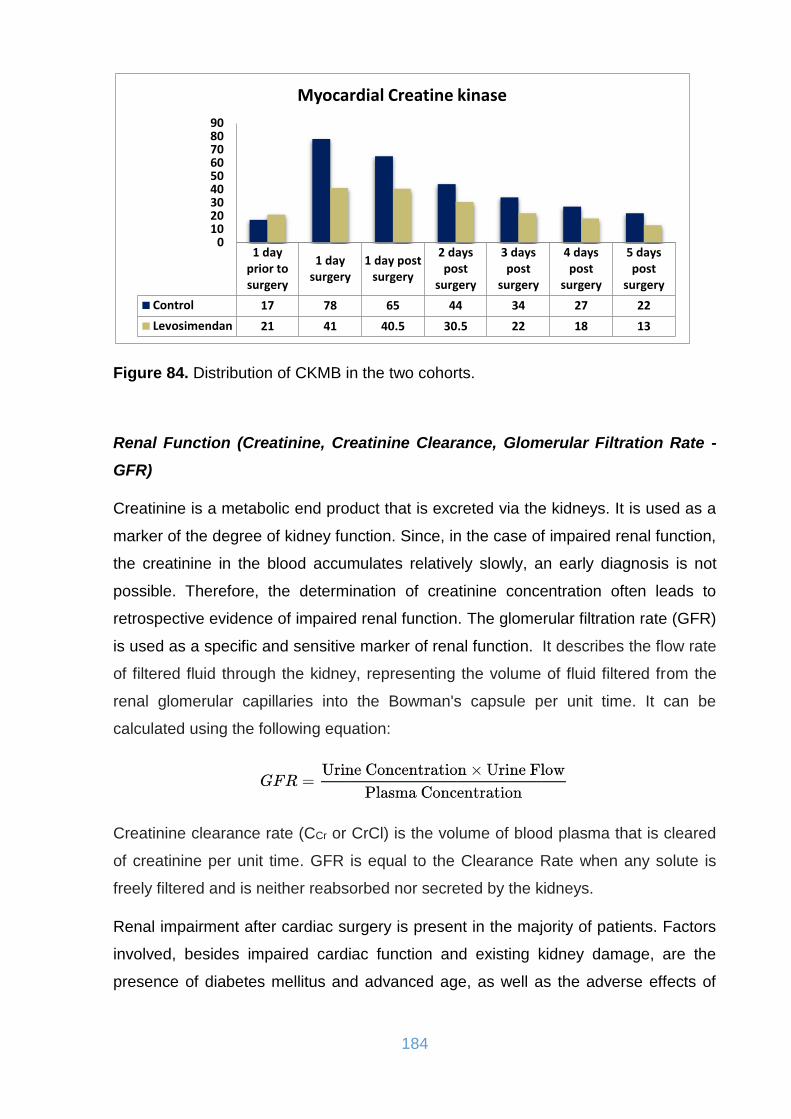
184
Figure 84. Distribution of CKMB in the two cohorts.
Renal Function (Creatinine, Creatinine Clearance, Glomerular Filtration Rate -
GFR)
Creatinine is a metabolic end product that is excreted via the kidneys. It is used as a
marker of the degree of kidney function. Since, in the case of impaired renal function,
the creatinine in the blood accumulates relatively slowly, an early diagnosis is not
possible. Therefore, the determination of creatinine concentration often leads to
retrospective evidence of impaired renal function. The glomerular filtration rate (GFR)
is used as a specific and sensitive marker of renal function. It describes the flow rate
of filtered fluid through the kidney, representing the volume of fluid filtered from the
renal glomerular capillaries into the Bowman's capsule per unit time. It can be
calculated using the following equation:
Creatinine clearance rate (CCr or CrCl) is the volume of blood plasma that is cleared
of creatinine per unit time. GFR is equal to the Clearance Rate when any solute is
freely filtered and is neither reabsorbed nor secreted by the kidneys.
Renal impairment after cardiac surgery is present in the majority of patients. Factors
involved, besides impaired cardiac function and existing kidney damage, are the
presence of diabetes mellitus and advanced age, as well as the adverse effects of
1 dayprior tosurgery
1 daysurgery
1 day postsurgery
2 dayspost
surgery
3 dayspost
surgery
4 dayspost
surgery
5 dayspost
surgery
Control 17 78 65 44 34 27 22
Levosimendan 21 41 40.5 30.5 22 18 13
0102030405060708090
Myocardial Creatine kinase
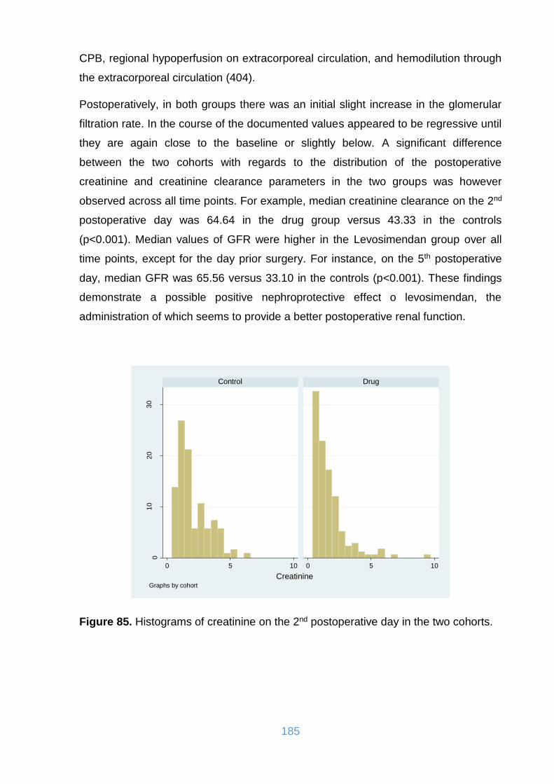
185
CPB, regional hypoperfusion on extracorporeal circulation, and hemodilution through
the extracorporeal circulation (404).
Postoperatively, in both groups there was an initial slight increase in the glomerular
filtration rate. In the course of the documented values appeared to be regressive until
they are again close to the baseline or slightly below. A significant difference
between the two cohorts with regards to the distribution of the postoperative
creatinine and creatinine clearance parameters in the two groups was however
observed across all time points. For example, median creatinine clearance on the 2nd
postoperative day was 64.64 in the drug group versus 43.33 in the controls
(p<0.001). Median values of GFR were higher in the Levosimendan group over all
time points, except for the day prior surgery. For instance, on the 5th postoperative
day, median GFR was 65.56 versus 33.10 in the controls (p<0.001). These findings
demonstrate a possible positive nephroprotective effect o levosimendan, the
administration of which seems to provide a better postoperative renal function.
Figure 85. Histograms of creatinine on the 2nd postoperative day in the two cohorts.
01
02
03
0
0 5 10 0 5 10
Control Drug
Pe
rcen
t
CreatinineGraphs by cohort

186
Figure 86. Distribution of creatine values in the two cohorts.
Figure 87. Histograms of creatinine clearance on the 2nd postoperative day in the two cohorts.
1 dayprior tosurgery
1 daysurgery
1 day postsurgery
2 dayspost
surgery
3 dayspost
surgery
4 dayspost
surgery
5 dayspost
surgery
Control 1.05 1.32 1.45 1.65 1.91 1.83 1.89
Levosimendan 1.1 1.11 1.24 1.15 1.22 1.2 1.14
0
0.5
1
1.5
2
2.5
Creatinine
01
02
03
0
0 100 200 300 0 100 200 300
Control Drug
Pe
rcen
t
Clearance creatinineGraphs by cohort
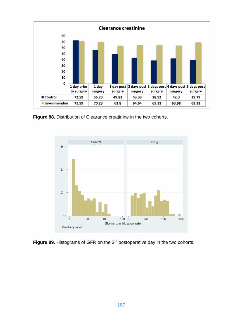
187
Figure 88. Distribution of Clearance creatinine in the two cohorts.
Figure 89. Histograms of GFR on the 3rd postoperative day in the two cohorts.
1 day priorto surgery
1 daysurgery
1 day postsurgery
2 days postsurgery
3 days postsurgery
4 days postsurgery
5 days postsurgery
Control 72.59 56.23 49.83 43.33 38.92 42.3 39.79
Levosimendan 71.59 70.23 63.8 64.64 65.13 63.98 69.13
0
10
20
30
40
50
60
70
80
Clearance creatinine
01
02
03
0
0 50 100 150 0 50 100 150
Control Drug
Pe
rcen
t
Glomerular filtration rateGraphs by cohort
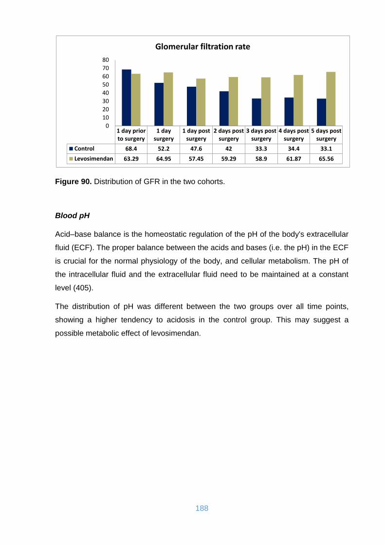
188
Figure 90. Distribution of GFR in the two cohorts.
Blood pH
Acid–base balance is the homeostatic regulation of the pH of the body's extracellular
fluid (ECF). The proper balance between the acids and bases (i.e. the pH) in the ECF
is crucial for the normal physiology of the body, and cellular metabolism. The pH of
the intracellular fluid and the extracellular fluid need to be maintained at a constant
level (405).
The distribution of pH was different between the two groups over all time points,
showing a higher tendency to acidosis in the control group. This may suggest a
possible metabolic effect of levosimendan.
1 day priorto surgery
1 daysurgery
1 day postsurgery
2 days postsurgery
3 days postsurgery
4 days postsurgery
5 days postsurgery
Control 68.4 52.2 47.6 42 33.3 34.4 33.1
Levosimendan 63.29 64.95 57.45 59.29 58.9 61.87 65.56
0
10
20
30
40
50
60
70
80
Glomerular filtration rate
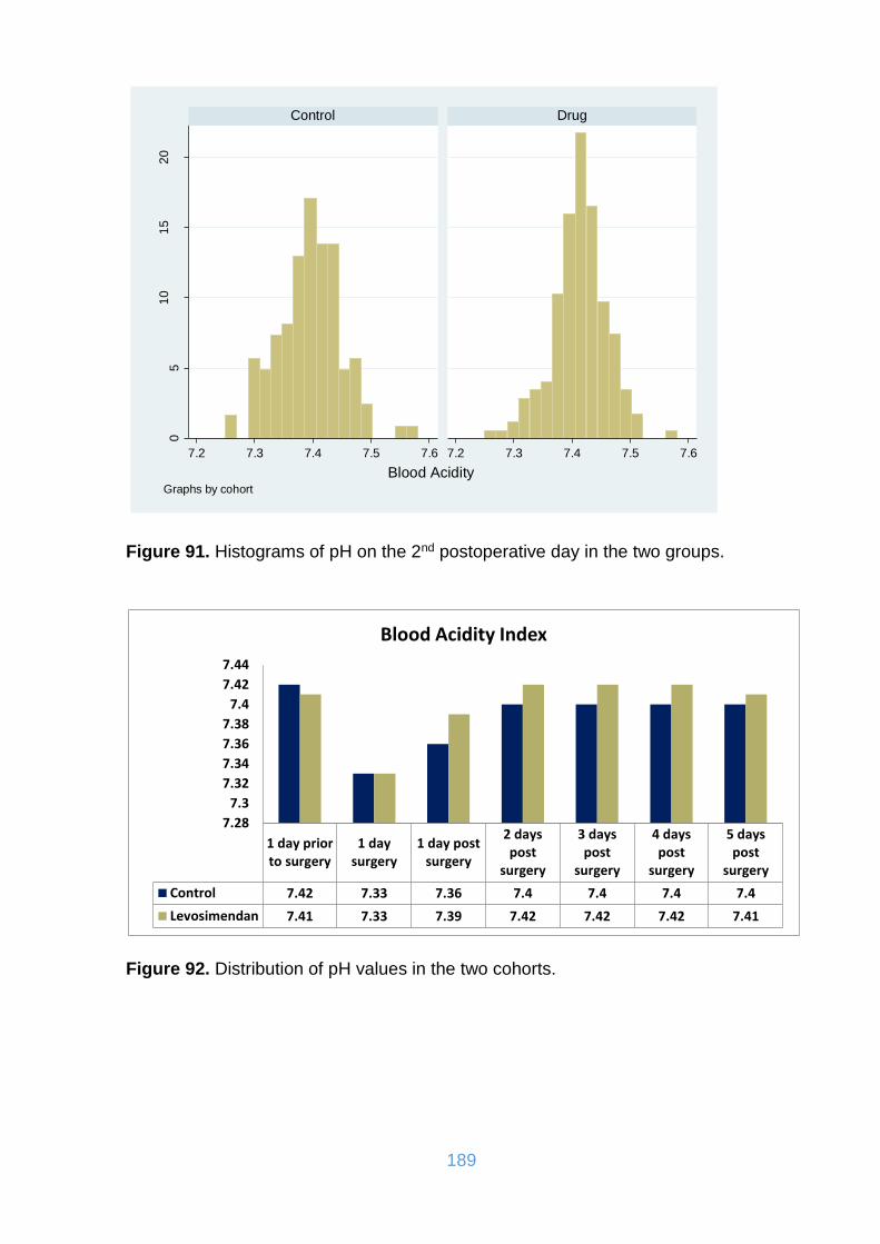
189
Figure 91. Histograms of pH on the 2nd postoperative day in the two groups.
Figure 92. Distribution of pH values in the two cohorts.
05
10
15
20
7.2 7.3 7.4 7.5 7.6 7.2 7.3 7.4 7.5 7.6
Control Drug
Pe
rcen
t
Blood AcidityGraphs by cohort
1 day priorto surgery
1 daysurgery
1 day postsurgery
2 dayspost
surgery
3 dayspost
surgery
4 dayspost
surgery
5 dayspost
surgery
Control 7.42 7.33 7.36 7.4 7.4 7.4 7.4
Levosimendan 7.41 7.33 7.39 7.42 7.42 7.42 7.41
7.28
7.3
7.32
7.34
7.36
7.38
7.4
7.42
7.44
Blood Acidity Index
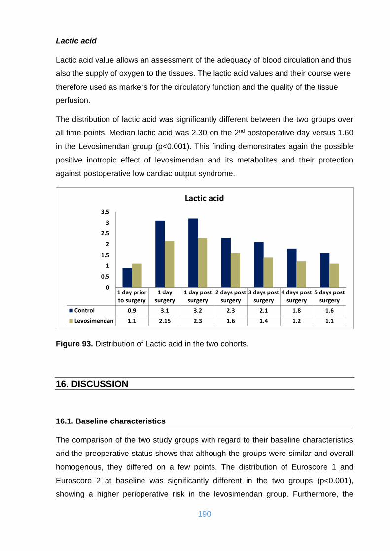
190
Lactic acid
Lactic acid value allows an assessment of the adequacy of blood circulation and thus
also the supply of oxygen to the tissues. The lactic acid values and their course were
therefore used as markers for the circulatory function and the quality of the tissue
perfusion.
The distribution of lactic acid was significantly different between the two groups over
all time points. Median lactic acid was 2.30 on the 2nd postoperative day versus 1.60
in the Levosimendan group (p<0.001). This finding demonstrates again the possible
positive inotropic effect of levosimendan and its metabolites and their protection
against postoperative low cardiac output syndrome.
Figure 93. Distribution of Lactic acid in the two cohorts.
16. DISCUSSION
16.1. Baseline characteristics
The comparison of the two study groups with regard to their baseline characteristics
and the preoperative status shows that although the groups were similar and overall
homogenous, they differed on a few points. The distribution of Euroscore 1 and
Euroscore 2 at baseline was significantly different in the two groups (p<0.001),
showing a higher perioperative risk in the levosimendan group. Furthermore, the
1 day priorto surgery
1 daysurgery
1 day postsurgery
2 days postsurgery
3 days postsurgery
4 days postsurgery
5 days postsurgery
Control 0.9 3.1 3.2 2.3 2.1 1.8 1.6
Levosimendan 1.1 2.15 2.3 1.6 1.4 1.2 1.1
0
0.5
1
1.5
2
2.5
3
3.5
Lactic acid

191
distribution of ASA score is significantly different in the two groups (p=0.048)
supporting the higher risk profile of the patients of the levosimendan group.
Occurrence of arrhythmia before surgery was significantly different in the two groups,
19.23% in the controls and 30.43% in the drug group (p=0.030). Similarly, the
appearance of pulmonary hypertension before surgery was significantly different in
the two groups, 8.46% in the controls and 30.43% in the drug group (p<0.001). The
same applies to preoperative renal function. In the controls, 13.85% of cases had
renal failure versus 25.54% in the drug group (p=0.012), 16.28% had arteriopathy
versus 31.52% in the levosimendan group (p=0.003), 4.62% had a cerebrovascular
accident versus 13.59% in the drug group (p=0.009). A significantly higher
distribution of ejection fraction was observed in the controls (Median of 30% for
control group vs. 20% for levosimendan group, p<0.001).
These are all essential factors that can significantly influence the outcome and thus,
despite the overall homogeneity of the two groups, such different values may lead to
a worse prognosis for patients of the study group.
16.2. Postoperative survival
The data collected on postoperative survival show significant differences between the
two groups. The Kaplan–Meier estimator shows the superior overall survival of the
levosimendan group compared to the control group (p=0.005). Survival estimates at
different time points for the two patient subgroups additionally show the consistently
higher overall survival probability of the levosimendan subgroup throughout follow-
up. The 10-days survival was similar in the two groups (82% for the control group vs.
85% for the levosimendan group). On the 30th day after surgery 64% of the control
patients and 76% of the levosimendan patients were alive (p=0.05). At 6 months after
surgery survival was estimated at 55% for the control group and 70% for the
levosimendan group, showing a significant difference at this time point (p=0.02). 1-
year follow-up showed a survival rate of 54% for the control group patients and 68%
for the levosimendan group patients (p=0.02). At 2 years the follow-up showed that
52% of the control and 65% of the levosimendan patients had survived (p=0.005). 3
years after surgery, 51% of cases were alive in the control group compared to 65% in
the drug group (p=0.005). It is worthy of consideration that the median survival

192
timepoint was not reached for the levosimendan group during follow-up, which adds
to the previous findings suggesting a positive effect of the drug on survival outcomes.
Several small trials suggest that levosimendan is associated with a favorable
outcome in patients undergoing cardiac surgery. However, as mentioned in chapter
12.8.2. recently published larger-scale trials did not provide evidence for a similar
benefit from levosimendan.
One of the first meta-analyses on levosimendan use in cardiac surgery patients
published by Zangrillo et al. in 2008 included a total of 139 patients from 5
randomized controlled studies and despite showing a significant reduction in
postoperative cardiac troponin peak release, it did not show any improvement of
survival in the patients treated with levosimendan (406). A meta-analysis of 10
randomized controlled trials by Landoni et al. in 2010 showed that the preoperative
administration of levosimendan has a highly significant effect. Immediate
postoperative mortality was significantly reduced in the levosimendan group
(p=0.003) (407). In a larger scale analysis from 2012, the Landoni group again
achieved similar results in 5480 patients from 45 studies. Here, the overall mortality
rate in the levosimendan group was 17.4%, in the control group 23.3% (408). Also
Maharaj et al. came to a similar conclusion in a meta-analysis of 17 studies involving
a total of 729 patients. The mortality rate in the study group was 4.9%, while in the
control group it was higher at 11.4% (409). A study by Levin et al. in 2012, in which
252 patients with preoperatively poor EF received CABG operations and were
randomized to either levosimendan or placebo administration 24 hours before
surgery, showed a marked improvement in 30-day survival in the preconditioned
group versus placebo group. The mortality rate here in the levosimendan group was
3.8%, whereas in the control group 12.4% (410). Hernandez et al. included 654
patients from 13 studies in their analysis. Levosimendan was associated with a
significant reduction in postoperative mortality, as 5.2% (18/344) versus 12.6%
(39/310) in the control arm (p=0.001) (411). Harrison et al. meta-analysed 1155
patients who participated in 14 randomized controlled trials of
perioperative Levosimendan administration. Pooled results demonstrated a reduction
in mortality with levosimendan (p=0.008). Subgroup analysis showed that this benefit
was confined to the low-EF studies (p<0.001). No benefit was observed in the
preserved-EF subgroup (p=0.66) (412). Another meta-analysis in 2015 by Lim et al.
included fourteen studies and demonstrated that levosimendan reduced early
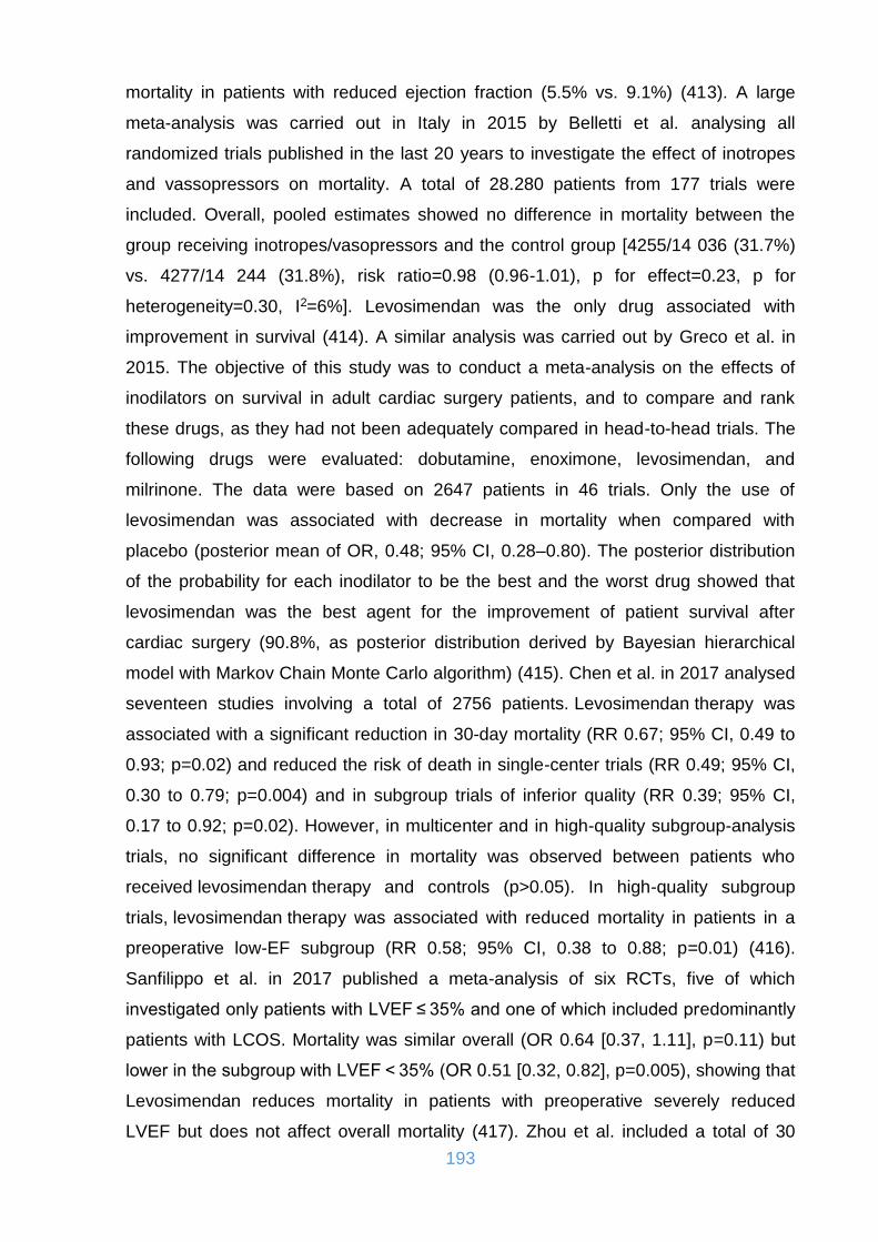
193
mortality in patients with reduced ejection fraction (5.5% vs. 9.1%) (413). A large
meta-analysis was carried out in Italy in 2015 by Belletti et al. analysing all
randomized trials published in the last 20 years to investigate the effect of inotropes
and vassopressors on mortality. A total of 28.280 patients from 177 trials were
included. Overall, pooled estimates showed no difference in mortality between the
group receiving inotropes/vasopressors and the control group [4255/14 036 (31.7%)
vs. 4277/14 244 (31.8%), risk ratio=0.98 (0.96-1.01), p for effect=0.23, p for
heterogeneity=0.30, I2=6%]. Levosimendan was the only drug associated with
improvement in survival (414). A similar analysis was carried out by Greco et al. in
2015. The objective of this study was to conduct a meta-analysis on the effects of
inodilators on survival in adult cardiac surgery patients, and to compare and rank
these drugs, as they had not been adequately compared in head-to-head trials. The
following drugs were evaluated: dobutamine, enoximone, levosimendan, and
milrinone. The data were based on 2647 patients in 46 trials. Only the use of
levosimendan was associated with decrease in mortality when compared with
placebo (posterior mean of OR, 0.48; 95% CI, 0.28–0.80). The posterior distribution
of the probability for each inodilator to be the best and the worst drug showed that
levosimendan was the best agent for the improvement of patient survival after
cardiac surgery (90.8%, as posterior distribution derived by Bayesian hierarchical
model with Markov Chain Monte Carlo algorithm) (415). Chen et al. in 2017 analysed
seventeen studies involving a total of 2756 patients. Levosimendan therapy was
associated with a significant reduction in 30-day mortality (RR 0.67; 95% CI, 0.49 to
0.93; p=0.02) and reduced the risk of death in single-center trials (RR 0.49; 95% CI,
0.30 to 0.79; p=0.004) and in subgroup trials of inferior quality (RR 0.39; 95% CI,
0.17 to 0.92; p=0.02). However, in multicenter and in high-quality subgroup-analysis
trials, no significant difference in mortality was observed between patients who
received levosimendan therapy and controls (p>0.05). In high-quality subgroup
trials, levosimendan therapy was associated with reduced mortality in patients in a
preoperative low-EF subgroup (RR 0.58; 95% CI, 0.38 to 0.88; p=0.01) (416).
Sanfilippo et al. in 2017 published a meta-analysis of six RCTs, five of which
investigated only patients with LVEF ≤ 35% and one of which included predominantly
patients with LCOS. Mortality was similar overall (OR 0.64 [0.37, 1.11], p=0.11) but
lower in the subgroup with LVEF < 35% (OR 0.51 [0.32, 0.82], p=0.005), showing that
Levosimendan reduces mortality in patients with preoperative severely reduced
LVEF but does not affect overall mortality (417). Zhou et al. included a total of 30
194
randomized controlled trials in their analysis in 2018. The pooled results indicated
that perioperative administration of levosimendan was associated with a reduction in
postoperative mortality [5.8% vs 8.5%; odds ratio 0.66, 95% confidence interval 0.50-
0.86, p=0.002; I2=17.1%; 25 trials; 3239 patients]. A subanalysis was conducted for
trials published after 2015, and it suggested that levosimendan could not reduce the
postoperative mortality (OR=0.91, 95% CI 0.63-1.31, p=0.626; I2=0.9%) (418). A
recent work by Chen et al. in 2018 showed that Levosimendan did not result in a
reduction in mortality in adult cardiac surgery patients. A total of 25 RCTs enrolling
2960 patients met the inclusion criteria; data from 15 placebo-controlled randomized
trials were included for meta-analysis. Pooled analysis showed that the all-cause
mortality rate was 6.4% (71 of 1106) in the levosimendan group and 8.4% (93 of
1108) in the placebo group (OR, 0.76; 95% CI, 0.55-1.04; p=0.09) (419). In one more
recent meta-analysis Wang et al. conclude that Levosimendan reduces the rate of
death and other adverse outcomes in patients with low EF undergoing cardiac
surgery, but results remain inconclusive. Data from 2152 patients in 15 randomized
clinical trials demonstrated a reduction in postoperative mortality in the levosimendan
group [RR = 0.53, 95% CI (0.38-0.73), I2=0]. However, the result showed that the
conclusion may be a false positive (420). Twenty-five studies (3247 patients) were
included in another meta-analysis by Qiang et al. in 2018. Pooled data indicated that
levosimendan reduced mortality after cardiac surgery (OR: 0.63, 95% CI: 0.47-0.84,
p=0.001). However, this reduction was restricted to patients with low (<50%) LVEF
(OR 0.49, 95% CI: 0.35-0.70, p=0.0001) (421). Finally in one of the latest meta-
analyses published in 2019 Ng et al. demonstrated that in comparison to the placebo
cohort, the levosimendan cohort showed a significant reduction in mortality
(TSA = inconclusive; p=0.002; I2=0%; FEM: OR 0.56; 95% CI 0.39, 0.80), especially
in the subgroups of preoperative severe low LVEF ≤ 30% (p=0.003; OR 0.33; 95% CI
0.16, 0.69), preoperative administering of levosimendan (p=0.001; OR 0.46; 95% CI
0.29, 0.74) and patients who had bolus followed by infusion of levosimendan
(p=0.005; OR 0.50; 95% CI 0.30, 0.81). However, the effect on mortality was not
significant in the subgroup analysis of high quality trials (p=0.14; OR 0.73; 95% CI
0.47, 1.12). Given the low level of evidence, the results of this meta-analysis neither
support nor oppose the use of levosimendan in cardiac patients with preoperative low
LVEF (422).

195
In conclusion it must be mentioned that the evidence from studies published in the
last 3 years questioned whether perioperative administration of levosimendan was
associated with better clinical outcomes in adult patients undergoing cardiac surgery.
For the future elucidation of this hypothesis, however, further prospective, if possible
randomized studies with a specific question about long-term implications are
necessary.
16.3. Effect on operative times and weaning from CPB
Total operation time, cardiopulmonary bypass time and cross-clamp time were
recorded in all patients. Distribution of cross-clamp time was significantly different in
the two groups where median was 78 minutes in the control group and 71 minutes in
the drug group, suggesting a trend towards longer cross-clamp time in the controls
(p=0.041). Duration of cardiopulmonary bypass (p=0.313) and the total duration of
surgery (p=0.220) were not significantly different in the two groups. The average total
operating times of the two patient groups hardly differ. Bias factors such as changes
in the team composition in the both areas of anaesthesia and surgical staff, the
variability of individual patients or any other individual factors were not able to be
taken into consideration, but these may also have contributed to the staggering
times. Overall, the proximity of the averages shows that the surgical treatment of the
two groups hardly differs.
These data are contradictory to the most literature reports in which levosimendan
seems to facilitate weaning from CPB (423, 424). Administration of levosimendan
significantly enhanced primary weaning from CPB compared with placebo in patients
undergoing 3-vessel on-pump CABG. The need for additional inotropic or mechanical
therapy during weaning was also decreased (424). A decisive criterion here is the
aggravating complexity of the interventions, which the patients had to undergo, in
conjunction with the preoperatively significantly impaired cardiac performance.
Taking this under consideration one can understand how difficult it is to standardize
such parameters. For this reason further studies designed and powered to detect
relevant clinical outcomes are needed before justifiable practice advisories can be
made.
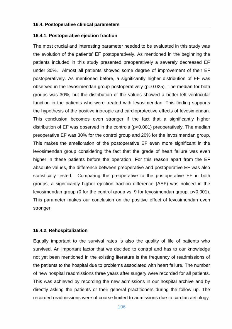
196
16.4. Postoperative clinical parameters
16.4.1. Postoperative ejection fraction
The most crucial and interesting parameter needed to be evaluated in this study was
the evolution of the patients’ EF postoperatively. As mentioned in the beginning the
patients included in this study presented preoperatively a severely decreased EF
under 30%. Almost all patients showed some degree of improvement of their EF
postoperatively. As mentioned before, a significantly higher distribution of EF was
observed in the levosimendan group postoperatively (p=0.025). The median for both
groups was 30%, but the distribution of the values showed a better left ventricular
function in the patients who were treated with levosimendan. This finding supports
the hypothesis of the positive inotropic and cardioprotective effects of levosimendan.
This conclusion becomes even stronger if the fact that a significantly higher
distribution of EF was observed in the controls (p<0.001) preoperatively. The median
preoperative EF was 30% for the control group and 20% for the levosimendan group.
This makes the amelioration of the postoperative EF even more significant in the
levosimendan group considering the fact that the grade of heart failure was even
higher in these patients before the operation. For this reason apart from the EF
absolute values, the difference between preoperative and postoperative EF was also
statistically tested. Comparing the preoperative to the postoperative EF in both
groups, a significantly higher ejection fraction difference (ΔEF) was noticed in the
levosimendan group (0 for the control group vs. 9 for levosimendan group, p<0.001).
This parameter makes our conclusion on the positive effect of levosimendan even
stronger.
16.4.2. Rehospitalization
Equally important to the survival rates is also the quality of life of patients who
survived. An important factor that we decided to control and has to our knowledge
not yet been mentioned in the existing literature is the frequency of readmissions of
the patients to the hospital due to problems associated with heart failure. The number
of new hospital readmissions three years after surgery were recorded for all patients.
This was achieved by recording the new admissions in our hospital archive and by
directly asking the patients or their general practitioners during the follow up. The
recorded readmissions were of course limited to admissions due to cardiac aetiology.
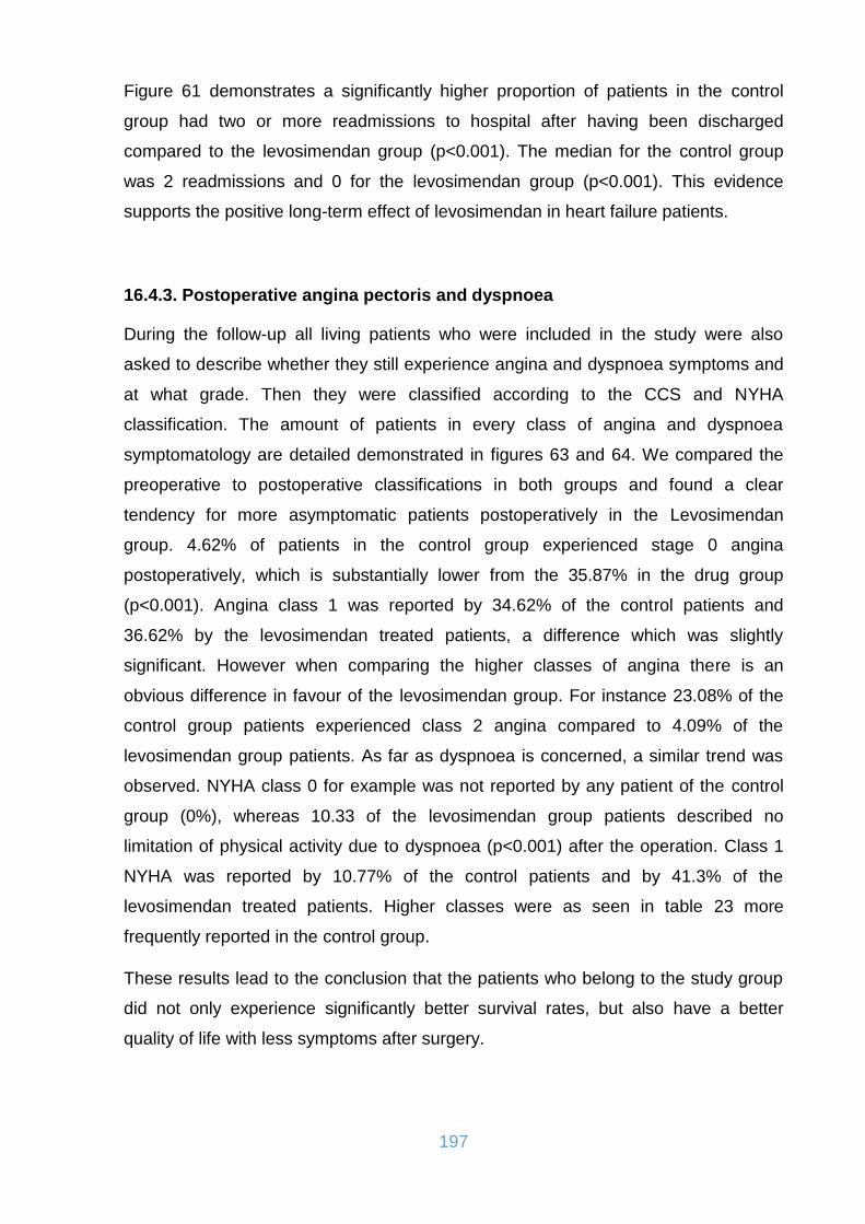
197
Figure 61 demonstrates a significantly higher proportion of patients in the control
group had two or more readmissions to hospital after having been discharged
compared to the levosimendan group (p<0.001). The median for the control group
was 2 readmissions and 0 for the levosimendan group (p<0.001). This evidence
supports the positive long-term effect of levosimendan in heart failure patients.
16.4.3. Postoperative angina pectoris and dyspnoea
During the follow-up all living patients who were included in the study were also
asked to describe whether they still experience angina and dyspnoea symptoms and
at what grade. Then they were classified according to the CCS and NYHA
classification. The amount of patients in every class of angina and dyspnoea
symptomatology are detailed demonstrated in figures 63 and 64. We compared the
preoperative to postoperative classifications in both groups and found a clear
tendency for more asymptomatic patients postoperatively in the Levosimendan
group. 4.62% of patients in the control group experienced stage 0 angina
postoperatively, which is substantially lower from the 35.87% in the drug group
(p<0.001). Angina class 1 was reported by 34.62% of the control patients and
36.62% by the levosimendan treated patients, a difference which was slightly
significant. However when comparing the higher classes of angina there is an
obvious difference in favour of the levosimendan group. For instance 23.08% of the
control group patients experienced class 2 angina compared to 4.09% of the
levosimendan group patients. As far as dyspnoea is concerned, a similar trend was
observed. NYHA class 0 for example was not reported by any patient of the control
group (0%), whereas 10.33 of the levosimendan group patients described no
limitation of physical activity due to dyspnoea (p<0.001) after the operation. Class 1
NYHA was reported by 10.77% of the control patients and by 41.3% of the
levosimendan treated patients. Higher classes were as seen in table 23 more
frequently reported in the control group.
These results lead to the conclusion that the patients who belong to the study group
did not only experience significantly better survival rates, but also have a better
quality of life with less symptoms after surgery.

198
16.4.4. Duration of mechanical ventilation, ICU and total hospital stay
In the present work, the duration of artificial ventilation presented a significant
difference between the two groups (p=0.007), whereas the ICU and total hospital
stay did not differ significantly.
The patients treated with levosimendan presented an easier and less complicated
weaning from the ventilator and were sooner extubated compared to the control
group patients (p=0.07). This suggests a possible lung protective effect of the drug.
There are experimental data in the literature which support the protective effect of
levosimendan against lung injury (425, 426).
On average, levosimendan treated patients spent 16 days in the hospital and 6 days
in the intensive care unit. Patients in the control group, on the other hand, remained
hospitalized for 15 days and spent 7 days in the ICU. Both Landoni et al. as well as
Lim et al. showed in their meta-analyses that there was a significant reduction in
hospital stay in patients treated with levosimendan (408, 413). Treskatsch has shown
in his study that early administration of levosimendan can also produce better results
in terms of ventilation time (427). Kodalli et al. however, in their double-blind,
randomized study with a total of 30 patients, showed no significant differences in
ventilation duration and duration of intensive care stay (428). Finally Qiang et al.
showed in their meta-analysis that levosimendan significantly shortened the duration
of the intensive care unit stay (weighted mean differences -0.49 day, 95% CI: -0.75 to
-0.24, p=0.0002) and mechanical ventilation use (weighted mean differences -2.30
hours, 95% CI: -3.76 to -0.84, p=0.002) (421). The data received from the literature
are to be discussed with regard to the current study. Administration of levosimendan
seemed in our study to have a positive effect as far the artificial ventilation and the
weaning from ventilator are concerned, but no significant influence on the reduction
of a patients stay in the ICU and overall in the hospital. On the one hand, we were
not able to identify any significant differences in terms of the parameters mentioned.
On the other hand, the treated group would expect much longer periods of ICU and
hospital stay regarding the higher risk profile of these patients as discussed above.
This suggested that prophylactic administration of levosimendan may have a positive
effect on postoperative ventilation and duration of ICU and hospital stay. However, a
more reliable statement can only be made by further studies with a special focus on
these parameters.

199
16.4.5. Support with inotropic agents and IABP
The need for inotropic support and administration of catecholamines was slightly, yet
not significantly less in the levosimendan group patients (p=0.076). If the higher
cardiovascular risk of the study group patients is taken into consideration, this fact
may support the hypothesis of the cardioprotective effect of levosimendan and shows
a faster recovery of myocardium after treatment.
These conclusions are generally supported by most literature reports in which a
reduced catecholamine requirement after preconditioning with levosimendan was
generally found. Several studies have shown that the early use of levosimendan
leads to a significantly reduced need for inotropic support. (429, 430) However, due
to the retrospective nature of this study, catecholamine control is not standardized. It
is difficult to find out how closely the i.v. fluid volume and catecholamine dose was
controlled in our patients. A more restrictive fluid infusion volume would be
conceivable in patients with more complex interventions and an initially worse EF,
which subsequently results in an increased vasopressor requirement. All these points
could be clarified within the framework of a prospective investigation with a broad
standardization of therapy control in order to investigate the actual effect of
levosimendan on the need for further circulatory support.
As far as the use of the IABP is concerned, no significant difference was observed
between the two groups (p=0.810). The duration of the mechanical circulatory
support and the efficacy of the weaning of IABP was similar in both groups.
16.4.6. Postoperative arrhythmias
Postoperative arrhythmias, mostly concerning postoperative atrial fibrillation (POAF)
are one of the most frequent complications after cardiac surgery. In patients
undergoing cardiothoracic surgery an incidence of 16–46% has been reported,
whereas in noncardiothoracic surgery, reported incidence of POAF varies between
0.4% and 12%. POAF can be observed during all the postoperative course, with a
peak between the 2nd and 5th postoperative day. POAF is a major complication after
cardiac surgery which can lead to high rates of morbidity and mortality, an enhanced
length of hospital stay, and an increased cost of care. Several studies have
attempted to identify predictors of POAF, with advanced age persisting as the most
potent and consistent risk factor, followed by a history of AF, chronic obstructive
200
pulmonary disease, and several operative characteristics (431). POAF is postulated
to be a multifactorial phenomenon; however, some major pathogeneses have been
proposed, including inflammatory pathways, oxidative stress, and autonomic
dysfunction (432).
Postoperative arrhythmias were recorded in many patients of both groups. The
frequency of such incidents was however significantly higher in the control group
(53%) compared to the group treated with levosimendan (40.76%) (p=0.031). In
order to adequately evaluate this finding the preoperative prevalence of arrhythmias
should also be taken under consideration. Occurrence of arrhythmias before surgery
was significantly different in the two groups, 19.23% in the controls and 30.43% in
the drug group (p=0.030). This comparison demonstrates that although the
occurrence of arrhythmias was higher in the patients of levosimendan group, they
presented significantly less arrhythmias postoperatively compared to the control
group.
According to Abacilar et al., the prophylactic administration of levosimendan leads to
a significant reduction in the incidence of POAF, especially after isolated CABG
intervention (433). This statement is also supported by many other studies and meta-
analyses. Thus, De Hert et al. reported similar results in 2008 in a randomized study
of 3 study groups. The group receiving levosimendan prior to surgery presented
significantly lower rates of atrial fibrillation postoperatively than the groups receiving
levosimendan intraoperatively during surgery, or never received levosimendan, but
were treated with milrinone instead (434). All these results lead to the suggestion of a
prophylactic antiarrhythmic effect of levosimendan.
16.4.7. Postoperative renal function and need for haemodialysis
Cardiac surgery is associated with a high risk for peri- and postoperative
complications. In consequence, protecting organ function in the operative setting
plays an important role for improving patient outcome. Postoperative renal
dysfunction is associated with increased mortality of 60-90% and prolonged stays on
the ICU and in the hospital (404, 435). Its incidence has been reported as up to 55%;
acute kidney injury is also associated with development of chronic kidney disease
(436, 437). The aetiology of acute kidney injury after cardiac surgery is known to be
201
multifactorial and includes inflammatory response, oxidative stress, transient
haemodynamic derangement and ischaemia‐reperfusion injury (438, 439).
In our study significant differences have been recorded between the levosimendan
and the control group in terms of postoperative renal function. Both the GFR,
creatinine clearance and the rest values for acute renal failure show significant
differences. 18 patients (13.85%) of the control group and 47 patients (25.54%) of
the levosimendan group presented an impaired renal function before surgery
presenting a significant difference (p=0.012) in favour of the control group. In both
groups an initial slight GFR decrease was observed directly after surgery. On the
following postoperative days all values of renal function appeared to be regressive
until they almost reached the baseline. A significant difference between the two
cohorts with regards to the distribution of the postoperative creatinine and creatinine
clearance parameters was recorded across all time points (p<0.001). Median values
of GFR were higher in the levosimendan group over all time points, except for the
day prior to surgery (p<0.001). A significantly higher need for postoperative renal
replacement therapy (RRT) was also observed in the control group (p<0.001). 38
patients of the control group (29.23%) and 21 patients of the levosimendan group
(11.41%) needed RRT after surgery. All above findings suggest a protective impact
of Levosimendan as far as renal function is concerned. It is also here important to
stress that preoperative impaired renal function is a key factor in acute postoperative
renal failure. With this knowledge, the results obtained are even more significantly
positive for the study group.
The nephroprotective effect of levosimendan is thought to be due to its
immunomodulatory, antiinflammatory, and antioxidant properties. Although
levosimendan's effect on blood flow to the kidneys during cardiopulmonary bypass is
not known, levosimendan significantly improved the ischaemia‐reperfusion injury in
renal tubules in previous animal studies and therefore has the potential to improve
renal function (440). The influence of levosimendan in the preglomerular vascular
resistance is shown in figure 94.

202
Figure 94. Differential effects of renal vasodilators on preglomerular (afferent arteriole) and postglomerular (efferent arteriole) vascular resistance sections.
(Source: Yilmaz MB, Grossini E, Silva Cardoso JC, et al. Renal effects of levosimendan: a consensus report. Cardiovasc Drugs Ther. 2013 Dec;27(6):581-90. doi: 10.1007/s10557-013-6485-6.)
A study by Bragadottir et al. in 2013, showed an effective vasodilation, especially of
the preglomerular vessels, leading to an improved renal blood flow and glomerular
filtration rate, without increasing the renal oxygen demand. This resulted in a reduced
incidence of acute renal failure and the need to RRT (441). Also the results of Baysal
et al. in 2014 support this hypothesis. In a prospective, double-blind, randomized
study with a total of 128 participants, significantly better creatinine and GFR values
were recorded in the levosimendan-treated group. The use of dialysis therapy was
also significantly less frequent in the study group (442). Lim et al. in their meta-
analysis of 14 controlled, randomized trials showed that postoperative acute renal
failure was lower with levosimendan therapy (7.4% vs. 11.5%) (413). In the meta-
analysis by Landoni et al. in 2010 on the contrary, no improvements in acute renal
failure could be found (407). Ristikankare et al. in 2012 came to a similar conclusion
that levosimendan did not have a significant influence on the kidney function
measuring specific kidney markers such as Urine N-acetyl-β-glucosaminidase (U-
NAG), Serum cystatin C and plasma creatinine (443). In 2013 a panel of 25 scientists
and clinicians from 15 European countries convened and reached a consensus on
the current interpretation of the renal effects of levosimendan described both in non-
clinical research and in clinical study reports. Most reports on the effect of
levosimendan indicated an improvement of renal function in heart failure, sepsis and

203
cardiac surgery settings. However, study designs differed from randomized,
controlled studies to uncontrolled ones. Importantly, in the largest HF study (REVIVE
I and II) no significant changes in the renal function were detected (444). Another
meta-analysis by Zhou et al. in 2015 included 13 trials with a total of 1345 study
patients. They concluded that levosimendan reduced the incidence of postoperative
AKI (40/460 vs 78/499; OR, 0.51; 95% CI, 0.34-0.76; p=0.001; I2=0.0%) and renal
replacement therapy (22/492 vs 49/491; OR, 0.43; 95% CI, 0.25-0.76; p=0.002;
I2=0.0%) (445). In 2018 Kim et al. performed an arm-based hierarchical Bayesian
network meta-analysis including 95 randomised controlled trials with 28,833
participants. Among other agents, levosimendan significantly decreased the rate of
postoperative renal dysfunction compared with placebo (446). Finally, a recent meta-
analysis by Qiang et al. in 2018 including 25 studies (3247 patients) demonstrated
that the administration of levosimendan significantly reduced the incidence of
postoperative acute kidney injury (OR 0.55, 95% CI: 0.41-0.74, p<0.0001) and renal
replacement therapy use (OR 0.56, 95% CI: 0.39-0.80, p=0.002) (421).
However it is important to mention that all existing meta-analyses came across with
the problem of significant heterogeneity regarding the criteria for diagnosing
postoperative renal dysfunction (AKIN criteria, RIFLE criteria, KDIGO criteria, etc.).
Given the lack of a standard definition of renal dysfunction, the results of these
meta‐analyses should be applied only cautiously to clinical practice. A
standardisation of renal outcome definitions in further studies is required in order to
establish more reliable evidence (446).
16.4.8. Haemoglobin and need for transfusion of blood products
CABG is a surgical procedure that causes sufficient hemoglobin loss to require
blood-product transfusion in around 50% of patients (447). Once recovery from
surgery has taken place, there is rarely any requirement for further blood transfusion.
CPB is associated with coagulopathy and platelet count reduction. In particular,
prolonged CPB and circulatory arrest with hypothermia result in more pronounced
alterations of the coagulation systems. Bleeding induced by coagulopathy requires
transfusion of allogeneic blood or blood products. Fresh frozen plasma (FFP) and
platelets include coagulation factors and thus ameliorate coagulopathy. However,
allogeneic blood transfusions, may have many adverse effects, including

204
anaphylactic shock and transfusion-related acute lung injury (TRALI). In addition,
transfusion-transmitted viral infections or graft-versus-host disease may occur in case
of allogenic blood transfusion. Horvath et al. described that each RBC unit transfused
was associated with a 29% increase in crude risk of major infection (p<0.001).
Among RBC recipients, the most common infections were pneumonia (3.6%) and
bloodstream infections (2%) (448). Moreover several studies have shown that
perioperative transfusions are associated with increased morbidity and mortality and
reduced long-term survival, mainly through immune modulation and tumor promotion
(447, 449). However Dardashti et al. showed that when pre-operative Hb levels and
renal function are taken into account, moderate transfusions of RBC after CABG
surgery do not seem to be associated with reduced long-term survival (450).
Therefore, increasing attention is being paid toward the use of blood-sparing
techniques and transfusion behaviors. Guidelines on transfusion practices in
cardiothoracic surgery have been published, but there still is a considerable variation
in transfusion practices nationally and internationally (451, 452). Efforts to change
transfusion behaviors and decrease transfusion rates in cardiac surgery have
persistent effects for several years, but more still needs to be done because the
transfusion rate is still high and inappropriate allogeneic transfusions may still be
common in different clinical scenarios (453).
In our study a significantly higher amount of erythrocytes and fresh frozen plasma
units transfusion was required after surgery in the Control group compared to the
Levosimendan group (p<0.001 for both parameters). Both groups presented a low
need for platelet unit transfusions and the difference was not significant between the
two groups (p=0.179).
Haemoglobin values were also recorded in all patients for the first 5 postoperative
days. As far as the preoperative values are concerned a significant difference was
observed in favour of the control group which presented a median value of 14.3 g/dl
whereas the levosimendan patients 13.4 g/dl (p=0.003). Significant differences were
observed on the operation day, directly after the transfer of the patient in the ICU (9.3
g/dl in the control group vs. 9.8 g/dl in the levosimendan group, p<0.001) and on the
first postoperative day (9.5 g/dl in the control group vs. 9.8 g/dl in the levosimendan
group, p=0.047) with the levosimendan group presenting better haemoglobin levels.
On the second, third and fourth postoperative day the differences were not significant
(p=0.956, 0.315 and 0.082 respectively). On the fifth postoperative day the difference
205
was significant again, however this time the control group presented higher levels
(10.4 g/dl in the control group vs. 9.9 g/dl in the levosimendan group, p=0.028). This
difference may be explained by the fact that the control patients had a higher
preoperative median value, and after recovery on the fifth postoperative day, both
groups reached values that reflect the baseline levels. The higher RBC transfusion
rates should also be taken into consideration while interpreting these results. There is
no obvious explanation of the reduced need for transfusions in the levosimendan
group, but this may be correlated with the possible limitation of the adverse effects of
CPB in this group, reducing the coagulopathy and therefore the perioperative
bleeding and transfusion need. The fact that the Control group consists of a historical
group of patients should also be considered, as other, probably more liberal
transfusion concepts may have been used at the time those patients were operated.
16.5. Postoperative laboratory parameters
16.5.1. Postoperative inflammatory response
CPB initiates a systemic inflammatory response syndrome (SIRS) after open-heart
surgery that is mediated by compartment activation, cytokine production and
neutrophil sequestration in the lungs and may lead to several complications. The
exact mechanism of this complex reaction has not been fully determined (454). The
mechanism of SIRS during CPB includes the interaction of blood and artificial surface
and endotoxemia. The interaction of blood and artificial surface is initiated by protein
adsorption. As a result of series of chain reactions, the numerous powerful
inflammatory mediators, including hormones and autacoids, are formed and
released. Subsequently, the contact system, coagulation system, complement
system, fibrinolysis system, and leukocytes, platelets, and endothelial cells, are all
activated to participate in the interaction of blood and artificial materials. These
activations of different systems and blood cells can interact and magnify each other.
CPB-associated endotoxemia has been demonstrated to intensify and deteriorate
SIRS during CPB (455).
The CRP values were in our study regularly determined for the detection or
assessment of inflammation. The preoperative values did not differ significantly
between the two groups (p=0.158). A significant difference was observed directly

206
after surgery (p<0.001), which may imply a better tolerance of the CBP in the
levosimendan group. A strong increase in CRP levels was recorded in both groups
on the second postoperative day and the differences on the first two postoperative
day were not significant (p=0.555 and p=0.422 respectively). Thereafter, the CRP
values were regressive in both groups. A slight difference was recorded on the third
postoperative day in favour of the levosimendan group (p=0.047). Significant
differences of the CRP values were recorded on the fourth and fifth day (p<0.001 for
both days), implying a faster recovery from the inflammatory response in the patients
treated with levosimendan.
Our conclusion is supported be the few existing studies on this issue. Adamopoulos
et al. and Trikas et al. in 2006, demonstrated a significant anti-inflammatory and anti-
apoptotic effect of levosimendan. Adamopoulos et al. studied 96 patients, one-third of
whom received levosimendan, dobutamine, or placebo, for proinflammatory and
apoptotic factors. Interleukin-6, TNF-α and soluble FAS ligand values were
measured. Here, only in the levosimendan group did a significant reduction of these
three parameters occur (456). Trikas et al. included in their study 27 patients and
were able to find a significant and long-lasting effect of levosimendan in terms of a
reduction of interleukin-6 as well as soluble FAS ligand and TNF-α and the
corresponding receptors TNF-R1 and -R2 (457).
16.5.2. Postoperative myocardial injury
A certain amount of myocardial injury is present in every patient undergoing cardiac
surgery. Cardiac damage after cardiac surgery could lead to prolonged hospital stay
as well as an increased perioperative mortality rate (458). Postoperative cardiac
troponin (cTn) is increased in all patients undergoing cardiac surgery. Numerous
factors can explain such an increase in cTn, even in the absence of postoperative MI
(459). The type of surgery, the choice of cardioplegic solution and its mode of
delivery, and the use of intraoperative aprotinin all influence postoperative cTn
release in patients scheduled for cardiac surgery (458). Several studies have
reported the prognostic significance of postoperative troponin measurements in
relation to both short- and long-term outcomes (460, 461). Data from patients
undergoing OPCAB surgery and aortic clamping suggest that coronary artery
anastomosis per se does not lead to an important release of cTn in contrast with
207
more complex procedures (e.g., mitral valve replacement with human mitral valve
homograft, Ross procedures, or some types of pediatric cardiac surgery) (462, 463).
Another issue concerning the interpretation of postoperative cTn concentrations in
terms of ischemic myocardial damage could be the frequent use of internal
defibrillation before the end of CPB. However, it has been shown previously that
internal defibrillation produces only early and minor elevations of cTn serum levels
(464). A meta-analysis by Zangrillo et al. showed that the cTn release peak was
lower in levosimendan-treated patients, a finding consistent with a beneficial
cardioprotective effect. Moreover, this reduction of myocardial damage
(postoperative cTn concentrations) appears to translate into outcome benefit
(reduced hospital length of stay) (458).
In order to assess the potential myocardial injury and to monitor the recovery after it
creatine kinase (CK) and its myocardial component (CKMB), as well as troponin I
(cTnI) were measured on a daily basis. The trend of all parameters was similar in
both study groups with a peak on the 1st postoperative day. cTnI levels differed
significantly between the two groups across all time points (p<0.001). Indicatively,
median of troponin I was 18.47 ng/ml and 9.90 ng/ml on the 1st postoperative day in
the control and levosimendan groups respectively (p<0.001). A significant difference
between the two cohorts with regards to the distribution of CKMB was observed
across all time points (p<0.001). A similar finding was seen for CK (p<0.001). In
summary, there is a perioperatively higher cardiac enzyme release in the control
group, especially in the first 48 h, which could be taken as a sign of significantly
higher heart damage in the control group. It can be hypothesized that the better
preservation of early cardiac function with levosimendan may result in an improved
global tissue perfusion with a better recovery from surgery.
16.5.3. Organ perfusion and metabolic balance
An arterial blood gas analysis was regularly performed during every day in the ICU,
providing an adequate monitoring of the respiratory and metabolic condition of every
patient. In order to assess the metabolic changes and the organ perfusion after
surgery the lowest pH and the highest lactic acid values of every day were recorded
and analyzed in all patients of the study. The distribution of pH appeared to be
different between the two groups over all time points measured, showing a higher

208
tendency to acidosis in the control group. This finding suggests a possible positive
metabolic effect of levosimendan reducing the degree of postoperative metabolic
acidosis.
The distribution of lactic acid values was also significantly different between the two
groups over all time points during the first five postoperative days. Median lactic acid
was 2.30 mmol/l in the control group on the 2nd postoperative versus 1.60 mmol/l in
the levosimendan group (p<0.001). In a study by Fang et al. in accordance to our
study, 36 patients who needed intensive care due to severe sepsis showed, in
addition to the improvement in the hemodynamic situation, lower measured lactate
values in the group treated with levosimendan (465). A similar recent work of Xu et
al. demonstrated that compared with dobutamine group, blood lactic acid at 24 h
decreased significantly in levosimendan group [(1.97±1.10)mmol/L vs. (2.73±2.06)
mmol/L, p=0.002]. Levosimendan seemed to improve cardiac systolic function and
tissue perfusion in elderly patients with septic myocardial contractility impairment
(466). To sum up our results implied once more the possible positive inotropic effect
of levosimendan and its metabolites and their protection against postoperative low
cardiac output syndrome.
17. LIMITATIONS OF THIS STUDY
As already discussed, the present work presents certain limitations, mostly because
of its retrospective character and the possible inequality of the studied groups. As
mentioned above there were significant differences in some of the parameters
measured or recorded preoperatively, showing some heterogeneity between the two
groups. These were attempted to minimize by making a consistent exclusion of
patients who had insufficient data documentation. However this heterogenity did not
seem to severely influence the conclusions that can be made when analysing the
results. Despite the limitations of the outcome of this work, it also brings certain
benefits. The data collected adequately reflect the use of levosimendan in everyday
clinical practice. The number of patients involved and the amount of parameters
recorded in every patient offer one more representative study to the existing literature
of this issue. Furthermore, long-term data on levosimendan use offered by this study,
sometimes even up to 5 years postoperatively so far haven’t been available in the
literature.

209
18. SUMMARY
18.1. Summary in English
The prevalence of postoperative myocardial dysfunction following cardiac surgery is
a relatively common phenomenon even in patients with a normal preoperative
ventricular function and its grade depends on factors such as patient age, the kind of
procedure performed and the existing co-morbidities. The phenomenon becomes
much more intense and more difficult to treat in patients with pre-existing heart
failure. This condition results in delayed recovery, organ failure, prolonged stay in the
ICU and in the hospital, increased morbidity and mortality and significantly increased
costs. When myocardial dysfunction is clinically significant it may jeopardize tissue
perfusion. In this case the use of inotropic agents is necessary in order to support
circulation until endogenous myocardial function is restored. The classic treatment
strategies have traditionally focused on the use of catecholamines and, in particular,
β-adrenergic agents, which are also characterized as first choice drugs due to their
beneficial effects on myocardial contractility and cardiac output. However, the use of
these sympathomimetic drugs can be accompanied by many side effects such as
tachycardia, arrhythmias, adverse effects on ventricular load conditions and
ultimately myocardial oxygen supply-consumption disorders. In addition, down-
regulation of β-adrenergic receptors in patients with chronic heart failure may
alleviate the response to exogenously administered catecholamines.
Levosimendan is a new inotropic drug, belonging to the class of calcium sensitizers.
It increases myofilament calcium sensitivity during cardiac systole by binding to
troponin C in a calcium-dependent manner. This interaction stabilizes the calcium-
induced conformational change of tropomyocin, thereby augmenting actin-myocin
cross-bridging without raising intracellular calcium concentrations. It also binds to and
opens ATP-dependent potassium (KATP) channels, having a direct effect on vascular
smooth muscle cells, causing vasodilation. The beneficial effects of levosimendan
despite the existing controversy have already been described in many studies of the
literature concerning internal medicine and surgical patients. The aim of this study is
to evaluate whether benefits of levosimendan could be extended to improve the
outcome of patients with low preoperative EF and myocardial dysfunction following
cardiac surgery.

210
The results of this work show substantial positive effects of levosimendan on the
short- and long-term outcome in patients with severely reduced ejection fraction
undergoing cardiac surgery. The prophylactic administration of levosimendan
significantly improved the short- and long-term outcomes of the treated patients. The
results of this study can be summarized in the following conclusions:
Levosimendan improves short- and long term survival after cardiac surgery.
It significantly facilitates postoperative improvement of the EF.
It provides a better hemodynamic stability and reduces the need for inotropic
support of the patients.
It does not affect the need for mechanical hemodynamic support with the
IABP.
It reduces mechanical ventilation time and facilitates a faster weaning from
ventilator.
It does not reduce the stay in the ICU and the total hospital stay.
It reduces rehospitalization and readmissions to the hospital due to cardiac
aetiology.
These patients reported a better clinical symptomatology with less angina and
dyspnea.
It reduces the need for transfusion of blood products.
It reduces the occurrence of postoperative arrhythmias.
It provides a renal protective effect and reduces the need for renal
replacement therapy.
It reduces the systemic inflammatory response and provides a faster recovery
from it.
It limits the amount and extense of the inevitable myocardial injury after
cardiac surgery.
It ameliorates organ and tissue perfusion and metabolic balance
postoperatively.
It is also important to mention that the purpose of this study was to demonstrate a
possible positive effect of levosimendan as an additive treatment and not as a
monotherapy, as the conventional medical, surgical and mechanical therapeutical
means remain the cornerstone of the treatment of a cardiac surgery patient.

211
The mentioned limitations of a retrospective analysis with unequal basic
characteristics were attempted to compensate with the help of a multivariate
regression analysis. In order to allow a more precise quantification of the positive or
potential negative effects of levosimendan, more prospective randomized studies
would be necessary, in which the documentation and control of the other
perioperative therapy are standardized and comprehensibly documented.
18.2. Summary in German / Zusammenfassung
Das Auftreten einer postoperativen myokardialen Dysfunktion nach einer
Herzoperation ist ein relativ häufiges Phänomen, auch bei Patienten mit einer
präoperativ normalen ventrikulären Funktion. Der Grad hängt von Faktoren wie dem
Alter des Patienten, der Art des durchgeführten Eingriffs und den bestehenden
Komorbiditäten ab. Das Phänomen ist bei Patienten mit vorbestehender
Herzinsuffizienz viel intensiver und schwieriger zu behandeln. Dieser Zustand führt
zu verzögerter Erholung, Organversagen, längerem Aufenthalt auf der Intensivstation
und im Krankenhaus, erhöhter Morbidität und Mortalität und signifikant erhöhten
Kosten. Wenn eine Myokardfunktionsstörung klinisch signifikant ist, kann sie die
Gewebeperfusion gefährden. In diesem Fall ist die Verwendung von Inotropika
erforderlich, um die Kreislaufverhältnisse zu verbessern, bis die endogene
Myokardfunktion wiederhergestellt ist. Die klassischen Behandlungsstrategien
konzentrierten sich traditionell auf die Verwendung von Katecholaminen und
insbesondere von β-adrenergen Wirkstoffen, die aufgrund ihrer vorteilhaften
Auswirkungen auf die Kontraktilität des Herzmuskels und das Herzzeitvolumen auch
als Arzneimittel erster Wahl gelten. Die Anwendung dieser Sympathomimetika kann
jedoch mit vielen Nebenwirkungen einhergehen, wie Tachykardien, Arrhythmien,
nachteiligen Auswirkungen auf die ventrikulären Belastungszustände und letztendlich
Störungen der Sauerstoffversorgung und des Sauerstoffverbrauchs des
Herzmuskels. Darüber hinaus kann eine Herunterregulierung der β-adrenergen
Rezeptoren bei Patienten mit chronischer Herzinsuffizienz die Reaktion auf exogen
verabreichte Katecholamine reduzieren.
Levosimendan ist ein neues inotropes Medikament, das zur Klasse der
Kalziumsensibilisatoren gehört. Es erhöht die Kalziumempfindlichkeit des
Myofilaments während der Herzsystole, indem es in kalziumabhängiger Weise an
212
Troponin C bindet. Diese Wechselwirkung stabilisiert die Kalzium-induzierte
Konformationsänderung von Tropomyocin, wodurch die Actin-Myocin-Überbrückung
verstärkt wird, ohne die intrazellulären Kalziumkonzentrationen zu erhöhen. Es
bindet und öffnet auch ATP-abhängige Kaliumkanäle (KATP), die eine direkte Wirkung
auf die glatten Gefäßmuskelzellen haben und eine Vasodilatation verursachen. Die
positive Wirkung von Levosimendan wurde trotz der bestehenden Kontroverse
bereits in vielen Literaturstudien zur Inneren Medizin und zu chirurgischen Patienten
beschrieben.
Ziel dieser Studie war es zu evaluieren, ob die Anwendung von Levosimendan
erweitert werden kann, um das Ergebnis von Patienten mit niedriger präoperativer
EF und Myokardfunktionsstörung nach einer Herzoperation zu verbessern.
Die Ergebnisse dieser Arbeit weisen deutliche positive Effekte von Levosimendan auf
das kurz- und langfristige Ergebnis bei Patienten mit stark reduzierter
Ejektionsfraktion, die sich einer Herzoperation unterziehen, nach. Die
prophylaktische Verabreichung von Levosimendan verbesserte die kurz- und
langfristigen Ergebnisse der behandelten Patienten signifikant. Die Ergebnisse dieser
Studie können in den folgenden Schlussfolgerungen zusammengefasst werden:
Levosimendan verbessert das kurz- und langfristige Überleben nach einer
Herzoperation bei Patienten mit schwer eingeschränkter Pumpfunktion.
Es unterstützt die postoperative Verbesserung der EF erheblich.
Es bietet eine bessere perioperative hämodynamische Stabilität und verringert
den Bedarf an inotroper Unterstützung für die Patienten.
Der Bedarf an mechanischer hämodynamischer Unterstützung mit dem IABP
wird dadurch nicht beeinträchtigt.
Es reduziert die Beatmungszeit und ermöglicht ein schnelleres Entwöhnen
vom Beatmungsgerät.
Der Aufenthalt auf der Intensivstation und der gesamte
Krankenhausaufenthalt werden nicht reduziert.
Es reduziert die Rehospitalisierung und die Reaufnahmen in das Krankenhaus
aufgrund von Herzinsuffizienz.
Die Patienten erleben eine bessere klinische Symptomatik postoperativ mit
weniger Angina und Dyspnoe.
Es reduziert die Notwendigkeit der Transfusion von Blutprodukten.

213
Es reduziert das Auftreten von postoperativen Arrhythmien.
Es bietet eine renale Schutzwirkung und reduziert den Bedarf an
Nierenersatztherapie.
Es reduziert die systemische Entzündungsreaktion und sorgt für eine
schnellere Erhohlung davon.
Es begrenzt das Ausmaß und die Intensität der unvermeidbaren
Myokardverletzung nach einer Herzoperation.
Es verbessert die Perfusion von Organen und Gewebe und den Stoffwechsel
nach der Operation.
Es ist wichtig zu erwähnen, dass der Zweck dieser Studie darin besteht, eine
mögliche positive Wirkung von Levosimendan als additive Behandlung und nicht als
Monotherapie aufzuzeigen, da die konventionellen medizinischen, chirurgischen und
mechanisch-therapeutischen Mittel die Eckpfeiler der Behandlung dieser
Patientengruppe bleiben.
Die genannten Einschränkungen einer retrospektiven Analyse mit ungleichen
Basismerkmalen wurden versucht, mit Hilfe einer multivariaten Regressionsanalyse
zu kompensieren. Um die positiven oder potenziellen negativen Wirkungen von
Levosimendan genauer quantifizieren zu können, wären prospektivere randomisierte
Studien erforderlich, in denen die Dokumentation und Kontrolle der anderen
perioperativen Therapie standardisiert und nachvollziehbar dokumentiert werden.

214
19. REFERENCES
1. Beaglehole R. International trends in coronary heart disease mortality and
incidence rates. J Cardiovasc Risk 1999;6(2):63-8.
2. Swedberg K, Cleland J, Dargie H, Drexler H, Follath F, Komajda M, et al.
Guidelines for the diagnosis and treatment of chronic heart failure: executive
summary (update 2005): The Task Force for the Diagnosis and Treatment of Chronic
Heart Failure of the European Society of Cardiology. Eur Heart J 2005;26(11):1115-
40.
3. Follath F. Ischemic versus non-ischemic heart failure: should the etiology be
determined? Heart Fail Monit 2001;1(4):122-5.
4. Wang T, Butany J. Pathogenesis of atherosclerosis. Diagnostic Histopathology
2017;23(11):473-478.
5. Tabas I, García-Cardena G, Owens GK. Recent insights into the cellular biology of
atherosclerosis. J Cell Biol 2015;209:13-22.
6. Brown RA, Shantsila E, Varma C, Lip GYH. Current understanding of
atherogenesis. Am J Med 2017;130(3):268-82.
7. Channon KM, Qian H, George SE. Nitric oxide synthase in atherosclerosis and
vascular injury: insights from experimental gene therapy. Arterioscler Thromb Vasc
Biol 2000;20(8):1873-81.
8. Umans JG, Levi R. Nitric oxide in the regulation of blood flow and arterial pressure.
Annu Rev Physiol 1995;57:771-90.
9. Gimbrone MA, García-Cardena G. Endothelial cell dysfunction and the
pathobiology of atherosclerosis. Circ Res 2016;118:620-36.
10. Berliner JA NM, Fogelman AM et al. Atherosclerosis: basic mechanisms.
Oxidation, inflammation and genetics. Circulation 1995;21:2488-2496.
11. Davignon J, Ganz P. Role of endothelial dysfunction in atherosclerosis.
Circulation 2004;109(23 Suppl 1):III27-32.
12. Kinlay S, Libby P, Ganz P. Endothelial function and coronary artery disease. Curr
Opin Lipidol 2001;12(4):383-9.
215
13. Hasdai D, Gibbons RJ, Holmes DR, Jr., Higano ST, Lerman A. Coronary
endothelial dysfunction in humans is associated with myocardial perfusion defects.
Circulation 1997;96(10):3390-5.
14. Selwyn AP, Kinlay S, Libby P, Ganz P. Atherogenic lipids, vascular dysfunction,
and clinical signs of ischemic heart disease. Circulation 1997;95(1):5-7.
15. Gimbrone Jr MA, García-Cardena G. Vascular endothelium, hemodynamics, and
the pathobiology of atherosclerosis. Cardiovasc Pathol 2013;22:9-15.
16. Landmesser U, Hornig B, Drexler H. Endothelial function: a critical determinant in
atherosclerosis? Circulation 2004;109(21 Suppl 1):II27-33.
17. Hurtubise J, McLellan K, Durr K, Onasanya O, Nwabuko D, Ndisang JF. The
different facets of dyslipidemia and hypertension in atherosclerosis. Curr Atheroscler
Rep 2016;18:82.
18. McLaren JE, Michael DR, Ashlin TG, Ramji DP. Cytokines, macrophage lipid
metabolism and foam cells: implications for cardiovascular disease therapy. Prog
Lipid Res 2011;50:331-47.
19. Fatkhullina AR, Peshkova IO, Koltsova EK. The role of cytokines in the
development of atherosclerosis. Biochem Biokhimiia 2016;81:1358-70.
20. Bennett MR, Sinha S, Owens GK. Vascular smooth muscle cells in
atherosclerosis. Circ Res 2016;118:692-702.
21. Ketelhuth DFJ, B€ack M. The role of matrix metalloproteinases in
atherothrombosis. Curr Atheroscler Rep 2011;13:162-9.
22. Nicoll R, Henein M. Arterial calcification: a new perspective? Int J Cardiol
2016;228:11-22.
23. Larsen SB, Grove EL, Wurtz M, Neergaard-Petersen S, Hvas AM, Kristensen SD.
The influence of low-grade inflammation on platelets in patients with stable coronary
artery disease. Thromb Haemost. 2015;114:519-529.
24. Musunuru K, Kathiresan S. Surprises from genetic analyses of lipid risk factors
for atherosclerosis. Circ Res 2016;118:579-85.
25. McPherson R, Tybjaerg-Hansen A. Genetics of coronary artery disease. Circ Res
2016;118:564-78.
216
26. Paynter NP, Ridker PM, Chasman DI. Are genetic tests for atherosclerosis ready
for routine clinical use? Circ Res 2016;118:607-19.
27. Davies MJ. The pathophysiology of acute coronary syndromes. Heart
2000;83(3):361-6.
28. Giuseppe M.C. Rosano, Massimo Fini, Giuseppe Caminiti and Giuseppe
Barbaro. Cardiac Metabolism in Myocardial Ischemia. Current Pharmaceutical
Design. Pages 2551-2562 (12).
29. Ferrari R. Metabolic disturbances during myocardial ischemia and reperfusion.
Am J Cardiol. 1995 Aug 24;76(6):17B-24B
30. Neely JR, Liedtke AJ, Whitmer JT, Rovetto MJ: Relationship between coronary
flow and adenosine triphosphate production from glycolysis and oxidative
metabolism. Recent Adv Stud Cardiac Struct Metab 1975;8:301-21.
31. Van der Vusse GJ, Glatz JF, Stam HC, Reneman RS: Fatty acid homeostasis in
the nor‐ moxic and ischemic heart. Physiol Rev 1992;72:881-940.
32. McHowat J, Yamada KA, Wu J, Yan GX, Corr PB: Recent insights pertaining to
sarco‐ lemmal phospholipid alterations underlying arrhythmogenesis in the ischemic
heart. J Cardiovasc Electrophysiol 1993;4:288-310.
33. Yamada KA, McHowat J, Yan GX, Donahue K, Peirick J, Kleber AG, Corr PB:
Cellu‐ lar uncoupling induced by accumulation of long-chain acylcarnitine during
ischemia. Circ Res 1994;74:83-95.
34. Nawada R, Murakami T, Iwase T, Nagai K, Morita Y, Kouchi I, Akao M,
Sasayama S: Inhibition of sarcolemmal Na+,K+-ATPase activity reduces the infarct
size-limiting effect of preconditioning in rabbit hearts. Circulation 1997;96:599-604.
35. Tani M, Neely JR: Role of intracellular Na+ in Ca2+ overload and depressed
recovery of ventricular function of reperfused ischemic rat hearts. Possible
involvement of H+- Na+ and Na+-Ca2+ exchange. Circ Res 1989;65:1045-56.
36. Granger DN: Role of xanthine oxidase and granulocytes in ischemia-reperfusion
injury. Am J Physiol 1988;255:H1269-75.
37. Maxwell SR, Lip GY. Reperfusion injury: a review of the pathophysiology, clinical
manifestations and therapeutic options. Int J Cardiol 1997;58:95-117.
217
38. Di Lisa F, Canton M, Menabo R, Kaludercic N, Bernardi P. Mitochondria and
cardio‐ protection. Heart Fail Rev 2007;12:249-60.
39. Turrens JF. Mitochondrial formation of reactive oxygen species. J Physiol
2003;552:335-44.
40. Krijnen PA, Nijmeijer R, Meijer CJ, Visser CA, Hack CE, Niessen HW. Apoptosis
in myocardial ischaemia and infarction. J Clin Pathol 2002;55(11):801-11.
41. Kloner RA, Ellis SG, Lange R, Braunwald E. Studies of experimental coronary
artery reperfusion. Effects on infarct size, myocardial function, biochemistry,
ultrastructure and microvascular damage. Circulation 1983;68(2 Pt 2):I8-15.
42. Abbate A, Biondi-Zoccai GG, Bussani R, Dobrina A, Camilot D, Feroce F,
Rossiello R, Baldi F, Silvestri F, Biasucci LM, Baldi A. Increased myocardial
apoptosis in patients with unfavorable left ventricular remodelling and early
symptomatic post-infarction heart failure. J Am Coll Cardiol 2003;41(5):753–60.
43. Teringova E, Tousek P. Apoptosis in ischemic heart disease. J Transl Med
2017;15:87.
44. Kerr JF, Wyllie AH, Currie AR. Apoptosis: a basic biological phenomenon with
wide-ranging implications in tissue kinetics. Br J Cancer 1972;26(4):239–57.
45. Renehan AG, Booth C, Potten CS. What is apoptosis, and why is it important?
BMJ 2001;322(7301):1536–8.
46. Cotter TG. Apoptosis and cancer: the genesis of a research feld. Nat Rev Cancer
2009;9(7):501–7.
47. Saraste A, Pulkki K, Kallajoki M, et al. Apoptosis in human acute myocardial
infarction. Circulation 1997;95:320–3.
48. Baldi A, Abbate A, Bussani R, Patti G, Melf R, Angelini A, Dobrina A, Rossiello R,
Silvestri F, Baldi F, Di Sciascio G. Apoptosis and post-infarction left ventricular
remodeling. J Mol Cell Cardiol 2002;34(2):165–74.
49. Yellon DM, Hausenloy DJ. Myocardial Reperfusion Injury. N Engl J Med
2007;357:1121-35.
50. Piper HM, Garcia-Dorado D, Ovize M. A fresh look at reperfusion injury.
Cardiovasc Res 1998;38:291-300.

218
51. Jennings RB, Sommers HM, Smyth GA, Flack HA, Linn H. Myocardial necrosis
induced by temporary occlusion of a coronary artery in the dog. Arch Pathol
1960;70:68-78.
52. Keeley EC, Boura JA, Grines CL. Primary angioplasty versus intravenous
thrombolytic therapy for acute myocardial infarction: a quantitative review of 23
randomised trials. Lancet 2003;361:13-20.
53. Braunwald E, Kloner RA. The stunned myocardium: prolonged, postischemic
ventricular dysfunction. Circulation 1982;66:1146-9.
54. Bolli R, Marbán E. Molecular and cellular mechanisms of myocardial stunning.
Physiol Rev 1999;79:609-34.
55. Ito H. No-reflow phenomenon and prognosis in patients with acute myocardial
infarction. Nat Clin Pract Cardiovasc Med 2006;3:499-506.
56. Manning AS, Hearse DJ. Reperfusion induced arrhythmias: mechanisms and
prevention. J Mol Cell Cardiol 1984;16:497-518.
57. Faxon DP, Gibbons RJ, Chronos NA, Gurbel PA, Sheehan F. The effect of
blockade of the CD11/CD18 integrin receptor on infarct size in patients with acute
myocardial infarction treated with direct angioplasty: the results of the HALT-MI
study. J Am Coll Cardiol 2002;40:1199-204.
58. Kloner RA, Jennings RB. Consequences of brief ischemia: stunning,
preconditioning, and their clinical implications: part 1. Circulation 2001;104(24):2981-
9.
59. Kloner RA, Jennings RB. Consequences of brief ischemia: stunning,
preconditioning, and their clinical implications: part 2. Circulation 2001 Dec
18;104(25):3158-67.
60. Gross GJ, Kersten JR, Warltier DC. Mechanisms of postischemic contractile
dysfunction. Ann Thorac Surg 1999 Nov;68(5):1898-904.
61. Bolli R. Basic and clinical aspects of myocardial stunning. Prog Cardiovasc Dis
1998 May-Jun;40(6):477-516.
62. Wijns W, Vatner SF, Camici PG. Hibernating myocardium. N Engl J Med. 1998
Jul 16;339(3):173-81.

219
63. Rahimtoola SH. Coronary bypass surgery for chronic angina. A perspective.
Circulation 1982;65:225–41.
64. Heusch G. Hibernating myocardium. Physiol Rev 1998 Oct;78(4):1055-85.
Review.
65. Heusch G, Schulz R. The biology of myocardial hibernation. Trends Cardiovasc
Med. 2000 Apr;10(3):108-14.
66. Bax JJ, Visser FC, Poldermans D, Elhendy A, Cornel JH, Boersma E, van Lingen
A, Fioretti PM, Visser CA. Time course of functional recovery of stunned and
hibernating segments after surgical revascularization. Circulation. 2001 Sep
18;104(12 Suppl 1):I314-8.
67. Cohen M, Charney R, Hershman R, Fuster V, Gorlin R. Reversal of chronic
ischemic myocardial dysfunction after transluminal coronary angioplasty. J Am Coll
Cardiol. 1988 Nov;12(5):1193-8.
68. Page BJ, Banas MD, Suzuki G, Weil BR, Young RF, Fallavollita JA, Palka BA,
Canty JM Jr. Revascularization of chronic hibernating myocardium stimulates
myocyte proliferation and partially reverses chronic adaptations to ischemia. J Am
Coll Cardiol 2015 Feb 24;65(7):684-97.
69. Suzuki G, Iyer V, Lee TC, et al. Autologous mesenchymal stem cells mobilize
cKit+ and CD133+ bone marrow progenitor cells and improve regional function in
hibernating myocardium. Circ Res 2011;109:1044–54.
70. Suzuki G, Lee TC, Fallavollita JA, et al. Adenoviral gene transfer of FGF-5 to
hibernating myocardium improves function and stimulates myocytes to hypertrophy
and reenter the cell cycle. Circ Res 2005;96:767–75.
71. Poulin MF, Deka A, Mohamedali B, Schaer GL. Clinical Benefits of Stem Cells for
Chronic Symptomatic Systolic Heart Failure: A Systematic Review of the Existing
Data and Ongoing Trials. Cell Transplant. 2016 Nov;25(11):1911-1923.
72. Afridi I, Kleiman NS, Raizner AE, Zoghbi WA. Dobutamine echocardiography in
myocardial hibernation. Optimal dose and accuracy in predicting recovery of
ventricular function after coronary angioplasty. Circulation 1995 Feb 1;91(3):663-70.
73. Allman KC. Noninvasive assessment myocardial viability: current status and
future directions. J Nucl Cardiol. 2013 Aug;20(4):618-37;quiz 638-9.

220
74. Rischpler C, Nekolla SG, Dregely I, Schwaiger M. Hybrid PET/MR imaging of the
heart: Potential, initial experiences, and future prospects. J Nucl Med 2013;54:402-
15.
75. Butler MJ, Chan W, Taylor AJ, Dart AM, Duffy SJ. Management of the no-reflow
phenomenon. Pharmacol Ther. 2011 Oct;132(1):72-85.
76. Reffelmann T, Kloner RA. The "no-reflow" phenomenon: basic science and
clinical correlates. Heart. 2002 Feb;87(2):162-8.
77. van der Weg K, Prinzen FW, Gorgels AP. Editor's Choice- Reperfusion cardiac
arrhythmias and their relation to reperfusion-induced cell death. Eur Heart J Acute
Cardiovasc Care. 2019 Mar;8(2):142-152.
78. Jurkovicová O, Cagán S. Reperfusion arrhythmias. Bratisl Lek Listy. 1998 Mar-
Apr;99(3-4):162-71.
79. Zweier JL. Measurement of superoxide-derived free radicals in the reperfused
heart: evidence for a free radical mechanism of reperfusion injury. J Biol Chem
1988;263:1353-7.
80. Hearse DJ, Humphrey SM, Chain EB. Abrupt reoxygenation of the anoxic
potassium-arrested perfused rat heart: a study of myocardial enzyme release. J Mol
Cell Cardiol 1973;5:395-407.
81. Zweier JL, Talukder MA. The role of oxidants and free radicals in reperfusion
injury. Cardiovasc Res 2006;70:181-90.
82. Piper HM, Garcia-Dorado D, Ovize M. A fresh look at reperfusion injury.
Cardiovasc Res 1998;38:291-300.
83. Lemasters JJ, Bond JM, Chacon E, Harper IS, Kaplan SH, Ohata H, Trollinger
DR, Herman B, Cascio WE. The pH paradox in ischemia-reperfusion injury to cardiac
myocytes. EXS 1996;76:99-114.
84. Vinten-Johansen J. Involvement of neutrophils in the pathogenesis of lethal
myocardial reperfusion injury. Cardiovasc Res 2004;61:481-97.
85. Apstein CS, Opie LH. A challenge to the metabolic approach to myocardial
ischaemia. Eur Heart J 2005;26:956-9.
221
86. Mehta SR, Yusuf S, Diaz R, et al. Effect of glucose-insulin-potassium infusion on
mortality in patients with acute ST-segment elevation myocardial infarction: the
CREATE-ECLA randomized controlled trial. JAMA 2005;293:437-46.
87. Halestrap AP, Richardson AP. The mitochondrial permeability transition: a current
perspective on its identity and role in ischaemia/reperfusion injury. J Mol Cell Cardiol.
2015 Jan;78:129-41.
88 . Halestrap AP, Pasdois P. The role of the mitochondrial permeability transition
pore in heart disease. Biochimica et Biophysica Acta 2009;1787:1402–1415.
89. Hausenloy DJ, Yellon DM. The mitochondrial permeability transition pore: its
fundamental role in mediating cell death during ischaemia and reperfusion. J Mol Cell
Cardiol. 2003 Apr;35(4):339-41.
90. Kim JS, Jin Y, Lemasters JJ. Reactive oxygen species, but not Ca2+ overloading,
trigger pH- and mitochondrial permeability transition-dependent death of adult rat
myocytes after ischemia-reperfusion. Am J Physiol Heart Circ Physiol
2006;290:H2024-H2034.
91. Hausenloy DJ, Yellon DM. Reperfusion injury salvage kinase signalling: taking a
RISK for cardioprotection. Heart Fail Rev. 2007 Dec;12(3-4):217-34.
92. Hausenloy DJ, Yellon DM. New directions for protecting the heart against
ischaemia-reperfusion injury: targeting the Reperfusion Injury Salvage Kinase (RISK)
- pathway. Cardiovasc Res 2004;61:448-60.
93. Hausenloy DJ, Tsang A, Yellon DM. The reperfusion injury salvage kinase
pathway: a common target for both ischemic preconditioning and postconditioning.
Trends Cardiovasc Med 2005;15:69-75.
94. Applegate RJ, Little WC. Systolic and diastolic left ventricular function. Prog in
Cardiol. 1991;4:63-77.
95. Packer M. Abnormalities of diastolic function as a potential cause of exercise
intolerance in chronic heart failure. Circulation. 1990 Feb;81(2 Suppl):III78-86.
Review.
96. Dougherty AH, Naccarelli GV, Gray EL, Hicks CH, Goldstein RA. Congestive
heart failure with normal systolic function. Am J Cardiol. 1984 Oct 1;54(7):778-82.

222
97. Sperelakis N, Kurachi Y, Terzic A, Cohen MV: Heart physiology and
pathophysiology. 4th ed., Elsevier, 2001. ISBN:978-0-12-656975-9.
98. Braunwald E. Heart Disease: A Textbook of Cardiovascular Medicine, 10th
edition; 2004. ISBN: 9780323294294
99. Pagel PS, Grossman W, Haering JM, Warltier DC. Left ventricular diastolic
function in the normal and diseased heart. Perspectives for the anesthesiologist (1).
Anesthesiology. 1993 Oct;79(4):836-54.
100. Pagel PS, Grossman W, Haering JM, Warltier DC.Left ventricular diastolic
function in the normal and diseased heart. Perspectives for the anesthesiologist (2).
Anesthesiology. 1993 Nov;79(5):1104-20.
101. Moss RL, Fitzsimons DP. Frank-Starling relationship=long on importance, short
on mechanism. Circ Res. 2002 Jan 11;90(1):11-3.
102. Fuchs F, Smith SH. Calcium, cross-bridges, and the Frank-Starling
Relationship. News Physiol Sci. 2001;16:5–10.
103. Leucke T, Roth H. Assessment of cardiac preload and left ventricular function
under increasing levels of positive end-expiratory pressure. Int Care Med.
2003;30(1):119–126.
104. Kasper, Dennis L; Braunwald, Eugene; Fauci, Anthony; et al. (2005). Harrison's
Principles of Internal Medicine (16th ed.). New York: McGraw-Hill. p. 1346. ISBN 0-
07-139140-1.
105. Ponikowski P, Voors AA, Anker SD, Bueno H, Cleland JGF, Coats AJS, Falk V,
González-Juanatey JR, Harjola VP, Jankowska EA, Jessup M, Linde C,
Nihoyannopoulos P, Parissis JT, Pieske B, Riley JP, Rosano GMC, Ruilope LM,
Ruschitzka F, Rutten FH, van der Meer P; ESC Scientific Document Group. 2016
ESC Guidelines for the diagnosis and treatment of acute and chronic heart failure:
The Task Force for the diagnosis and treatment of acute and chronic heart failure of
the European Society of Cardiology (ESC)Developed with the special contribution of
the Heart Failure Association (HFA) of the ESC. Eur Heart J. 2016 Jul
14;37(27):2129-2200.

223
106. Meyer TE, Perlini S, Foëx P. Regional nonischemic performance as assessed
by end-systolic measures of shortening and thickening. J Am Coll Cardiol. 1994
Dec;24(7):1797-805.
107. Meyer TE, Föex P, Ryder WA.Effect of critical coronary stenosis on regional
function of a segment remote from the acute ischemic bed. Coron Artery Dis. 1994
Jun;5(6):471-9.
108. Schuster EH, Bulkley BH. Ischemia at a distance after acute myocardial
infarction: A cause of early postinfarction angina. Circulation 1980;62:509.
109. Marsch SC, Wanigasekera VA, Ryder WA, Wong LS, Foëx P.Graded
myocardial ischemia is associated with a decrease in diastolic distensibility of the
remote nonischemic myocardium in the anesthetized dog. J Am Coll Cardiol. 1993
Sep;22(3):899-906.
110. Alonso DR, Scheidt S, Post M, Killip T. Pathophysiology of cardiogenic shock.
Quantification of myocardial necrosis, clinical, pathologic and electrocardiographic
correlations. Circulation. 1973 Sep;48(3):588-96.
111. Harizi RC1, Bianco JA, Alpert JS. Diastolic function of the heart in clinical
cardiology. Arch Intern Med. 1988 Jan;148(1):99-109.
112. Hollenberg SM, Kavinsky CJ, Parrillo JE. Cardiogenic shock. Ann Intern Med.
1999 Jul 6;131(1):47-59.
113. Forrester JS, Wyatt HL, Da Luz PL, et al: Functional significance of regional
ischemic contraction abnormalities. Circulation 1976;54:64.
114. White HD, Norris RM, Brown MA, et al. Left ventricular end-systolic volume as
the major determinant of survival after recovery from myocardial infarction.
Circulation 1987;76:44.
115. Sadanandan S, Buller C, Menon V, et al. The late open artery hypothesis-a
decade later. Am Heart J 2001;142:411.
116. McMurray J, Solomon S, Pieper K, et al. The effect of valsartan, captopril, or
both on atherosclerotic events after acute myocardial infarction: An analysis of the
Valsartan in Acute Myocardial Infarction Trial (VALIANT). J Am Coll Cardiol
2006;47:726.
224
117. Grossman W. Diastolic dysfunction and congestive heart failure. Circulation.
1990 Feb;81(2 Suppl):III1-7.
118. Grossman W. Diastolic function and heart failure: an overview. Eur Heart J.
1990 May;11 Suppl C:2-7.
119. Sutton MG, Sharpe N. Left ventricular remodeling after myocardial infarction:
pathophysiology and therapy. Circulation 2000;101(25):2981-8.
120. Auer J, Berent R, Eber B. Pathophysiology and therapeutic aspects of left
ventricular "remodeling" in the post-infarct phase. Acta Med Austriaca.
2001;28(5):117-22.
121. Erlebacher JA, Weiss JL, Weisfeldt ML, Bulkley BH. Early dilation of the
infarcted segment in acute transmural myocardial infarction: role of infarct expansion
in acute left ventricular enlargement. J Am Coll Cardiol 1984;4(2):201-8.
122. Cleutjens JP, Kandala JC, Guarda E, Guntaka RV, Weber KT. Regulation of
collagen degradation in the rat myocardium after infarction. J Mol Cell Cardiol
1995;27(6):1281-92.
123. Warren SE, Royal HD, Markis JE, Grossman W, McKay RG. Time course of left
ventricular dilation after myocardial infarction: influence of infarct-related artery and
success of coronary thrombolysis. J Am Coll Cardiol 1988;11(1):12-9.
124. Packer M. The neurohormonal hypothesis: a theory to explain the mechanism of
disease progression in heart failure. J Am Coll Cardiol 1992;20(1):248-54.
125. Pfeffer MA, Braunwald E. Ventricular remodeling after myocardial infarction.
Experimental observations and clinical implications. Circulation 1990;81(4):1161-72.
126. Knowlton AA, Connelly CM, Romo GM, Mamuya W, Apstein CS, Brecher P.
Rapid expression of fibronectin in the rabbit heart after myocardial infarction with and
without reperfusion. J Clin Invest 1992;89(4):1060-8.
127. Mancia G. Neurohumoral activation in congestive heart failure. Am Heart J.
1990 Dec;120(6 Pt 2):1532-7.
128. Hirsch AT, Pinto YM, Schunkert H, Dzau VJ. Potential role of the tissue renin-
angiotensin system in the pathophysiology of congestive heart failure. Am J Cardiol.
1990 Oct 2;66(11):22D-30D; discussion 30D-32D.
225
129. Gibbons RJ, Abrams J, Chatterjee K, Daley J, Deedwania PC, Douglas JS, et
al. ACC/AHA 2002 guideline update for the management of patients with chronic
stable angina--summary article: a report of the American College of
Cardiology/American Heart Association Task Force on Practice Guidelines
(Committee on the Management of Patients With Chronic Stable Angina). Circulation
2003;107(1):149-58.
130. Freedman SB, Wong CK. Triggers of daily life ischaemia. Heart 1998;80(5):489-
92.
131. Christie LG, Jr., Conti CR. Systematic approach to evaluation of angina-like
chest pain: pathophysiology and clinical testing with emphasis on objective
documentation of myocardial ischemia. Am Heart J 1981;102(5):897-912.
132. Campeau L. The Canadian Cardiovascular Society grading of angina pectoris
revisited 30 years later. Can J Cardiol 2002;18(4):371-9.
133. Cohn PF, Fox KM, Daly C. Silent myocardial ischemia. Circulation
2003;108(10):1263-77.
134. Gill JB, Cairns JA, Roberts RS, Costantini L, Sealey BJ, Fallen EF, et al.
Prognostic importance of myocardial ischemia detected by ambulatory monitoring
early after acute myocardial infarction. N Engl J Med 1996;334(2):65-70.
135. Grech ED, Ramsdale DR. Acute coronary syndrome: unstable angina and non-
ST segment elevation myocardial infarction.BMJ 2003; 326: 1259-1261.
136. Crea F, Liuzzo G. Pathogenesis of acute coronary syndromes. J Am Coll
Cardiol. 2013 Jan 8;61(1):1-11.
135. Hamm CW, Braunwald E. A classification of unstable angina revisited.
Circulation 2000;102(1):118-22.
136 104. Keller KB, Lemberg L. Prinzmetal's angina. Am J Crit Care 2004;13(4):350-
4.
137. Teragawa H, Kato M, Kurokawa J, Yamagata T, Matsuura H, Chayama K.
Endothelial dysfunction is an independent factor responsible for vasospastic angina.
Clin Sci (Lond) 2001;101(6):707-13.

226
138. Lanza GA, De Candia E, Romagnoli E, Messano L, Sestito A, Landolfi R, et al.
Increased platelet sodiumhydrogen exchanger activity in patients with variant angina.
Heart 2003;89(8):935-6.
139. Chutkow WA, Pu J, Wheeler MT, Wada T, Makielski JC, Burant CF, et al.
Episodic coronary artery vasospasm and hypertension develop in the absence of
Sur2 K(ATP) channels. J Clin Invest 2002;110(2):203-8.
140. Boersma E, Mercado N, Poldermans D, Gardien M, Vos J, Simoons ML. Acute
myocardial infarction. Lancet 2003;361(9360):847-58.
141. Tunstall-Pedoe H, Kuulasmaa K, Amouyel P, Arveiler D, Rajakangas AM, Pajak
A. Myocardial infarction and coronary deaths in the World Health Organization
MONICA Project. Registration procedures, event rates, and case-fatality rates in 38
populations from 21 countries in four continents. Circulation 1994;90(1):583-612.
142. Myocardial infarction redefined--a consensus document of The Joint European
Society of Cardiology/American College of Cardiology Committee for the redefinition
of myocardial infarction. Eur Heart J 2000;21(18):1502-13.
143. Thygesen K, Alpert JS, White HD. Universal definition of myocardial infarction. J
Am Coll Cardiol 2007;50:2173–95.
144. White H, Thygesen K, Alpert JS, Jaffe A. Universal MI definition update for
cardiovascular disease. Curr Cardiol Rep. 2014;16(6):492.
145. GRACE Investigators. Rationale and design of the GRACE (Global Registry of
Acute Coronary Events) project: a multinational registry of patients hospitalized with
acute coronary syndromes. Am Heart J 2001;141:190–199.
146. Birkhead JS, Walker L, Pearson M, Weston C, Cunningham AD, Rickards AF.
Improving care for patients with acute coronary syndromes: initial results from the
National Audit of Myocardial Infarction Project (MINAP). Heart 2004;90:1004–1009.
147. Eagle KA, Lim MJ, Dabbous OH, Pieper KS, Goldberg RJ, Van de Werf F,
Goodman SG, Granger CB, Steg PG, Gore JM, Budaj A, Avezum A, Flather MD, Fox
KA. A validated prediction model for all forms of acute coronary syndrome: estimating
the risk of 6-month postdischarge death in an international registry. JAMA 2004;
291:2727–2733.
227
148. Bogaty P, Poirier P, Simard S, Boyer L, Solymoss S, Dagenais GR. Biological
profiles in subjects with recurrent acute coronary events compared with subjects with
long-standing stable angina. Circulation 2001;103:3062–3068.
149. Cheitlin MD, McAllister HA, de Castro CM. Myocardial infarction without
atherosclerosis. JAMA. 1975 Mar 3;231(9):951-9.
150. Mosterd A, Hoes AW. Clinical epidemiology of heart failure. Heart
2007;93:1137–1146.
151. van Riet EES, Hoes AW, Limburg A, Landman MAJ, van der Hoeven H, Rutten
FH. Prevalence of unrecognized heart failure in older persons with shortness of
breath on exertion. Eur J Heart Fail 2014;16:772–777.
152. Bleumink GS, Knetsch AM, Sturkenboom MCJM, Straus SMJM, Hofman A,
Deckers JW, Witteman JCM, Stricker BHC. Quantifying the heart failure epidemic:
prevalence, incidence rate, lifetime risk and prognosis of heart failure The Rotterdam
Study. Eur Heart J England; 2004;25:1614–1619.
153. Owan TE, Hodge DO, Herges RM, Jacobsen SJ, Roger VL, Redfield MM.
Trends in prevalence and outcome of heart failure with preserved ejection fraction. N
Engl J Med 2006;355:251–259.
154. Meta-analysis Global Group in Chronic Heart Failure (MAGGIC). The survival of
patients with heart failure with preserved or reduced left ventricular ejection fraction:
an individual patient data meta-analysis. Eur Heart J 2012;33:1750–1757.
155. Maggioni AP, Dahlstrom U, Filippatos G, Chioncel O, Leiro MC, Drozdz J,
Fruhwald F, Gullestad L, Logeart D, Fabbri G, Urso R, Metra M, Parissis J, Persson
H, Ponikowski P, Rauchhaus M, Voors AA, Nielsen OW, Zannad F, Tavazzi L,
Crespo Leiro M, Drozdz J, Fruhwald F, Gullestad L, Logeart D, Fabbri G, Urso R,
Metra M, Parissis J, Persson H, Ponikowski P, Rauchhaus M, Voors AA, Nielsen
OW, Zannad F, Tavazzi L. EURObservational Research Programme: regional
differences and 1-year follow-up results of the Heart Failure Pilot Survey (ESC-HF
Pilot). Eur J Heart Fail 2013;15:808–817.
156. Pocock SJ, Ariti CA, McMurray JJV, Maggioni A, Køber L, Squire IB, Swedberg
K, Dobson J, Poppe KK, Whalley GA, Doughty RN. Predicting survival in heart
failure: a risk score based on 39 372 patients from 30 studies. Eur Heart J
2013;34:1404–1413.

228
157. Mosterd A, Hoes AW. Clinical epidemiology of heart failure. Heart. 2007
Sep;93(9):1137-46.
158. Hoppe E. (2009). "Pocket Leitlinien, Therapie der chronischen und akuten
Herzinsuffizienz, Update 2009."
159. Zugck C, Müller A, Helms TM, Wildau HJ, Becks T, Hacker J, Haag S,
Goldhagen K, Schwab JO. Health economic impact of heart failure: An analysis of
the nationwide German database. Dtsch Med Wochenschr. 2010 Apr;135(13):633-8.
160. Lam CS, Donal E, Kraigher-Krainer E, Vasan RS. Epidemiology and clinical
course of heart failure with preserved ejection fraction. Eur J Heart Fail. 2011
Jan;13(1):18-28.
161. Butler J, Fonarow GC, Zile MR, Lam CS, Roessig L, Schelbert EB, Shah SJ,
Ahmed A, Bonow RO, Cleland JGF, Cody RJ, Chioncel O, Collins SP, Dunnmon P,
Filippatos G, Lefkowitz MP, Marti CN, McMurray JJ, Misselwitz F. Acute and Chronic
Heart Failure 2012 of the European Society of Cardiology. Developed in collaboration
with the Heart. Eur J Heart Fail 2012;14:803–869.
162. Ackerman MJ, Priori SG, Willems S, Berul C, Brugada R, Calkins H, Camm AJ,
Ellinor PT, Gollob M, Hamilton R, Hershberger RE, Judge DP, Le Marec H, McKenna
WJ, Schulze-Bahr E, Semsarian C, Towbin JA, Watkins H, Wilde A, Wolpert C, Zipes
DP; Heart Rhythm Society (HRS); European Heart Rhythm Association (EHRA).
HRS/EHRA expert consensus statement on the state of genetic testing for the
channelopathies and cardiomyopathies: this document was developed as a
partnership between the Heart Rhythm Society (HRS) and the European Heart
Rhythm Association (EHRA). Europace. 2011 Aug;13(8):1077-109.
163. Killip T, Kimball JT. Treatment of myocardial infarction in a coronary care unit. A
two year experience with 250 patients. Am J Cardiol. 1967 Oct;20(4):457-464.
164. Tsai TH, Chua S, Hussein H, Leu S, Wu CJ, Hang CL, Fang HY, Chung SY, Fu
M, Chen HC, Chang LT, Yeh KH, Yip HK. Outcomes of patients with Killip class III
acute myocardial infarction after primary percutaneous coronary intervention. Crit
Care Med. 2011 Mar; 39(3):436-442.
165. Forrester JS, Diamond GA, Swan HJ. Correlative classification of clinical and
hemodynamic function after acute myocardial infarction. Am J Cardiol. 1977 Feb;
39(2):137-145.
229
166. Dickstein K, Cohen-Solal A, Filippatos G, McMurray JJ, Ponikowski P, Poole-
Wilson PA, Strömberg A, van Veldhuisen DJ, Atar D, Hoes AW, Keren A, Mebazaa
A, Nieminen M, Priori SG, Swedberg K. ESC Committee for Practice Guidelines.
ESC Guidelines for the diagnosis and treatment of acute and chronic heart failure
2008: the Task Force for the Diagnosis and Treatment of Acute and Chronic Heart
Failure 2008 of the European Society of Cardiology. Eur Heart J. 2008
Oct;29(19):2388-2442.
167. Nohria A, Tsang SW, Fang JC, Lewis EF, Jarcho JA, Mudge GH, Stevenson
LW. Clinical assessment identifies hemodynamic profiles that predict outcomes in
patients admitted with heart failure. J Am Coll Cardiol. 2003 May;41(10):1797-1804.
168. Bennett JA, Riegel B, Bittner V, Nichols J. Validity and reliability of the NYHA
classes for measuring research outcomes in patients with cardiac disease. Heart
Lung. 2002 Jul-Aug;31(4):262-270.
169. The Criteria Committee of the New York Heart Association. Functional capacity
and objective assessment. Nomenclature and criteria for diagnosis of diseases of the
heart and great vessels. 9th ed. Little, Brown and Company; Boston, MA 1994;253-
255.
170. McMurray JJ. Clinical practice. Systolic heart failure. N Engl J Med. 2010 Jan;
362(3):228-236.
171. Scrutinio D, Lagioia R, Ricci A, Clemente M, Boni L, Rizzon P. Prediction of
mortality in mild to moderately symptomatic patients with left ventricular dysfunction.
The role of the New York Heart Association classification, cardiopulmonary exercise
testing, two-dimensional echocardiography and holter monitoring. Eur Heart J. 1994
Aug;15(8):1089-1095.
172. Yancy CW, Jessup M, Bozkurt B, Butler J, Casey DE Jr, Drazner MH, Fonarow
GC, Geraci SA, Horwich T, Januzzi JL, Johnson MR, Kasper EK, Levy WC, Masoudi
FA, McBride PE, McMurray JJ, Mitchell JE, Peterson PN, Riegel B, Sam F,
Stevenson LW, Tang WH, Tsai EJ, Wilkoff BL; American College of Cardiology
Foundation/American Heart Association Task Force on Practice Guidelines. 2013
ACCF/AHA Guideline for the Management of Heart Failure. Circulation. 2013
Oct;128(16):e240-e327.

230
173. Stewart S, Jenkins A, Buchan S, McGuire A, Capewell S, McMurray JJJV. The
current cost of heart failure to the National Health Service in the UK. Eur J Heart Fail
2002;4:361–371.
174. Gheorghiade M, Shah AN, Vaduganathan M, Butler J, Bonow RO, Rosano
GMC, Taylor S, Kupfer S, Misselwitz F, Sharma A, Fonarow GC. Recognizing
hospitalized heart failure as an entity and developing new therapies to improve
outcomes: academics’, clinicians’, industry’s, regulators’, and payers’ perspectives.
Heart Fail Clin 2013;9:285–290, v–vi.
175. Garg R, Yusuf S. Overview of randomized trials of angiotensin-converting
enzyme inhibitors on mortality and morbidity in patients with heart failure. JAMA
1995;273:1450–1456.
176. Packer M, Poole-Wilson PA, Armstrong PW, Cleland JGF, Horowitz JD, Massie
BM, Ryden L, Thygesen K, Uretsky BF, Ryde´n L, Thygesen K, Uretsky BF, ATLAS
Study Group. Comparative effects of low and high doses of the angiotensin-
converting enzyme inhibitor, lisinopril, on morbidity and mortality in chronic heart
failure. Circulation 1999;100:2312–2318.
177. Fukuta H, Goto T, Wakami K, Ohte N. Effects of drug and exercise intervention
on functional capacity and quality of life in heart failure with preserved ejection
fraction: a meta-analysis of randomized controlled trials. Eur J Prev Cardiol 2016;
23:78–85.
178. Lewis EF, Lamas GA, O’Meara E, Granger CB, Dunlap ME, McKelvie RS,
Probstfield JL, Young JB, Michelson EL, Halling K, Carlsson J, Olofsson B, McMurray
JJV, Yusuf S, Swedberg K, Pfeffer MA. Characterization of health-related quality of
life in heart failure patients with preserved versus low ejection fraction in CHARM.
Eur J Heart Fail 2007;9:83–91.
179. Packer M, Coats AJ, Fowler MB, Katus HA, Krum H, Mohacsi P, Rouleau JL,
Tendera M, Castaigne A, Roecker EB, Schultz MK, DeMets DL. Effect of carvedilol
on survival in severe chronic heart failure. N Engl J Med 2001;344:1651–1658.
180. CIBIS-II Investigators and Committees. The Cardiac Insufficiency Bisoprolol
Study II (CIBIS-II): a randomised trial. Lancet 1999;353:9–13.
181. Willenheimer R, van Veldhuisen DJ, Silke B, Erdmann E, Follath F, Krum H,
Ponikowski P, Skene A, van de Ven L, Verkenne P, Lechat P, CIBIS III Investigators.

231
Effect on survival and hospitalization of initiating treatment for chronic heart failure
with bisoprolol followed by enalapril, as compared with the opposite sequence:
results of the randomized Cardiac Insufficiency Bisoprolol Study (CIBIS) III.
Circulation 2005;112:2426–2435.
182. Pitt B, Zannad F, Remme WJ, Cody R, Castaigne A, Perez A, Palensky J,
Wittes J. The effect of spironolactone on morbidity and mortality in patients with
severe heart failure. N Engl J Med 1999;341:709–717.
183. Zannad F, McMurray JJV, Krum H, Van Veldhuisen DJ, Swedberg K, Shi H,
Vincent J, Pocock SJ, Pitt B. Eplerenone in patients with systolic heart failure and
mild symptoms. N Engl J Med 2011;364:11–21.
184. Faris RF, Flather M, Purcell H, Poole-Wilson PA, Coats AJ. Diuretics for heart
failure. Cochrane Database Syst Rev 2012;2:CD003838.
185. King JB, Bress AP, Reese AD, Munger MA. Neprilysin inhibition in heart failure
with reduced ejection fraction: a clinical review. Pharmacother J Hum Pharmacol
Drug Ther 2015;35:823–837.
186. Swedberg K, Komajda M, Bo¨hm M, Borer JS, Ford I, Dubost-Brama A,
Lerebours G, Tavazzi L. Ivabradine and outcomes in chronic heart failure (SHIFT): a
randomised placebo-controlled study. Lancet 2010;376:875–885.
187. Granger CB, McMurray JJV, Yusuf S, Held P, Michelson EL, Olofsson B,
Ostergren J, Pfeffer MA, Swedberg K. Effects of candesartan in patients with chronic
heart failure and reduced left-ventricular systolic function intolerant to angiotensin-
converting-enzyme inhibitors: the CHARM-Alternative trial. Lancet 2003;362:772–
776.
188. Cohn JN, Tognoni G. A randomized trial of the angiotensin-receptor blocker
valsartan in chronic heart failure. N Engl J Med 2001;345:1667–1675.
189. Ouyang A-J, Lv Y-N, Zhong H-L,Wen J-H,Wei X-H, Peng H-W, Zhou J, Liu L-L.
Meta-analysis of digoxin use and risk of mortality in patients with atrial fibrillation. Am
J Cardiol 2015;115:901–906.
190. Bavishi C, Khan AR, Ather S. Digoxin in patients with atrial fibrillation and heart
failure: a meta-analysis. Int J Cardiol 2015;188:99–101.

232
191. Theuns DAMJ, Smith T, Hunink MGM, Bardy GH, Jordaens L. Effectiveness of
prophylactic implantation of cardioverter-defibrillators without cardiac
resynchronization therapy in patients with ischaemic or non-ischaemic heart disease:
a systematic review and meta-analysis. Europace 2010;12:1564–1570.
192. Moss AJ, Hall WJ, Cannom DS, Daubert JP, Higgins SL, Klein H, Levine JH,
Saksena S, Waldo AL, Wilber D, Brown MW, Heo M. Improved survival with an
implanted defibrillator in patients with coronary disease at high risk for ventricular
arrhythmia. Multicenter Automatic Defibrillator Implantation Trial Investigators. N Engl
J Med 1996;335:1933–1940.
193. Sohaib SMMA, Finegold JA, Nijjer SS, Hossain R, Linde C, Levy WC, Sutton R,
Kanagaratnam P, Francis DP, Whinnett ZI. Opportunity to increase life span in
narrow QRS cardiac resynchronization therapy recipients by deactivating ventricular
pacing: evidence from randomized controlled trials. JACC Heart Fail 2015;3:327–
336.
194. Cleland JGF, Mareev Y, Linde C. Reflections on EchoCRT: sound guidance on
QRS duration and morphology for CRT? Eur Heart J 2015;36:1948–1951.
195. Linde C, Stahlberg M, Benson L, Braunschweig F, Edner M, Dahlstrom U,
Alehagen U, Lund LH. Gender, underutilization of cardiac resynchronization therapy,
and prognostic impact of QRS prolongation and left bundle branch block in heart
failure. Europace 2015;17:424–431.
196. Curtis AB,Worley SJ, Adamson PB, Chung ES, Niazi I, Sherfesee L, Shinn T, St
John Sutton M. Biventricular pacing for atrioventricular block and systolic dysfunction.
N Engl J Med 2013;368:1585–1593.
197. leumink GS, Knetsch AM, Sturkenboom MC, Straus SM, Hofman A,
DeckersJW, Witteman JC, Stricker BH. Quantifying the heart failure epidemic:
prevalence,incidence rate, lifetime risk and prognosis of heart failure. The Rotterdam
Study.Eur Heart J2004;25:1614–1619.
198. Rahmel A, ed. Eurotransplant International Foundation Annual Report 2013.
Leiden, The Netherlands: CIP-Gegevens Koninklijke Bibliotheek, 2013.
199. Gustafsson F, Rogers JG. Left ventricular assist device therapy in advanced
heart failure: patient selection and outcomes. Eur J Heart Fail. 2017 May;19(5):595-
602.
233
200. Trivedi JR, Cheng A, Singh R, Williams ML, Slaughter MS. Survival on the heart
transplant waiting list: impact of continuous flow left ventricular assist device as
bridge to transplant. Ann Thorac Surg 2014;98:830–834.
201. Mehra MR, Canter CE, Hannan MM, Semigran MJ, Uber PA, Baran DA,
Danziger-Isakov L, Kirklin JK, Kirk R, Kushwaha SS, Lund LH, Potena L, Ross HJ,
Taylor DO, Verschuuren EAM, Zuckermann A. The 2016 International Society for
Heart Lung Transplantation listing criteria for heart ransplantation: a 10-year update.
J Heart Lung Transplant 2016;35:1–23.
202. Bristow M, Ginsburg R, Minobe W, Cubicciotti RS, Sageman WS, Lurie K,
Billingham ME, Harrison DC, Stinson EB. Decreased Catecholamine Sensitivity and
β-Adrenergic-Receptor Density in Failing Human Hearts. N Engl J Med. 1982
Jul;307(4):205-211.
203. Schwinger RH, Böhm M, Schmidt U, Karczewski P, Bavendiek U, Flesch M,
Krause EG, Erdmann E. Unchanged protein levels of SERCA II and phospholamban
but reduced Ca uptake and CaATPase activity of cardiac sarcoplasmic reticulum
from dilated cardiomyopathy patients compared with patients with nonfailing hearts.
Circulation. 1995 Dec;92(11):3220-3228.
204. Hummel A, Empen K, Dörr M, Felix SB. De novo acute heart failure and acute
decompensated chronic heart failure. Dtsch Arztebl Int. 2015 Apr;112(17):298-310.
205. Cotter G, Felker GM, Adams KF, Milo-Cotter O, O'Connor CM. The
pathophysiology of acute heart failure-is it all about fluid accumulation? Am Heart J.
2008 Jan;155(1):9-18.
206. Vincent JL, De Backer D. Circulatory shock. N Engl J Med. 2013
Oct;369(18):1726-1734.
207. Hochman JS, Buller CE, Sleeper LA, Boland J, Dzavik V, Sanborn TA, Godfrey
E, White HD, Lim J, LeJemtel T. Cardiogenic shock complicating acute myocardial
infarction – etiologies, management and outcome: a report from the SHOCK Trial
Registry. J Am Coll Cardiol. 2000 Sep;36(3 Suppl A):1063-1070.
208. Goldberg RJ, Samad NA, Yarzebski J, Gurwitz J, Bigelow C, Gore JM.
Temporal trends in cardiogenic shock complicating acute myocardial infarction. N
Engl J Med. 1999 Apr;340(15):1162-1168.
234
209. Babaev A, Frederick PD, Pasta DJ, Every N, Sichrovsky T, Hochman JS; NRMI
Investigators. Trends in management and outcomes of patients with acute
myocardial infarction complicated by cardiogenic shock. JAMA. 2005 Jul;294(4):448-
454.
210. Goldberg RJ, Spencer FA, Gore JM, Lessard D, Yarzebski J. Thirty-year trends
(1975 to 2005) in the magnitude of, management of, and hospital death rates
associated with cardiogenic shock in patients with acute myocardial infarction: a
population-based perspective. Circulation. 2009 Mar;119(9):1211-1219.
211. Bridgewater B. Mortality data in adult cardiac surgery for named surgeons:
Retrospective examination of prospectively collected data on coronary artery surgery
and aortic valve replacement. BMJ 2005;330:506–10.
212. Vincent J-L, DeBacker D. Circulatory shock. N Engl J Med 2013;369:1726–34.
213. Algarni KD, Maganti M, Yau TM. Predictors of low cardiac output syndrome
after isolated coronary artery bypass surgery: Trends over 20 years. Ann Thorac
Surg 2011;92:1678–84.
214. Landoni G, Bove T, Crivellari M, Poli D, Fochi O, Marchetti C, Romano A,
Marino G, Zangrillo A. Acute renal failure after isolated CABG surgery: six years of
experience. Minerva Anestesiol. 2007 Nov;73(11):559-65.
215. Hamad MA1, van Straten AH, Schönberger JP, ter Woorst JF, de Wolf AM,
Martens EJ, van Zundert AA. Preoperative ejection fraction as a predictor of survival
after coronary artery bypass grafting: comparison with a matched general population.
J Cardiothorac Surg. 2010 Apr 23;5:29.
216. Lomivorotov VV, Efremov SM, Kirov MY, Fominskiy EV, Karaskov AM. Low-
Cardiac-Output Syndrome After Cardiac Surgery. J Cardiothorac Vasc Anesth. 2017
Feb;31(1):291-308.
217. Royster RL. Myocardial dysfunction following cardiopulmonary bypass: recovery
patterns, predictors of inotropic need, theoretical concepts of inotropic administration.
J Cardiothorac Vasc Anesth. 1993 Aug;7(4 Suppl 2):19-25.
218. Via G, Veronesi R, Maggio G, Braschi A. The need for inotropic drugs in
anesthesiology and intensive care. Ital Heart J. 2003 May;4 Suppl 2:50S-60S.
Review.
235
219. Gold JP, Roberts AJ, Hoover EL, Blank S, Gay WA Jr, Subramanian VA. Effects
of prolonged aortic cross-clamping with potassium cardioplegia on myocardial
contractility in man. Surg Forum. 1979;30:252-4.
220. Swanson DK, Myerowitz PD. Effect of reperfusion temperature and pressure on
the functional and metabolic recovery of preserved hearts. J Thorac Cardiovasc
Surg. 1983 Aug;86(2):242-51.
221. Casey LC. Role of cytokines in the pathogenesis of cardiopulmonary-induced
multisystem organ failure. Ann Thorac Surg. 1993 Nov;56(5 Suppl):S92-6.
222. Vinten-Johansen J, Johnston WE, Mills SA, Faust KB, Geisinger KR, DeMasi
RJ, Cordell AR. Reperfusion injury after temporary coronary occlusion. J Thorac
Cardiovasc Surg. 1988 Jun;95(6):960-8.
223. Lazar HL, Buckberg GD, Foglia RP, Manganaro AJ, Maloney JV Jr. Detrimental
effects of premature use of inotropic drugs to discontinue cardiopulmonary bypass. J
Thorac Cardiovasc Surg. 1981 Jul;82(1):18-25.
224. Bolli R, Jeroudi MO, Patel BS, DuBose CM, Lai EK, Roberts R, McCay
PB.Direct evidence that oxygen-derived free radicals contribute to postischemic
myocardial dysfunction in the intact dog. Proc Natl Acad Sci U S A. 1989
Jun;86(12):4695-9.
225. Vinten-Johansen J, Nakanishi K. Postcardioplegia acute cardiac dysfunction
and reperfusion injury. J Cardiothorac Vasc Anesth. 1993 Aug;7(4 Suppl 2):6-18.
Review.
226. Mangano DT. Biventricular function after myocardial revascularization in
humans: deterioration and recovery patterns during the first 24 hours.
Anesthesiology. 1985 May;62(5):571-7.
227. Royster RL, Butterworth JF 4th, Prough DS, Johnston WE, Thomas JL, Hogan
PE, Case LD, Gravlee GP. Preoperative and intraoperative predictors of inotropic
support and long-term outcome in patients having coronary artery bypass grafting.
Anesth Analg. 1991 Jun;72(6):729-36.
228. Kass DA., Bronzwaer JGF, and Paulus WJ. What mechanisms underlie diastolic
dysfunction in heart failure? Circ Res 2004;94:1533-1542.
236
229. Lappas DG, Skubas NJ, Lappas GD, et al. Prevalence of left ventricular
diastolic filling abnormalities in adult cardiac surgical patients: An intraoperative
echocardiographic study. Semin Thorac Cardiovasc Surg 1999;11:125-133.
230. Rodeheffer R, Miller W, Burnett J. Pathophysiology of circulatory failure. In
Giuliani E., Gersh B., and Megoon M. (eds): Mayo Clinic practice of clinical
cardiology, ed 3. St Louis, MO: Mosby, 1996.
231. Zochios V, and Jones N. Acute right heart syndrome in the critically ill patient.
Heart Lung Vessel 2014;6:157-170.
232. Brooks H, Kirk ES, Vokonas PS, et al. Performance of the right ventricle under
stress: Relation to right coronary flow. J Clin Invest 1971;50:2176-2183.
233. Price LC, Wort SJ, Finney SJ, et al. Pulmonary vascular and right ventricular
dysfunction in adult critical care: Current and emerging options for management: A
systematic literature review. Crit Care 2010;14:ppR169.
234. Pfisterer M. Right ventricular involvement in myocardial infarction and
cardiogenic shock. Lancet 2003;362:392-394.
235. Chambers DJ, Fallouh HB. Cardioplegia and cardiac surgery: pharmacological
arrest and cardioprotection during global ischemia and reperfusion. Pharmacol Ther.
2010 Jul;127(1):41-52.
236. Grieshaber P, Böning A. Myokardprotektion. Z Herz- Thorax- Gefäßchir
2017;31:315-327.
237. Warner KG, Josa M, Marston W, Butler MD, Gherardi PC, Assousa SN et al.
Reduction in myocardial acidosis using blood cardioplegia. J Surg Res 1897;42:247–
256.
238. Jacob S, Kallikourdis A, Sellke F, Dunning J. Is blood cardioplegia superior to
crystalloid cardioplegia? Interact Cardiovasc Thorac Surg 2008;7:491–498.
239. Buckberg GD, Brazier JR, Nelson RL, Goldstein SM, McConnell DH, Cooper N.
Studies of the effects of hypothermia on regional myocardial blood flow and
metabolism during cardiopulmonary bypass. I. The adequately perfused beating,
fibrillating, and arrested heart. J Thorac Cardiovasc Surg 1977;73:87–94.

237
240. Abah U, Roberts PG, Ishaq M, De Silva R. Is cold or warm blood cardioplegia
superior for myocardial protection? Interact Cardiovasc Thorac Surg 2012;14:848–
855.
241. Pehkonen EJ, Honkonen EL, Mäkynen PJ, Kataja MJ, Tarkka MR. Conduction
disturbances after different blood cardioplegia modes in coronary artery bypass
grafting. Including comparison with an earlier patient series. Scand J Thorac
Cardiovasc Surg 1996;30:149–155.
242. Böning A, Hagmüller S, Heep M, Rohrbach S, Niemann B, Mühlfeld C. Is warm
or cold Calafiore blood cardioplegia better? Hemodynamic, metabolic, and electron
microscopic differences. Thorac Cardiovasc Surg 2014;62:683–689.
243. Fan Y, Zhang A‐M, Xiao Y‐B, Weng Y‐G, Hetzer R. Warm versus cold
cardioplegia for heart surgery: a meta-analysis. Eur J Cardiothorac Surg
2010;37:912–919.
244. Chambers DJ, Hearse DJ. Developments in cardioprotection: "polarized" arrest
as an alternative to "depolarized" arrest. Ann Thorac Surg 1999 Nov;68(5):1960-6.
245. Maruyama Y, Chambers DJ, Ochi M. Future perspective of cardioplegic
protection in cardiac surgery. J Nippon Med Sch. 2013;80(5):328-41.
246. Hillis L.D., Smith P.K., Anderson J.L., et al: 2011 ACCF/AHA Guideline for
Coronary Artery Bypass Graft Surgery: Executive summary: A report of the American
College of Cardiology Foundation/American Heart Association Task Force on
Practice Guidelines. Circulation 2011;124:2610-2642.
247. Landoni G., Biondi-Zoccai G.G.L., Zangrillo A., et al: Desflurane and
sevoflurane in cardiac surgery: A meta-analysis of randomized clinical trials. J
Cardiothorac Vasc Anesth 2007;21:502-511.
248. Hausenloy DJ, Boston-Griffiths E, Yellon DM.Cardioprotection during cardiac
surgery. Cardiovasc Res 2012 May 1;94(2):253-65.
249. Hausenloy DJ, Yellon DM. The therapeutic potential of ischemic conditioning: an
update. Nat Rev Cardiol 2011;8:619–629.
250. Murry CE, Jennings RB, Reimer KA. Preconditioning with ischemia: a delay of
lethal cell injury in ischemic myocardium. Circulation 1986;74:1124–1136.

238
251. Schmidt MR, Smerup M, Konstantinov IE, Shimizu M, Li J, Cheung M, et al.
Intermittent peripheral tissue ischemia during coronary ischemia reduces myocardial
infarction through a KATP-dependent mechanism: first demonstration of remote
ischemic perconditioning. Am J Physiol Heart Circ Physiol 2007;292:H1883–H1890.
252. Zhao ZQ, Corvera JS, Halkos ME, Kerendi F, Wang NP, Guyton RA, et al.
Inhibition of myocardial injury by ischemic postconditioning during reperfusion:
comparison with ischemic preconditioning. Am J Physiol Heart Circ
Physiol 2003;285:H579–H588.
253. Yellon DM, Alkhulaifi AM, Pugsley WB. Preconditioning the human
myocardium. Lancet 1993;342:276–277.
254. Jenkins DP, Pugsley WB, Alkhulaifi AM, Kemp M, Hooper J, Yellon DM.
Ischaemic preconditioning reduces troponin T release in patients undergoing
coronary artery bypass surgery. Heart 1997;77:314–318.
255. Walsh SR, Tang TY, Kullar P, Jenkins DP, Dutka DP, Gaunt ME. Ischaemic
preconditioning during cardiac surgery: systematic review and meta-analysis of
perioperative outcomes in randomised clinical trials. Eur J Cardiothorac
Surg 2008;34:985–994.
256. Jennings RB, Sebbag L, Schwartz LM, Crago MS, Reimer KA. Metabolism of
preconditioned myocardium: effect of loss and reinstatement of cardioprotection. J
Mol Cell Cardiol 2001 Sep;33(9):1571-88.
257. Yellon DM, Baxter GF. A "second window of protection" or delayed
preconditioning phenomenon: future horizons for myocardial protection? J Mol Cell
Cardiol 1995 Apr;27(4):1023-34.
258. Rubino A, Yellon DM. Ischaemic preconditioning of the vasculature: an
overlooked phenomenon for protecting the heart? Trends Pharmacol Sci 2000
Jun;21(6):225-30.
259. Cohen MV, Baines CP, Downey JM. Ischemic preconditioning: from adenosine
receptor to KATP channel. Annu Rev Physiol 2000;62:79-109.
260. Bolli R. The late phase of preconditioning. Circ Res 2000 Nov 24;87(11):972-83.
239
261. Przyklenk K, Bauer B, Ovize M, Kloner RA, Whittaker P. Regional ischemic
‘preconditioning’ protects remote virgin myocardium from subsequent sustained
coronary occlusion. Circulation 1993;87:893–899.
262. Hausenloy DJ, Yellon DM. Remote ischaemic preconditioning: underlying
mechanisms and clinical application. Cardiovasc Res 2008;79:377–386.
263. Lim SY, Yellon DM, Hausenloy DJ. The neural and humoral pathways in remote
limb ischemic preconditioning. Basic Res Cardiol 2010;105:651–655.
264. Cheung MM, Kharbanda RK, Konstantinov IE, Shimizu M, Frndova H, Li J, et al.
Randomized controlled trial of the effects of remote ischemic preconditioning on
children undergoing cardiac surgery: first clinical application in humans. J Am Coll
Cardiol 2006;47:2277–2282.
265. Hausenloy DJ, Mwamure PK, Venugopal V, Harris J, Barnard M, Grundy E, et
al. Effect of remote ischaemic preconditioning on myocardial injury in patients
undergoing coronary artery bypass graft surgery: a randomised controlled
trial. Lancet 2007;370:575–579.
266. Huang Z, Zhong X, Irwin MG, Ji S, Wong GT, Liu Y, et al. Synergy of isoflurane
preconditioning and propofol postconditioning reduces myocardial reperfusion injury
in patients. Clin Sci (Lond) 2011;121:57–69.
267. Ovize M, Baxter GF, Di Lisa F, Ferdinandy P, Garcia-Dorado D, Hausenloy DJ,
et al. Postconditioning and protection from reperfusion injury: where do we stand?
Position paper from the Working Group of Cellular Biology of the Heart of the
European Society of Cardiology. Cardiovasc Res 2010;87:406–423.
268. Luo W, Li B, Lin G, Huang R. Postconditioning in cardiac surgery for tetralogy of
Fallot. J Thorac Cardiovasc Surg 2007;133:1373–1374.
269. Li B, Chen R, Huang R, Luo W. Clinical benefit of cardiac ischemic
postconditioning in corrections of tetralogy of Fallot. Interact Cardiovasc Thorac
Surg 2009;8:17–21.
270. Nashef SA, Roques F, Michel P, Gauducheau E, Lemeshow S, Salamon R.
European system for cardiac operative risk evaluation (EuroSCORE). Eur J
Cardiothorac Surg 1999 Jul;16(1):9-13.
240
271. Roques F, Michel P, Goldstone AR, Nashef SA. The logistic EuroSCORE. Eur
Heart J 2003;24:881-2.
271. Roques F, Nashef SAM, Michel P, et al. Risk factors and outcome in European
cardiac surgery: analysis of the EuroSCORE multinational databse of 19030 patients.
European Journal of Cardio-thoracic Surgery 1999;15:816-23.
272. Michel P, Roques F, Nashef SA. Logistic or additive EuroSCORE for high-risk
patients? Eur J Cardiothorac Surg 2003;23:684-7; discussion 7.
273. Surgeons TSoT. Executive Summary: Society of Thoracic Surgeons Spring
2007 Report. 2007.
274. Metzler B. SYNTAX-, STS- und EuroSCORE - Wie genau sind sie in der
Risikobewertung bei Herzerkrankungen? Austrian Jornal of Cardiology 2011;18:355-
61.
275. Magne J. EuroSCORE II and STS score: Are they similar. In: EuroValve
Congress; 2014 October 24-25, 2014; 2014.
276. Moulopoulos SD, Topaz S, Kolff WJ. Diastolic balloon pumping (with carbon
dioxide) in the aorta--a mechanical assistance to the failing circulation. Am Heart J.
1962 May;63:669-75.
277. Moulopoulos SD, Topaz SR, Kolff WJ. Extracorporeal assistance to the
circulation and intraaortic balloon pumping. Trans Am Soc Artif Intern Organs.
1962;8:85-9.
278. Scheidt S, Wilner G, Mueller H, Summers D, Lesch M, Wolff G, Krakauer J,
Rubenfire M, Fleming P, Noon G, Oldham N, Killip T, Kantrowitz A. Intra-aortic
balloon counterpulsation in cardiogenic shock. Report of a co-operative clinical trial.
N Engl J Med. 1973 May;288(19):979-984.
279. Barron HV, Every NR, Parsons LS, Angeja B, Goldberg RJ, Gore JM, Chou TM.
The use of intra-aortic balloon counterpulsation in patients with cardiogenic shock
complicating acute myocardial infarction: data from the National Registry of
Myocardial Infarction. Am Heart J 2001 Jun;141(6):933-939.
280. Kantrowitz A, Tjonneland S, Freed PS, Phillips SJ, Butner AN, Sherman JL Jr.
Intraaortic balloon pumping. JAMA 1968 Mar;203(11):988.
241
281. Werdan K, Gielen S, Ebelt H, Hochman JS. Mechanical circulatory support in
cardiogenic shock. Eur Heart J. 2014 Jan;35(3):156-167.
282. Krishna M, Zacharowski K. Principles of intra-aortic balloon pump
counterpulsation. Contin Educ Anaesth Crit Care Pain 2009;9(1):24-28.
283. Prondzinsky R, Lemm H, Swyter M, Wegener N, Unverzagt S, Carter JM, Russ
M, Schlitt A, Buerke U, Christoph A, Schmidt H, Winkler M, Thiery J, Werdan K,
Buerke M. Intra-aortic balloon counterpulsation in patients with acute myocardial
infarction complicated by cardiogenic shock: the prospective, randomized IABP
SHOCK Trial for attenuation of multiorgan dysfunction syndrome. Crit Care Med 2010
Jan;38(1):152-160.
284. Steg PG, James SK, Atar D, Badano LP, Blömstrom-Lundqvist C, Borger MA, Di
Mario C, Dickstein K, Ducrocq G, Fernandez-Aviles F, Gershlick AH, Giannuzzi P,
Halvorsen S, Huber K, Juni P, Kastrati A, Knuuti J, Lenzen MJ, Mahaffey KW,
Valgimigli M, van 't Hof A, Widimsky P, Zahger D.Task Force on the management of
ST-segment elevation acute myocardial infarction of the European Society of
Cardiology (ESC), ESC Guidelines for the management of acute myocardial
infarction in patients presenting with ST-segment elevation. Eur Heart J 2012
Oct:33(20):2569-2619.
285. Antman EM, Anbe DT, Armstrong PW, Bates ER, Green LA, Hand M, Hochman
JS, Krumholz HM, Kushner FG, Lamas GA, Mullany CJ, Ornato JP, Pearle DL, Sloan
MA, Smith SC Jr, Alpert JS, Anderson JL, Faxon DP, Fuster V, Gibbons RJ,
Gregoratos G, Halperin JL, Hiratzka LF, Hunt SA, Jacobs AK; American College of
Cardiology/American Heart Association Task Force on Practice Guidelines (Writing
Committee to Revise the 1999 Guidelines for the Management of Patients With
Acute Myocardial Infarction). ACC/AHA guidelines for the management of patients
with ST-elevation myocardial infarction – executive summary. Circulation 2004
Aug;110(5):588–636.
286. Zeymer U, Vogt A, Zahn R, Weber MA, Tebbe U, Gottwik M, Bonzel T, Senges
J, Neuhaus KL. Predictors of in-hospital mortality in 1333 patients with acute
myocardial infarction complicated by cardiogenic shock treated with primary
percutaneous coronary intervention (PCI); Results of the primary PCI registry of the
Arbeitsgemeinschaft Leitende Kardiologische Krankenhausärzte (ALKK). Eur Heart J
2004 Feb;25(4):322-328.
242
287. Thiele H, Zeymer U, Neumann FJ, Ferenc M, Olbrich HG, Hausleiter J, de
Waha A, Richardt G, Hennersdorf M, Empen K, Fuernau G, Desch S, Eitel I,
Hambrecht R, Lauer B, Böhm M, Ebelt H, Schneider S, Werdan K, Schuler G.
Intraaortic Balloon Pump in cardiogenic shock II (IABP-SHOCK II) trial investigators.
Intraaortic balloon counterpulsation in acute myocardial infarction complicated by
cardiogenic shock (IABP-SHOCK II): final 12 month results of a randomised, open-
label trial. Lancet. 2013 Nov;382(9905):1638-1645.
288. Thiele H, Zeymer U, Neumann FJ, Ferenc M, Olbrich HG, Hausleiter J, Richardt
G, Hennersdorf M, Empen K, Fuernau G, Desch S, Eitel I, Hambrecht R, Fuhrmann
J, Böhm M, Ebelt H, Schneider S, Schuler G, Werdan K; IABP-SHOCK II Trial
Investigators. Intraaortic balloon support for myocardial infarction with cardiogenic
shock. N Engl J Med 2012 Oct;367(14):1287-1296.
289. Thiele H, Schuler G, Neumann FJ, Hausleiter J, Olbrich HG, Schwarz B,
Hennersdorf M, Empen K, Fuernau G, Desch S, de Waha S, Eitel I, Hambrecht R,
Böhm M, Kurowski V, Lauer B, Minden HH, Figulla HR, Braun-Dullaeus RC, Strasser
RH, Rochor K, Maier SK, Möllmann H, Schneider S, Ebelt H, Werdan K, Zeymer U.
Intraaortic balloon counterpulsation in acute myocardial infarction complicated by
cardiogenic shock: design and rationale of the Intraaortic Balloon Pump in
Cardiogenic Shock II (IABP-SHOCK II) trial. Am Heart J 2012 Jun;163(6):938-945.
290. Ferrari M. Die akute Herzinsuffizienz- Mechanische Kreislaufunterstützung.
Dtsch Med Wochenschr 2009 Jul;134(28-29):1474-1477.
291. Meyns B, Dens J, Sergeant P, Herijgers P, Daenen W, Flameng W. Initial
experiences with the Impella device in patients with cardiogenic shock -Impella
support for cardiogenic shock. The Thoracic and Cardiovascular Surgeon 2003
Dec;51(6):312-317.
292. Raess DH, Weber DM. Impella 2.5. J Cardiovasc Transl Res 2009 Jun;2(2):168-
172.
293. Kawashima D, Gojo S, Nishimura T, Itoda Y, Kitahori K, Motomura N, Morota T,
Murakami A, Takamoto S, Kyo S, Ono M. Left ventricular mechanical support with
Impella provides more ventricular unloading in heart failure than extracorporeal
membrane oxygenation. ASAIO J. 2011 May-Jun;57(3):169-176.

243
294. Manzo-Silberman S, Fichet J, Mathonnet A, Varenne O, Ricome S, Chaib A,
Zuber B, Spaulding C, Cariou A. Percutaneous left ventricular assistance in post
cardiac arrest shock: comparison of intraaortic blood pump and IMPELLA Recover
LP2.5. Resuscitation 2013 May;84(5):609-615.
295. Seyfarth M, Sibbing D, Bauer I, Fröhlich G, Bott-Flügel L, Byrne R, Dirschinger
J, Kastrati A, Schömig A. A randomized clinical trial to evaluate the safety and
efficacy of a percutaneous left ventricular assist device versus intra-aortic balloon
pumping for treatment of cardiogenic shock caused by myocardial infarction. J Am
Coll Cardiol. 2008 Nov;52(19):1584-1588.
296. Henriques JPS, Remmelink M, Baan J, Jr, van der Schaaf RJ, Vis MM, Koch
KT, Scholten EW, de Mol BAJM, Tijssen JGP, Piek JJ, de Winter RJ. Safety and
feasibility of elective high-risk percutaneous coronary intervention procedures with
left ventricular support of the Impella Recover LP 2.5. Am J Cardiol. 2006
Apr;97(7):990-992.
297. Thiele H, Smalling RW, Schuler GC. Percutaneous left ventricular assist devices
in acute myocardial infarction complicated by cardiogenic shock. Eur Heart J. 2007
Sep;28(17):2057-2063.
298. Vranckx P, Foley DP, de Feijter PJ, Vos J, Smits P, Serruys PW. Clinical
introduction of the Tandemheart, a percutaneous left ventricular assist device, for
circulatory support during high-risk percutaneous coronary intervention. Int J
Cardiovasc Intervent. 2003;5(1):35-39.
299. Burkhoff D, Cohen H, Brunckhorst C, O'Neill WW. A randomized multicenter
clinical study to evaluate the safety and efficacy of the TandemHeart percutaneous
ventricular assist device versus conventional therapy with intraaortic balloon pumping
for treatment of cardiogenic shock. Am Heart J. 2006 Sep;152(3):469.e1-8.
300. Thiele H, Sick P, Boudriot E, Diederich KW, Hambrecht R, Niebauer J, Schuler
G. Randomized comparison of intra-aortic balloon support with a percutaneous left
ventricular assist device in patients with revascularized acute myocardial infarction
complicated by cardiogenic shock. Eur Heart J. 2005 Jul;26(13):1276-1283.
301. C. L. Meuwese, F. Z. Ramjankhan, S. A. Braithwaite, N. de Jonge, M. de Jong,
M. P. Buijsrogge, J. G. D. Janssen, C. Klöpping, J. H. Kirkels, D. W. Donker.

244
Extracorporeal life support in cardiogenic shock: indications and management in
current practice. Neth Heart J. 2018 Feb;26(2):58–66.
302. Karagiannidis C, Brodie D, Strassmann S, et al. Extracorporeal membrane
oxygenation: evolving epidemiology and mortality. Intensive Care
Med 2016;42(5):889–896.
303. Ouweneel DM, Schotborgh JV, Limpens J, et al. Extracorporeal life support
during cardiac arrest and cardiogenic shock: a systematic review and meta-analysis.
Intensive Care Med 2016;42(12):1922–1934.
304. Pranikoff T, Hirschl RB, Remenapp R, et al. Venovenous extracorporeal life
support via percutaneous cannulation in 94 patients. Chest 1999;115(3):818–822.
305. Windecker S, Kolh P, Alfonso F, et al. 2014 ESC/EACTS guidelines on
myocardial revascularization: the Task Force on Myocardial Revascularization of the
European Society of Cardiology (ESC) and the European Association for Cardio-
Thoracic Surgery (EACTS). Developed with the special contribution of the European
Association of Percutaneous Cardiovascular Interventions (EAPCI) Eur Heart J
2014;35(37):2541–2619.
306. Yancy CW, Jessup M, Bozkurt B, et al. 2013 ACCF/AHA guideline for the
management of heart failure: a report of the American College of Cardiology
Foundation/American Heart Association task force on practice guidelines. J Am Coll
Cardiol 2013;62(16):e147–e239.
307. 14. Werdan K, Gielen S, Ebelt H, Hochman JS. Mechanical circulatory support
in cardiogenic shock. Eur Heart J 2014;35(3):156–167.
308. Riebandt J, Haberl T, Mahr S, et al. Preoperative patient optimization using
extracorporeal life support improves outcomes of INTERMACS Level I patients
receiving a permanent ventricular assist device. Eur J Cardiothorac Surg
2014;46(3):486–492.
309. Griffin MJ, Hines RL. Management of perioperative ventricular dysfunction. J
Cardiothorac Vasc Anesth 2001 Feb;15(1):90-106.
310. Santman FW. Catecholamines in critical care. The commonly used
catecholamines: receptor and clinical profile, indications and dosages. Pharm
Weekbl Sci 1992 Oct 16;14(5):290-6.
245
311. Steen PA, Tinker JH, Pluth JR, Barnh;rst DA, Tarhan S. Efficacy of dopamine,
dobutamine, and epinephrine during emergence from cardiopulmonary bypass in
man. Circulation 1978 Feb;57(2):378-84.
312. Overgaard CB, Dzavík V. Inotropes and vasopressors: review of physiology and
clinical use in cardiovascular disease. Circulation 2008 Sep 2;118(10):1047-56.
313. Santman FW. Prolonged infusion of varied doses of dopexamine hydrochloride
for low cardiac output after cardiac surgery. J Cardiothorac Vasc Anesth 1992
Oct;6(5):568-72.
314. Ruffolo RR Jr. The pharmacology of dobutamine. Am J Med Sci 1987;294:244 –
248.
315. Communal C, Singh K, Pimentel DR, Colucci WS. Norepinephrine stimulates
apoptosis in adult rat ventricular myocytes by activation of the β-adrenergic pathway.
Circulation 1998;98:1329 –1334.
316. Singh K, Xiao L, Remondino A, Sawyer DB, Colucci WS. Adrenergic regulation
of cardiac myocyte apoptosis. J Cell Physiol 2001;189:257–265.
317. Ruffolo RR Jr, Kopia GA. Importance of receptor regulation in the
pathophysiology and therapy of congestive heart failure. Am J Med. 1986 Feb
28;80(2B):67-72.
318. Holmes CL. Vasoactive drugs in the intensive care unit. Curr Opin Crit Care
2005 Oct;11(5):413-7.
319. Colucci WS. Cardiovascular effects of milrinone. Am Heart J 1991;121(6 Pt
2):1945-7.
320. Lowes BD, Higginbotham M, Petrovich L, DeWood MA, Greenberg MA, Rahko
PS, et al. Low-dose enoximone improves exercise capacity in chronic heart failure.
Enoximone Study Group. J Am Coll Cardiol 2000;36(2):501-8.
321. Feldman AM, Bristow MR, Parmley WW, Carson PE, Pepine CJ, Gilbert EM, et
al. Effects of vesnarinone on morbidity and mortality in patients with heart failure.
Vesnarinone Study Group. N Engl J Med 1993;329(3):149-55.

246
322. Cohn JN, Goldstein SO, Greenberg BH, Lorell BH, Bourge RC, Jaski BE, et al.
A dose-dependent increase in mortality with vesnarinone among patients with severe
heart failure. Vesnarinone Trial Investigators. N Engl J Med 1998;339(25):1810-6.
323. Turner RA, Pierce JG, du Vigneaud V. The purification and the amino acid
content of vasopressin preparations. J Biol Chem 1951;191:21–28.
324. Guillon G, Grazzini E, Andrez M, Breton C, Trueba M, Serradeil-LeGal C,
Boccara G, Derick S, Chouinard L, Gallo-Payet N. Vasopressin: a potent
autocrine/paracrine regulator of mammal adrenal functions. Endocr Res
1998;24:703–710.
325. Kwak YL. Calcium sensitizers, a new class of inodilators. Int Anesthesiol Clin
2005 Spring;43(2):115-33.
326. Cleland JG, McGowan J. Levosimendan: a new era for inodilator therapy for
heart failure? Curr Opin Cardiol 2002 May;17(3):257-65.
327. Endoh M. Mechanisms of action of novel cardiotonic agents. J Cardiovasc
Pharmacol 2002 Sep;40(3):323-38.
328. Toller WG, Metzler H. Acute perioperative heart failure. Curr Opin Anaesthesiol
2005 Apr;18(2):129-35.
329. Lehtonen LA. Levosimendan: A parenteral calcium-sensitising drug with
additional vasodilatory properties. Expert Opin Investig Drugs 2001;10(5): 955-70.
330. Papp Z, Edes I, Fruhwald S, et al. Levosimendan: molecular mechanisms and
clinical implications: consensus of experts on the mechanisms of action of
levosimendan. Int J Cardiol 2012;159(2): 82-7.
331. Sorsa T, Pollesello P and Solaro RJ. The contractile apparatus as a target for
drugs against heart failure: interaction of levosimendan, a calcium sensitiser, with
cardiac troponin c. Mol Cell Biochem 2004;266(1-2):87-107.
332. Robertson IM, Baryshnikova OK, Li MX and Sykes BD. Defining the binding site
of levosimendan and its analogues in a regulatory cardiac troponin C-troponin I
complex. Biochemistry 2008;7(28):7485-95.
247
333. Kaheinen P, Pollesello P, Levijoki J and Haikala H. Levosimendan increases
diastolic coronary flow in isolated guinea-pig heart by opening ATP-sensitive
potassium channels. J Cardiovasc Pharmacol 2001;37(4):367-74.
334. Ozdem SS, Yalcin O, Meiselman HJ, et al. The role of potassium channels in
relaxant effect of levosimendan in rat small mesenteric arteries. Cardiovasc Drugs
Ther 2006;20(2):123-7.
335. Pollesello P and Papp Z. The cardioprotective effects of levosimendan:
preclinical and clinical evidence. J Cardiovasc Pharmacol 2007;50(3):257-63.
336. Kopustinskiene DM, Pollesello P and Saris NE. Potassium-specific effects of
levosimendan on heart mitochondria. Biochem Pharmacol 2004;68(5):807-12.
337. Haikala H, Kaivola J, Nissinen E, et al. Cardiac troponin C as a target protein for
a novel calcium sensitizing drug, levosimendan. J Mol Cell Cardiol 1995;27(9):1859-
66.
338. Kersten JR, Montgomery MW, Pagel PS and Warltier DC. Levosimendan, a new
positive inotropic drug, decreases myocardial infarct size via activation of K(ATP)
channels. Anesth Analg 2000;90(1):5-11.
339. Nieminen MS, Pollesello P, Vajda G and Papp Z. Effects of levosimendan on
the energy balance: preclinical and clinical evidence. J Cardiovasc Pharmacol
2009;53(4):302-10.
340. 19. P ollesello P, Ovaska M, Kaivola J, et al. Binding of a new Ca2+ sensitizer,
levosimendan, to recombinant human cardiac troponin C. A molecular modelling,
fluorescence probe, and proton nuclear magnetic resonance study. J Biol Chem
1994;269(46):28584-90.
341. Szilagyi S, Pollesello P, Levijoki J, et al. The effects of levosimendan and OR-
1896 on isolated hearts, myocyte-sized preparations and phosphodiesterase
enzymes of the guinea pig. Eur J Pharmacol 2004;486(1):67-74.
342. Lancaster MK and Cook SJ. The effects of levosimendan on [Ca2+]i in guinea-
pig isolated ventricular myocytes. Eur J Pharmacol 1997;339(1):97-100.
343. Banfor PN, Preusser LC, Campbell TJ, et al. Comparative effects of
levosimendan, OR-1896, OR-1855, dobutamine, and milrinone on vascular

248
resistance, indexes of cardiac function, and O2 consumption in dogs. Am J Physiol
Heart Circ Physiol 2008;294(1):238-48.
344. Masutani S, Cheng HJ, Hyttila-Hopponen M, et al. Orally available
levosimendan doserelated positive inotropic and lusitropic effect in conscious
chronically instrumented normal and heart failure dogs. J Pharmacol Exp Ther
2008;325(1):236-47.
345. Jorgensen K, Bech-Hanssen O, Houltz E and Ricksten SE. Effects of
levosimendan on left ventricular relaxation and early filling at maintained preload and
afterload conditions after aortic valve replacement for aortic stenosis. Circulation
2008;117(8):1075-81.
346. Ukkonen H, Saraste M, Akkila J, et al. Myocardial efficiency during
levosimendan infusion in congestive heart failure. Clin Pharmacol Ther
2000;68(5):522-31.
347. Deschodt-Arsac V, Calmettes G, Raffard G, et al. Absence of mitochondrial
activation during levosimendan inotropic action in perfused paced guinea pig hearts
as demonstrated by modular control analysis. Am J Physiol Regul Integr Comp
Physiol 2010 Sep;299(3):R786-92.
348. Stump GL, Wallace AA, Gilberto DB, et al. Arrhythmogenic potential of positive
inotropic agents. Basic Res Cardiol 2000;95(3):186-98.
349. Haikala H and Linden IB. Mechanisms of action of calcium-sensitizing drugs. J
Cardiovasc Pharmacol 1995;26 Suppl 1:S10-9.
350. Yokoshiki H, Katsube Y, Sunagawa M, Sperelakis N. Levosimendan, a novel
Ca2+ sensitizer, activates the glibenclamidesensitive K+ channel in rat arterial
myocytes. Eur J Pharmacol 1997;333(2-3):249-59.
351. Pataricza J, Hohn J, Petri A, et al. Comparison of the vasorelaxing effect of
cromakalim and the new inodilator, levosimendan, in human isolated portal vein. J
Pharm Pharmacol 2000;52(2):213-7.
352. Hohn J, Pataricza J, Petri A, et al. Levosimendan interacts with potassium
channel blockers in human saphenous veins. Basic Clin Pharmacol Toxicol
2004;94(6):271-3.
249
353. Erdei N, Papp Z, Pollesello P, et al. (2006) The levosimendan metabolite OR-
1896 elicits vasodilation by activating the K(ATP) and BK(Ca) channels in rat isolated
arterioles. Br J Pharmacol 148 (5):696-702.
354. Adamopoulos S, Parissis JT, Iliodromitis EK, et al. Effects of levosimendan
versus dobutamine on inflammatory and apoptotic pathways in acutely
decompensated chronic heart failure. Am J Cardiol 2006;98(1):102-6.
355. Louhelainen M, Vahtola E, Kaheinen P, et al. Effects of levosimendan on
cardiac remodeling and cardiomyocyte apoptosis in hypertensive Dahl/Rapp rats. Br
J Pharmacol 2007;150(7):851-61.
356. Szilágyi S, Pollesello P, Levijoki J, Kaheinen P, Haikala H, Edes I, Papp Z. The
effects of levosimendan and OR-1896 on isolated hearts, myocyte-sized preparations
and phosphodiesterase enzymes of the guinea pig. Eur J Pharmacol 2004
Feb;486(1):67-74.
357. Antoniades C, Tousoulis D, Koumallos N, Marinou K, Stefanadis C.
Levosimendan: beyond its simple inotropic effect in heart failure. Pharmacol Ther
2007 May;114(2):184-197.
358. Seta, Y, Shan K, et al. Basic mechanisms in heart failure: the cytokine
hypothesis. J Card Fail 1996;2(3):243-249.
359. Plenz, G, Song ZF, et al. Activation of the cardiac interleukin-6 system in
advanced heart failure. Eur J Heart Fail 2001;3(4):415-421.
360. Parissis, JT, Adamopoulos S, et al. Effects of levosimendan on circulating pro-
inflammatory cytokines and soluble apoptosis mediators in patients with
decompensated advanced heart failure. Am J Cardiol 2004;93(10):1309-1312.
361. Avgeropoulou, C, Andreadou I, et al. The Ca2+-sensitizer levosimendan
improves oxidative damage, BNP and pro-inflammatory cytokine levels in patients
with advanced decompensated heart failure in comparison to dobutamine. Eur J
Heart Fail 2005;7(5):882-887.
362. De Witt BJ, Ibrahim IN, Bayer E, Fields AM, Richards TA, Banister RE, Kaye
AD. An analysis of responses to levosimendan in the pulmonary vascular bed of the
cat. Anesth Analg 2002;94(6):1427-33.

250
363. Oldner A, Konrad D, Weitzberg E, Rudehill A, Rossi P, Wanecek M. Effects of
levosimendan, a novel inotropic calcium-sensitizing drug, in experimental septic
shock. Crit Care Med 2001;29(11):2185-93.
364. Boost KA, Hoegl S, Dolfen A, Czerwonka H, Scheiermann P, Zwissler B,
Hofstetter C. Inhaled levosimendan reduces mortality and release of proinflammatory
mediators in a rat model of experimental ventilator-induced lung injury. Crit Care Med
2008;36(6):1873-9.
365. Yasa H, Yakut N, Emrecan B, Ergunes K, Ortac R, Karahan N, Ozbek C,
Gurbuz A. Protective effects of levosimendan and iloprost on lung injury induced by
limb ischemia-reperfusion: a rabbit model. J Surg Res 2008;147(1):138-42.
366. Virág L, Hála O, Marton A, Varró A, Papp JG. Cardiac electrophysiological
effects of levosimendan, a new calcium sensitizer. Gen Pharmacol 1996;27(3):551-6.
367. Yokoshiki H, Katsube Y, Sunagawa M, Sperelakis N. The novel calcium
sensitizer levosimendan activates the ATP-sensitive K+ channel in rat ventricular
cells. J Pharmacol Exp Ther 1997;283(1):375-83.
368. Toivonen L, Viitasalo M, Sundberg S, Akkila J, Lehtonen L. Electrophysiologic
effects of a calcium sensitizer inotrope levosimendan administered intravenously in
patients with normal cardiac function. J Cardiovasc Pharmacol 2000;35(4):664-9.
369. Antila S, Huuskonen H, Nevalainen T, et al. Site dependent bioavailability and
metabolism of levosimendan in dogs. Eur J Pharm Sci 1999;9:85-91.
370. Pagel PS, Haikala H, Pentikäinen PJ, et al. Pharmacology of levosimendan: a
new myofilament calcium sensitizer. Cardiovasc Drug Rev 1996;14(3):286-316.
371. Sandell E-P, Häyhä M, Antila S, et al. Pharmacokinetics of levosimendan in
healthy volunteers and patients with congestive heart failure. J Cardiovasc
Pharmacol 1995;26(1):S57-S62.
372. Louhelainen M, Merasto S, Finckenberg P, et al. Effects of calcium sensitizer
OR-1986 on a cardiovascular mortality and myocardial remodelling in hypertensive
Dahl/Rapp rats. J Physiol Pharmacol 2009;60(3):41-7.
373. Kivikko M, Antila S, Eha J, et al. Pharmacodynamics and safety of a new
calcium sensitizer, levosimendan, and its metabolites during an extended infusion in
patients with severe heart failure. J Clin Pharmacol 2002;42(1):43-51.

251
374. Kivikko M, Antila S, Eha J, et al. Pharmacokinetics of levosimendan and its
metabolites during and after a 24-hour continuous infusion in patients with severe
heart failure. Int J Clin Pharmacol Ther 2002;40(10):465-71.
375. Meyer UA and Zanger UM. Molecular mechanisms of genetic polymorphisms of
drug metabolism. Annu Rev Pharmacol Toxicol 1997;37:269-96.
376. Kivikko M, Sundberg S, Karlsson MO, et al. Acetylation status does not affect
levosimendan’s hemodynamic effects in heart failure patients. Scand Cardiovasc J
2010;45(2):86-90.
377. Eriksson HI, Jalonen JR, Heikkinen LO, et al. Levosimendan facilitates weaning
from cardiopulmonary bypass in patients undergoing coronary artery bypass grafting
with impaired left ventricular function. Ann Thorac Surg 2009;87(2):448-54.
378. Archan S, Toller W. Levosimendan: Current status and future prospects. Curr
Opin Anaesthesiol 2008 Feb;21(1):78-84.
379. Tasouli A, Papadopoulos K, Antonioub T, Kriarasa I, Stavridisc G, Degiannisd
D, Geroulanosa S.. Efficacy and safety of perioperative infusion of levosimendan in
patients with compromised cardiac function undergoing open-heart surgery:
importance of early use. European Journal of Cardio-thoracic Surgery 2007;32:629-
33.
380. Packer M. REVIVE II: Multicenter placebo-controlled trial of levosimendan on
clinical status in acutely decompensated heart failure. Circulation. 2005; 112: 3363.
381. Damman K, Voors AA. Levosimendan improves renal function in acute
decompensated heart failure: cause and clinical application. Cardiovasc Drugs Ther.
2007 Dec;21(6):403-404.
382. Nieminen MS, Akkila J, Hasenfuss G, Kleber FX, Lehtonen LA, Mitrovic V,
Nyquist O, Remme WJ. Hemodynamic and neurohumoral effects of continuous
infusion of levosimendan in patients with congestive heart failure. J Am Coll Cardiol
2000 Nov;36(6):1903-1912.
383. Slawsky MT, Colucci WS, Gottlieb SS, Greenberg BH, Haeusslein E, Hare J,
Hutchins S, Leier CV, LeJemtel TH, Loh E, Nicklas J, Ogilby D, Singh BN, Smith W.
Acute hemodynamic and clinical effects of levosimendan in patients with severe
heart failure. Study Investigators. Circulation. 2000 Oct;102(18):2222-2227.
252
384. Moiseyev VS, Põder P, Andrejevs N, Ruda MY, Golikov AP, Lazebnik LB,
Kobalava ZD, Lehtonen LA, Laine T, Nieminen MS, Lie KI; RUSSLAN Study
Investigators. Safety and efficacy of a novel calcium sensitizer, levosimendan, in
patients with left ventricular failure due to an acute myocardial infarction. A
randomized, placebocontrolled, double-blind study (RUSSLAN). Eur Heart J. 2002
Sep;23(18):1422-1432.
385. Figgitt DP, Gillies PS, Goa KL. Levosimendan. Drugs 2001;61(5):613-27
386. Nanas JN, Papazoglou PP, Terrovitis JV, Kanakakis J, Dalianis A, Tsolakis E,
Tsagalou EP, Agrios N, Christodoulou K, Anastasiou-Nana MI. Hemodynamic effects
of levosimendan added to dobutamine in patients with decompensated advanced
heart failure refractory to dobutamine alone. Am J Cardiol 2004 Nov;94(10):1329-
1332.
387. Follath F, Cleland JG, Just H, Papp JG, Scholz H, Peuhkurinen K, Harjola VP,
Mitrovic V, Abdalla M, Sandell EP, Lehtonen L; Steering Committee and Investigators
of the Levosimendan Infusion versus Dobutamine (LIDO) Study. Efficacy and safety
of intravenous levosimendan compared with dobutamine in severe low output heart
failure (the LIDO study): a randomized double-blind trial. Lancet 2002
Jul;360(9328):196-202.
388. Delle Karth G, Buberl A, Geppert A, Neunteufl T, Huelsmann M, Kopp C,
Nikfardjam M, Berger R, Heinz G. Hemodynamic effects of a continuous infusion of
levosimendan in critically ill patients with cardiogenic shock requiring
catecholamines. Acta Anaesthesiol Scand. 2003 Nov;47(10):1251-1256.
389. Tritapepe L, De Santis V, Vitale D, et al. Levosimendan pre-treatment improves
outcomes in patients undergoing coronary artery bypass graft surgery. British Journal
of Anaesthesia 2009;102:198-204.
390. Aksun M, Karahan N, Adanir T, et al. Timing of Levosimendan in cardiac
surgery. The Anatolian Journal of Cardiology 2009;9:223-30.
391. Eris C, Yavuz S, Toktas F, et al. Preoperative usages of levosimendan in
patients undergoing coronary artery bypass grafting. International journal of clinical
and experimental medicine 2014;7:219-29.
392. Garratt C, Packer M, Colucci W, Fisher L, Massie B, Teerlink J, Young J.
Development of a comprehensive new endpoint for the evaluation of new treatments

253
for acute decompensated heart failure: Results with levosimendan in the REVIVE 1
study. Crit Care 2004;8:89.
393. Zairis MN, Apostolatos C, Anastasiadis P, Mytas D, Katsaris C, Kouris N et al.
The effect of a calcium sensitizer or an inotrope or none in chronic low output
decompensated heart failure: results from the calcium sensitizer or inotrope or none
in low output heart failure study (CASINO). J Am Col Cardiol. 2004;43:A206-207.
394. Mebazaa A, Nieminen MS, Packer M, Cohen-Solal A, Kleber FX, Pocock SJ,
Thakkar R, Padley RJ, Põder P, Kivikko M; SURVIVE Investigators. Levosimendan
vs dobutamine for patients with acute decompensated heart failure: the SURVIVE
Randomized Trial. JAMA 2007 May;297(17):1883-1891.
395. Mebazaa A, Nieminen MS, Filippatos GS, Cleland JG, Salon JE, Thakkar R,
Padley RJ, Huang B, Cohen-Solal A. Levosimendan vs. dobutamine: outcomes for
acute heart failure patients on beta-blockers in SURVIVE. Euro J Heart Fail 2009
Mar;11(3):304-311.
396. Bocchi EA, Vilas-Boas F, Moreira Mda C, Barretto AC, Lage S, Albuquerque D,
Baima J, Rassi S, Ribeiro JP; Investigators of BELIEF Study; Heart Failure Working
Group of Brazilian Society of Cardiology. Levosimendan in decompensated heart
failure patients: efficacy in a Brazilian cohort. Results of the BELIEF study. Arq Bras
Cardiol 2008 Mar;90(3):182-190.
397. Landoni G, Lomivorotov VV, Alvaro G, Lobreglio R, Pisano A, Guarracino F,
Calabrò MG, Grigoryev EV, Likhvantsev VV, Salgado-Filho MF, Bianchi A, Pasyuga
VV, Baiocchi M, Pappalardo F, Monaco F, Boboshko VA, Abubakirov MN, Amantea
B, Lembo R, Brazzi L, Verniero L, Bertini P, Scandroglio AM, Bove T, Belletti A,
Michienzi MG, Shukevich DL, Zabelina TS, Bellomo R, Zangrillo A; CHEETAH Study
Group. Levosimendan for Hemodynamic Support after Cardiac Surgery. N Engl J
Med 2017 May 25;376(21):2021-2031.
398. Mehta RH, Leimberger JD, van Diepen S, Meza J, Wang A, Jankowich R,
Harrison RW, Hay D, Fremes S, Duncan A, Soltesz EG, Luber J, Park S, Argenziano
M, Murphy E, Marcel R, Kalavrouziotis D, Nagpal D, Bozinovski J, Toller W,
Heringlake M, Goodman SG, Levy JH, Harrington RA, Anstrom KJ, Alexander JH;
LEVO-CTS Investigators. Levosimendan in Patients with Left Ventricular Dysfunction
Undergoing Cardiac Surgery. N Engl J Med 2017 May 25;376(21):2032-2042.
254
399. Cholley B, Caruba T, Grosjean S, Amour J, Ouattara A, Villacorta J, Miguet B,
Guinet P, Lévy F, Squara P, Aït Hamou N, Carillion A, Boyer J, Boughenou MF,
Rosier S, Robin E, Radutoiu M, Durand M, Guidon C, Desebbe O, Charles-Nelson A,
Menasché P, Rozec B, Girard C, Fellahi JL, Pirracchio R, Chatellier G. Effect of
Levosimendan on Low Cardiac Output Syndrome in Patients With Low Ejection
Fraction Undergoing Coronary Artery Bypass Grafting With Cardiopulmonary Bypass:
The LICORN Randomized Clinical Trial. JAMA 2017 Aug 8;318(6):548-556.
400. Meier J, Müller MM, Lauscher P, Sireis W, Seifried E, Zacharowski K.
Perioperative Red Blood Cell Transfusion: Harmful or Beneficial to the Patient?
Transfus Med Hemother 2012 Apr;39(2):98-103.
401. Mannu GS, The non-cardiac use and significance of cardiac troponins. Scott
Med J 2014;59(3):172-8.
402. Thompson D, Pepys MB, Wood SP. The physiological structure of human C-
reactive protein and its complex with phosphocholine. Structure 1999 Feb
15;7(2):169-77.
403. Griselli M, Herbert J, Hutchinson WL, Taylor KM, Sohail M, Krausz T, Pepys
MB. C-reactive protein and complement are important mediators of tissue damage in
acute myocardial infarction. J Exp Med 1999 Dec 20;190(12):1733-40.
404. Baraki H, Gohrbandt B, Del Bagno B, Haverich A, Boethig D, Kutschka I. Does
pulsatile perfusion improve outcome after cardiac surgery? A propensity-matched
analysis of 1959 patients. Perfusion 2012 May;27(3):166-74.
405. Hamm LL, Nakhoul N, Hering-Smith KS. Acid-Base Homeostasis. Clin J Am Soc
Nephrol 2015 Dec 7;10(12):2232-42.
406. Zangrillo A, Biondi-Zoccai G, Mizzi A, Bruno G, Bignami E, Gerli C, De Santis V,
Tritapepe L, Landoni G.Levosimendan reduces cardiac troponin release after cardiac
surgery: a meta-analysis of randomized controlled studies. J Cardiothorac Vasc
Anesth 2009 Aug;23(4):474-8.
407. Landoni G, Mizzi A, Biondi-Zoccai G, et al. Reducing mortality in cardiac surgery
with levosimendan: a meta-analysis of randomized controlled trials. J Cardiothorac
Vasc Anesth 2010;24:51-7.

255
408. Landoni G, Biondi-Zoccai G, Greco M, et al. Effects of levosimendan on
mortality and hospitalization. A meta-analysis of randomized controlled studies. Crit
Care Med 2012;40:634-46.
409. Maharaj R, Metaxa V. Levosimendan and mortality after coronary
revascularisation: a meta-analysis of randomised controlled trials. Crit Care 2011 Jun
8;15(3):R140.
410. Levin R, Degrange M, Del Mazo C, Tanus E, Porcile R. Preoperative
levosimendan decreases mortality and the development of low cardiac output in
highrisk patients with severe left ventricular dysfunction undergoing coronary artery
bypass grafting with cardiopulmonary bypass. Exp Clin Cardiol 2012;17:125-30.
411. Hernández A, Miranda A, ParadaA. Levosimendan reduces mortality in cardiac
surgery: a systematic review and meta-analysis. Rev. Esp. Anestesiol. Reanim
2012;59(1):6–11.
412. Harrison RW, Hasselblad V, Mehta RH, Levin R, Harrington RA, Alexander JH.
Effect of levosimendan on survival and adverse events after cardiac surgery: a meta-
analysis. J Cardiothorac Vasc Anesth 2013 Dec;27(6):1224-32.
413. Lim JY, Deo SV, Rababa'h A, Altarabsheh SE, Cho YH, Hang D, McGraw M,
Avery EG, Markowitz AH, Park SJ. Levosimendan Reduces Mortality in Adults with
Left Ventricular Dysfunction Undergoing Cardiac Surgery: A Systematic Review and
Meta-analysis. J Card Surg 2015 Jul;30(7):547-54.
414. Belletti A, Castro ML, Silvetti S, Greco T, Biondi-Zoccai G, Pasin L, Zangrillo A,
Landoni G. The Effect of inotropes and vasopressors on mortality: a meta-analysis of
randomized clinical trials. Br J Anaesth 2015 Nov;115(5):656-75.
415. Greco T, Calabrò MG, Covello RD, Greco M, Pasin L, Morelli A et al., A
Bayesian network meta-analysis on the effect of inodilatory agents on mortality, Br. J.
Anaesth 2015;114(5):746–756.
416. Chen QH, Zheng RQ, Lin H, Shao J, Yu JQ, Wang HL. Effect of levosimendan
on prognosis in adult patients undergoing cardiac surgery: a meta-analysis of
randomized controlled trials. Crit Care 2017 Oct 17;21(1):253.
417. Sanfilippo F, Knight JB, Scolletta S, Santonocito C, Pastore F, Lorini FL,
Tritapepe L, Morelli A, Arcadipane A. Levosimendan for patients with severely
256
reduced left ventricular systolic function and/or low cardiac output syndrome
undergoing cardiac surgery: a systematic review and meta-analysis. Crit Care 2017
Oct 19;21(1):252.
418. Zhou X, Hu C, Xu Z, Liu P, Zhang Y, Sun L, Wang Y, Gao X. Effect of
levosimendan on clinical outcomes in adult patients undergoing cardiac surgery: a
meta-analysis of randomized controlled trials. Interact Cardiovasc Thorac Surg 2018
Jun 1;26(6):1016-1026.
419. Chen P, Wu X, Wang Z, Li Z, Tian X, Wang J, Yan T. Effects of levosimendan
on mortality in patients undergoing cardiac surgery: A systematic review and meta-
analysis. J Card Surg 2018 Jun;33(6):322-329.
420. Wang B, He X, Gong Y, Cheng B. Levosimendan in Patients with Left
Ventricular Dysfunction Undergoing Cardiac Surgery: An Update Meta-Analysis and
Trial Sequential Analysis. Biomed Res Int 2018 May 8;2018:7563083.
421. Qiang H, Luo X, Huo JH, Wang ZQ. Perioperative Use of Levosimendan
Improves Clinical Outcomes in Patients After Cardiac Surgery: A Systematic Review
and Meta-Analysis. J Cardiovasc Pharmacol 2018 Jul;72(1):11-18.
422. Ng KT, Chan XL, Tan W, Wang CY. Levosimendan use in patients with
preoperative low ejection fraction undergoing cardiac surgery: A systematic review
with meta-analysis and trial sequential analysis. J Clin Anesth 2019 Feb;52:37-47.
423. Eriksson HI, Jalonen JR, Heikkinen LO, Kivikko M, Laine M, Leino KA, Kuitunen
AH, Kuttila KT, Peräkylä TK, Sarapohja T, Suojaranta-Ylinen RT, Valtonen M,
Salmenperä MT.Levosimendan facilitates weaning from cardiopulmonary bypass in
patients undergoing coronary artery bypass grafting with impaired left ventricular
function. Ann Thorac Surg 2009 Feb;87(2):448-54.
424. Akgul A, Mavioglu L, Katircioglu SF, Pac M, Cobanoglu A.Levosimendan for
weaning from cardiopulmonary bypass after coronary artery bypass grafting. Heart
Lung Circ 2006 Oct;15(5):320-4.
425. Sezen ŞC, Kucuk A, Özer A, Kılıç Y, Mardin B, Alkan M, Erkent FD, Arslan M,
Ünal Y, Oktar GL, Tosun M. Assessment of the effects of levosimendan and
thymoquinone on lung injury after myocardial ischemia reperfusion in rats. Drug Des
Devel Ther 2018 May 22;12:1347-1352.

257
426. Feng J, Liu H, Chen J, Wang J, Liu Z, Ge S. Levosimendan Reduces Lung
Injury in a Canine Model of Cardiopulmonary Bypass. Korean Circ J 2016
May;46(3):402-7.
427. Treskatsch S, Balzer F, Geyer T, et al. Early levosimendan administration is
associated with decreased mortality after cardiac surgery. J Crit Care 2015;30:859
e1-6.
428. Kodalli RK, Sundar AS, Vakamudi M, et al. Effect of levosimendan on
hemodynamic changes in patients undergoing off-pump coronary artery bypass
grafting: a randomized controlled study. Ann Card Anaesth 2013;16:94-9.
429. Malliotakis P, Xenikakis T, Linardakis M, Hassoulas J. Haemodynamic Effects of
Levosimendan for Low Cardiac Output After Cardiac Surgery: A Case Series. The
Hellenic Journal of Cardiology 2007;48:80-8.
430. Tritapepe L, De Santis V, Vitale D, et al. Levosimendan pre-treatment improves
outcomes in patients undergoing coronary artery bypass graft surgery. British Journal
of Anaesthesia 2009;102:198-204.
431. Steinberg BA, Zhao Y, He X, Hernandez AF, Fullerton DA, Thomas KL, Mills R,
Klaskala W, Peterson ED, Piccini JP. Management of postoperative atrial fibrillation
and subsequent outcomes in contemporary patients undergoing cardiac surgery:
insights from the Society of Thoracic Surgeons CAPS-Care Atrial Fibrillation Registry.
Clin Cardiol 2014 Jan;37(1):7-13.
432. Rezaei Y, Peighambari MM, Naghshbandi S, Samiei N, Ghavidel AA, Dehghani
MR, Haghjoo M, Hosseini S. Postoperative Atrial Fibrillation Following Cardiac
Surgery: From Pathogenesis to Potential Therapies. Am J Cardiovasc Drugs. 2019
Sep 10;20(1):19-49.
433. Abacilar AF, Dogan OF. Levosimendan use decreases atrial fibrillation in
patients after coronary artery bypass grafting: a pilot study. The heart surgery forum
2013;16:E287-94.
434. De Hert SG, Lorsomradee S, vanden Eede H, Cromheecke S, Van der Linden
PJ. A randomized trial evaluating different modalities of levosimendan administration
in cardiac surgery patients with myocardial dysfunction. J Cardiothorac Vasc Anesth
2008;22:699-705.
258
435. Sear JW. Kidney dysfunction in the postoperative period. Br J Anaesth.
2005;95:20–32.
436. Hobson CE, Yavas S, Segal MS, et al. Acute kidney injury is associated with
increased long‐term mortality after cardiothoracic surgery. Circulation
2009;119:2444–53.
437. Chawla LS, Eggers PW, Star RA, Kimmel PL. Acute kidney injury and chronic
kidney disease as interconnected syndromes. New England Journal of Medicine
2014;371:58–66.
438. Lannemyr L, Bragadottir G, Krumbholz V, Redfors B, Sellgren J, Ricksten SE.
Effects of cardiopulmonary bypass on renal perfusion, filtration, and oxygenation in
patients undergoing cardiac surgery. Anesthesiology 2017;126:205–13.
439. Salmasi V, Maheshwari K, Yang D, et al. Relationship between intraoperative
hypotension, defined by either reduction from baseline or absolute thresholds, and
acute kidney and myocardial injury after noncardiac surgery: a retrospective cohort
analysis. Anesthesiology 2017;126:47–65.
440. Yakut N, Yasa H, Bahriye Lafci B, et al. The influence of levosimendan and
iloprost on renal ischemia‐reperfusion: an experimental study. Interactive
Cardiovascular and Thoracic Surgery 2008;7:235–9.
441. Bragadottir G, Redfors B, Ricksten SE. Effects of levosimendan on glomerular
filtration rate, renal blood flow, and renal oxygenation after cardiac surgery with
cardiopulmonary bypass: a randomized placebo-controlled study. Crit Care Med
2013;41:2328-35.
442. Baysal A, Yanartas M, Dogukan M, Gundogus N, Kocak T, Koksal C.
Levosimendan Improves Renal Outcome in Cardiac Surgery: A Randomized Trial.
Journal of cariothoracic and vascular anesthesia 2014;28:586-94.
443. Ristikankare A, Pöyhiä R, Eriksson H, Valtonen M, Leino K, Salmenperä M.
Effects of levosimendan on renal function in patients undergoing coronary artery
surgery. J Cardiothorac Vasc Anesth 2012 Aug;26(4):591-5.
444. Yilmaz MB, Grossini E, Silva Cardoso JC, Édes I, Fedele F, Pollesello P,
Kivikko M, Harjola VP, Hasslacher J, Mebazaa A, Morelli A, le Noble J, Oldner A,
Oulego Erroz I, Parissis JT, Parkhomenko A, Poelzl G, Rehberg S, Ricksten SE,

259
Rodríguez Fernández LM, Salmenperä M, Singer M, Treskatsch S, Vrtovec B,
Wikström G. Renal effects of levosimendan: a consensus report. Cardiovasc Drugs
Ther 2013 Dec;27(6):581-90.
445. Zhou C, Gong J, Chen D, Wang W, Liu M, Liu B. Levosimendan for Prevention
of Acute Kidney Injury After Cardiac Surgery: A Meta-analysis of Randomized
Controlled Trials. Am J Kidney Dis 2016 Mar;67(3):408-16.
446. Kim WH, Hur M, Park SK, Jung DE, Kang P, Yoo S, Bahk JH. Pharmacological
interventions for protecting renal function after cardiac surgery: a Bayesian network
meta-analysis of comparative effectiveness. Anaesthesia 2018 Aug;73(8):1019-1031.
447. Weightman WM, Gibbs NM, Sheminant MR, Newman MA, Grey DE. Moderate
exposure to allogeneic blood products is not associated with reduced long-term
survival after surgery for coronary artery disease. Anesthesiology 2009
Aug;111(2):327-33.
448. Horvath KA, Acker MA, Chang H, Bagiella E, Smith PK, Iribarne A, Kron IL,
Lackner P, Argenziano M, Ascheim DD, Gelijns AC, Michler RE, Van Patten D,
Puskas JD, O'Sullivan K, Kliniewski D, Jeffries NO, O'Gara PT, Moskowitz AJ,
Blackstone EH. Blood transfusion and infection after cardiac surgery. Ann Thorac
Surg 2013 Jun;95(6):2194-201.
449. Koch CG, Li L, Duncan AI, et al. Transfusion in coronary artery bypass grafting
is associated with reduced long-term survival. Ann Thorac Surg 2006;81:1650-7.
450. Dardashti A, Ederoth P, Algotsson L, Brondén B, Lührs C, Bjursten H. Blood
transfusion after cardiac surgery: is it the patient or the transfusion that carries the
risk? Acta Anaesthesiol Scand 2011 Sep;55(8):952-61.
451. Ferraris VA, Ferraris SP, et al. Perioperative blood transfusion and blood
conservation in cardiac surgery: The Society of Thoracic Surgeons and the Society of
Cardiovascular Anesthesiologists clinical practice guideline. Ann Thorac Surg
2007;83:S27-S86.
452. Rogers MA, Blumberg N, Saint S, Langa KM, Nallamothu BK. Hospital variation
in transfusion and infection after cardiac surgery: a cohort study. BMC Med 2009 Jul
31;7:37.

260
453. Andreasen JJ, Sindby JE, Brocki BC, Rasmussen BS, Dethlefsen C. Efforts to
change transfusion practice and reduce transfusion rates are effective in coronary
artery bypass surgery. J Cardiothorac Vasc Anesth. 2012 Aug;26(4):545-9.
454. Varan B, Tokel K, Mercan S, Dönmez A, Aslamaci S. Systemic inflammatory
response related to cardiopulmonary bypass and its modification by methyl
prednisolone: high dose versus low dose. Pediatr Cardiol. 2002 Jul-Aug;23(4):437-
41.
455. Li S, Price R, Phiroz D, Swan K, Crane TA. Systemic inflammatory response
during cardiopulmonary bypass and strategies. J Extra Corpor Technol. 2005
Jun;37(2):180-8.
456. Adamopoulos S, Parissis JT, Iliodromitis EK, et al. Effects of levosimendan
versus dobutamine on inflammatory and apoptotic pathways in acutely
decompensated chronic heart failure. Am J Cardiol 2006;98:102-6.
457. Trikas A, Antoniades C, Latsios G, et al. Long-term effects of levosimendan
infusion on inflammatory processes and sFas in patients with severe heart failure.
Eur J Heart Fail 2006;8:804-9.
458. Zangrillo A, Biondi-Zoccai G, Mizzi A, Bruno G, Bignami E, Gerli C, De Santis V,
Tritapepe L, Landoni G. Levosimendan reduces cardiac troponin release after
cardiac surgery: a meta-analysis of randomized controlled studies. J Cardiothorac
Vasc Anesth 2009 Aug;23(4):474-8.
459. Jaffe AS, Ravkilde J, Roberts R, et al. It's time for a change to a troponin
standard. Circulation 2000;102:1216-1220.
460. Fellahi JL, Gue X, Richomme X, et al. Short- and long- term prognostic value of
postoperative cardiac troponin I concentration in patients undergoing coronary artery
bypass grafting. Anesthesiology 2003;99:270-274.
461. Croal BL, Hillis GS, Gibson PH, et al. Relationship between postoperative
cardiac troponin I levels and outcome of cardiac surgery. Circulation 2006;114: 1468-
1475.
462. Birdi I, Caputo M, Hutter JA, et al. Troponin I release during minimally invasive
coronary artery surgery. J Thorac Cardiovasc Surg 1997;114:509-510.

261
463. Dehoux M, Provenchère S, Benessiano J, et al. Utility of cardiac troponin
measurement after cardiac surgery. Clin Chim Acta 2001;311:41-44.
464. Boriani G, Biffi M, Cervi V, et al. Evaluation of myocardial injury following
repeated internal atrial shocks by monitoring serum cardiac troponin I levels. Chest
2000;118:342-347.
465. Fang M, Dong S. Effects of levosimendan on hemodynamics and cardiac
function in patients with septic shock. Zhonghua Wei Zhong Bing Ji Jiu Yi Xue. 2014
Oct;26(10):692-6.
466. Xu CX, Li L, Gong SJ, Yu YH, Yan J. The effects of levosimendan on the
cardiac function and prognosis in elderly patients with septic shock and myocardial
contractility impairment. Zhonghua Nei Ke Za Zhi. 2018 Jun 1;57(6):423-428.

262
20. NOTE OF THANKS / DANKSAGUNG
Dem Chefarzt der Klinik für Thorax-, Herz- und Gefßchirurgie des Westpfalz-
Klinikums in Kaiserslautern danke ich ganz herzlich für die Vergabe des
interessanten und aktuellen Promotionsthemas und für die Möglichkeit meine
wissenschaftliche Arbeit neben meiner klinischen Aufgaben in durchführen zu dürfen.
Weiterhin danke ich ihm für die umgehende konstruktive und wertvolle Kritik an
dieser Arbeit, ohne die eine Vollendung nicht möglich gewesen wäre.
Mein besonderer Dank gilt meinen Kollegen der Klinik für Thorax-, Herz- und
Gefßchirurgie des Westpfalz-Klinikums in Kaiserslautern die mir mit Rat und Tat zur
Seite standen und mir immer durch ihr fachspezifisches Wissen und ihre freundliche
aufgeschlossene Art die nötige Motivation gaben, um diese Arbeit zu vollenden. Ihre
gute Erreichbarkeit sowie unsere interessanten und angeregten Diskussionen
werden mir immer in guter Erinnerung bleiben.
Weiterhin danke ich meiner Ehefrau, die immer ein offenes Ohr für meine Probleme
und Bedenken hatte und durch aufmunternde Worte einen großen Anteil am
Gelingen dieser Dissertation hat. Ohne ihre Unterstützung und ihre Geduld wäre die
Durchführung dieser Arbeit nicht möglich gewesen.
«Ἓν οἶδα ὅτι οὐδὲν οἶδα -
Η μόνη αληθινή σοφία είναι η γνώση ότι δεν ξέρεις τίποτα. »
‚Ich weiß dass ich nichts weiß -
Die einzig wahre Weisheit ist das Wissen, dass du nichts weißt. ‘
Σωκράτης (470 - 399 π.Χ.), Έλλην Φιλόσοφος
Sokrates (470 - 399 v. Chr.), griechischer Philosoph

263
21. CURRICULUM VITAE / LEBENSLAUF
21.1. Angaben zur Person
Name Leivaditis
Vorname Vasileios
E-mail [email protected]
21.2. Arbeitserfahrung
Vom 01.03.2019 bis
heute
Oberarzt der Klinik für Thorax-, Herz und Gefäßchirurgie des
Westpfalz-Klinikums in Kaiserslautern, Rheinland-Pfalz.
Vom 01.08.2018 bis
28.02.2019
Funktionsoberarzt der Klinik für Thorax-, Herz und Gefäßchirurgie
des Westpfalz-Klinikums in Kaiserslautern, Rheinland-Pfalz.
Vom 27.06.2018 bis
31.07.2018
Facharzt in der Klinik für Thorax-, Herz und Gefäßchirurgie des
Westpfalz-Klinikums in Kaiserslautern, Rheinland-Pfalz.
Vom 07.02.2013 bis
26.06.2018
Assistenzarzt in der Klinik für Thorax-, Herz und Gefäßchirurgie
des Westpfalz-Klinikums in Kaiserslautern, Rheinland-Pfalz.

264
Vom 06.08.2012 bis
06.02.2013
Assistenzarzt in der Abteilung für Allgemeinchirurgie des Caritas
Krankenhauses (CTT) in Lebach, Saarland.
Vom 07.06.2012 bis
05.08.2012
Gastarzt in der Abteilung für Allgemeinchirurgie des Caritas
Krankenhauses (CTT) in Lebach, Saarland.
Vom 06.06.2011 bis
06.06.2012
Assistenzarzt in der Klinik für Allgemein- und Viszeralchirurgie
des “Agios Andreas” Maximalversorgungs-Krankenhauses in
Patras, Griechenland.
Parallel dazu Arzt in der Abteilung für Herz- und Thoraxchirurgie
des Universitätsklinikums in Patras; Teilnahme an Diensten.
Vom 10.05.2010 bis
10.02.2011
Wehrdienst als Militärarzt in Arta und Patras, Griechenland und in
Mathiatis, Zypern.
Vom 14.04.2009 bis
10.05.2010
Allgemeinmedizin (Landsarzt) im “Chatzikosta” Krankenhaus in
Mesolonghi und im Gesundheitszentrum in Aetoliko, Griechenland.
Parallel dazu Arzt in der Abteilung für Herz- und Thoraxchirurgie
des Universitätsklinikums in Patras; Teilnahme an Diensten.
Vom 01.08.2008 bis
13.04.2009
Sportmedizin, Arzt in den Fußballspielen der Liga des Westlichen
Griechenlands (IV) und der Achaia-Liga (V).
21.3. Ausbildung - Weiterbildung
27.06.2018 Facharzt für Herzchirurgie
01.12.2017 Fachkunde im Strahlenschutz – Röntgendiagnostik des Thorax
Von 17.11.2014 bis
21.11.2014
Praktikum (Clinical attachment) in Manchester Heart Centre,
Manchester Royal Infirmary, Manchester, Großbritanien.
02.08.2012
Approbation als Arzt in Deutschland.
265
Vom 01.08.2007 bis
31.08.2007
Praktikum in der Abteilung für Herz- und Thoraxchirurgie -
Herzzentrum des TAYS Universitätsklinikums in Tampere,
Finnland.
Von 2002 bis 2008 Medizinstudium an der Universität Patras, in Patras, Griechenland.
Von 2003 bis 2012 Arzt und Sanitäter des Griechischen Roten Kreuzes.
Von 1999 bis 2002 Lyzeum in Patras, Griechenland.
Von 1996 bis 1999 Gymnasium in Patras, Griechenland.
Von 1990 bis 1996 Grundschule in Patras, Griechenland.
266
21.4. Kurse - Seminare
08.01.2019 Strahlenschutzkurs - Spezialkurs Interventionsradiologie
28.09.-06.10.2018 Seminarkongress Notfallmedizin, Arbeitsgemeinschaft
Intensivmedizin e.V. (AIM), Arnsberg.
09-10.11.2017 Repetitorium zum Erwerb der Facharztanerkennung Herzchirurgie
– Praxis, DGTHG, Aeskulap Akademie, Tuttlingen.
06-07.11.2017 Grundlagen der Medizinischen Begutachtung, Akademie für
ärztliche Fortbildung in Rheinland-Pfalz, Mainz.
28-30.09.2017 Repetitorium zum Erwerb der Facharztanerkennung Herzchirurgie
– Theorie, DGTHG, Aeskulap Akademie, Berlin.
21-24.03.2017 37th International Symposium on Intensive Care and Emergency
Medicine, Brussel, Belgium.
20-21.01.2017 Gerinnung in Anästhesie und Intensivmedizin, Krankenhaus
Nordwest GmbH, Köningstein.
07-08.11.2016 Enterale und parenterale Ernährungstherapie in der
Intensivmedizin, Zertifizierung als Ernährungsbeauftragten
‚Ernährungsmanagement im Intensivbereich‘, Fresenius
Konzernzentrale, Bad Homburg.
03-06.11.2016 Echo-, und Dopperechokardiographie Aufabaukurs, Deutsches
Herzzentrum, Berlin.
08-11.09.2016 Echo-, und Dopperechokardiographie Grundkurs, Deutsches
Herzzentrum, Berlin.
04.06.2016 Bronchoskopiekurs, Unniversitätsklinikum Mannheim.

267
Von 04.03.2016 bis
11.03.2016
Einführungskurs Intensivmedizin, Arbeitsgemeinschaft
Intensivmedizin e.V. (AIM), Arnsberg.
24-25.04.2015 Spezialkurs Röntgendiagnostik, Westpfalz-Klinikum,
Kaiserslautern.
20-22.03.2015 Kombi-Kurs Strahlenschutz, Westpfalz-Klinikum, Kaiserslautern.
Von 16.03.2015 bis
29.03.2015
Forschung chirurgischer Aktivitäten, einschließlich physiologischen
Studien über die ex-vivo Schweineherz und Praktikum bezüglich
der Aortenklappenimplantationen und aortocoronaren
Bypassoperationen am Schweineherzen, in Surgical Skills Centre,
Manchester Science Park, Manchester, Großbritanien.
Von 24.11.2914 bis
28.11.2014
Fortbildung in aortocoronaren Anastomosen,
Aortenklappenimplantation, End-zu.End Anastomosen der Aorta an
Schweineherzen in Surgical Skills Centre, Manchester Science
Park, Manchester, Großbritanien.
13-15.06.2013 Kurs der mechanischer Kreislaufunterstützung (IABP und
ECMO/ECLS), Maquett Akademie, Rastatt.
05.2013 ALS (Advanced Life Support) Kurs (Reanimationskurs) in
Kaiserslautern.
05.2009 ATLS (Advanced Trauma Life Support) Kurs in Patras,
Griechenland.
2003 BLS (Basic Life Support) und PHTLS (Prehospital Trauma Life
Support) Kurs. Griechisches Rotes Kreuz, Patras, Griechenland.

268
21.5. Publikationen – Veröffentlichungen
1. Massive pulmonary embolism due to hydatid cysts: A rare postoperative
complication of liver echinococcosis. Kotoulas S, Grapatsas K, Leivaditis V,
Panagiotou I, Spiridakis E, Le UT, Osei-Agyemang T, Kotoulas C. Respir Med Case
Rep. 2020 Apr 20;30:101054.
2. Common swelling of the dorsal thoracic wall brings an unexpected finding:
elastofibroma dorsi. Kotoulas S, Grapatsas K, Georgiou C, Tsilogianni Z, Leivaditis
V, Rufino MBL, Kotoulas C. Monaldi Arch Chest Dis. 2020 Jan 21;90(1).
3. The π-Circuit Technique in Coronary Surgery: Analysis of 1359 Consecutive
Cases. Prapas SN, Pangiotopoulos IA, Leivaditis VN, Katsavrias KP, Prapa VS,
Linardakis IN, Koletsis EN, Grapatsas K. Open J Cardiovasc Surg. 2019 Aug
27;11:1179065219871948.
4. Sclerosing Mediastinitis Causing Unilateral Pulmonary Edema Due to Left Atrial
and Pulmonary Venous Compression. A Case Report and Literature Review.
Panagopoulos N, Leivaditis V, Kraniotis P, Ravazoula P, Koletsis E, Dougenis D.
Braz J Cardiovasc Surg. 2019 Jan-Feb;34(1):85-92.
5. Deep accidental hypothermia accompanied with cardiac arrest after alcohol and
drug poisoning treated with extracorporeal life support. Grapatsas K, Leivaditis V,
Panagiotopoulos I, Spiliotopoulos K, Koletsis E, Dahm M, Kosmidis C, Laskou S,
Zarogoulidis P, Katsaounis A, Pavlidis E, Giannakidis D, Koulouris C, Mantalovas S,
Konstantinou F, Amaniti A, Munteanu A, Surlin V, Sapalidis K, Kesisoglou I. Respir
Med Case Rep. 2018 Jun 19;25:66-67.
6. A rare case of giant cell lung carcinoma with intracardiac extension via the
pulmonary vein and thrombus formation. Leivaditis V, Koletsis E, Spiliotopoulos K,
Grapatsas K, Tzelepi V, Dougenis D. J Surg Case Rep. 2018 Jun 25;2018(6):rjy144.
7. Hamman's syndrome (spontaneous pneumomediastinum presenting as
subcutaneous emphysema): A rare case of the emergency department and review of
the literature. Grapatsas K, Tsilogianni Z, Leivaditis V, Kotoulas S, Kotoulas C,
Koletsis E, Iliadis IS, Dahm M, Trakada G, Veletza L, Kallianos A, Huang H, Kosmidis
C, Karanikas M, Thomaidis V, Porpodis K, Zarogoulidis P. Respir Med Case Rep.
2017 Dec 11;23:63-65.

269
8. Successful treatment of postoperative massive pulmonary embolism with
paradoxal arterial embolism through extracorporeal life support and thrombolysis.
Grapatsas K, Leivaditis V, Zarogoulidis P, Tsilogianni Z, Kotoulas S, Kotoulas C,
Koletsis E, Iliadis IS, Spiliotopoulos K, Trakada G, Veletza L, Kallianos A, Tsiouda T,
Kosmidis C, Hohenforst-Schmidt W, Huang H, Haussmann R, Haussmann E, Dahm
M. Respir Med Case Rep. 2017 Oct 31;23:1-3.
9. Malignant Pleura Mesothelioma: Clinical Perspectives. Grapatsas K, Leivaditis V,
Zarogoulidis P, Dahm M, Iliadis IS, Tsilogianni Z, Koletsis E, Dimopoulos E, Vasilikos
I, Rizos G, Gazos E, Baikoussis NG, Tsiouda T, Mparpmetakis N, Man YG, Kotoulas
C. Oncomedicine 2017; 2: 121-125.
10. Community-acquired pneumonia: current data. Tsilogianni Z, Grapatsas K,
Leivaditis V, Zarogoulidis P, Katsikogiannis N, Sarika E, Barbetakis N, Sarafis P,
Paliouras D, Karapantzos I, Karapantzou C, Trakada G, Bakakos P. Ann Res Hosp
2017;1:25.
11. Smoking habit of children and adolescents: an overview. Grapatsas K,
Tsilogianni Z, Leivaditis V, Dimopoulos E, Zarogoulidis P, Karapantzos I, Tsiouda T,
Barbetakis N, Paliouras D, Chatzinikolaou F, Trakada G, Skouras V. Ann Res Hosp
2017;1:26.
12. Solitary fibrous tumor: A center's experience and an overview of the
symptomatology, the diagnostic and therapeutic procedures of this rare tumor.
Hohenforst-Schmidt W, Grapatsas K, Dahm M, Zarogoulidis P, Leivaditis V,
Kotoulas C, Tomos P, Koletsis E, Tsilogianni Z, Benhassen N, Huang H, Kosmidis C,
Kosan B. Respiratory Medicine Case Reports, Volume 21, 2017, Pages 99-104.
13. A rare case of unruptured aneurysm of left coronary sinus of Valsalva
accompanied with patent foramen ovale and atrial fibrillation detected after cardiac
etiology stroke. Hohenforst-Schmidt W, Grapatsas K, Zarogoulidis P, Leivaditis V,
Kotoulas C, Tomos P, Benhassen N, Tsilogianni Z, Koletsis E, Haussmann E,
Haussmann R, Huang H, Kosmidis C, Kosan B, Dahm M, Sinha A. Respiratory
Medicine Case Reports, Volume 21, 2017, Pages 105-107.
14. Prevalence of Pectus Excavatum, Pectus Carinatum and the Most Common
Occurring Simultaneous Diseases in About 9,000 Greek Male Soldiers”. Grapatsas
K, Zarogoulidis P, Tomos P, Kaselouris K, Sakellaridis T, Neofotistos K, Leivaditis V,

270
Grapatsa E, Skouras V, Papanikolaou Z, Tsilogianni Z, Karnesis L, Tsantilas A,
Huang H, Chong B, Haussmann E, Kosan B, Dahm M, Piyis A. EC Pulmonology and
Respiratory Medicine 3.3 (2017): 71-80.
15. Characteristics of smoking among young Greek males at the time of their
recruitment in the army. Grapatsas K, Leivaditis V, Dimopoulos E, Tsilogianni Z,
Grapatsa E, Dahm M, Zisis P, Hohenforst-Schmidt W, Kioumis I, Pataka A, Trakada
G, Zarogoulidis P. J Res Hosp 2016.
16. Epidemiology, risk factors, symptomatology, TNM classification of Non Small Cell
Lung Cancer. An overview while waiting the 8th TNM classification. Grapatsas K,
Leivaditis V, Tsilogianni Z, HaussmannE, Kaplunov V, Dahm M, Zarogoulidis P,
Hohenforst-Schmidt W, Tsakiridis K, Foroulis C, Paliouras D, Barbetakis N, Kosan B.
Oncomedicine 2017; 2: 14-23.
17. Solitäres Pleurafibrom: Eine seltene Differentialdiagnose eines intrathorakalen
Tumors. Was der Thoraxchirurg beachten sollte. Grapatsas K, Leivaditis V, Dula D,
Dahm M, Kosan B. Zentralbl Chir 2016; 141.
18. Therapeutic modalities for Pancoast tumors. Panagopoulos N, Leivaditis V,
Koletsis E, Alexopoulos P, Prokakis C, Baltayiannis N, Hatzimichalis A, Tsakiridis K,
Zarogoulidis P, Zarogoulidis K, Katsikogiannis N, Kougioumtzi I, Machairiotis N,
Tsiouda T, Machairiotis N, Madesis A, Vretzakis G, Kolettas A, Dougenis D. J Thorac
Dis. 2014 Mar;6 Suppl 1:S180-93. doi: 10.3978/j.issn.2072-1439.2013.12.31.
19. Pancoast tumors: characteristics and preoperative assessment. Panagopoulos N,
Leivaditis V, Koletsis E, Prokakis C, Alexopoulos P, Baltayiannis N, Hatzimichalis A,
Tsakiridis K, Zarogoulidis P, Zarogoulidis K, Katsikogiannis N, Kougioumtzi I,
Machairiotis N, Tsiouda T, Kesisis G, Siminelakis S, Madesis A, Dougenis D. J
Thorac Dis. 2014 Mar;6 Suppl 1:S108-15. doi: 10.3978/j.issn.2072-1439.2013.12.29.
Review.
20. Beta2-Adrenergic activation via administration of Atenolol/Formoterol
combination increases contractility and coronary blood flow in isolated rat hearts.
Watson DC, Sargianou M, Leivaditis V, Anagnostopoulos C. Hellenic J Cardiol.
2013 Sep-Oct;54(5):341-347.
271
21. Right-sided infective endocarditis: surgical management. Akinosoglou K,
Apostolakis E, Koutsogiannis N, Leivaditis V, Gogos CA. Eur J Cardiothorac Surg.
2012 Mar 16.
22. Sutureless technique to support anastomosis during thoracic aorta replacement.
Apostolakis EE, Leivaditis VN, Anagnostopoulos C. J Cardiothorac Surg. 2009 Nov
13;4:66.
23. Repair of injured right inferior pulmonary vein during mitral valve replacement.
Apostolakis E, Leivaditis VN, Kallikourdis A, Dedeilias P. J Cardiothorac Surg. 2009
Nov 7;4:64.
24. Deep hypothermia and circulatory arrest in the surgical management of renal
tumors with cavoatrial extension. Dedeilias P, Koletsis E, Rousakis AG, Kouerinis I,
Zaragkas S, Grigorakis A, Leivaditis V, Malovrouvas D, Apostolakis E. J Card Surg.
2009 Nov-Dec;24(6):617-23. Epub 2009 Sep 2.
25. A safe technique of exposing of a "hidden" left anterior descending artery.
Apostolakis E, Koletsis E, Leivaditis V, Lozos V, Dougenis D. J Card Surg. 2007
Nov-Dec;22(6):505-6; discussion 507.
21.6. Präsentationen in Kongresse
21.6.1. Vorträge
1. Molecular Approach to the Pathogenesis of Pulmonary Embolism. 10th
Postgraduate Seminar on Cardiothoracic Surgery, 12 May 2006, Patras, Greece.
2. Molecular biology of the pathogenesis and progression of Barrett's esophagus .
3rd Scientific Symposium on Aortic and Esophageal Surgery, 4-5 May 2007, Patras,
Greece.
3. The method of isolated heart perfusion according to the principle of Langendorff,
as a means of studying cardiac function in modern research. 4th Panhellenic
Scientific Symposium "Developments in Cardiothoracic Surgery with emphasis on
modern research in Greece", 12-13 December 2009, Patras, Greece.
4. Dysphagia and shoulder pain bring an unexpected diagnosis: Pancreatitis of
ectopic pancreatic tissue in mediastinum, Athens Crossroad, 12th Congress of the
272
Hellenic Society of Thoracic and Cardiovascular Surgerons, 8-10 November 2018.
Athens, Greece.
5. Surgical repair of or a spontaneous lung herniation due to sternoclavicular joint
dislocation, Athens Crossroad, 12th Congress of the Hellenic Society of Thoracic and
Cardiovascular Surgerons, 8-10 November 2018. Athens, Greece.
21.6.2. Posterpräsentationen
1. Complications of the lower extremities after aortic bypass: Risk factors and
prevention. Apostolakis E, Koletsis E, Leivaditis V, Prokakis C, Panagopoulos N,
Charoulis N, Dougenis D. 7th Congress of the Hellenic Society of Thoracic &
Cardiovascular Surgeons. 28-30 November 2008, Thessaliniki, Greece.
2. Ex vivo physiological study of isolated heart function with high-resolution CCD
camera. Anagnostopoulos K, Grivas K, Leivaditis V, Moschovas A, Dougenis D. 4th
Panhellenic Scientific Symposium "Developments in Cardiothoracic Surgery with
Emphasis on Modern Research in Greece", 12-13 December 2009, Patras, Greece.
3. Indication and safety of fiberoptic bronchoscopy in the cardiothoracic intensive
care unit. Grapatsas K, Leivaditis V, Tsilogianni Z. 22nd Congress of the balkan
military medical committee, 25-28 September 2017, Belgrade, Serbia.
4. Synchronous lung and esophageal cancer: Current clinical experience over the
treatment of this rare oncological entity. Grapatsas K, Dimopoulos E, Tsilogianni Z,
Leivaditis V. 22nd Congress of the balkan military medical committee, 25-28
September 2017, Belgrade, Serbia.
5. Can sternoclavicular septic arthritis be treateted successfully with negative
pressure wound therapy? Grapatsas K, Leivaditis V, Dimopoulos E, Tsilogianni Z.
22nd Congress of the balkan military medical committee, 25-28 September 2017,
Belgrade, Serbia.
6. Successful treatment withi ECMO of a fulminant pulmonary embolism with acute
respiratory insufficiency and at the same time total thromboembolism of the right
popliteal artery by paradoxal embolism after thromboembolectomy of the left femoral
artery. Grapatsas K, Leivaditis V, Tsilogianni Z. 22nd Congress of the balkan military
medical committee, 25-28 September 2017, Belgrade, Serbia.

273
7. R0 successful surgical treatment of a monofocal hepatocellular carcinoma with
cava intrusion and infiltration of the right atrium. Grapatsas K, Leivaditis V,
Dimoloulos E, Tsilogianni Z. 22nd Congress of the balkan military medical committee,
25-28 September 2017, Belgrade, Serbia.
8. Transaortic transcatheter aortic valve replacement as alternative access.
Grapatsas K, Leivaditis V, Dimopoulos E, Tsilogianni Z. 22nd Congress of the
balkan military medical committee, 25-28 September 2017, Belgrade, Serbia.
9. Screening for pectus excavatum in a homogenous Greek male population.
Grapatsas K, Dimopoulos E, Tsilogianni Z, Trelopoulos A, Leivaditis V. 22nd
Congress of the balkan military medical committee, 25-28 September 2017,
Belgrade, Serbia.
10. Second hand smoking in a homogenous Greek male population. Grapatsa E,
Kotoulas S, Tsilogianni Z, Leivaditis V, Grapatsas K. 22nd Congress of the balkan
military medical committee, 25-28 September 2017, Belgrade, Serbia.
11. Pneumonectomy with the use of extracorporeal circulation due to intracardiac
extention of lung carcinoma. Leivaditis V, Koletsis E, Grapatsas K, Panagiotopoulos
I, Stefatou KM, Spiliotopoulos K, Eforakopoulos F, Prokakis C, Stroumpos C,
Margaritis E, Dougenis D. Athens Crossroad, 12th Congress of the Hellenic Society
of Thoracic and Cardiovascular Surgerons, 8-10 November 2018, Athens, Greece.
12. Inflammation and stenosis of the trachea due to a Corynobacterium stratum
infection. Leivaditis V, Koletsis E, Xaplanteri P, Spiliopoulou I, Grapatsas K,
Eforakopoulos F, Prokakis C, Stroumpos C, Margaritis E, Stefatou KM, Dougenis D,
Kolonitsiou F. Athens Crossroad, 12th Congress of the Hellenic Society of Thoracic
and Cardiovascular Surgerons, 8-10 November 2018, Athens, Greece.
13. Hemostasis of a recurrent endobronchial bleeding through bronchial artery
embolism. Leivaditis V, Koletsis E, Grapatsas K, Panagiotopoulos I, Stefatou KM,
Spiliotopoulos K, Eforakopoulos F, Prokakis C, Stroumpos C, Margaritis E, Dougenis
D. Athens Crossroad, 12th Congress of the Hellenic Society of Thoracic and
Cardiovascular Surgerons, 8-10 November 2018, Athens, Greece.
14. The use of aortic vascular stent for the treatment of an endobronchial in-stent
stenosis. Leivaditis V, Koletsis E, Grapatsas K, Panagiotopoulos I, Stefatou KM,
Spiliotopoulos K, Eforakopoulos F, Prokakis C, Stroumpos C, Margaritis E, Dougenis
274
D. Athens Crossroad, 12th Congress of the Hellenic Society of Thoracic and
Cardiovascular Surgerons, 8-10 November 2018, Athens, Greece.
15. Emergency treatment of an accidental deep hypothermia using an ECLS
(Extracorporeal Life Support) system. Leivaditis V, Grapatsas K, Koletsis E,
Panagiotopoulos I, Stefatou KM, Spiliotopoulos K, Dougenis D, Dahm M. Athens
Crossroad, 12th Congress of the Hellenic Society of Thoracic and Cardiovascular
Surgerons, 8-10 November 2018, Athens, Greece.
17. Simultaneous thrombus formation in left and right atrium in a patient with an
embolic cerebrovascular accident. Leivaditis V, Grapatsas K, Koletsis E,
Haussmann E, Haussmann R, Panagiotopoulos I, Spiliotopoulos K, Stefatou KM,
Dougenis D, Dahm M. Athens Crossroad, 12th Congress of the Hellenic Society of
Thoracic and Cardiovascular Surgerons, 8-10 November 2018, Athens, Greece.
18. Endovascular repair of a traumatic aortic isthmus rupture in a polytrauma patient.
Leivaditis V, Karagkouni E, Kraniotis P, Koletsis E, Grapatsas K, Stroumpos C,
Prokakis C, Eforakopoulos F, Margaritis E, Charoulis N, Panagiotopoulos I, Stefatou
KM, Dougenis D. Athens Crossroad, 12th Congress of the Hellenic Society of
Thoracic and Cardiovascular Surgerons, 8-10 November 2018, Athens, Greece.
19. Massive pulmonary embolism (MPE) is a diasastrous and fatal postoperative
complication. Thrombolysis is the treatment if choice, although the interventional of
surgical thrombectomy is the alternative one. Kotoulas S, Grapatasas K, Leivaditis
V, Koletsis E, Dahm M. Athens Crossroad, 12th Congress of the Hellenic Society of
Thoracic and Cardiovascular Surgerons, 8-10 November 2018, Athens, Greece.
21.7. Übrige wissentschaftliche Arbeit
Komplette Illustration der Monographie "The Lung Transplantation" von Efstratios
Apostolakis, Dimitrios Dougenis, Efstratios Koletsis, Patras, 2008. ISBN: 978-960-
930661-4.