original research article clinico -laboratory and
TRANSCRIPT

IP Indian Journal of Clinical and Experimental Dermatology 5 (2019) 195–201
Content available at: iponlinejournal.com
IP Indian Journal of Clinical and Experimental Dermatology
Journal homepage: www.innovativepublication.com
Original Research Article
Clinico -laboratory and trichoscopic evaluation of various patternsof female baldness – A prospective case control study
Sappa Ramatulasi1, Manjari Annapurna Malladi1,*, Ruttala Sri Satya1, Kashetty SrujanKumar1, Anand Acharya2
1Dermatology Venereology leprosy, KIMS, Amalapuram, Andhra Pradesh, India2Pharmacology, KIMS, Amalapuram, Andhra Pradesh, India
A R T I C L E I N F O
Article history:Received 24-06-2019Accepted 31-07-2019Available online 14-09-2019
Keywords:Trichoscopic featuresPatterns of female BaldnessCase control study
A B S T R A C T
Introduction: Female pattern hair loss (FPHL) is a form of non scarring diffuse hair loss and hair thinningin a pattern fashion. The role of hyperandrogenemia is not clear in FPHL. An association between irondeficiency and FPHL is also debated. Literature investigating trichoscopic features in this condition arefew.Aim : To analyse the clinico laboratory findings in women with pattern baldness and to correlate thetrichoscopic parameters in the three forms of FPHL namely Ludwig, Olsen, Hamilton & Norwood.Materials and Methods : This was a prospective case control study. Seventy (70) female patients of agebetween 18-48 years with pattern baldness and thirty(30) age matched females were included in the study.Trichoscopy was done for all the cases. Laboratory evaluation was done for both cases and controls. Thedata was analysed using SPSS 16.0 version. Chi Square and ‘ t ’ tests were used and the results expressedin mean, standard deviation.Results: A statistically significantly difference was observed between the mean values of total testosterone,DHEAS, TSH of cases when compared to controls. The serum ferritin level less than the cut off level of40 mic.gram/L was seen in 52% of cases and the mea n serum ferritin values of cases is less than that ofcontrols. Hair diameter diversity( 90%), peripilar sign(91.4%) were the most frequent trichoscopic findings.Conclusion: The biochemical findings in our study support the role of hormones, Iron deficiency in theetiology of FPHL. The trichoscopic findings did not differ significantly between the three forms of FPHL.
© 2019 Published by Innovative Publication.
1. Introduction
Hair loss can potentially result in low self- esteem and poorbody imge especially in women. Female pattern hair loss(FPHL) is a form of non scarring diffuse hair loss andhair thinning in a pattern fashion and can affect womenas early as in their teen years.1 Genetic predisposition &hormonal abnormalities like hyperandrogenism, hair cycledefects and follicular miniaturization have been implicatedas etiopathogenetic factors for androgenic alopecia. Thereis a difference in the pathogenesis of patterned baldness inmen and women. The role of hyperandrogenemia is not veryclear in FPHL.2 An association between iron deficiency and
* Corresponding author.E-mail address: [email protected] (M. Annapurna
Malladi).
FPHL is also debated. Trichoscopy as a tool for evaluatinghair loss disorders had gained momentum for the past onedecade. However, studies investigating the features ofpatterned hair loss in females are few. This study was aimedto analyse the clinico- laboratory findings in women withpattern hair loss and to correlate the trichoscopic parametersin the three form s of female pattern hair loss namelyLudwig, Olsen, Hamilton & Norwood.
2. Materials and methods
2.1. Cases
Seventy female (70) patients of age between 18 to 48 yearswith history of hair loss over the scalp with the clinicalfeatures of pattern hair loss like thinning of hair over the
https://doi.org/10.18231/j.ijced.2019.0422581-4710/© 2019 Published by Innovative Publication. 195

196 Ramatulasi et al. / IP Indian Journal of Clinical and Experimental Dermatology 5 (2019) 195–201
crown, temporal area, recession of hair line, widening ofcentral partition were included in the study after takinginformed consent. The study has been approved by theinstitutional ethics committee.
2.2. Exclusion Criteria
Pregnant women, lactating mothers, postmenopausal orhysterectomised patients were excluded.
Patients with known endocrine disorders or those whoare on hormonal therapies like oral contraceptive pills wereexcluded.
Patients on oral or topical treatments for hair loss werenot included in the study.
Patients on drugs causing hair loss such as steroids,immunosuppressives, retinoids, anti cancer drugs were alsoexcluded.
Patients with other types of cicatricial, non cicatricialalopecia and any hair shaft disorders were excluded
2.3. Controls
Thirty (30) female patients of age between 18 to 48 yearswith no clinical signs of hair loss or hyperandrogenism wereincluded in the control group after taking informed consent.
History was obtained from each patient using a standardproforma which included age, marital status, parity, age ofonset of hair loss, duration, menstrual irregularities.
Cutaneous examination was done to look for signsof hyperandrogenism like acne, hirsutism, acanthosisnigricans, striae. Scalp and hair were examined to ruleout any signs of inflammation or scarring or any hair shaftabnormalities. The extent and pattern of hair loss wasassessed by the severity of hair loss on the crown, recessionof frontal hair line, involvement of fronto- temporal area andthe patient s were then classified i n to Ludwig,3 Olsen,4
Hamilton & Norwood5 types.Trichoscopy was done using Dermaindia Wifi 2.0
dermoscope. Frontal, temporal, vertex, occipital areas wereobserved and images captured with the help of iphone 6 foranalysis. Image capturing was performed by a single personto avoid variation. Morning blood sample was collected forhormonal and biochemical analysis on 3rd to 5th day of themenstrual cycle from the patients with regular cycles or onany other day from the patient who haven’t menstruated inthe past 2months.
Total testosterone, dehydroepiandrosteroneacetate(DHEAS), LH / FSH, prolactin, T3, T4, TSH,serum ferritin were done.
Ultrasound pelvis was done between 3rd and 5th day ofmenstrual cycle. A diagnosis of PCOS was done based onmodified National I nstitute of Health (NIH) criteria.6
The data was tabulated in excel sheets and analysed usingSPSS 16.0 version. The parametric data was analysed byunpaired ‘t’ test and non parametric data by Chi Square test.
P value <0.05 was considered statistically significant. Theresults were expressed in mean, standard deviation.
3. Results
Out of the 70 patients, majority 42(60 % ) were in 29 t o 38year age group followed by 15(21.43%) in 18 to 28 year agegroup and 13(18.57%) in 39 to 48year age group (Table 1 ).T he mean age at presentation was 33 years (sd ± 4.5 ). Themean age of control group was 31.7 years (sd ± 3.2).
The mean age of onset of hair fall was 26.6(sd ± 2.4)years and the mean duration of hair loss was 4.8 (sd ± 1.8)years. In more than half of the patients,(53%) the durationof hair fall was 2 to 4 years where as it was less than 2 yearsin 30% of patients and the rest 17% of patients had hair lossfor more than 4 years.
Family history of pattern hair loss was positive in 56% ofpatients. After detailed examination of scalp, t he patientswere classified in to the three patterns of FPHL (Table 2 ) .
Fig. 1: Hamilton & Norwood pattern
PCOS was diagnosed by modified NIH criteria in 19patients (27.14 %) of the total cases.
The parameters observed on trichoscopy were (Figures 1,2, 3, 4 and 5 )
Ramatulasi et al. / IP Indian Journal of Clinical and Experimental Dermatology 5 (2019) 195–201 197
Table 1: Age distribution of cases and controls
Age in years Cases (n= 70) Controls (n=30)18-28 15 (31.43 % ) 6 (20%)29-38 42 (60%) 16 (53.3%)39-48 13 (18.57 %) 8 (26.66 %)
Table 2: classification of patients according to Pattern of hair loss and Assosciated cutaneous findings
Variables Number Percentage
Pattern of hairlossLudwing 48/70 68.5%Olseci 18/70 25.8%Hamilton and Narwood 4/70 5.7%
Cutaneous findingsAcne 20/70 28.5%Acanthosis nigracans 26/70 37%Hirsutism 28/70 40%
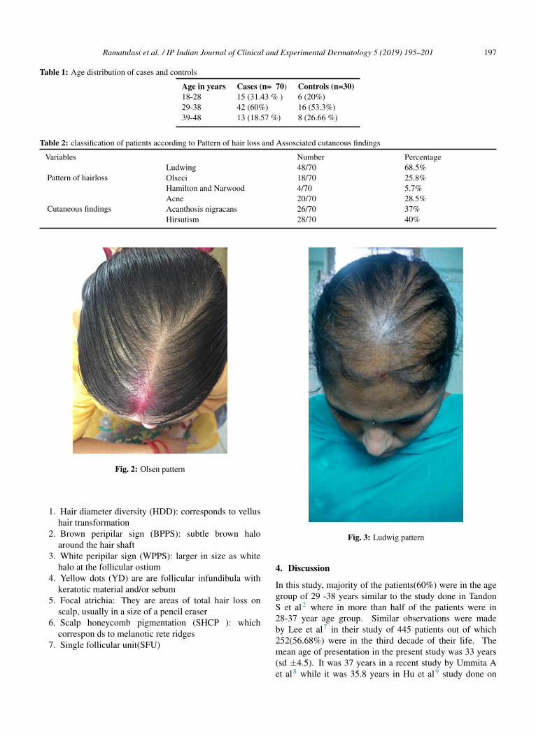
Fig. 2: Olsen pattern
1. Hair diameter diversity (HDD): corresponds to vellushair transformation
2. Brown peripilar sign (BPPS): subtle brown haloaround the hair shaft
3. White peripilar sign (WPPS): larger in size as whitehalo at the follicular ostium
4. Yellow dots (YD) are are follicular infundibula withkeratotic material and/or sebum
5. Focal atrichia: They are areas of total hair loss onscalp, usually in a size of a pencil eraser
6. Scalp honeycomb pigmentation (SHCP ): whichcorrespon ds to melanotic rete ridges
7. Single follicular unit(SFU)
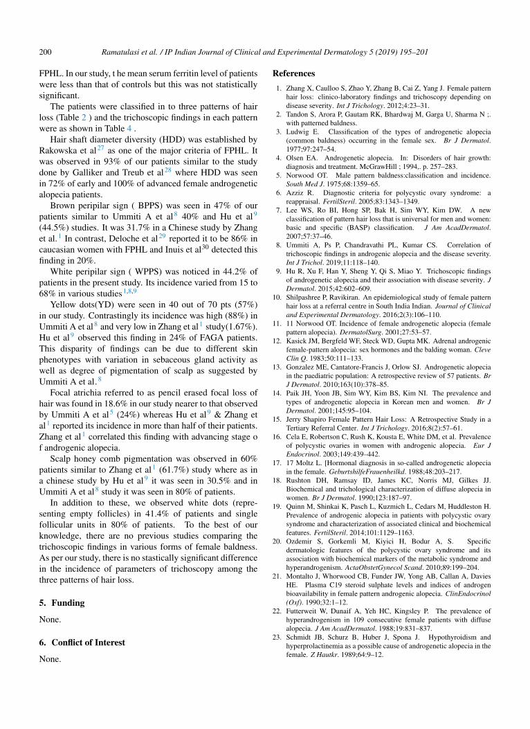
Fig. 3: Ludwig pattern
4. Discussion
In this study, majority of the patients(60%) were in the agegroup of 29 -38 years similar to the study done in TandonS et al2 where in more than half of the patients were in28-37 year age group. Similar observations were madeby Lee et al7 in their study of 445 patients out of which252(56.68%) were in the third decade of their life. Themean age of presentation in the present study was 33 years(sd ±4.5). It was 37 years in a recent study by Ummita Aet al8 while it was 35.8 years in Hu et al9 study done on

198 Ramatulasi et al. / IP Indian Journal of Clinical and Experimental Dermatology 5 (2019) 195–201
Table 3: Trichoscopic findings
Total no. of cases (n=70) Ludwig (n=48) Olsen (n=18) Hamilton &Norwood (n=4) ‘P’ valueYellow dots (40) 27 (56.21) 10 (55%) 3 (75%) 0.82(>0.05)BPPS (33) 23 (48%) 8 (44.4) 2(50%) 0.96(>0.05)WPPS (31) 20 (41.6%) 9(50%) 2(50%) o.51(>0.05)HDD (63) 43 (90%) 16 (89.5%) 4 (100%) 0.79(>0.05)SFU (56) 37(77%) 16 (88%) 3 (75%) 0.56(>0.05)WD (29) 19 (39.5%) 9 (50%) 1 (25%) 0.64(>0.05)Scalp pigmentation(42) 29 (63%) 11 (66%) 2 (50%) 0.91(>0.05)Focal atrichia (11) 7 (14.5) 3 (16.6%) 1 (25%) 0.97(>0.05)
Table 4: Laboratory findings
HORMONE Units Cases Mean±sd Controls Mean±sd P VALUETotal testosterone ng/dl 52.7 ±19.75 35.2 ±12.36 <0.05 #DHEAS mic.gm/dl 179 ± 61.27 154.56 ±52.14 <0.05 #T3 ng/dl 121.52 ±48.07 123.56 ± 36.04 >0.05T4 mic.gm/dl 7.9 ±2.72 7.72±1.98 >0.05TSH Micro IU/ml 6.87±1 3.17±1.97 <0.05 #LH/FSH ratio 0.8±0.32 0.75±0.58 >0.05PROLACTIN ng/lit 16.95 ±6.22 14.53 ± 7.26 >0.05S.FERRITIN microgm/lit 43.74 ±25.77 51.03 ± 29.2 >0.05
# statistically significant
Fig. 4: HDD
Fig. 5: YD & WD
Fig. 6: BPPS
Fig. 7: F ocal atrichia & SHCP

Ramatulasi et al. / IP Indian Journal of Clinical and Experimental Dermatology 5 (2019) 195–201 199
Fig. 8: WPPS
Chinese population.The mean age of onset of hair fall was 26.6(sd ±2.4) y
ears in our study which is nearer to the study b y Tandon S etal2(26.1 years). In a study conducted by Shilpashree et al10
mean age of onset was 28.4±8.2 years which is very similarto our study. Norwood11 reported that Caucasian womenhad FPHL which begins in late 20s which is in accordancewith this study. The mean age of onset was little high (29.8years)in a study by Zhang et al1 on Chinese women and itwas little less (23 years) in the study done by Kasick et al12
The mean duration of hair fall was 4.8 years (sd±1.8 ) inthe present study which is well in agreement with the studiesof Zhang et al1 and Tandon S et al2 where the mean durationwas 5.1 years and 4.49 years respectively.
Family history of hair loss was noticed in 56% ofour patients which is similar to the study conducted byShilpashree et al10 at 51%. It varied from 19.2 to 32.4%in various Chinese studies.11 13 The incidence of familyhistory of baldness was 45.2% in the study by Paik et al14
on Korean women and 45% in the study by Zhang et al1
on Chinese women which are in near agreement with ourstudy. In the study by Tee Wei Siah et al15 85% of patientshad family history of baldness.
The cutaneous findings of hyperandrogenism observedin assosciation with FPHL in our patients were Hirsutism(40%), Acanthosis nigracans (37%) and acne (28.5 %)
Most of the previous studies reported hirsutism in 12-22% of patients.16–18 where as Tandon S et al2 reportedhirsutism in 66.6% of their patients.
Cela et al16 reported acne in 38 of 80 patients (43%) andMoltz17 and Tandon S et al2 observed acne in 41.6% and36.6% of their patients respectively.
43.3% of patients with FPHL had acanthosis nigracansin the study by Tandon S et al2 which is little higher thanthat observed in our study (37%).
P COS was diagnosed based on modified NIH criteriain 19 of the total 70 patients (27.14%). Cela et al16 atthe reproductive endocrinology service in London reporteda higher prevalence of PCOS (67)% while Quinn et al19
and2 Tandon S et al2 reported 22% and 26.6% respectively
which is similar to the present study. This difference inthe prevalence could be due to different population groupsstudied, difference in sample size and screening methodsused in diagnosis.
Various studies have evaluated the role of hyperandro-genemia in FPHL but the results were discordant. MollyQuinn et al19 did not find a difference in overall biochemicalhyperandrogenemia between subjects with and withoutAGA. Similarly, Ozdemir et al20 concluded that AGA wasnot a marker for hyperandrogenemia. In contrast, Cela etal16 reported a higher total testosterone, androstenedioneand free testosterone index in 89 women with AGAcompared with 73 control women.
In the present study, the difference between the meanvalues of total testosterone, DHEAS and TSH of cases werestatistically significant when compared with that of controls(Z-test p<0.05) which is similar to the observations madeby Tandon et al.2 Kasick et al12also reported a significantincrease in DHEAS levels in cases compared to controls.However, total serum testosterone levels were normal in alltheir subjects whereas 19% of our patients had serum totaltestosterone above the normal range.
DHEAS, apart from conversion to potent androgens, hasa direct action on hair follicle as inhibitor of G6PD (glucose6 phosphate dehydrogenase) there by inhibiting nucleic acidsynthesis. This suggests that the se low potent adrenalandrogens are sufficient to cause FPHL with or withoutother findings of hyperandrogenism. However, Montalto etal21 and Rushton et al18 did not find a significant increasein the plasma levels of DHEAS in their studies.
TSH and prolactin can interact with androgenmetabolism at various levels. Futterweit et al22 reportedthat two patients had hyperprolactinemia out of the 109female patients with diffuse alopecia. Schimdt et al23
had studied hypoth yroidism and hyperprolactinemia asa possible cause of androgenetic alopecia in females andfound that 23% of their patients had raised TSH which is inaccordance with our study where 28.5% patients had raisedTSH levels. One third of (2 9%) their patients had raisedprolactin. In contrast, all our patients had normal prolactinlevels.
The relationship between serum ferritin and hair losshas been investigated in a number of studies howeverwith relatively discrepant findings. Sinclair24 in his studyfound no direct relationship between low serum ferritin andhairloss. Rasheed et al25 found that serum ferritin levelsin TE and FPHL cases were significantly lower than incontrols. There is a wide range of serum ferritin level (6-160 mic.gm/l ). However, as per the literature a cut off of 41mic.gm/l yields specificity of 98% and sensitivity of 98%.When this cut off level was taken, more than half (52%)of our patients fall under this group which is comparableto Olsen et al26 (58.8%) and Rushton et al18 studies(65%) indicating an association between Iron deficiency and
200 Ramatulasi et al. / IP Indian Journal of Clinical and Experimental Dermatology 5 (2019) 195–201
FPHL. In our study, t he mean serum ferritin level of patientswere less than that of controls but this was not statisticallysignificant.
The patients were classified in to three patterns of hairloss (Table 2 ) and the trichoscopic findings in each patternwere as shown in Table 4 .
Hair shaft diameter diversity (HDD) was established byRakowska et al27 as one of the major criteria of FPHL. Itwas observed in 93% of our patients similar to the studydone by Galliker and Treub et al28 where HDD was seenin 72% of early and 100% of advanced female androgeneticalopecia patients.
Brown peripilar sign ( BPPS) was seen in 47% of ourpatients similar to Ummiti A et al8 40% and Hu et al9
(44.5%) studies. It was 31.7% in a Chinese study by Zhanget al.1 In contrast, Deloche et al29 reported it to be 86% incaucasian women with FPHL and Inuis et al30 detected thisfinding in 20%.
White peripilar sign ( WPPS) was noticed in 44.2% ofpatients in the present study. Its incidence varied from 15 to68% in various studies1,8,9
Yellow dots(YD) were seen in 40 out of 70 pts (57%)in our study. Contrastingly its incidence was high (88%) inUmmiti A et al8 and very low in Zhang et al1 study(1.67%).Hu et al9 observed this finding in 24% of FAGA patients.This disparity of findings can be due to different skinphenotypes with variation in sebaceous gland activity aswell as degree of pigmentation of scalp as suggested byUmmiti A et al.8
Focal atrichia referred to as pencil erased focal loss ofhair was found in 18.6% in our study nearer to that observedby Ummiti A et al5 (24%) whereas Hu et al9 & Zhang etal1 reported its incidence in more than half of their patients.Zhang et al1 correlated this finding with advancing stage of androgenic alopecia.
Scalp honey comb pigmentation was observed in 60%patients similar to Zhang et al1 (61.7%) study where as ina chinese study by Hu et al9 it was seen in 30.5% and inUmmiti A et al8 study it was seen in 80% of patients.
In addition to these, we observed white dots (repre-senting empty follicles) in 41.4% of patients and singlefollicular units in 80% of patients. To the best of ourknowledge, there are no previous studies comparing thetrichoscopic findings in various forms of female baldness.As per our study, there is no stastically significant differencein the incidence of parameters of trichoscopy among thethree patterns of hair loss.
5. Funding
None.
6. Conflict of Interest
None.
References1. Zhang X, Caulloo S, Zhao Y, Zhang B, Cai Z, Yang J. Female pattern
hair loss: clinico-laboratory findings and trichoscopy depending ondisease severity. Int J Trichology. 2012;4:23–31.
2. Tandon S, Arora P, Gautam RK, Bhardwaj M, Garga U, Sharma N ;.with patterned baldness.
3. Ludwig E. Classification of the types of androgenetic alopecia(common baldness) occurring in the female sex. Br J Dermatol.1977;97:247–54.
4. Olsen EA. Androgenetic alopecia. In: Disorders of hair growth:diagnosis and treatment. McGrawHill ; 1994,. p. 257–283.
5. Norwood OT. Male pattern baldness:classification and incidence.South Med J. 1975;68:1359–65.
6. Azziz R. Diagnostic criteria for polycystic ovary syndrome: areappraisal. FertilSteril. 2005;83:1343–1349.
7. Lee WS, Ro BI, Hong SP, Bak H, Sim WY, Kim DW. A newclassification of pattern hair loss that is universal for men and women:basic and specific (BASP) classification. J Am AcadDermatol.2007;57:37–46.
8. Ummiti A, Ps P, Chandravathi PL, Kumar CS. Correlation oftrichoscopic findings in androgenic alopecia and the disease severity.Int J Trichol. 2019;11:118–140.
9. Hu R, Xu F, Han Y, Sheng Y, Qi S, Miao Y. Trichoscopic findingsof androgenetic alopecia and their association with disease severity. JDermatol. 2015;42:602–609.
10. Shilpashree P, Ravikiran. An epidemiological study of female patternhair loss at a referral centre in South India Indian. Journal of Clinicaland Experimental Dermatology. 2016;2(3):106–110.
11. 11 Norwood OT. Incidence of female androgenetic alopecia (femalepattern alopecia). DermatolSurg. 2001;27:53–57.
12. Kasick JM, Bergfeld WF, Steck WD, Gupta MK. Adrenal androgenicfemale-pattern alopecia: sex hormones and the balding woman. CleveClin Q. 1983;50:111–133.
13. Gonzalez ME, Cantatore-Francis J, Orlow SJ. Androgenetic alopeciain the paediatric population: A retrospective review of 57 patients. BrJ Dermatol. 2010;163(10):378–85.
14. Paik JH, Yoon JB, Sim WY, Kim BS, Kim NI. The prevalence andtypes of androgenetic alopecia in Korean men and women. Br JDermatol. 2001;145:95–104.
15. Jerry Shapiro Female Pattern Hair Loss: A Retrospective Study in aTertiary Referral Center. Int J Trichology. 2016;8(2):57–61.
16. Cela E, Robertson C, Rush K, Kousta E, White DM, et al. Prevalenceof polycystic ovaries in women with androgenic alopecia. Eur JEndocrinol. 2003;149:439–442.
17. 17 Moltz L. [Hormonal diagnosis in so-called androgenetic alopeciain the female. GeburtshilfeFrauenheilkd. 1988;48:203–217.
18. Rushton DH, Ramsay ID, James KC, Norris MJ, Gilkes JJ.Biochemical and trichological characterization of diffuse alopecia inwomen. Br J Dermatol. 1990;123:187–97.
19. Quinn M, Shinkai K, Pasch L, Kuzmich L, Cedars M, Huddleston H.Prevalence of androgenic alopecia in patients with polycystic ovarysyndrome and characterization of associated clinical and biochemicalfeatures. FertilSteril. 2014;101:1129–1163.
20. Ozdemir S, Gorkemli M, Kiyici H, Bodur A, S. Specificdermatologic features of the polycystic ovary syndrome and itsassociation with biochemical markers of the metabolic syndrome andhyperandrogenism. ActaObstetGynecol Scand. 2010;89:199–204.
21. Montalto J, Whorwood CB, Funder JW, Yong AB, Callan A, DaviesHE. Plasma C19 steroid sulphate levels and indices of androgenbioavailability in female pattern androgenic alopecia. ClinEndocrinol(Oxf). 1990;32:1–12.
22. Futterweit W, Dunaif A, Yeh HC, Kingsley P. The prevalence ofhyperandrogenism in 109 consecutive female patients with diffusealopecia. J Am AcadDermatol. 1988;19:831–837.
23. Schmidt JB, Schurz B, Huber J, Spona J. Hypothyroidism andhyperprolactinemia as a possible cause of androgenetic alopecia in thefemale. Z Hautkr. 1989;64:9–12.

Ramatulasi et al. / IP Indian Journal of Clinical and Experimental Dermatology 5 (2019) 195–201 201
24. Sinclair R. There is no clear association between low serum ferritinand chronic diffuse telogen hair loss. Br J Dermatol. 2002;147:982–986.
25. Rasheed H, Mahgoub D, Hegazy R, El-Komy M, Hay A, et al. Serumferritin and Vitamin D in female hair loss: Do they play a role? SkinPharmacolPhysiol. 2013;26:101–108.
26. Olsen EA, Reed KB, Cacchio PB, Caudill L. Iron deficiency in femalepattern hair loss, chronic telogen effluvium, and control groups. J AmAcadDermatol. 2010;63:991–1000.
27. Rakowska A, Slowinska M, Kowalska-Oledzka E, Olszewska M,Rudnicka L. Dermoscopy in female androgenic alopecia: Methodstandardization and diagnostic criteria. Int J Trichology. 2009;1:123–153.
28. Galliker NA, Treb RM. Value of trichoscopy versus trichogramfor diagnosis of female androgenetic alopecia. Int J Trichology.2012;4:19–22.
29. Deloche C, Lacharrire OD, Misciali C, Piraccini BM, Vincenzi C,Bastien P. Histological features of peripilar signs associated withandrogenetic alopecia. Arch Dermatol Res. 2004;29:422–430.
Author biography
Sappa Ramatulasi Assistant Professor
Manjari Annapurna Malladi Senior Resident
Ruttala Sri Satya Post Graduates
Kashetty Srujan Kumar Post Graduates
Anand Acharya Professor and Head
Cite this article: Ramatulasi S, Annapurna Malladi M, Sri Satya R,Srujan Kumar K, Acharya A. Clinico -laboratory and trichoscopicevaluation of various patterns of female baldness – A prospective casecontrol study. Indian J Clin Exp Dermatol 2019;5(3):195-201.