mature b-cell neoplasms - hemepathreview home...
TRANSCRIPT

Introduction
Mature B-cell Neoplasms
DEFINITION
• Clonal proliferations of B cells Naïve B cells -> Mature plasma cells
• Recapitulate stages of normal differentiation

EPIDEMIOLOGY
• >90% of lymphoid neoplasms worldwide;
annually 4% of all cancers
• More common in developed countries
• Annual incidence – 15/100,000 (USA) , 1.2/100,000 (China)
• Follicular lymphoma & DLBCL (66% of NHL)
are most common
EPIDEMIOLOGY
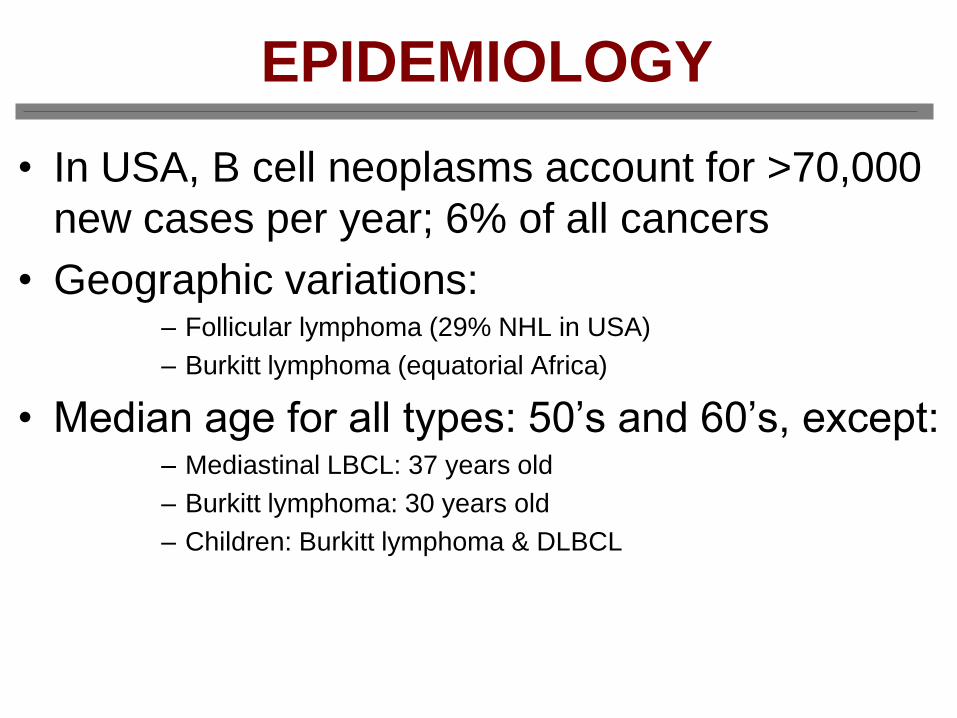
• In USA, B cell neoplasms account for >70,000
new cases per year; 6% of all cancers
• Geographic variations: – Follicular lymphoma (29% NHL in USA)
– Burkitt lymphoma (equatorial Africa)
• Median age for all types: 50’s and 60’s, except: – Mediastinal LBCL: 37 years old
– Burkitt lymphoma: 30 years old
– Children: Burkitt lymphoma & DLBCL
EPIDEMIOLOGY
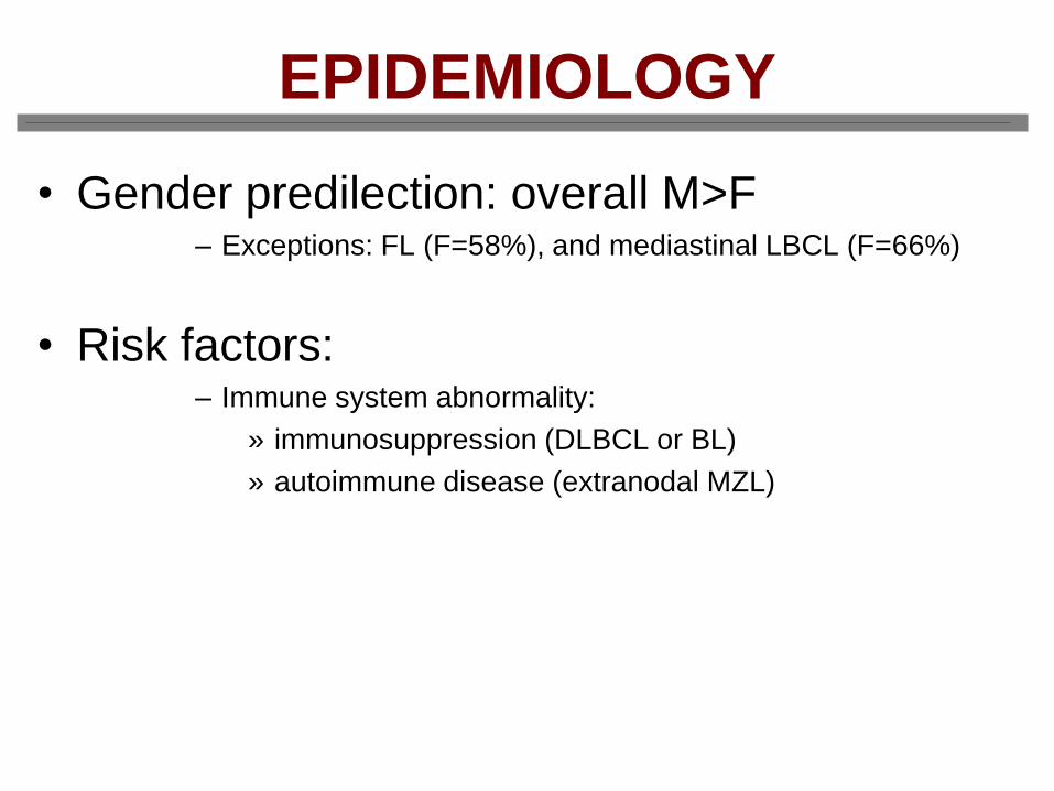
• Gender predilection: overall M>F – Exceptions: FL (F=58%), and mediastinal LBCL (F=66%)
• Risk factors: – Immune system abnormality:
» immunosuppression (DLBCL or BL)
» autoimmune disease (extranodal MZL)

ETIOLOGY
• Epstein-Barr virus • Burkitt lymphoma:
– Endemic (100%)
– Sporadic & HIV-associated (40%)
• Majority of B-cell lymphomas in iatrogenically immunosuppressed
patients
• KSHV/HHV8 • PEL
• Multicentric Castleman disease-associated lymphomas in HIV
patients
• Hepatitis C virus • Lymphoplasmacytic lymphoma associated with type II
cryoglobulinemia
• Some lymphomas of liver and salivary glands
ETIOLOGY
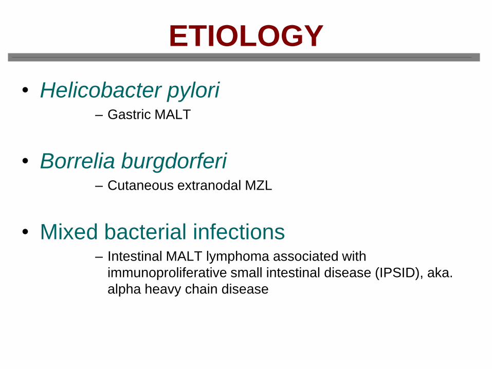
• Helicobacter pylori – Gastric MALT
• Borrelia burgdorferi – Cutaneous extranodal MZL
• Mixed bacterial infections – Intestinal MALT lymphoma associated with
immunoproliferative small intestinal disease (IPSID), aka.
alpha heavy chain disease

ANTIGEN
PATHOPHYSIOLOGY
Precursor B cell lymphoblasts
Naïve B cell (sIgM+/sIgD+; CD5+)
VDJ gene rearrangement
Follicular blast
Mantle cell
Centroblast
Centrocyte
Marginal zone
Plasma cell
Plasmacytoid lymphocyte
Immunoblast

ANTIGEN
PATHOPHYSIOLOGY
Precursor B cell lymphoblasts
Naïve B cell (sIgM+/sIgD+; CD5+)
VDJ gene rearrangement
Follicular blast
Mantle cell
Centroblast
Centrocyte
Marginal zone
Plasma cell
Plasmacytoid
lymphocyte
Immunoblast
B-CLL/SLL
Lymphoplasmacytic
lymphoma
Mantle cell lymphoma
LBCL
Follicular lymphoma
LBCL
Burkitt lymphoma
Precursor B-ALL
Marginal zone lymphomas
Plasma cell myeloma
PATHOPHYSIOLOGY
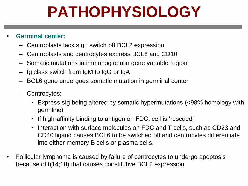
• Germinal center:
– Centroblasts lack sIg ; switch off BCL2 expression
– Centroblasts and centrocytes express BCL6 and CD10
– Somatic mutations in immunoglobulin gene variable region
– Ig class switch from IgM to IgG or IgA
– BCL6 gene undergoes somatic mutation in germinal center
– Centrocytes:
• Express sIg being altered by somatic hypermutations (<98% homology with
germline)
• If high-affinity binding to antigen on FDC, cell is ‘rescued’
• Interaction with surface molecules on FDC and T cells, such as CD23 and
CD40 ligand causes BCL6 to be switched off and centrocytes differentiate
into either memory B cells or plasma cells.
• Follicular lymphoma is caused by failure of centrocytes to undergo apoptosis
because of t(14;18) that causes constitutive BCL2 expression
PATHOPHYSIOLOGY
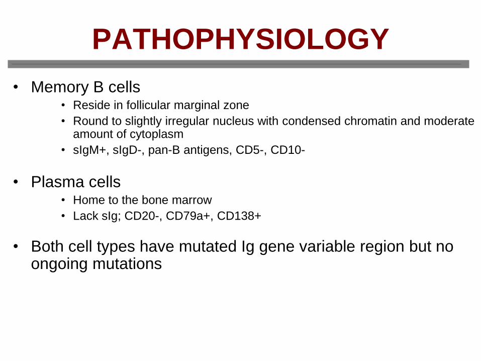
• Memory B cells • Reside in follicular marginal zone
• Round to slightly irregular nucleus with condensed chromatin and moderate amount of cytoplasm
• sIgM+, sIgD-, pan-B antigens, CD5-, CD10-
• Plasma cells • Home to the bone marrow
• Lack sIg; CD20-, CD79a+, CD138+
• Both cell types have mutated Ig gene variable region but no ongoing mutations

GENETICS
• t(11;14) in mantle cell lymphoma
• t(14;18) in follicular lymphoma
• t(8;14) in Burkitt lymphoma
• t(11;18) in MALT lymphoma
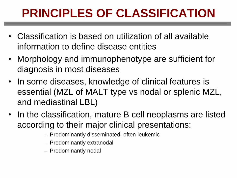
PRINCIPLES OF CLASSIFICATION
• Classification is based on utilization of all available
information to define disease entities
• Morphology and immunophenotype are sufficient for
diagnosis in most diseases
• In some diseases, knowledge of clinical features is
essential (MZL of MALT type vs nodal or splenic MZL,
and mediastinal LBL)
• In the classification, mature B cell neoplasms are listed
according to their major clinical presentations: – Predominantly disseminated, often leukemic
– Predominantly extranodal
– Predominantly nodal
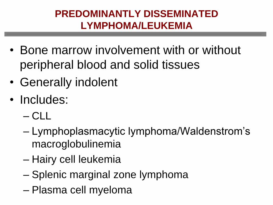
PREDOMINANTLY DISSEMINATED
LYMPHOMA/LEUKEMIA
• Bone marrow involvement with or without
peripheral blood and solid tissues
• Generally indolent
• Includes:
– CLL
– Lymphoplasmacytic lymphoma/Waldenstrom’s
macroglobulinemia
– Hairy cell leukemia
– Splenic marginal zone lymphoma
– Plasma cell myeloma
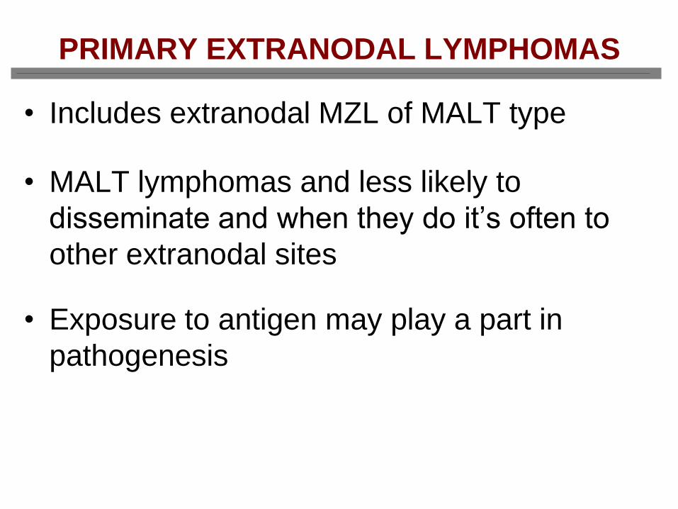
PRIMARY EXTRANODAL LYMPHOMAS
• Includes extranodal MZL of MALT type
• MALT lymphomas and less likely to
disseminate and when they do it’s often to
other extranodal sites
• Exposure to antigen may play a part in
pathogenesis
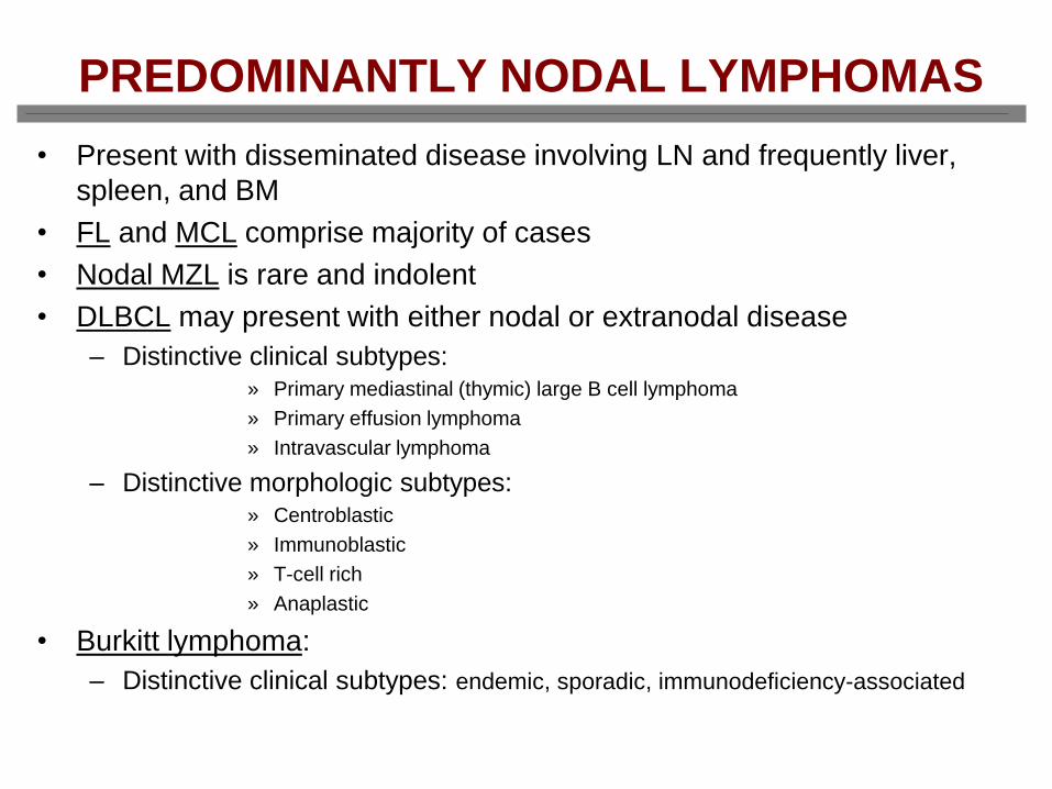
PREDOMINANTLY NODAL LYMPHOMAS
• Present with disseminated disease involving LN and frequently liver,
spleen, and BM
• FL and MCL comprise majority of cases
• Nodal MZL is rare and indolent
• DLBCL may present with either nodal or extranodal disease
– Distinctive clinical subtypes: » Primary mediastinal (thymic) large B cell lymphoma
» Primary effusion lymphoma
» Intravascular lymphoma
– Distinctive morphologic subtypes: » Centroblastic
» Immunoblastic
» T-cell rich
» Anaplastic
• Burkitt lymphoma:
– Distinctive clinical subtypes: endemic, sporadic, immunodeficiency-associated

Chronic Lymphocytic
Leukemia/
Small Lymphocytic Lymphoma

WHO CLL/SLL
• Neoplasm of monomorphic small, round B
lymphocytes in blood, BM and lymph nodes, admixed
with prolymphocytes and paraimmunoblasts
(pseudofollicles), usually expressing CD5 and CD23.
• CLL: restricted to BM involvement, PB monoclonal B
cell count > 5x10⁹/l)
• SLL: restricted to non-leukemic cases (PB
monoclonal B cell count < 5x10⁹/l)
• Healthy pts with a B cell clone, PB monoclonal B cell
count < 5x10⁹/l -> monoclonal B cell lymphocytosis
(3.5% of pts > 40 y/o)
Synonyms
• WF: small lymphocytic, consistent with
CLL.
• REAL: B-cell chronic lymphocytic
leukemia.
• FAB: B-cell chronic lymphocytic
leukemia.
Epidemiology
• Comprises 90% of chronic lymphoid
leukemias in USA and Europe.
• 6.7% of non-Hodgkin´s lymphoma.
• Majority of patients >50 y/o (median
65).
• M:F ratio 2:1.
Sites of involvement
• CLL: by definition, BM and PB. Monoclonal B cell count > 5x10⁹/l.
• Dx can be done with < 5x10⁹/l with the proper BM morphology and immunophenotype.
• SLL: lymph nodes, liver and spleen are typically infiltrated.
• Skin, breast and ocular adnexae may be involved.
Clinical features
• Most patients asymptomatic. Some may
present with fatigue, autoimmune hemolytic
anaemia, infections, splenomegaly,
hepatomegaly, lymphadenopathy or
extranodal infiltrates.
• Small M-component in some pats.

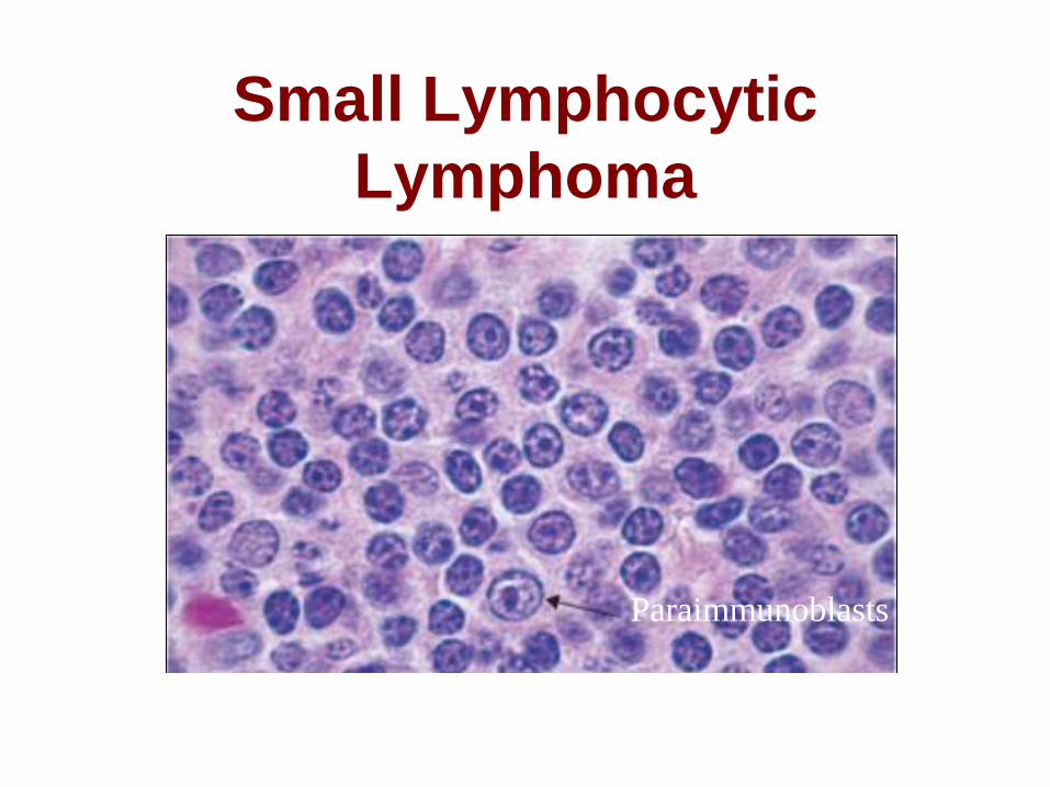
Morphology
• Lymph nodes.
– Effacement of architecture, pseudofollicular
pattern of pale areas of large cells in a dark
background of small cells. Ocasionally is
interfollicular.
– The predominant cell is a small lymphocyte
with clumped chromatin, round nucleus,
ocassionally a nucleolus.
– Mitotic activity usually very low.
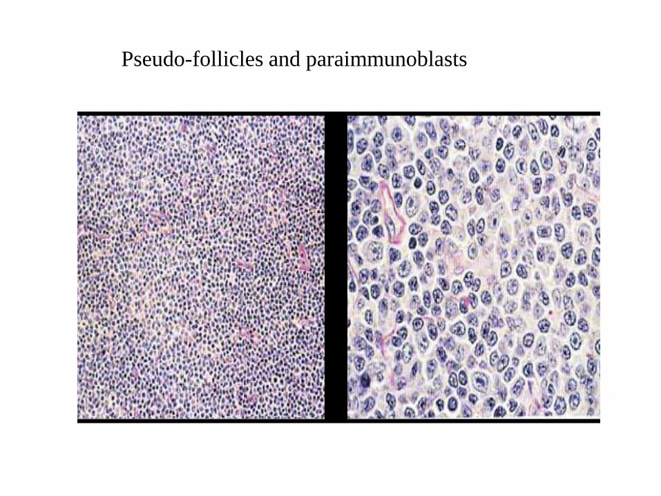
Pseudo-follicles and paraimmunoblasts

(Diffuse) Small Lymphocytic
Lymphoma
Pseudo-follicle
Small Lymphocytic
Lymphoma
Paraimmunoblasts
Morphology
- Pseudofollicles or proliferation centers contain
small, medium and large cells.
- Prolymphocytes are medium-sized with
dispersed chromatin and small nucleoli.
- Paraimmunoblasts are medium to large cells
with round to oval nuclei, dispersed chromatin,
central eosinophilic nucleoli and slightly
basophilic cytoplasm.
Morphology
• Spleen.
- White pulp involvement predominant. Red pulp
also involved.
- Pseudofollicles less prominent.
- Sometimes there is some nuclear irregularity
mimicking MCL. Pseudofollicles, prolymphocytes
and paraimmunoblasts help to rule out MCL.
- Sometimes plasmacytoid differentiation.

Morphology • BM and Blood.
– CLL cells are small lymphocytes with clumped chromatin.
Cytoplasm is scanty, clear to basophilic with regular outline.
– Smudge or basket cells are typically seen in the blood
(alleviated with albumin treatment)).
– Proportion of prolymphocytes in the smear usually < 2%.
Incresing numbers correlate with more aggressive disease, p53
abnormalities and trisomy 12.
– With increased prolymphocytes-> (CLL/PLL) defined by
prolymphyctes between 10% and 55%.
Morphology
– BM involvement can be interstitial, nodular
or diffuse or combinations.
– Pseudofollicles less common in BM.
– Paratrabecular aggregates not typical.
– Nodular and interstitial patterns are seen in
early disease.
– Diffuse pattern associated with advanced
disease and BM failure.

Chronic Lymphocytic
Leukemia
P.B.: Chronic lymphocytic leukemia
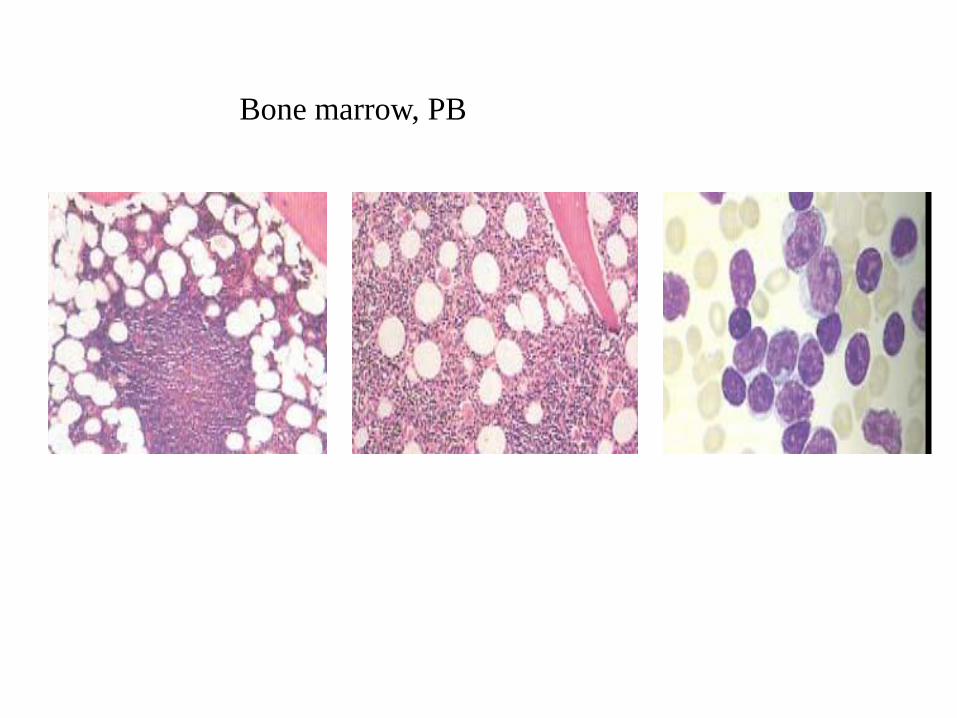
Bone marrow, PB
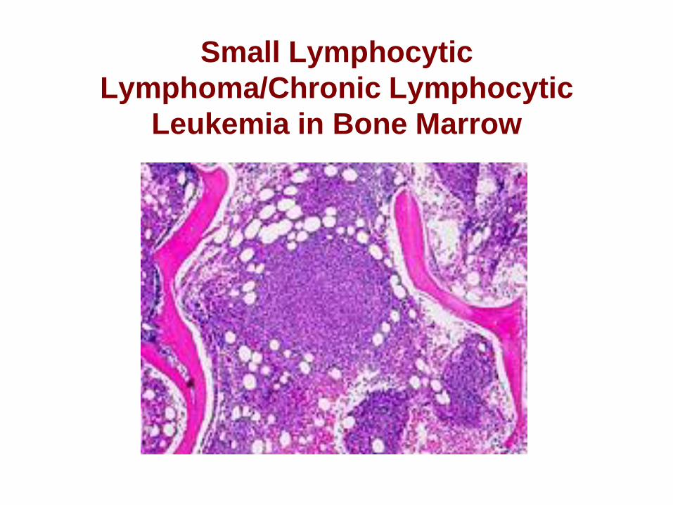
Small Lymphocytic
Lymphoma/Chronic Lymphocytic
Leukemia in Bone Marrow

Small Lymphocytic Lymphoma
in Liver
Morphology
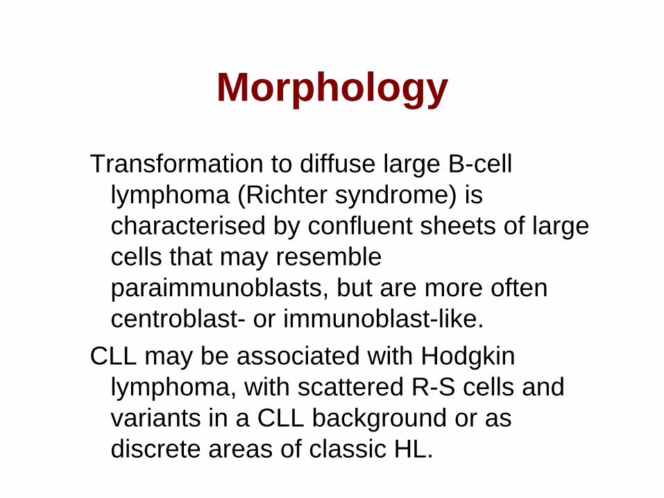
Transformation to diffuse large B-cell
lymphoma (Richter syndrome) is
characterised by confluent sheets of large
cells that may resemble
paraimmunoblasts, but are more often
centroblast- or immunoblast-like.
CLL may be associated with Hodgkin
lymphoma, with scattered R-S cells and
variants in a CLL background or as
discrete areas of classic HL.
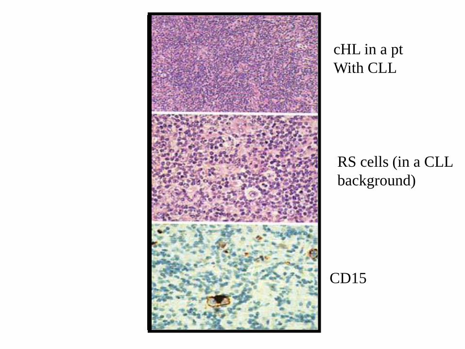
CD15
cHL in a pt
With CLL
RS cells (in a CLL
background)

Immunophenotype
• Express weak or dim surface IgM or
IgM and IgD, CD5, CD19, CD20 (weak),
CD22 (weak), CD79a, CD23, CD43,
CD11c (weak).
• CD10-, cyclin D1-.
• FMC7 and CD79b negative or weak.

Immunophenotype
• Cases with unmutated Ig variable region genes are reported to be CD38+ and ZAP70+.
Immunophenotype
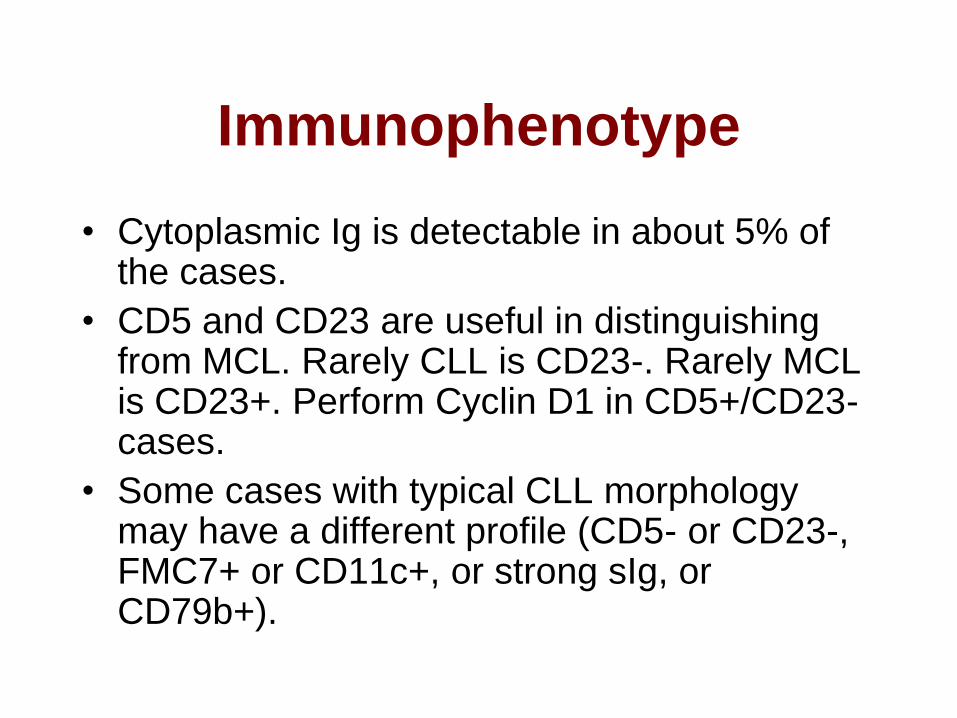
• Cytoplasmic Ig is detectable in about 5% of the cases.
• CD5 and CD23 are useful in distinguishing from MCL. Rarely CLL is CD23-. Rarely MCL is CD23+. Perform Cyclin D1 in CD5+/CD23- cases.
• Some cases with typical CLL morphology may have a different profile (CD5- or CD23-, FMC7+ or CD11c+, or strong sIg, or CD79b+).
Genetics • Antigen receptor genes:
– Ig heavy and light genes are rearranged.
Suggestion of 2 distinct types of CLL defined by
the mutational status of the IgVH genes: 40-50%
show no somatic mutations of their variable region
genes (naïve cells, unmutated). 50-60% have
somatic mutations consistent with derivation from
post-germinal center B-cells.
– DNA sequencing shows hypermutation if there is
<98% homology with germline
– Unmuted: poor prognosis; hypermutated: better
prognosis

Genetics
• Cytogenetic abns and oncogenes:
– About 80% of the cases have abnormal karyotypes by FISH.
– Trisomy 12 reported in 20% of cases. Have predominantly unmutated Ig variable region genes.
– Deletions at 13q14.3 in up to 50%. Have mutations more often (Ig variable region genes).

Genetics
• Cytogenetic abns and oncogenes:
– Deletions at 11q22-23 are found in 20% of cases. Most often unmutated.
– Deletions at 17p13 (p53 locus) are seen in 10% of cases, respectively. Most often unmutated.
– t(11;14) and BCL1 gene rearrangement have been reported. These cases may have been leukemic MCL and misdiagnosed as CLL.
Postulated cell of origin
• Thought to correspond to recirculating CD5+
CD23+ IgM+ IgD+ naïve B cells., found in PB,
primary follicle, and follicular mantle zone.
• Cases that show V region mutations may
correspond to a subset of PB CD5+ IgM+ B
cells that appear to be memory B cells.
Prognosis and predictive
factors
• Clinical course is indolent, not
considered to be curable with available
therapy.
• Purine nucleoside analogues, such as
fludarabine, may result in sustained
remissions.
• 5 year OS of SLL was 51% with a FFS
of 25%. Overall median survival is 7 yrs.
Prognosis and predictive
factors
• Clinical staging systems (Rai 0-IV and
Binet A-C) are the best predictors of
survival.
• Cases of CLL/PLL and diffuse BM
involvement may have a worse
prognosis.

Prognosis and predictive
factors
• Rapid doubling time (< 12 months) is a
prediction of poor prognosis
• +12 correlates with atypical morphology
and aggressive clinical course.
• Abns of 13q14 are reported associated
with long survival.
Prognosis and predictive
factors
• Mutations in Ig genes variable regions
have a better prognosis than those with
germline VH regions (median survival 7
yrs vs 3 yrs).
• CD38, ZAP70 expression appears to
have worse prognosis.
• 11q22-23 deletions have extensive
lymphadenopathy and poor survival.
Prognosis and predictive
factors
• Transformation to high grade lymphoma
(Richter syndrome) occurs in aprox.
3.5% of cases. Usually DLBCL (3%).
HD (0.5%), particularly in pats treated
with purine nucleotide analogues.

Prognosis and predictive
factors
• Molecular genetic analysis suggests
about in 50% cases the aggressive
lymphoma represents transformation of
the original neoplastic clone.
• In the remainder the lymphoma maybe
a second, unrelated neoplasm.

Prognosis and Predictive
Factors • Variant: Mu heavy chain disease.
– Usually associated with neoplasm resembling CLL, in which a defective mu heavy chain lacking a variable region is produced.
– BM has characteristic vacuolated plasma cells, admixed with small, round lymphocytes.
– Pats are adults with hepato-splenomegaly, absence of peripheral lymphadenopathy, and a slowly progressive course.
B-cell Prolymphocytic
Leukemia
B-PLL

Definition
• Malignancy of B-prolymphocytes
– Round lymphoid cells
– Medium-sized
– Prominent nucleoli
• Affects PB, BM, and spleen

Definition
• Prolymphocytes: >55% of PB lymphoid
cells
• Exclude
– Transformed CLL
– CLL with increased prolymphocytes
Epidemiology
• Extremely rare (~1% of lymphocytic
leukemias)
• Elderly (most >60 y/o; median 70)
• Male predominance (M:F = 1.6:1)

Clinical features • Most patients
– Marked splenomegaly
– No peripheral lymphadenopathy
– Rapidly rising lymphocyte count (usually >100 x 109/L)
• 50% of patients – Anemia
– Thrombocytopenia
• Some patients – Serum M-component

Morphology in PB and BM • PB Prolymphocytes
– >55% of circulating cells
(usually >90%)
– Medium-sized (2X size of a
lymphocyte)
– Round nucleus (+/-
indentation)
– Moderately condensed
chromatin
– Prominent central nucleolus
– Small amount of faintly
basophilic cytoplasm
• BM
– Diffuse intertrabecular
infiltrate
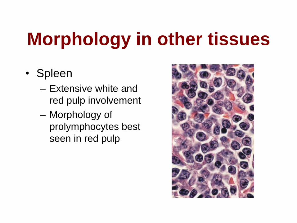
Morphology in other tissues
• Spleen
– Extensive white and
red pulp involvement
– Morphology of
prolymphocytes best
seen in red pulp

Morphology in other tissues
• LN
– Diffuse or vaguely nodular infiltrates of
prolymphocytes
– No pseudofollicles

Immunophenotype
• Strong positive for – sIgM (+/- IgD)
• Positive for – CD19/20/22/79a/79b
– FMC7
• CD5 positive in 1/3 of cases
• Negative for CD23

Genetics • t(11;14)(q13;q32)
– In 20% of typical cases
– Some might be cases of leukemic blastoid variant of MCL (misdiagnosed as PLL)
• Abnormalities of TP53 (including 17p del) – 53% of cases (highest in lymphoid malignancies)
– p53 protein expression
• Deletions at 11q23 and 13q14 (by FISH)

Prognosis • Prognosis is not dependent on: CD38, ZAP70, 17p del, or
mutational status of Ig gene.
• Poor response to CLL therapy
• Short survival
• May respond to
– CHOP
– Fludarabine
– Cladribine
– Splenic irradiation
• Splenectomy
– May improve patient’s general condition
– Does not delay disease progression