dna origami delivery system for cancer therapy with ... · pdf file1 dna origami delivery...
TRANSCRIPT
1 DNA Origami Delivery System for2 Cancer Therapy with Tunable3 Release Properties4 Yong-Xing Zhao,†,‡ Alan Shaw,† Xianghui Zeng,† Erik Benson,† AndreasM. Nystrom,† and Bjorn Hogberg†,*
5†Swedish Medical Nanoscience Center, Department of Neuroscience, Karolinska Institute, SE-171 77 Stockholm, Sweden and ‡School of Pharmaceutical Sciences,
6 Zhengzhou University, Zhengzhou 450001, Henan Province, People's Republic of China
78
In the assembly of DNA nanostructures,
9 the specificity of Watson�Crick base10 pairing is used to control matter at the11 nanoscale.1�7 With this technology, nano-12 scale assemblies of drugs, ligands, and other13 functionalities can be organized with un-14 precedented precision for targeting8 and15 even to perform simple logic to deliver16 payloads only when needed.8,9 This tech-17 nique will allow researchers to move closer18 to the goal of building the magic bullet for19 cancer, a concept introduced by Paul Erhlich20 in the 20th century. Compared to many21 other nanoscale systems designed for drug22 delivery such as polymer micelles and inor-23 ganic particles, the DNA origami based con-24 struction has several advantages: (i) same25 size, shape, and charge for each particle26 instead of the size distribution often seen27 for self-assembled nanostructures; (ii) per-28 fect control of the placement of functional-29 ities on the structure using specific oligos.30 The above features suggest that these31 nanostructures should be considered a pro-32 mising tool for cancer nanotechnology10 in33 the future. So far, DNA origami nanostruc-34 tures have been used to successfully deliver35 different cargos to cells, such as immuno-36 stimulatory oligonucleotides11 or apoptosis-37 inducing antibodies.8 Anthracyclines for38 cancer therapy12 intercalate DNA,13,14 and39 since DNA nanotechnology allows such a40 high degree of customization, the question41 is whether it is possible to tune the DNA42 nanostructures to optimize the delivery of43 doxorubicin (Dox) to human cancer cells.44 Recently Ke et al.15 demonstrated that by45 designing DNA origami nanostructures to46 have a certain twist density, the structures47 would fold correctly only when a certain48 amount of intercalator was added to the49 folding reaction.Here,we investigatewhether50 such an alternation in the origami design51 would enable a change in the drug loading
52
53
54
55
56
57585960616263646566676869707172737475and release properties. To analyze how the76in vitrodrug kinetics correspond to changes in77cellular delivery, we also investigate how the78different designs affect the viability of three79common breast cancer cell lines: MDA-MB-80213, MDA-MB-468, and MCF-7.
81RESULTS AND DISCUSSION
82To investigate the feasibility of using DNA83origami nanostructures as a drug delivery84system for Dox, we designed two 18-helix
* Address correspondence [email protected].
Received for review May 22, 2012and accepted September 5, 2012.
Published online10.1021/nn3022662
ABSTRACT
In the assembly of DNA nanostructures, the specificity of Watson�Crick base pairing is used to
control matter at the nanoscale. Using this technology for drug delivery is a promising route
toward the magic bullet concept, as it would allow the realization of complex assemblies that co-
localize drugs, targeting ligands and other functionalities in one nanostructure. Anthracyclines'
mechanism of action in cancer therapy is to intercalate DNA, and since DNA nanotechnology
allows for such a high degree of customization, we hypothesized that this would allow us to tune
the DNA nanostructures for optimal delivery of the anthracycline doxorubicin (Dox) to human
breast cancer cells. We have tested two DNA origami nanostructures on three different breast
cancer cell lines (MDA-MB-231, MDA-MB-468, and MCF-7). The different nanostructures were
designed to exhibit varying degrees of global twist, leading to different amounts of relaxation in
the DNA double-helix structure. By tuning the nanostructure design we are able to (i) tune the
encapsulation efficiency and the release rate of the drug and (ii) increase the cytotoxicity and
lower the intercellular elimination rate when compared to free Dox. Enhanced apoptosis induced
by the delivery system in breast cancer cells was investigated using flow cytometry. The findings
indicate that DNA origami nanostructures represent an efficient delivery system for Dox, resulting
in high degrees of internalization and increased induction of programmed cell death in breast
cancer cells. In addition, by designing the structures to exhibit different degrees of twist, we are
able to rationally control and tailor the drug release kinetics.
KEYWORDS: DNA nanotechnology . cancer drug delivery . DNA origami .breast cancer cells . doxorubicin . cell uptake
ARTIC
LEACS Nano | 3b2 | ver.9 | 7/9/012 | 21:51 | Msc: nn-2012-022662 | TEID: deb00 | BATID: 00000 | Pages: 7.94
ZHAO ET AL . VOL. XXX ’ NO. XX ’ 000–000 ’ XXXX
www.acsnano.org
A
CXXXX American Chemical Society
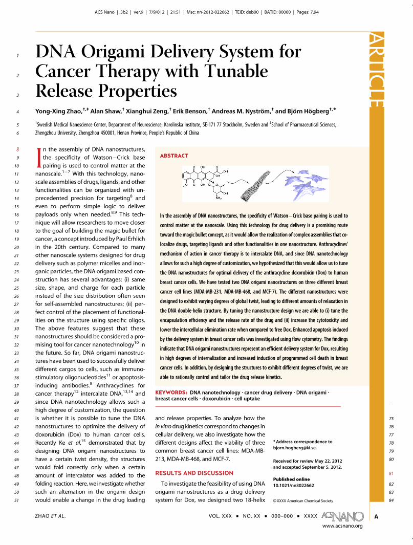
85 bundle nanotubes using the honeycomb lattice frame-86 work3 and using caDNAno16 for the design work87 (complete design schematics and sequences in supple-88 mentary Figures S1 and S2). The first of these test89 structures was a straight nanotube (S-Nano) using a90 conventional number of 10.5 bases per helical turn of91 DNA to determine crossover positions between neigh-92 boring helices. The S-Nano design, depicted schemati-93 cally in Figure 1F1 C, was designed to use a 7560 nt long94 scaffold3 and to be 138 nm long with a diameter of95 13 nm and folds correctly in the absence of Dox; see96 supplementary Figure S3. Using the same S-Nano de-97 sign as a starting point, a twisted version (T-Nano) was98 designed by adding one insertion every seventh base99 pair following the technique introduced by Dietz et al.6
100 To accommodate the additional base pair requirements101 after adding insertions, an 8634 nt long ssDNA scaffold3
102 was used in this design. By designing the structure to103 have 12 bp per turn, it is expected that the entire104 structure would adopt a global right-handed twist to105 partially relieve the stress induced by imposing an106 unnatural twist density on the DNA. Indeed, we find107 that structure predictions and TEM data show an ex-108 tremely twisted shape when this structure is folded in109 the absence of Dox, Figure 1E. When the structures are110 folded in the presence of Dox, the S-Nano, designed for
11110.5 bp per turn, exhibits decreased folding quality, as112can be observed by lower gel mobility (Figure 1C).113Conversely, the T-Nano, designed to have 12 bp/turn,114exhibits a higher yield and an increased folding quality115when Dox is added; see Figure 1D. Further, TEM data of116the T-Nano structures loaded with Dox (Dox/T-Nano), in117Figure 1F, show that these structures are more com-118pact, straighter, and more elongated with a general119appearance of superior folding quality than the120T-Nano folded without Dox in Figure 1E. This observa-121tion can be understood by examining known crystal122structures of DNA. In Figure 1A and B, we show123crystallography data for six base pairs of DNA from124normal DNA17 and DNA intercalated with Dox.18 By125plotting these data together with a simplified DNA126model on the same scale, it becomes apparent that a127more elongated DNA with a 12 bp/turn twist density128should become the dominant structure after Dox inter-129calation. The gel data presented are captured after spin130filtration to remove excess staple oligonucleotides; see131Materials and Methods.132Next, we proceeded to examine whether the twist133design was able to encapsulate and retain Dox at a134higher rate, via measurements of the in vitro drug135release properties of the different nanotubes. As an136additional control, a normal double-stranded DNA
Figure 1. Nanostructure design and characterization. (A and B) Dox intercalation shifts the pitch and twist density of DNA.Crystal structure data of six base pairs of DNA without, A (from ref 17), and with Dox, B (from ref 18), intercalation at the sitesmarked with * (to scale, rendered with Molecular Maya29 from PDB structures). Ball-and-stick simplified DNA models of24 base pairs are shown in the same scale. Without Dox, normal B-type DNA has a twist density of 10.5 bp per turn,leading to origami designs where 21 bp separate two consecutive crossovers. With Dox intercalation the twist densityappears to shift to 12 bp per turn, and origami designs where 24 bp separate two consecutive twists would constitute arelaxed design. (C) Straight nanotube (S-Nano) design using 10.5bp per turn twist density. 3D model where each dsDNAhelix is represented by a cylinder. 2% Agarose gel elctrophoresis showing the folding quality at different concentrationsof Dox: (1) p7560 scaffold alone, (2) S-Nano no Dox, (3) S-Nano with 16 μM Dox, (4) S-Nano 32 μM Dox, (5) S-Nano 64 μMDox, (6) S-Nano 96 μM Dox. (D) Twisted, 12bp/turn, design (T-Nano). Simple 3D model (not based on simulation) andgel data: (7) p8634 scaffold alone, (8) T-Nano no Dox, (9) T-Nano 16 μM Dox, (10) T-Nano 32 μM Dox, (11) T-Nano 64 μMDox, (12) T-Nano 96 μM Dox. (E) CanDo prediction30 and TEM micrographs of T-Nano folded without Dox. (F) TEMmicrographs of T-Nano folded with 96 μM Dox. In both E and F: 50� 200 nm close-ups and a 800� 800 nm field of view,100 nm scale bars.
ARTIC
LE
ZHAO ET AL . VOL. XXX ’ NO. XX ’ 000–000 ’ XXXX
www.acsnano.org
B
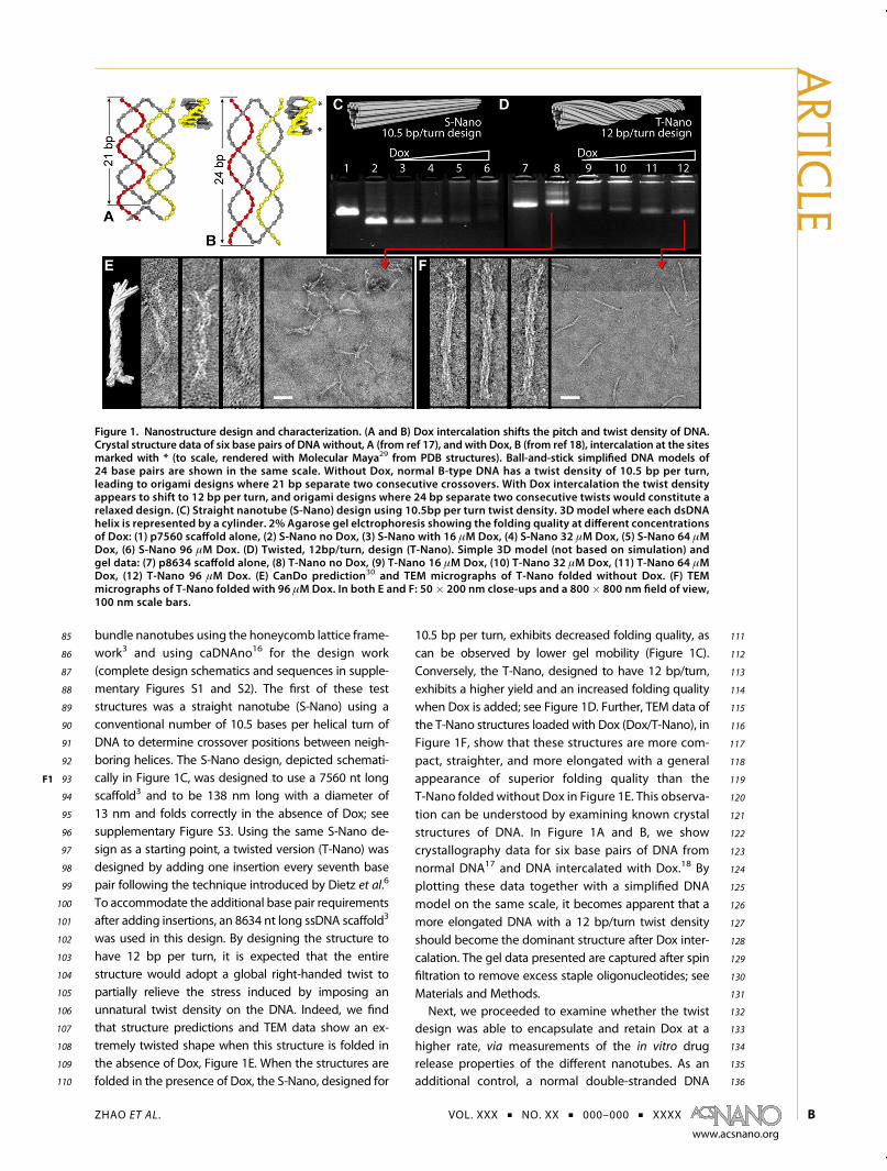
137 sample (dsDNA) was also used to verify if the nano-138 structured DNA exhibited any different properties than139 just plain double-stranded DNA, previously suggested140 as a carrier for Dox.19,20 For this experiment a sample of141 M13mp18 RF DNAwas used as a negative control since142 the sequence of this DNA to a large extent is the same143 as the sequence in the different scaffold DNA samples144 used. The in vitro release rate of Dox from the struc-145 tures was studied by measuring the Dox fluorescence146 at the other side of a semipermeable membrane (see147 schematic in Figure 2F2 A). One can see large differences148 in the release kinetics when comparing the T-Nano149 design with the S-Nano design. The T-Nano design150 manages to retain the drug to a greater extent and151 exhibits a slower release profile. Importantly, 50% of152 the Dox still remains bound to the T-Nano after several153 hours. In contrast, the straight, S-Nano design shows154 little or no significant difference in drug retention155 when compared to a simple, nonstructured dsDNA156 sample. The inset in Figure 2A shows the release data157 plotted in a log�log diagram. After analyzing the data158 using theHiguchimodel21 and the Korsmeyer�Peppas159 model,22 the r2 values of the Higuchi fit were found160 to be less than 0.9 (corresponding plot can be found161 in supplementary Figure S6), and the best fit for162 the various DNA samples could be obtained using163 Korsmeyer�Peppas' model for kinetic release. In this164 model the fraction of released drug is exponential165 with respect to time and the release time-exponent is
166dependent on the type of diffusion displayed by the167material. From the log�log plot we find that the slopes168of the release curves are 0.16, 0.31, and 0.79 for the169Dox/S-Nano, Dox/dsDNA, andDox/T-Nano, respectively,170indicating that the Dox/dsDNA and Dox/S-Nano171tended to exhibit Fickian diffusion characteristics,172whereas the Dox/T-Nano indicated a non-Fickian173release behavior.23
174The loading capacity, or encapsulation efficiency, of175the two types of nanostructured DNA was also mea-176sured and are presented in Figure 2B. We observe177that the T-Nano is able to encapsulate significantly178more Dox per structure compared to the S-Nano. The179difference in loading capacity is 33% higher for180T-Nano at a 96 μM loading concentration compared181to S-Nano. This increase can only partially be ex-182plained by the fact that T-Nano contains 14% more183base pairs per structure, but apart from that, the184increased loading capacity is probably an effect of a185higher affinity for Dox in the 12bp/turn design due to186the relaxation of the structure as Dox is intercalated187(see Figure 1).188After in vitro release for 24 h, the T-Nano structures189look very similar (see Figure 2C) to the structures folded190without Dox (Figure 1E). Moreover, gel analysis of the191Dox/T-Nano complexes strongly suggests that the struc-192tures are stable in the pH range used in the cell-based193assays (Figure 2D). By looking at the stability of the194T-Nano without loaded Dox in a gel shift assay, it was
Figure 2. DNA nanostructures as a drug delivery carrier. (A) The release rates of the different carriers were measured bymeasuring the fluorescence intensity of Dox after diffusion through a dialysis membrane permeable only to small molecules.Linear plot where the release curve of free Dox is shown as a reference baseline. Inset: log�log plot of the same release datawith a linear regression fit. S-Nano: green; dsDNA: blue; and T-Nano: red. (B) Loading capacity of the T-Nano vs the S-Nanostructures with linear regression. The concentration on the x-axis is used to equilibrate the structures with Dox. Immediatelyafter washing away the excess Dox, the amount of Dox bound to the nanostructures ismeasured and plotted on the y-axis. (C)TEM micrographs of T-Nano folded in 96 μM Dox after in vitro release for 24 h. 50 � 200 nm close-up and 350 � 350 nmoverviewwith 100 nm scale bar. (D) 2% agarose gel electrophoresis image showing the stability of the Dox-loaded T-Nano invarious pH buffers for 30 min: (1) 1 kb ladder, (2) p8634 scaffold alone, Dox/T-Nano in (3) pH 4.0, (4) pH 5.0, (5) pH 6.0, (6) pH7.0, (7) pH 7.8, (8) pH 9.0, (9) pH 10. (E) 2% agarose gel showing the T-Nano (no Dox) stability in cell culturemediumwith 10%fetal bovine serum: (10) p8634 scaffold alone, (11) T-Nano not incubated, (12) 30min incubation, (13) 1 h, (14) 3 h, (15) 6 h, (16)12 h, (17) 24 h, (18) 48 h incubation.
ARTIC
LE
ZHAO ET AL . VOL. XXX ’ NO. XX ’ 000–000 ’ XXXX
www.acsnano.org
C
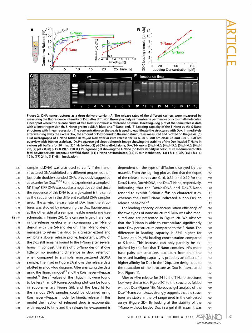
195 possible to rule out gel shift and the implied struc-196 tural change attributable to Dox release. The T-Nano197 structure seems to be stable over the time spans198 used in this report; see Figure 2E (see supplementary199 Figure S7 for a gel of Dox-loaded T-Nano over the200 same time span). Interestingly, the S-Nano structure201 appears not to reversibly return to its ground state202 after 24 h release as T-Nano does; see supplementary203 Figure S3 for additional TEM images and agarose204 gels of the structures at various stages after release205 of Dox.206 Similarly to most controlled release systems24 and207 as verified by our release rate mesurements, Dox will208 start to diffuse out of the nanostructures immediately209 upon transfer to an environment with a low Dox210 concentration, such as the cell culture. But because211 of the retention capabilities of the T-Nano, the Dox is212 released primarily at the cells due to cell binding and213 uptake of the nanostructures. The current opinion is214 that DNA structures get degraded after endocytosis215 followed by the release of Dox inside endocytotic216 vesicles.14 Our data, although not sufficient to directly217 support this effect, are consistent with such an218 interpretation.219 The cytotoxicity of Dox/T-Nano was examined by220 the sulforhodamine B colorimetric assay (SRB).25 Three221 breast cancer cell lines were examined: MDA-MB-231,
222MDA-MB-468, and MCF-7. The cell viability was mea-223sured after 48 h incubations with drug or drug-224loaded nanostructures. As can be seen in Figure 3 F3,225the half-maximal inhibitory concentration, IC50, of226Dox is significantly lower when delivered encapsu-227lated in T-Nano structures than when added as free228Dox. This fact, together with the relatively slow229release profile of the T-Nano, is encouraging, as it230constitutes a solid foundation for the continued231development of a nanostructure-based targeted232delivery system for cancer therapy. It should be233noted that the Dox/S-Nano and Dox/dsDNA showed234higher IC50 values in the cases where these could be235compared with Dox/T-Nano (see supplementary236Figure S4 B). Since these systems also exhibited237faster release profiles and less loading (Figure 2A238and B), we maintain that a Dox/DNA nanostructure239delivery system not using underwinding like in240T-Nano is no more efficient than a system based on241simple double-stranded plasmid DNA. Empty nano-242tubes without Dox were nontoxic (data shown in243supplementary Figure S4 A).244Confocal microscopy was performed to investigate245cellular uptake of the Dox/T-Nano system. To follow246the nanostructures, one of the staple oligonucleo-247tides was labeled with Alexa 488. Since DAPI binds to248DNA, and would thus stain our nanostructures, we
Figure 3. Cytotoxicity of free Dox and Dox/T-Nano against MDA-MB-231 cells (A), MDA-MB-468 cells (B), and MCF-7 cells(C) after 48 h incubation. The half-maximal inhibitory concentration, IC50, values (D) were calculated by GraphPad Prismusing nonlinear regression analysis. The sulforhodamine B (SRB) assay suggested that Dox/T-Nano was more cytotoxicthan free Dox to breast cancer cells. t test: ***p < 0.001, **p < 0.01 when compared to free Dox. Data represent mean ( SD(n = 4).
ARTIC
LE
ZHAO ET AL . VOL. XXX ’ NO. XX ’ 000–000 ’ XXXX
www.acsnano.org
D

249 decided not to use DAPI for nuclear staining after250 verifying this fact. Results from the confocal imaging251 can be found in Figure 4F4 A, and the Alexa 488-labeled252 T-Nano is clearly taken up by the cells. Moreover, the253 Dox/T-Nano seems to efficiently deliver Dox to the254 cell nuclei.255 Similarly to earlier studies of DNA nanostructure256 uptake by cells,11,26,27 only one of the components257 was labeled (in this case, one staple with Alexa 488), so258 similarly to these reports, the data do not exclude a259 scenario where the structures would be bound to the260 surface, degraded, or otherwise destabilized followed261 by uptake of some of the components. Nevertheless,262 the data are also consistent with the hypothesis that263 large (g7kb) DNA nanostructures are completely en-264 docytosed by mammalian cells,11 and our opinion is265 that this is the most likely scenario.266 To quantitatively investigate the uptake of Dox in267 the case of free drug or bound in Dox/T-Nano, living268 cells in culture were examined by measuring the Dox269 fluorescence intensity after incubation for 0.5 or 2 h270 (see Figure 4B). After these relatively short incubation
271times, the levels of intracellular Dox appear to be272higher in the case of free drug than when delivered273in T-Nano. However, if excess Dox/T-Nano or free Dox274is washed away after 2 h, the intracellular levels of Dox275are significantly higher in the case of Dox/T-Nano276after continued growth in fresh culturemedium for 12277or 24 h (see Figure 4C). This series of observations278indicate that the cellular elimination is lower for Dox279bound to T-Nano compared to the free drug. Our280interpretation of the results is that free Dox as well as281Dox/T-Nano is endocytosed as discussed above, fol-282lowed by diffusion of the small Dox molecules from283the endosomes to the cytosol and finally to the284nucleus. The difference in depletion rates for the free285drug and Dox/T-Nano can most likely be attributed to286an intracellular depot effect. Here we hypothesize287that the DNA nanostructures are slowly degraded in288the endosomes and the Dox gets released over an289extended period of time, with the endosomes acting290as local depots. In contrast, free Dox would get291transported in quickly, but reversely it would also292diffuse out of the cells at a higher rate. This is,
Figure 4. Internalization and intracellular Dox level after incubation with free Dox and Dox/T-Nano. (A) Confocal microscopyimages of monolayer MDA-MB-231 cells exposed to free Dox and Dox-loaded Alexa 488-labeled twisted nanotubes for 2 h.Confocal images certified that Dox could be delivered to the nucleus through Dox/T-Nano. (B) Intracellular Dox level in MDA-MB-231 cells, MDA-MB-468 cells, and MCF-7 cells exposed to free Dox and Dox/T-Nano for 0.5 and 2 h. (C) Elimination ofintracellular Dox.MDA-MB-231 cells,MDA-MB-468 cells, andMCF-7 cellswere incubatedwith freeDox andDox/T-Nano for 2 hand then continued to be cultured in fresh medium (absent Dox). The Dox level was determined at 12 and 24 h. Theconcentration of Dox (free or equivalent) in cell culture was 5 μM in all the experiments. t test: **p < 0.01, *p < 0.05 whencompared with free Dox. Data represent mean ( SD (n = 3).
ARTIC
LE
ZHAO ET AL . VOL. XXX ’ NO. XX ’ 000–000 ’ XXXX
www.acsnano.org
E
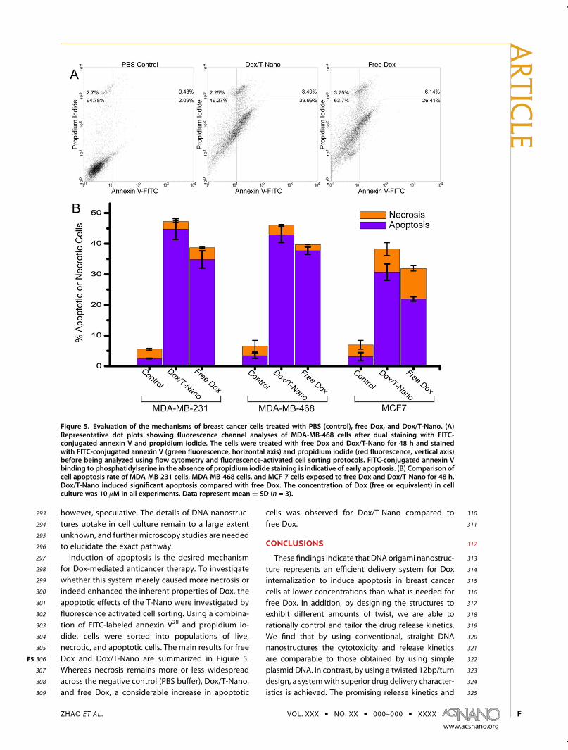
293 however, speculative. The details of DNA-nanostruc-294 tures uptake in cell culture remain to a large extent295 unknown, and further microscopy studies are needed296 to elucidate the exact pathway.297 Induction of apoptosis is the desired mechanism298 for Dox-mediated anticancer therapy. To investigate299 whether this system merely caused more necrosis or300 indeed enhanced the inherent properties of Dox, the301 apoptotic effects of the T-Nano were investigated by302 fluorescence activated cell sorting. Using a combina-303 tion of FITC-labeled annexin V28 and propidium io-304 dide, cells were sorted into populations of live,305 necrotic, and apoptotic cells. The main results for free306 Dox and Dox/T-Nano are summarized in Figure 5F5 .307 Whereas necrosis remains more or less widespread308 across the negative control (PBS buffer), Dox/T-Nano,309 and free Dox, a considerable increase in apoptotic
310cells was observed for Dox/T-Nano compared to311free Dox.
312CONCLUSIONS
313These findings indicate that DNA origami nanostruc-314ture represents an efficient delivery system for Dox315internalization to induce apoptosis in breast cancer316cells at lower concentrations than what is needed for317free Dox. In addition, by designing the structures to318exhibit different amounts of twist, we are able to319rationally control and tailor the drug release kinetics.320We find that by using conventional, straight DNA321nanostructures the cytotoxicity and release kinetics322are comparable to those obtained by using simple323plasmid DNA. In contrast, by using a twisted 12bp/turn324design, a systemwith superior drug delivery character-325istics is achieved. The promising release kinetics and
Figure 5. Evaluation of the mechanisms of breast cancer cells treated with PBS (control), free Dox, and Dox/T-Nano. (A)Representative dot plots showing fluorescence channel analyses of MDA-MB-468 cells after dual staining with FITC-conjugated annexin V and propidium iodide. The cells were treated with free Dox and Dox/T-Nano for 48 h and stainedwith FITC-conjugated annexin V (green fluorescence, horizontal axis) and propidium iodide (red fluorescence, vertical axis)before being analyzed using flow cytometry and fluorescence-activated cell sorting protocols. FITC-conjugated annexin Vbinding to phosphatidylserine in the absence of propidium iodide staining is indicative of early apoptosis. (B) Comparison ofcell apoptosis rate of MDA-MB-231 cells, MDA-MB-468 cells, and MCF-7 cells exposed to free Dox and Dox/T-Nano for 48 h.Dox/T-Nano induced significant apoptosis compared with free Dox. The concentration of Dox (free or equivalent) in cellculture was 10 μM in all experiments. Data represent mean ( SD (n = 3).
ARTIC
LE
ZHAO ET AL . VOL. XXX ’ NO. XX ’ 000–000 ’ XXXX
www.acsnano.org
F
326 cytotoxicity of the Dox/T-Nano system, in combination327 with the well-known flexibility of the DNA origami328 method to decorate the structures with targeting
329ligands,10 make this a promising candidate platform330for active targeting of nanostructures intended for331anticancer therapy.332
333 MATERIALS AND METHODS334 Folding of DNA Origami Shapes. Each sample was prepared by335 combining a 5 nM scaffold (p7560 or p8634, derived from336 M13mp18), 25 nM of each staple oligonucleotide, buffer, and337 salts including 5 mM Tris, 1 mM EDTA (pH 7.8 at 20 �C), and338 10 mM MgCl2. Folding was carried out by rapid heat denatura-339 tion followed by slow cooling from 80 to 60 �C over 80min, then340 60 to 24 �C over 173 h. To remove the excess staples and/or free341 doxorubicin, the DNA nanostructure suspensions were added342 to VIVAspin 500 (molecular cutoff of 100 kD, Sartorius Stedim343 Biotech GmbH) and centrifuged at 3000g for 30min followed by344 suspension in Tris buffer (5 mM Tris, 1 mM EDTA, and 13 mM345 MgCl2). Samples were electrophoresed on 2% agarose gels346 (0.5� TBE, 11 mM MgCl2, 0.5 μg/mL ethidium bromide) at 70 V347 for 3 h in an ice�water bath.348 Electron Microscopy. A 3 μL aliquot of Dox/T-NanoT sample349 was spotted on a glow-discharged, carbon-coated Formvar grid350 (Electron Microscopy Sciences), incubated for 20 s, blotted off351 with filter paper, and then stainedwith 2% (w/v) aqueous uranyl352 formate solution. EM analysis was performed using a FEI353 Morgagni 268(D) transmission electron microscope at 80 kV354 with nominal magnifications between 12 000 and 44 000.355 Images were recorded digitally by using the Advanced Micro-356 scopy Techniques Image Capture Engine 5.42.357 Stability of Doxorubicin-Intercalated Nanotube. TheDox-intercalated358 nanotube was incubated with culture medium (including 10%359 fetal bovine serum) at 37 �C for 1, 15, 30, 60, 120, and 240 min360 or in various pH (4.0, 5.0, 6.0, 7.0, 7.8, 9.0, and 10.0) Tris buffer361 (5 mM Tris, 1 mM EDTA) for 30 min, and then the stability was362 verified using agarose (2%) gel electrophoresis (0.5� TBE, 11mM363 MgCl2, 0.5 μg/mL ethidium bromide) at 70 V for 3 h in an ice�364 water bath.365 Drug Intercalation Efficiency. To determine Dox intercalaction366 efficiency, 60 μL of Dox-intercalated nanotube was added into367 VIVaspin 500 (molecular cutoff of 100 kD, Biotech) and centri-368 fuged at 3000g for 30 min. Dox content in the filtrate as free369 drug was measured using a microplate reader. The drug inter-370 calaction efficiency (%) was calculated by the following equa-371 tion: Intercalaction efficiency (%) = (Dtotal � Dfree)/Dtotal � 100.372 Dtotal is the Dox content in Dox-intercalated nanotube solution;373 Dfree is the Dox content in the filtrate.374 In Vitro Drug Release Behaviors of Doxorubicin-Intercalated Nanotube.375 The dialysis units with molecular weight cutoff of 20 000 Da376 were used to carry out the drug release experiments. Phosphate-377 buffered saline (PBS, pH = 7.4) was used as the drug release378 media. The sample Dox-intercalated nanotube or Dox solution379 (30 μL) was put into the dialysis unit. The sealed dialysis units380 were put into a polystyrene flotation device, floating on a381 surface of 1000 mL of release media in a beaker. The release382 medium was stirred at a speed of 200 rpm at 37 �C protected383 from light. At certain time intervals, the dialysis unit was taken384 out and 10 μL of Dox-intercalated nanotube was taken out from385 the dialysis unit for measuring the nonreleased drug concen-386 trations by the microplate reader.387 In Vitro Cytotoxicity Assay. The cytotoxicity of empty nano-388 tubes, free Dox, Dox/S-Nano, Dox/T-Nano, and Dox/dsDNA was389 tested against breast cancer cell lines MCF-7, MDA-MB-231, and390 MDA-MB-468 (MCF-7 human breast adenocarcinoma cell line391 ER pos., 231 adenocarcinoma ER neg., 468 adenocarcinoma ER392 neg.). The cells were obtained from ATCC (American Tissue393 Culture Collection, Manassas, VA, USA) and maintained accord-394 ing to instructions. The cytotoxicity was evaluated by the395 sulforhodamine B cell cytotoxicity assay (G-Biosciences, St396 Louis, MO, USA). Cells were cultured in Dulbecco's modified397 Eagle medium supplemented with 10% fetal bovine serum,398 100 units/mL penicillin, and 100mg/mL streptomycin under 5%399 CO2 at 37 �C and collected by trypsinization using 0.25% trypsin
400solution. Cells were seeded in 96-well plates (Costa, Corning401Incorporated) at a density of 1� 104 cells/well and incubated for40224 h to allow for cell attachment. The cells were incubated with403Dox/T-Nano or Dox at equivalent drug concentrations ranging404from 50 to 15 000 nM for 48 h. At the end of the experiment, the405SRB assay was performed according to the manufacturer's406instructions. The absorbance was measured at 565 nm using a407microplate reader. Cell viability rate was calculated by the408following equation:
Cell viability rate (%) ¼ (Adrug � Ablank)=(Acontrol � Ablank)� 100
409Adrug is the absorbance of the cells incubated with Dox/T-Nano410or Dox; Acontrol is the absorbance of the cells incubated with the411culturemedium only; and Ablank is the absorbance of the culture412medium.413Intracellular Uptake of Doxorubicin-Intercalated Twisted Nanotube.414For the Dox/T-Nano uptake study, 12-well plates were seeded415with breast cancer cells at 2 � 105 per well, and the cells were416allowed to attach for 24 h. The medium was replaced with 1 mL417of medium containing Dox/T-Nano or Dox solution (final Dox418concentration 5 μM) and incubated for 0.5 and 2 h. Cells were419washed three times to remove the free Dox/T-Nano or Dox with420PBS buffer and lysed in 100 μL of cell lysis buffer for 10 min.421A 10 μL amount of cell lysate was used to quantitate protein422concentration by the BCA assay. The remaining portion was423extracted by dissolving each sample in 0.2 mL of acidified424methanol (0.1 M HCL, 90% methanol) solution, and the super-425natant was analyzed for Dox level using the microplate reader426(excitation wavelength: 485 nm, emission wavelength: 591 nm)427after centrifugation at 16800g for 10 min. The data were428normalized to per milligram cell protein.429Intracellular Elimination of Doxorubicin. For the Dox/T-Nano430elimination study, 12-well plates were seeded with breast431cancer cells at 2 � 105 per well, and the cells were allowed to432attach for 24 h. Themediumwas replaced with 1mL of medium433containingDox/T-Nano or Dox solution (final Dox concentration4345 μM) and incubated for 2 h. Cells were washed three times to435remove the free Dox or Dox/T-Nano with PBS buffer and436continued to incubate with fresh medium. Intracellular Dox437level was determined at 12 and 24 h using the same method as438that described above for intracellular uptake assay.439Internalization of Doxorubicin-Intercalated Twisted Nanotube. Con-440focal laser scanningmicroscopy (CLSM, Olympus FV1000) further441confirmed the cellular uptake of Dox/T-Nano. MDA-MB-231 cells442were plated at a density of 1 � 105 cells/well containing 22 mm443sterile coverslips for 24 h. Dox and the Dox/T-Nano with equiva-444lent Dox concentration (5 μM) were incubated for 2 h, and445then the cells were washed three times with PBS and fixed with4463.7% formaldehyde in PBS. Vectashield (Vector Laboratories, Inc.447Burlingame, CA, USA) was dropped on the slides to seal the cell448samples after the cells were washed three times with PBS. The449stained coverslips were imaged using CLSM.450Cell Apoptosis Analysis. Apoptotic cells were determined by451dual staining with an annexin V-FITC and propidium iodide kit452(Invitrogen, CA, USA) according to the manufacturer's instruc-453tions. In brief, the breast cancer cells were seeded into six-well454culture plates at a concentration of 1 � 106 cells/well and455incubated for 24 h to allow cell attachment. The cells were456pretreated with Dox, Dox/T-Nano at equivalent drug concen-457trations (10 μM), or empty nanotubes for 48 h and collected by458trypsinization using a 0.125% trypsin solution. After that, the459cells were washed twice with PBS (pH = 7.4) and resuspended in460100 μL of binding buffer at a density of 1� 105/mL. Then 2 μL of461annexin V-FITC was added, and cells were incubated at room462temperature for 20 min in darkness. Then 5 μL of propidium463iodide stock solution (100 μg/mL) was added after adding464400 μL of PBS, and cells were incubated for another 5 min in
ARTIC
LE
ZHAO ET AL . VOL. XXX ’ NO. XX ’ 000–000 ’ XXXX
www.acsnano.org
G
465 darkness. The cells were analyzed by flow cytometry (Becton466 Dickinson, Sunnyvale, CA, USA) with CellQuest software within467 1 h. The numbers of cells undergoing necrosis (positive for468 propidium iodide), early apoptosis (positive for annexin V), and469 late apoptosis (double-positive for annexin V and propidium470 iodide) stages of apoptosiswere quantified using flow cytometry.471 Statistical Analysis. All datawere expressed asmean( SD. IC50472 values were calculated by GraphPad Prism using nonlinear473 regression analysis. The statistical significance was determined474 using a t test. Ap value less than0.05 (i.e.,p<0.05)was considered475 to indicate statistical significance for all comparisons.476 Conflict of Interest: The authors declare no competing477 financial interest.
478 Supporting Information Available: Structure designs, DNA479 sequences, additional TEM images, cytotoxicity data, and flow480 cytometry data are available free of charge via the Internet at481 http://pubs.acs.org.
482 Acknowledgment. The project was funded by the Swedish483 Research Council (VR) through a repatriation grant and a project484 grant to B.H. (grants 2010-6296 and 2010-5060). B.H. and A.M.N.485 are recipients of assistant professorships with startup funding486 funded by Carl Bennet AB, Karolinska Institutet, and Vinnova. A.487 S. is partially funded by a Karolinska Institutet faculty grant (KID).488 Funding support to A.M.N. by the Royal Swedish Academy of489 Sciences, Falk Foundation, Jeanssons Foundation, Axel and Eva490 Wallströms Foundation, and the Swedish Research Council (VR)491 under grants 2011-3720 and 2009-3259 is acknowledged. Y.X.Z.492 is funded by the China Scholarship Council (CSC). We acknowl-493 edge Klas Udekwu and Ana Teixeira for helpful discussions and494 Xiaoyuan Ren for helping Y.X.Z. get started.
495 REFERENCES AND NOTES496 1. Seeman, N. C. Nucleic-Acid Junctions and Lattices. J. Theor.497 Biol. 1982, 99, 237–247.498 2. Rothemund, P. W. K.; Papadakis, N.; Winfree, E. Algorithmic499 Self-Assembly of DNA Sierpinski Triangles. PLoS Biol. 2004,500 6, 240–252.501 3. Douglas, S. M.; Dietz, H.; Liedl, T.; Högberg, B.; Graf, F.;502 Shih, W. M. Self-Assembly of DNA into Nanoscale Three-503 Dimensional Shapes. Nature 2009, 459, 414–418.504 4. Rothemund, P. W. K. Folding DNA to Create Nanoscale505 Shapes and Patterns. Nature 2006, 440, 297–302.506 5. Liedl, T.; Högberg, B.; Tytell, J.; Ingber, D. E.; Shih, W. M. Self-507 Assembly of Three-Dimensional Prestressed Tensegrity508 Structures from DNA. Nat. Nanotechnol. 2010, 5, 520–524.509 6. Dietz, H.; Douglas, S. M.; Shih, W. M. Folding DNA into510 Twisted and Curved Nanoscale Shapes. Science 2009, 325,511 725–730.512 7. Högberg, B.; Liedl, T.; Shih, W. M. Folding DNA Origami513 from a Double-Stranded Source of Scaffold. J. Am. Chem.514 Soc. 2009, 131, 9154–55.515 8. Douglas, S. M.; Bachelet, I.; Church, G. M. A Logic-Gated516 Nanorobot for Targeted Transport of Molecular Payloads.517 Science 2012, 335, 831–834.518 9. Andersen, E. S.; Dong, M.; Nielsen, M. M.; Jahn, K.;519 Subramani, R.; Mamdouh, W.; Golas, M. M.; Sander, B.;520 Stark, H.; Oliveira, C. L. P.; et al. Self-Assembly of a Nano-521 scale DNA Box with a Controllable Lid. Nature 2009, 459,522 73–76.523 10. Peer, D.; Karp, J. M.; Hong, S.; Farokhzad, O. C.; Margalit, R.;524 Langer, R. Nanocarriers as an Emerging Platform for525 Cancer Therapy. Nat. Nanotechnol. 2007, 2, 751–760.526 11. Schüller, V. J.; Heidegger, S.; Sandholzer, N.; Nickels, P. C.;527 Suhartha, N. A.; Endres, S.; Bourquin, C.; Liedl, T. Cellular528 Immunostimulation by CpG-Sequence-Coated DNA529 Origami Structures. ACS Nano 2011, 5, 9696–9702.530 12. Arora, H. C.; Jensen, M. P.; Yuan, Y.; Wu, A.; Vogt, S.;531 Paunesku, T.; Woloschak, G. E. Nanocarriers Enhance532 Doxorubicin Uptake in Drug-Resistant Ovarian Cancer533 Cells. Cancer Res. 2012, 72, 769–778.534 13. Kellogg, G. E.; Scarsdale, J. N.; Fornari, F. A., Jr. Identification535 and Hydropathic Characterization of Structural Features
536Affecting Sequence Specificity for Doxorubicin Intercala-537tion into DNA Double-Stranded Polynucleotides. Nucleic538Acids Res. 1998, 26, 4721–4732.53914. Chang, M; Yang, C.-S.; Huang, D.-M. Aptamer-Conjugated540DNA Icosahedral Nanoparticles As a Carrier of Doxorubicin541for Cancer Therapy. ACS Nano 2011, 8, 6156–6163.54215. Ke, Y.; Bellot, G.; Voigt, N. V.; Fradkov, E.; Shih, W. M. Two543Design Strategies for Enhancement of Multilayer�DNA-544Origami Folding: Underwinding for Specific Intercalator545Rescue and Staple-Break Positioning. Chem. Sci. 2012, 3,5462587–2597.54716. Douglas, S. M.; Marblestone, A. H.; Teerapittayanon, S.;548Vazquez, A.; Church, G.M.; Shih,W.M. Rapid Prototyping of5493D DNA-Origami Shapes with caDNAno. Nucleic Acids Res.5502009, 37, 5001–5006.55117. Ortiz-Lombardia, M.; Gonzalez, A.; Eritja, R.; Aymami, J.;552Azorin, F.; Coll, M. Crystal Structure of a DNA Holliday553Junction. Nat. Struct. Biol. 1999, 6, 913–917.55418. Frederick, C. A.; Williams, L. D.; Ughetto, G.; van der Marel,555G. A.; van Boom, J. H.; Rich, A.; Wang, A. H. Structural556Comparison of Anticancer Drug-DNA Complexes: Adria-557mycin and Daunomycin. Biochemistry 1990, 29, 2538–5582549.55919. Bagalkot, V.; Lee, I.-H.; Yu, M. K.; Lee, E.; Park, S.; Lee, J.-H.;560Jon, S. A Combined Chemoimmunotherapy Approach561Using a Plasmid-Doxorubicin Complex.Mol. Pharmaceutics5622009, 6, 1019–1028.56320. Bagalkot, V.; Farokhzad, O. C.; Langer, R.; Jon, S. An564Aptamer�Doxorubicin Physical Conjugate as a Novel565Targeted Drug-Delivery Platform. Angew. Chem., Int. Ed.5662006, 45, 8149–8152.56721. Higuchi, T. Mechanism of Sustained-Action Medication:568Theoretical Analysis of Rate of Release of Solid Drugs569Dispersed in Solid Matrices. J. Pharm. Sci. 1963, 52,5701145–1149.57122. Korsmeyer, R. W.; Gurnya, R.; Doelkera, E.; Buria, P.; Peppas,572N. A. Mechanisms of Solute Release from Porous Hydro-573philic Polymers. Int. J. Pharm. 1983, 15, 25–35.57423. Lin, L. Y.; Lee, N. S.; Zhu, J.; Nyström, A. M.; Pochan, D. J.;575Dorshow, R. B.; Wooley, K. L. Tuning Core vs. Shell Dimen-576sions to Adjust the Performance of Nanoscopic Containers577for the Loading and Release of Doxorubicin. J. Controlled578Release 2011, 152, 37–48.57924. Charrois, J. R.; Allen, T. M. Drug Release Rate Influences the580Pharmacokinetics, Biodistribution, Therapeutic Activity,581and Toxicity of Pegylated Liposomal Doxorubicin Formu-582lations in Murine Breast Cancer. Biochim. Biophys. Acta5832004, 1663, 167–177.58425. Skehan, P.; Storeng, R.; Scudiero, D.; Monks, A.; McMahon,585J.; Vistica, D.; Warren, T. W.; Bokesch, H.; Kenney, S.; Boyd,586M. R. New Colorimetric Cytotoxicity Assay for Anticancer-587Drug Screening. J. Nat. Cancer Inst. 1990, 82, 1107–1112.58826. Hamblin, G. D.; Carneiro, K. M. M.; Fakhoury, J. F.; Bujold,589K. E.; Sleiman, H. F. Rolling Circle Amplification-Templated590DNA Nanotubes Show Increased Stability and Cell Pene-591tration Ability. J. Am. Chem. Soc. 2012, 134, 2888–2891.59227. Ko, S.-H.; Liu, H.; Chen, Y.; Mao, C. DNA Nanotubes as593Combinatorial Vehicles for Cellular Delivery. Biomacromo-594lecules 2008, 9, 3039–3043.59528. Koopman, G.; Reutelingsperger, C. P.; Kuijten, G.A.; Keehnen,596R. M.; Pals, S. T.; van Oers, M. H. Annexin V for Flow597Cytometric Detection of Phosphatidylserine Expression on598B Cells Undergoing Apoptosis. Blood 1994, 84, 1415–1420.59929. www.molecularmovies.com.60030. Castro, C. E.; Kilchherr, F; Kim, D.-N.; Shiao, E. L.; Wauer, T;601Wortmann, P.; Bathe, M.; Dietz, H. A Primer to Scaffolded602DNA Origami. Nat. Methods 2011, 8, 221–229.
ARTIC
LE
ZHAO ET AL . VOL. XXX ’ NO. XX ’ 000–000 ’ XXXX
www.acsnano.org
H