dna origami-enabled biosensors
TRANSCRIPT
sensors
Review
DNA Origami-Enabled Biosensors
Shuang Wang 1,2,†, Zhaoyu Zhou 2,†, Ningning Ma 2, Sichang Yang 2, Kai Li 1,2, Chao Teng 1,*,Yonggang Ke 3 and Ye Tian 2,4
1 Institute of Marine Biomedicine, Shenzhen Polytechnic, Shenzhen 518055, China; [email protected] (S.W.);[email protected] (K.L.)
2 State Key Laboratory of Analytical Chemistry for Life Science, College of Engineering and Applied Sciences,Nanjing University, Nanjing 210023, China; [email protected] (Z.Z.);[email protected] (N.M.); [email protected] (S.Y.); [email protected] (Y.T.)
3 Wallace H. Coulter Department of Biomedical Engineering, Georgia Institute of Technology andEmory University, Atlanta, GA 30322, USA; [email protected]
4 Shenzhen Research Institute of Nanjing University, Shenzhen 518000, China* Correspondence: [email protected]† These authors contributed equally to this work.
Received: 23 November 2020; Accepted: 30 November 2020; Published: 3 December 2020 �����������������
Abstract: Biosensors are small but smart devices responding to the external stimulus, widely used inmany fields including clinical diagnosis, healthcare and environment monitoring, etc. Moreover, there isstill a pressing need to fabricate sensitive, stable, reliable sensors at present. DNA origami technologyis able to not only construct arbitrary shapes in two/three dimension but also control the arrangement ofmolecules with different functionalities precisely. The functionalization of DNA origami nanostructureendows the sensing system potential of filling in weak spots in traditional DNA-based biosensor.Herein, we mainly review the construction and sensing mechanisms of sensing platforms based onDNA origami nanostructure according to different signal output strategies. It will offer guidance forthe application of DNA origami structures functionalized by other materials. We also point out somepromising directions for improving performance of biosensors.
Keywords: DNA origami; sensor; DNA nanotechnology; FRET; chirality; SERS; nanopore
1. Introduction
1.1. DNA Biosensors
Biosensors, as a kind of product of interdisciplinary combinations such as chemistry, physics,medicine, and engineering, are able to complete rapid analysis and tracking for targets with highspecificity, sensitivity, and low cost. They are widely applied in various fields including clinicaldiagnosis, food and drug analysis, healthcare, immunology, and environmental monitoring [1–9].Its core components consist of a recognition unit (establishes a relationship with targets) and a transducerunit (converts the interaction between sensors and analyte into a measurable physical signals such asoptical, electrochemical, piezoelectric, thermal, mechanical, acoustic, and magnetic signals [10–13]).The main materials used in biosensors include enzymes, proteins, DNAs, antibodies and antigens, cells,tissues, etc. All of them work as sensitive elements to recognize analyte. According to the differentmaterials, sensors are divided into enzyme-based, cell-based, DNA-based, and tissue-based sensorsand immunosensors, etc.
DNA-based sensors have got substantial development in recent years because DNA has numerousmerits such as good stability and recognizable ability, strong predictability, excellent programmability,and easy synthesis. As the development of DNA nanotechnology, structural polymorphisms further
Sensors 2020, 20, 6899; doi:10.3390/s20236899 www.mdpi.com/journal/sensors
Sensors 2020, 20, 6899 2 of 18
enriched the biosensors. The biosensing platform developed gradually from simple duplex to morecomplex three-dimensional nanostructure as well as the function integrated from single target detectionto multiplex detection. Moreover, the performance got much better.
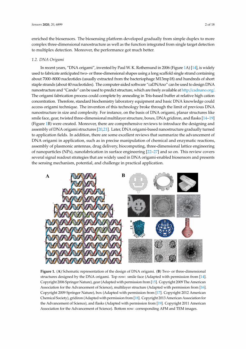
1.2. DNA Origami
In recent years, “DNA origami”, invented by Paul W. K. Rothemund in 2006 (Figure 1A) [14], is widelyused to fabricate anticipated two- or three-dimensional shapes using a long scaffold single strand containingabout 7000–8000 nucleotides (usually extracted from the bacteriophage M13mp18) and hundreds of shortstaple strands (about 40 nucleotides). The computer-aided software “caDNAno” can be used to design DNAnanostructure and “Cando” can be used to predict structure, which are freely available at http://cadnano.org/.The origami fabrication process could complete by annealing in Tris-based buffer at relative high cationconcentration. Therefore, standard biochemistry laboratory equipment and basic DNA knowledge couldaccess origami technique. The invention of this technology broke through the limit of previous DNAnanostructure in size and complexity. For instance, on the basis of DNA origami, planar structures likesmile face, gear, twisted three-dimensional multilayer structure, boxes, DNA gridiron, and flasks [14–19](Figure 1B) were created. Moreover, there are comprehensive reviews to introduce the designing andassembly of DNA origami structures [20,21]. Later, DNA origami-based nanostructure gradually turnedto application fields. In addition, there are some excellent reviews that summarize the advancement ofDNA origami in application, such as in precise manipulation of chemical and enzymatic reactions,assembly of plasmonic antennas, drug delivery, biocomputing, three-dimensional lattice engineeringof nanoparticles (NPs), nanofabrication in surface engineering [22–27] and so on. This review coversseveral signal readout strategies that are widely used in DNA origami-enabled biosensors and presentsthe sensing mechanism, potential, and challenge in practical application.
Sensors 2020, 20, x FOR PEER REVIEW 2 of 18
polymorphisms further enriched the biosensors. The biosensing platform developed gradually from simple duplex to more complex three-dimensional nanostructure as well as the function integrated from single target detection to multiplex detection. Moreover, the performance got much better.
1.2. DNA Origami
In recent years, “DNA origami”, invented by Paul W. K. Rothemund in 2006 (Figure 1A) [14], is widely used to fabricate anticipated two- or three-dimensional shapes using a long scaffold single strand containing about 7000–8000 nucleotides (usually extracted from the bacteriophage M13mp18) and hundreds of short staple strands (about 40 nucleotides). The computer-aided software “caDNAno” can be used to design DNA nanostructure and “Cando” can be used to predict structure, which are freely available at http://cadnano.org/. The origami fabrication process could complete by annealing in Tris-based buffer at relative high cation concentration. Therefore, standard biochemistry laboratory equipment and basic DNA knowledge could access origami technique. The invention of this technology broke through the limit of previous DNA nanostructure in size and complexity. For instance, on the basis of DNA origami, planar structures like smile face, gear, twisted three-dimensional multilayer structure, boxes, DNA gridiron, and flasks [14–19] (Figure 1B) were created. Moreover, there are comprehensive reviews to introduce the designing and assembly of DNA origami structures [20,21]. Later, DNA origami-based nanostructure gradually turned to application fields. In addition, there are some excellent reviews that summarize the advancement of DNA origami in application, such as in precise manipulation of chemical and enzymatic reactions, assembly of plasmonic antennas, drug delivery, biocomputing, three-dimensional lattice engineering of nanoparticles (NPs), nanofabrication in surface engineering [22–27] and so on. This review covers several signal readout strategies that are widely used in DNA origami-enabled biosensors and presents the sensing mechanism, potential, and challenge in practical application.
Figure 1. (A) Schematic representation of the design of DNA origami. (B) Two- or three-dimensional structures designed by the DNA origami. Top row: smile face (Adapted with permission from [14]. Copyright 2006 Springer Nature), gear (Adapted with permission from [15]. Copyright 2009 The American Association for the Advancement of Science), multilayer structure (Adapted with permission from [16]. Copyright 2009 Springer Nature), box (Adapted with permission from [17]. Copyright 2012 American Chemical Society), gridiron (Adapted with permission from [18]. Copyright 2013 American Association for the Advancement of Science), and flasks (Adapted with permission from [19]. Copyright 2011 American Association for the Advancement of Science). Bottom row: corresponding AFM and TEM images.
Figure 1. (A) Schematic representation of the design of DNA origami. (B) Two- or three-dimensionalstructures designed by the DNA origami. Top row: smile face (Adapted with permission from [14].Copyright 2006 Springer Nature), gear (Adapted with permission from [15]. Copyright 2009 The AmericanAssociation for the Advancement of Science), multilayer structure (Adapted with permission from [16].Copyright 2009 Springer Nature), box (Adapted with permission from [17]. Copyright 2012 AmericanChemical Society), gridiron (Adapted with permission from [18]. Copyright 2013 American Association forthe Advancement of Science), and flasks (Adapted with permission from [19]. Copyright 2011 AmericanAssociation for the Advancement of Science). Bottom row: corresponding AFM and TEM images.
Sensors 2020, 20, 6899 3 of 18
2. Sensing Strategies of DNA-Origami-Enabled Sensors
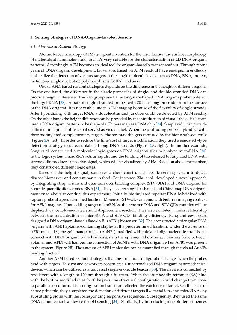
2.1. AFM-Based Readout Strategy
Atomic force microscopy (AFM) is a great invention for the visualization the surface morphologyof materials at nanometer scale, thus it’s very suitable for the characterization of 2D DNA origamipatterns. Accordingly, AFM becomes an ideal tool for origami-based biosensor readout. Through recentyears of DNA origami development, biosensors based on AFM readout have emerged in endlesslyand realize the detection of various targets at the single molecule level, such as DNA, RNA, protein,metal ions, single nucleotide polymorphisms (SNPs), and so on.
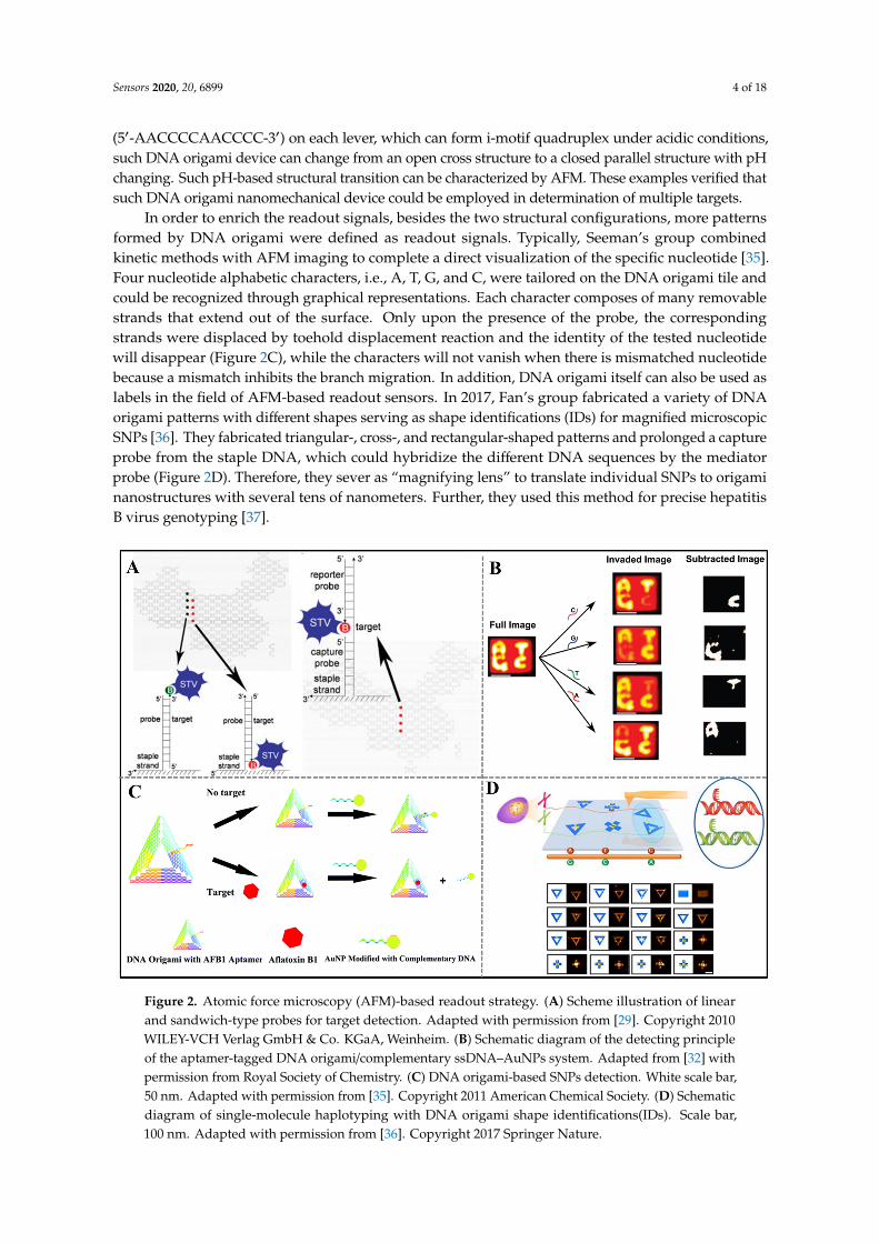
One of AFM-based readout strategies depends on the difference in the height of different regions.On the one hand, the difference in the elastic properties of single- and double-stranded DNA canprovide height difference. The Yan group used a rectangular-shaped DNA origami probe to detectthe target RNA [28]. A pair of single-stranded probes with 20-base long protrude from the surfaceof the DNA origami. It is not visible under AFM imaging because of the flexibility of single strands.After hybridizing with target RNA, a double-stranded junction could be detected by AFM readily.On the other hand, the height difference can be provided by the introduction of visual labels. He’s teamused a DNA origami pattern in the shape of a Chinese map as a DNA chip [29]. Streptavidin can providesufficient imaging contrast, so it served as visual label. When the protruding probes hybridize withtheir biotinylated complementary targets, the streptavidin gets captured by the biotin subsequently(Figure 2A, left). In order to reduce the time/cost of target modification, they used a sandwich-typedetection strategy to detect unlabeled long DNA strands (Figure 2A, right). In another example,Song et al. constructed a molecular logic gates on DNA origami tiles to analyze microRNA [30].In the logic system, microRNA acts as inputs, and the binding of the released biotinylated DNA withstreptavidin produces a positive signal, which will be visualized by AFM. Based on above mechanism,they constructed different logic gates.
Based on the height signal, some researchers constructed specific sensing system to detectdisease biomarker and contaminants in food. For instance, Zhu et al. developed a novel approachby integrating streptavidin and quantum dots binding complex (STV-QDs) and DNA origami foraccurate quantification of microRNA [31]. They used rectangular-shaped and China-map DNA origamimentioned above to conduct this experiment. Initially, biotinylated reporter DNA hybridized withcapture probe at a predetermined location. Moreover, STV-QDs can bind with biotin as imaging contrastfor AFM imaging. Upon adding target microRNAs, the reporter DNA and STV-QDs complex will bedisplaced via toehold-mediated strand displacement reaction. They also exhibited a linear relationshipbetween the concentration of microRNA and STV-QDs binding efficiency. Pang and coworkersdesigned a DNA origami-based aflatoxin B1 (AFB1) biosensor [32]. They constructed a triangular DNAorigami with AFB1 aptamer-containing staples at the predetermined location. Under the absence ofAFB1 molecules, the gold nanoparticles (AuNPs) modified with thiolated oligonucleotide strands canconnect with DNA origami by hybridizing with the aptamer. The stronger binding force betweenaptamer and AFB1 will hamper the connection of AuNPs with DNA origami when AFB1 was presentin the system (Figure 2B). The amount of AFB1 molecules can be quantified through the visual AuNPsbinding fraction.
Another AFM-based readout strategy is that the structural configuration changes when the probesbind with targets. Kuzuya and coworkers constructed a functionalized DNA origami nanomechanicaldevice, which can be utilized as a universal single-molecule beacon [33]. The device is connected bytwo levers with a length of 170 nm through a fulcrum. When the streptavidin tetramer (SA) bindwith the biotins modified in each of the jaws, the structural configuration could change from crossto parallel closed form. The configuration transition reflected the existence of target. On the basis ofabove principle, they completed the detection of different targets like metal ions and microRNAs bysubstituting biotin with the corresponding responsive sequences. Subsequently, they used the sameDNA nanomechanical device for pH sensing [34]. Similarly, by introducing nine binder sequences
Sensors 2020, 20, 6899 4 of 18
(5′-AACCCCAACCCC-3′) on each lever, which can form i-motif quadruplex under acidic conditions,such DNA origami device can change from an open cross structure to a closed parallel structure with pHchanging. Such pH-based structural transition can be characterized by AFM. These examples verified thatsuch DNA origami nanomechanical device could be employed in determination of multiple targets.
In order to enrich the readout signals, besides the two structural configurations, more patternsformed by DNA origami were defined as readout signals. Typically, Seeman’s group combinedkinetic methods with AFM imaging to complete a direct visualization of the specific nucleotide [35].Four nucleotide alphabetic characters, i.e., A, T, G, and C, were tailored on the DNA origami tile andcould be recognized through graphical representations. Each character composes of many removablestrands that extend out of the surface. Only upon the presence of the probe, the correspondingstrands were displaced by toehold displacement reaction and the identity of the tested nucleotidewill disappear (Figure 2C), while the characters will not vanish when there is mismatched nucleotidebecause a mismatch inhibits the branch migration. In addition, DNA origami itself can also be used aslabels in the field of AFM-based readout sensors. In 2017, Fan’s group fabricated a variety of DNAorigami patterns with different shapes serving as shape identifications (IDs) for magnified microscopicSNPs [36]. They fabricated triangular-, cross-, and rectangular-shaped patterns and prolonged a captureprobe from the staple DNA, which could hybridize the different DNA sequences by the mediatorprobe (Figure 2D). Therefore, they sever as “magnifying lens” to translate individual SNPs to origaminanostructures with several tens of nanometers. Further, they used this method for precise hepatitisB virus genotyping [37].
Sensors 2020, 20, x FOR PEER REVIEW 4 of 18
parallel structure with pH changing. Such pH-based structural transition can be characterized by AFM. These examples verified that such DNA origami nanomechanical device could be employed in determination of multiple targets.
In order to enrich the readout signals, besides the two structural configurations, more patterns formed by DNA origami were defined as readout signals. Typically, Seeman’s group combined kinetic methods with AFM imaging to complete a direct visualization of the specific nucleotide [35]. Four nucleotide alphabetic characters, i.e., A, T, G, and C, were tailored on the DNA origami tile and could be recognized through graphical representations. Each character composes of many removable strands that extend out of the surface. Only upon the presence of the probe, the corresponding strands were displaced by toehold displacement reaction and the identity of the tested nucleotide will disappear (Figure 2C), while the characters will not vanish when there is mismatched nucleotide because a mismatch inhibits the branch migration. In addition, DNA origami itself can also be used as labels in the field of AFM-based readout sensors. In 2017, Fan’s group fabricated a variety of DNA origami patterns with different shapes serving as shape identifications (IDs) for magnified microscopic SNPs [36]. They fabricated triangular-, cross-, and rectangular-shaped patterns and prolonged a capture probe from the staple DNA, which could hybridize the different DNA sequences by the mediator probe (Figure 2D). Therefore, they sever as “magnifying lens” to translate individual SNPs to origami nanostructures with several tens of nanometers. Further, they used this method for precise hepatitis B virus genotyping [37].
Figure 2. Atomic force microscopy (AFM)-based readout strategy. (A) Scheme illustration of linear and sandwich-type probes for target detection. Adapted with permission from [29]. Copyright 2010 WILEY-VCH Verlag GmbH & Co. KGaA, Weinheim. (B) Schematic diagram of the detecting principle of the aptamer-tagged DNA origami/complementary ssDNA–AuNPs system. Adapted from [32] with permission from Royal Society of Chemistry. (C) DNA origami-based SNPs detection. White scale bar, 50 nm. Adapted with permission from [35]. Copyright 2011 American Chemical Society. (D) Schematic diagram of single-molecule haplotyping with DNA origami shape identifications(IDs). Scale bar, 100 nm. Adapted with permission from [36]. Copyright 2017 Springer Nature.
Figure 2. Atomic force microscopy (AFM)-based readout strategy. (A) Scheme illustration of linearand sandwich-type probes for target detection. Adapted with permission from [29]. Copyright 2010WILEY-VCH Verlag GmbH & Co. KGaA, Weinheim. (B) Schematic diagram of the detecting principleof the aptamer-tagged DNA origami/complementary ssDNA–AuNPs system. Adapted from [32] withpermission from Royal Society of Chemistry. (C) DNA origami-based SNPs detection. White scale bar,50 nm. Adapted with permission from [35]. Copyright 2011 American Chemical Society. (D) Schematicdiagram of single-molecule haplotyping with DNA origami shape identifications(IDs). Scale bar,100 nm. Adapted with permission from [36]. Copyright 2017 Springer Nature.
Sensors 2020, 20, 6899 5 of 18
2.2. SERS-Based Readout Strategy
Surface-enhanced Raman scattering (SERS) is a phenomenon in which traditional Raman signalsare amplified by several orders of magnitude. It is believed that this enhancement is due to theamplification of the local electromagnetic (EM) field excited by the surface plasmon resonance [38].SERS has many advantages, such as narrow Raman bands, little environmental interference [39], and noneed for complicated sample preparation, making it a powerful tool for biosensing. After recent yearsof development, researchers have constructed a variety of SERS-based biosensors that can be used todetect small molecules, proteins, nucleic acids and even cells, viruses and a series of substances [40].Over the past few decades, researchers have conducted extensive research on SERS biosensors basedon metal NPs. The biggest disadvantage of this method is that single NPs are not enough to providesufficient Raman scattering signals, thereby constructing nanoparticle assemblies with specific spatialarrangements is crucial. The excellent programmability and addressability of DNA origami arebeneficial to effectively control the position of NPs, so SERS sensors based on DNA origami show greatapplication prospects.
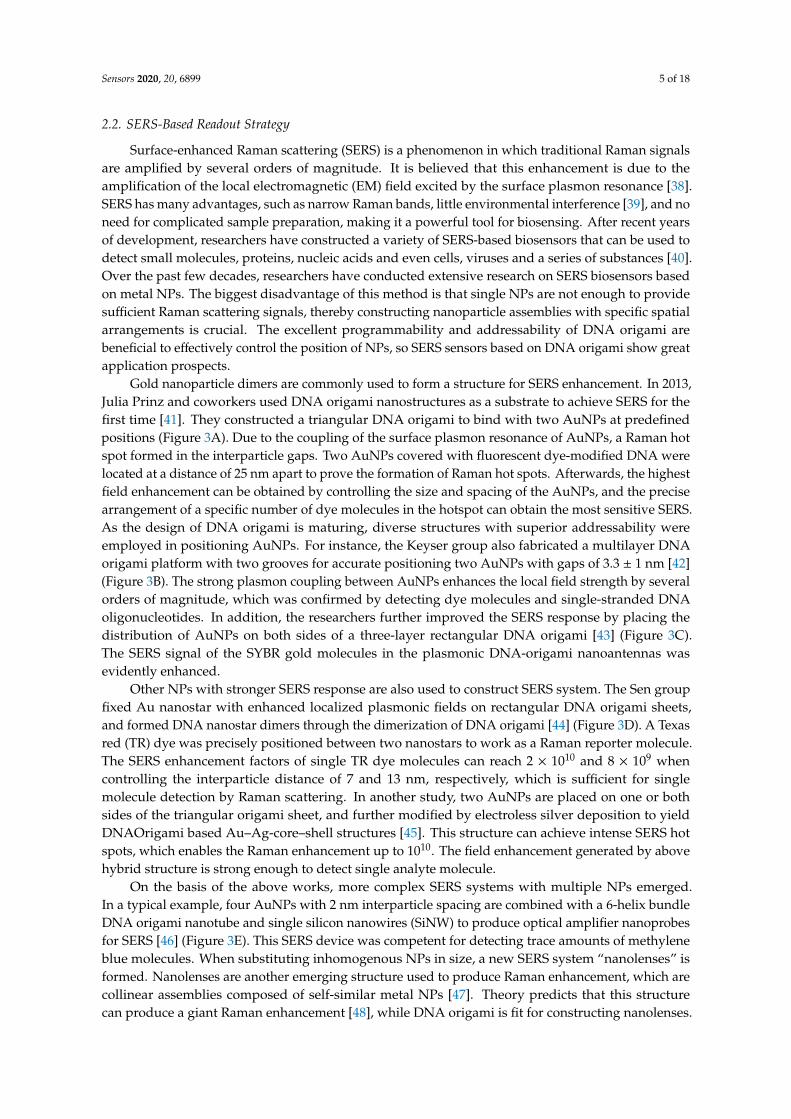
Gold nanoparticle dimers are commonly used to form a structure for SERS enhancement. In 2013,Julia Prinz and coworkers used DNA origami nanostructures as a substrate to achieve SERS for thefirst time [41]. They constructed a triangular DNA origami to bind with two AuNPs at predefinedpositions (Figure 3A). Due to the coupling of the surface plasmon resonance of AuNPs, a Raman hotspot formed in the interparticle gaps. Two AuNPs covered with fluorescent dye-modified DNA werelocated at a distance of 25 nm apart to prove the formation of Raman hot spots. Afterwards, the highestfield enhancement can be obtained by controlling the size and spacing of the AuNPs, and the precisearrangement of a specific number of dye molecules in the hotspot can obtain the most sensitive SERS.As the design of DNA origami is maturing, diverse structures with superior addressability wereemployed in positioning AuNPs. For instance, the Keyser group also fabricated a multilayer DNAorigami platform with two grooves for accurate positioning two AuNPs with gaps of 3.3 ± 1 nm [42](Figure 3B). The strong plasmon coupling between AuNPs enhances the local field strength by severalorders of magnitude, which was confirmed by detecting dye molecules and single-stranded DNAoligonucleotides. In addition, the researchers further improved the SERS response by placing thedistribution of AuNPs on both sides of a three-layer rectangular DNA origami [43] (Figure 3C).The SERS signal of the SYBR gold molecules in the plasmonic DNA-origami nanoantennas wasevidently enhanced.
Other NPs with stronger SERS response are also used to construct SERS system. The Sen groupfixed Au nanostar with enhanced localized plasmonic fields on rectangular DNA origami sheets,and formed DNA nanostar dimers through the dimerization of DNA origami [44] (Figure 3D). A Texasred (TR) dye was precisely positioned between two nanostars to work as a Raman reporter molecule.The SERS enhancement factors of single TR dye molecules can reach 2 × 10¹⁰ and 8 × 10⁹ whencontrolling the interparticle distance of 7 and 13 nm, respectively, which is sufficient for singlemolecule detection by Raman scattering. In another study, two AuNPs are placed on one or bothsides of the triangular origami sheet, and further modified by electroless silver deposition to yieldDNAOrigami based Au–Ag-core–shell structures [45]. This structure can achieve intense SERS hotspots, which enables the Raman enhancement up to 1010. The field enhancement generated by abovehybrid structure is strong enough to detect single analyte molecule.
On the basis of the above works, more complex SERS systems with multiple NPs emerged.In a typical example, four AuNPs with 2 nm interparticle spacing are combined with a 6-helix bundleDNA origami nanotube and single silicon nanowires (SiNW) to produce optical amplifier nanoprobesfor SERS [46] (Figure 3E). This SERS device was competent for detecting trace amounts of methyleneblue molecules. When substituting inhomogenous NPs in size, a new SERS system “nanolenses” isformed. Nanolenses are another emerging structure used to produce Raman enhancement, which arecollinear assemblies composed of self-similar metal NPs [47]. Theory predicts that this structurecan produce a giant Raman enhancement [48], while DNA origami is fit for constructing nanolenses.
Sensors 2020, 20, 6899 6 of 18
In 2017, Bald’s team successfully constructed gold nanolenses (AuNLs) using triangular DNA origamiand AuNPs of different sizes, and tested the SERS capability of single AuNLs [49]. In this study,10, 20, and 60 nm AuNPs were arranged in the predicted positions of triangular DNA origami byextending different capture strands to form AuNLs. Three different spatial arrangements of AuNPsgenerated different SERS signals in which 20-10-60 arrangement could produce the strongest Ramanenhancement. Moreover, the field enhancement located in the gap between the 20 and 10 nm AuNPs isstrongest, which is consistent with the theoretical prediction. Based on this research, they constructedsilver nanolenses subsequently [50]. It is expected that silver nanolenses can provide superior fieldenhancements over gold nanolenses. Similarly, triangular DNA origami scaffolds were used toassemble 10, 20, and 60 nm silver NPs into 20-10-60 structures, which generated the highest filedenhancement in the research mentioned above. Single protein streptavidin labeled with alkyne groupswere placed in the gap between the 20 and 10 nm particles through a noncovalent bond with biotingroup, which modified in the DNA origami scaffolds. Thus, the signal of streptavidin could be detectedthrough SERS spectra.
Sensors 2020, 20, x FOR PEER REVIEW 6 of 18
20, and 60 nm AuNPs were arranged in the predicted positions of triangular DNA origami by extending different capture strands to form AuNLs. Three different spatial arrangements of AuNPs generated different SERS signals in which 20-10-60 arrangement could produce the strongest Raman enhancement. Moreover, the field enhancement located in the gap between the 20 and 10 nm AuNPs is strongest, which is consistent with the theoretical prediction. Based on this research, they constructed silver nanolenses subsequently [50]. It is expected that silver nanolenses can provide superior field enhancements over gold nanolenses. Similarly, triangular DNA origami scaffolds were used to assemble 10, 20, and 60 nm silver NPs into 20-10-60 structures, which generated the highest filed enhancement in the research mentioned above. Single protein streptavidin labeled with alkyne groups were placed in the gap between the 20 and 10 nm particles through a noncovalent bond with biotin group, which modified in the DNA origami scaffolds. Thus, the signal of streptavidin could be detected through SERS spectra.
Figure 3. Surface-enhanced Raman scattering (SERS)-based readout strategy. (A) Triangular DNA origami binds with two gold nanoparticles (AuNPs). Adapted with permission from [41]. Copyright 2013 American Chemical Society. (B) A schematic of the NP dimers assembled on the DNA origami platform. Adapted with permission from [42]. Copyright 2014 Springer Nature. (C) Plasmonic DNA-origami nanoantennas. Adapted with permission from [43]. Copyright 2014 American Chemical Society. (D) Au nanostar dimers on dimerized rectangular origami structures. Adapted with permission from [44]. Copyright 2017 American Chemical Society. (E) Schematic diagram of a DNA origami-based SERS probe-decorated single SiNW. Adapted with permission from [46]. Copyright 2019 American Chemical Society.
2.3. Chirality-Based Readout Strategy
Optically active materials can induce the rotation of circularly polarized light. Moreover, we consider these materials to have optical chirality, which is described by circular dichroism (CD). The characteristic peaks of CD spectra reflect the chiral geometry and conformation of materials; thus, CD is widely utilized in recording the conformational changes of protein and DNA in ultraviolet range. Recently, surface plasmon resonance in chiral assemblies of metal NPs shows strong CD response at visible wavelengths [51–53]. The addressability and programmability of DNA origami are competent to arrange NPs accurately and enable fabricate plasmonic structures with novel optical properties. Many functionalized plasmonic systems are established and show great potential in mediating optical signal transduction, leading a new generation of readout strategy for biosensor.
In 2012, Liedl’s group employed DNA origami technique to control the arrangement of metal NPs and chiral geometries, which broke through the limits in fabricating plasmonic materials [51] (Figure 4A). They designed nine helically arranged attachment sites on a 24-helix-bundle origami nanostructure in different helical directions. The left- and right-handed helical arrangement of plasmonic particles exhibited the anticipated signature and agreed well with the theoretical
Figure 3. Surface-enhanced Raman scattering (SERS)-based readout strategy. (A) Triangular DNAorigami binds with two gold nanoparticles (AuNPs). Adapted with permission from [41]. Copyright 2013American Chemical Society. (B) A schematic of the NP dimers assembled on the DNA origamiplatform. Adapted with permission from [42]. Copyright 2014 Springer Nature. (C) PlasmonicDNA-origami nanoantennas. Adapted with permission from [43]. Copyright 2014 American ChemicalSociety. (D) Au nanostar dimers on dimerized rectangular origami structures. Adapted with permissionfrom [44]. Copyright 2017 American Chemical Society. (E) Schematic diagram of a DNA origami-basedSERS probe-decorated single SiNW. Adapted with permission from [46]. Copyright 2019 AmericanChemical Society.
2.3. Chirality-Based Readout Strategy
Optically active materials can induce the rotation of circularly polarized light. Moreover, we considerthese materials to have optical chirality, which is described by circular dichroism (CD). The characteristicpeaks of CD spectra reflect the chiral geometry and conformation of materials; thus, CD is widelyutilized in recording the conformational changes of protein and DNA in ultraviolet range. Recently,surface plasmon resonance in chiral assemblies of metal NPs shows strong CD response at visiblewavelengths [51–53]. The addressability and programmability of DNA origami are competent toarrange NPs accurately and enable fabricate plasmonic structures with novel optical properties.Many functionalized plasmonic systems are established and show great potential in mediating opticalsignal transduction, leading a new generation of readout strategy for biosensor.
Sensors 2020, 20, 6899 7 of 18
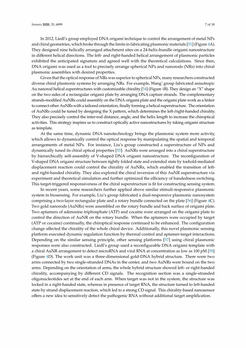
In 2012, Liedl’s group employed DNA origami technique to control the arrangement of metal NPsand chiral geometries, which broke through the limits in fabricating plasmonic materials [51] (Figure 4A).They designed nine helically arranged attachment sites on a 24-helix-bundle origami nanostructurein different helical directions. The left- and right-handed helical arrangement of plasmonic particlesexhibited the anticipated signature and agreed well with the theoretical calculations. Since then,DNA origami was used as a tool to precisely arrange spherical NPs and nanorods (NRs) into chiralplasmonic assemblies with desired properties.
Given that the optical response of NRs was superior to spherical NPs, many researchers constructeddiverse chiral plasmonic systems by arranging NRs. For example, Wang’ group fabricated anisotropicAu nanorod helical superstructures with customizable chirality [54] (Figure 4B). They design an “X” shapeon the two sides of a rectangular origami plate by arranging DNA capture strands. The complementarystrands-modified AuNRs could assembly on the DNA origami plate and the origami plate work as a linkerto connect other AuNRs with a tailored orientation, finally forming a helical superstructure. The orientationof AuNRs could be tuned by designing the X pattern, which determines the left-/right-handed chirality.They also precisely control the inter-rod distance, angle, and the helix length to increase the chiropticalactivities. This strategy inspires us to construct optically active nanostructures by taking origami structureas template.
At the same time, dynamic DNA nanotechnology brings the plasmonic system more activity,which allows to dynamically control the optical response by manipulating the spatial and temporalarrangements of metal NPs. For instance, Liu’s group constructed a superstructure of NPs anddynamically tuned its chiral optical properties [55]. AuNRs were arranged into a chiral superstructureby hierarchically self-assembly of V-shaped DNA origami nanostructure. The reconfiguration ofV-shaped DNA origami structure between tightly folded state and extended state by toehold-mediateddisplacement reaction could control the chirality of AuNRs, which enabled the transition of left-and right-handed chirality. They also explored the chiral inversion of this AuNR superstructure viaexperiment and theoretical simulation and further optimized the efficiency of handedness switching.This target-triggered responsiveness of the chiral superstructure is fit for constructing sensing system.
In recent years, some researchers further applied above similar stimuli-responsive plasmonicsystem in biosensing. For example, Liu’s group fabricated a dual-responsive plasmonic nanosystemcomprising a two-layer rectangular plate and a rotary bundle connected on the plate [56] (Figure 4C).Two gold nanorods (AuNRs) were assembled on the rotary bundle and back surface of origami plate.Two aptamers of adenosine triphosphate (ATP) and cocaine were arranged on the origami plate tocontrol the direction of AuNR on the rotary bundle. When the aptamers were occupied by target(ATP or cocaine) continually, the chiroptical response continued to be enhanced. The configurationchange affected the chirality of the whole chiral device. Additionally, this novel plasmonic sensingplatform executed dynamic regulation function by thermal control and aptamer-target interactions.Depending on the similar sensing principle, other sensing platforms [57] using chiral plasmonicresponses were also constructed. Liedl’s group used a reconfigurable DNA origami template witha chiral AuNR arrangement to detect microRNA and viral RNA at concentration as low as 100 pM [58](Figure 4D). The work unit was a three-dimensional gold-DNA hybrid structure. There were twoarms connected by two single-stranded DNAs in the center, and two AuNRs were bound on the twoarms. Depending on the orientation of arms, the whole hybrid structure showed left- or right-handedchirality, accompanying by different CD signals. The recognition section was a single-strandedoligonucleotides set at the end of each arm. When target was not in the system, the structure waslocked in a right-handed state, whereas in presence of target RNA, the structure turned to left-handedstate by strand displacement reaction, which led to a strong CD signal. This chirality-based nanosensoroffers a new idea to sensitively detect the pathogenic RNA without additional target amplification.
Sensors 2020, 20, 6899 8 of 18Sensors 2020, 20, x FOR PEER REVIEW 8 of 18
Figure 4. Chirality-based readout strategies. (A) Fabrication of chiral plasmonic materials based on DNA origami technique. Adapted with permission from [51]. Copyright 2012 Springer Nature. (B) DNA origami-based chiral superstructure. Adapted with permission from [54]. Copyright 2015 American Chemical Society. (C) Dual-responsive plasmonic nanosystem. Adapted with permission from [56]. Copyright 2018 American Chemical Society. (D) Reconfigurable DNA origami plasmonic sensing system used in RNA sensing. Adapted with permission from [58]. Copyright 2018 Wiley-VCH.
2.4. Fluorescence-Based Readout Strategy
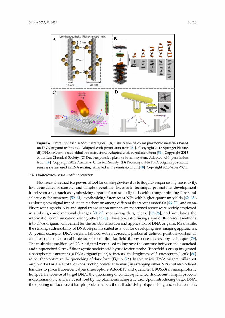
Fluorescent method is a powerful tool for sensing devices due to its quick response, high sensitivity, low abundance of sample, and simple operation. Metrics in technique promote its development in relevant areas such as synthesizing organic fluorescent ligands with stronger binding force and selectivity for structure [59–61], synthesizing fluorescent NPs with higher quantum yields [62–65], exploring new signal transduction mechanism among different fluorescent materials [66–70], and so on. Fluorescent ligands, NPs and signal transduction mechanism mentioned above were widely employed in studying conformational changes [71,72], monitoring drug release [73–76], and simulating the information communication among cells [77,78]. Therefore, introducing superior fluorescent methods into DNA origami will benefit for the functionalization and application of DNA origami. Meanwhile, the striking addressability of DNA origami is suited as a tool for developing new imaging approaches. A typical example, DNA origami labeled with fluorescent probes at defined position worked as a nanoscopic ruler to calibrate super-resolution far-field fluorescence microscopy technique [79]. The multiplex positions of DNA origami were used to improve the contrast between the quenched and unquenched form of fluorogenic nucleic acid hybridization probe. Tinnefeld’s group integrated a nanophotonic antennas (a DNA origami pillar) to increase the brightness of fluorescent molecule [80] rather than optimize the quenching of dark form (Figure 5A). In this article, DNA origami pillar not only worked as a scaffold for constructing optical antennas (by arranging silver NPs) but also offered handles to place fluorescent dyes (fluorophore Atto647N and quencher BBQ650) in nanophotonic hotspot. In absence of target DNA, the quenching of contact-quenched fluorescent hairpin probe is more remarkable and is not reduced by the plasmonic
Figure 4. Chirality-based readout strategies. (A) Fabrication of chiral plasmonic materials basedon DNA origami technique. Adapted with permission from [51]. Copyright 2012 Springer Nature.(B) DNA origami-based chiral superstructure. Adapted with permission from [54]. Copyright 2015American Chemical Society. (C) Dual-responsive plasmonic nanosystem. Adapted with permissionfrom [56]. Copyright 2018 American Chemical Society. (D) Reconfigurable DNA origami plasmonicsensing system used in RNA sensing. Adapted with permission from [58]. Copyright 2018 Wiley-VCH.
2.4. Fluorescence-Based Readout Strategy
Fluorescent method is a powerful tool for sensing devices due to its quick response, high sensitivity,low abundance of sample, and simple operation. Metrics in technique promote its developmentin relevant areas such as synthesizing organic fluorescent ligands with stronger binding force andselectivity for structure [59–61], synthesizing fluorescent NPs with higher quantum yields [62–65],exploring new signal transduction mechanism among different fluorescent materials [66–70], and so on.Fluorescent ligands, NPs and signal transduction mechanism mentioned above were widely employedin studying conformational changes [71,72], monitoring drug release [73–76], and simulating theinformation communication among cells [77,78]. Therefore, introducing superior fluorescent methodsinto DNA origami will benefit for the functionalization and application of DNA origami. Meanwhile,the striking addressability of DNA origami is suited as a tool for developing new imaging approaches.A typical example, DNA origami labeled with fluorescent probes at defined position worked asa nanoscopic ruler to calibrate super-resolution far-field fluorescence microscopy technique [79].The multiplex positions of DNA origami were used to improve the contrast between the quenchedand unquenched form of fluorogenic nucleic acid hybridization probe. Tinnefeld’s group integrateda nanophotonic antennas (a DNA origami pillar) to increase the brightness of fluorescent molecule [80]rather than optimize the quenching of dark form (Figure 5A). In this article, DNA origami pillar notonly worked as a scaffold for constructing optical antennas (by arranging silver NPs) but also offeredhandles to place fluorescent dyes (fluorophore Atto647N and quencher BBQ650) in nanophotonichotspot. In absence of target DNA, the quenching of contact-quenched fluorescent hairpin probe ismore remarkable and is not reduced by the plasmonic nanostructure. Upon introducing target DNA,the opening of fluorescent hairpin probe realizes the full additivity of quenching and enhancement.
Sensors 2020, 20, 6899 9 of 18
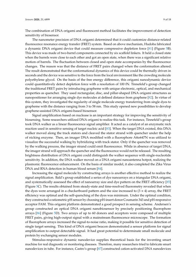
The combination of DNA origami and fluorescent method facilitates the improvement of detectionsensitivity of biosensor.
The nanometer precision of DNA origami determined that it could customize distance-relatedfluorescence resonance energy transfer (FRET) system. Based on above mechanism, Hudoba fabricateda dynamic DNA origami device that could measure compressive depletion force [81] (Figure 5B).This device was made of two-barrel components connected by six scaffold linkers. It held a closed statewhen the barrels were close to each other and got an open state, when there was a significant relativemotion of barrels. The fluctuation between closed and open state accompanies by the fluorescencechanges. The reason was that the distance of FRET pairs changed when the conformation altered.The result demonstrated that the conformational dynamics of this device could be thermally driven inseconds and the device was sensitive to the force from the local environment like the crowding moleculepolyethylene glycol. On the basis of the free energy difference, this origami nanodynamic devicecould quantitatively detect depletion force with a resolution of 100 fN. Tinnefeld’s group changedthe traditional FRET pairs by introducing grapheme with unique electronic, optical, and mechanicalproperties as quencher. They used rectangular, disc, and pillar-shaped DNA origami structures asnanopositioner for arranging single dye molecules at defined distance from graphene [82]. In virtue ofthis system, they investigated the regularity of single molecule energy transferring from single dyes tographeme with the distance ranging from 3 to 58 nm. This study opened new possibilities to developgraphene-assisted DNA origami-based biosensor.
Signal amplification based on nuclease is an important strategy for improving the sensitivity ofbiosensing. Some researchers utilized DNA origami to realize this task. For instance, Tinnefeld’s grouptook DNA walker as a linear fluorescence signal amplifier. It acted as a catalyst of an enzymatic nickreaction used in sensitive sensing of target nucleic acid [83]. When the target DNA existed, this DNAwalker moved along the track stators and cleaved the stator strand with quencher under the helpof nicking enzyme. Then, an imager DNA modified with a fluorophore Atto647N was utilized tovisualize the successful walking by hybridizing with track stator. Only if the quencher was removedby the walking process, the imager strand could emit fluorescence. While in absence of target DNA,the imager strand will approach the quencher and the fluorescence would not be released. The resultingbrightness distribution of DNA origami could distinguish the walker sequence with single-nucleotidesensitivity. In addition, the DNA walker moved on a DNA origami nanoantenna hotpot, realizing theplasmonic fluorescence enhancement. On the basis of similar model, it also completed the Zika VirusDNA and RNA detection in human blood serum [84].
Increasing the signal molecule by constructing arrays is another effective method to realize thesignal amplification. Bald’s group established a series of dye nanoarrays on a triangular DNA origami,and systematically assessed the effect of nanoarray size and dye pattern on the FRET efficiency [85](Figure 5C). The results obtained from steady-state and time-resolved fluorometry revealed that whenthe dyes were arranged in a checkerboard pattern and the size increased to (3 × 4) array, the FRETefficiency was optimal and the self-quenching of the dyes was minimum. Under the optimal condition,they constructed a ratiometric pH sensor by choosing pH-insert donor Coumarin 343 and pH-responsiveacceptor FAM. This origami platform demonstrated a good prospect in sensing scheme. Andersen’sgroup constructed an optical DNA origami nanobiosensor by precisely positioning fluorophorearrays [86] (Figure 5D). Two arrays of up to 60 donors and acceptors were composed of multipleFRET pairs, giving high-output signal with a mainstream fluorescence microscope. The formationof fluorophore arrays increased the signal-to-noise ratio, making it possible for sensitive detection ofsingle target sensing. This kind of DNA origami beacon demonstrated a sensor platform for signalamplification to output detectable signal. It had great potential to determinate small molecule andprotein by exchanging sensor modules.
Stimulus-responsive dynamic nanodevice supplies theoretical basis for the inventing smartmachine for real diagnostic or monitoring diseases. Therefore, many researchers tried to fabricate smartnanodevices in tube. For instance, Castro’s group [87] constructed cation-activated DNA nanodevices
Sensors 2020, 20, 6899 10 of 18
with subsecond actuation response time (Figure 5E). They applied a DNA origami hinges composed oftwo rectangular arms, which were connected by several flexible single-strand scaffold at one end ofrectangular arm. In order to endow these nanodevices ion-triggered actuation function between openand close conformations, they modified short single-strand staple overhangs along the inner face of eacharm. These staple overhangs rapidly hybridize or dehybridize when the cation concentration changes.Additionally, they explored the effects of the number of overhang connections, the strength of theseconnections, the torsional stiffness of hinge, and cations on the actuation response using single-moleculeFRET assay. As the opening and closing of these nanodevices, the fluorescence changes due to thedistance changes between fluorophore and quencher. This externally driven nanodevice is likely todevelop to be future generations of DNA nanorobots that could be used in biosensing, imaging anddiagnosis and treatment of disease. Keyser’s group fabricated a new type of voltage sensors byconverting the change of electric potential into optical signals [88]. They designed a two-layeredDNA origami rectangular template containing a double-strand leash and an aperture in the center.A pair of FRET pairs was marked on this origami plate and reversibly fixed on a nanocapillary tipto monitor the surrounding potential. The FRET pairs were vertical to the direction of the electricfield or parallel to the electric field. When the electric field strength increased, the deformation oforigami plate led to the changes in the positions of the dyes and the FRET efficiency. Additionally,coarse-grained Brownian dynamics simulation gave an assisted proof that the voltage could cause thedistance change between dyes. This voltage-sensitive FRET sensing mechanism shows enough latentcapacity in live-cell imaging of transmembrane potential.
Sensors 2020, 20, x FOR PEER REVIEW 10 of 18
hinges composed of two rectangular arms, which were connected by several flexible single-strand scaffold at one end of rectangular arm. In order to endow these nanodevices ion-triggered actuation function between open and close conformations, they modified short single-strand staple overhangs along the inner face of each arm. These staple overhangs rapidly hybridize or dehybridize when the cation concentration changes. Additionally, they explored the effects of the number of overhang connections, the strength of these connections, the torsional stiffness of hinge, and cations on the actuation response using single-molecule FRET assay. As the opening and closing of these nanodevices, the fluorescence changes due to the distance changes between fluorophore and quencher. This externally driven nanodevice is likely to develop to be future generations of DNA nanorobots that could be used in biosensing, imaging and diagnosis and treatment of disease. Keyser’s group fabricated a new type of voltage sensors by converting the change of electric potential into optical signals [88]. They designed a two-layered DNA origami rectangular template containing a double-strand leash and an aperture in the center. A pair of FRET pairs was marked on this origami plate and reversibly fixed on a nanocapillary tip to monitor the surrounding potential. The FRET pairs were vertical to the direction of the electric field or parallel to the electric field. When the electric field strength increased, the deformation of origami plate led to the changes in the positions of the dyes and the FRET efficiency. Additionally, coarse-grained Brownian dynamics simulation gave an assisted proof that the voltage could cause the distance change between dyes. This voltage-sensitive FRET sensing mechanism shows enough latent capacity in live-cell imaging of transmembrane potential.
Figure 5. Fluorescence strategies of DNA-origami-enabled sensors. (A) DNA origami-based nanophotonic antennas were utilized to improve sensing performance by increasing signal-noise ratio. Adapted with permission from [80]. Copyright 2017 American Chemical Society. (B) A dynamic DNA origami device that could measure compressive depletion force. Adapted with permission from [81]. Copyright 2017 American Chemical Society. (C) Nanoarrays with different patterns and sizes used in pH sensing. Reprinted with permission from [85]. Copyright 2017 American Chemical Society. (D) DNA origami beacon array was used to amplify signal and execute detection of single molecule. Adapted with permission from [86]. Copyright 2018 American Chemical Society. (E) A cation-responsive sensor fabricated by an origami nanostructure and FRET pairs. Adapted with permission from [87]. Copyright 2018 American Chemical Society.
Figure 5. Fluorescence strategies of DNA-origami-enabled sensors. (A) DNA origami-based nanophotonicantennas were utilized to improve sensing performance by increasing signal-noise ratio. Adapted withpermission from [80]. Copyright 2017 American Chemical Society. (B) A dynamic DNA origami devicethat could measure compressive depletion force. Adapted with permission from [81]. Copyright 2017American Chemical Society. (C) Nanoarrays with different patterns and sizes used in pH sensing.Reprinted with permission from [85]. Copyright 2017 American Chemical Society. (D) DNA origamibeacon array was used to amplify signal and execute detection of single molecule. Adapted with permissionfrom [86]. Copyright 2018 American Chemical Society. (E) A cation-responsive sensor fabricated by anorigami nanostructure and FRET pairs. Adapted with permission from [87]. Copyright 2018 AmericanChemical Society.

Sensors 2020, 20, 6899 11 of 18
2.5. DNA Origami Nanopore Readout Strategy
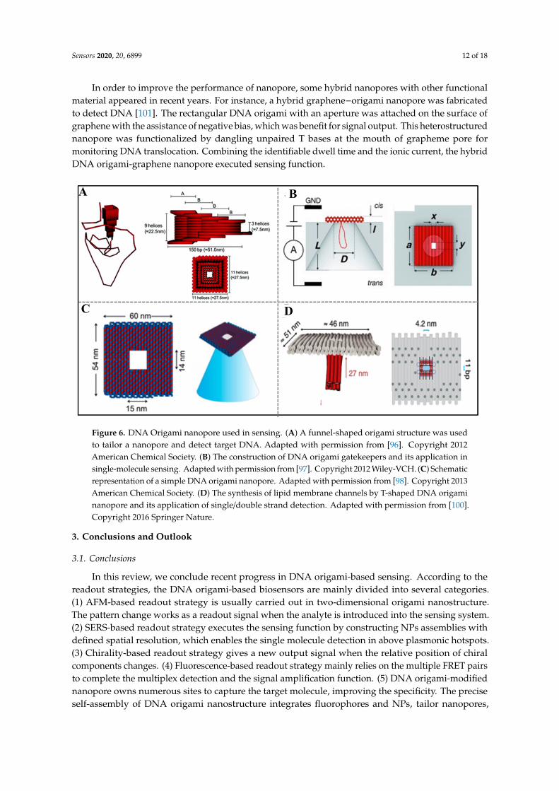
Nanopores sensing has shown good potential for a label-free monitoring molecular interaction,identifying structural polymorphisms at the single-molecular level [89]. The pulse modulations in ioniccurrent indicate the target translocation through the nanopore [90]. Nanopores are typically dividedinto two categories including biological (fabricated from proteins [91]) and solid-state (formed byinorganic or organic materials) nanopores [92]. The sizes of solid state nanopores are tunable, but thecontrollability of surface functionality (precisely positioning single binding sites for analytes) limits theirdevelopment. Although the biological nanopores like α-hemolysin are superior to similar solid-statenanopores in surface functionality by genetic engineering, most of their narrowest dimensions areless than 2 nm, which limited the range of the detecting targets to single-stranded DNA or smallmolecule [93,94]. DNA origami can address above challenge since it can tailor a size-tunable nanoporeand precisely control the position of target-responsive probes as well.
Inspired by Hall’s report [95] about integrating α-hemolysin protein pore on solid-state nanopore,Keyser et al. firstly chose DNA origami nanostructure to synthesize nanopore with tailored geometries.For example, they constructed a funnel-shaped DNA origami structure and demonstrated the repeatedassembly of hybrid nanopore by reversing the applied potential for the first time. This hybrid nanoporewas also applied in detecting λ-DNA [96] (Figure 6A), which proved that DNA origami-based nanoporehas potential as resistive-pulse sensors.
The tailored dimension of nanopore could be realized by virtue of DNA origami technique,but analyte-specific modifications of cavities require considerable effort. Aiming at this challenge,Dietz’s group invented a DNA origami gatekeepers on the basis of solid nanopores [97] (Figure 6B).The nanopore consisted of an insulating silicon nitride membrane containing a single conical pore,an origami rectangular with a central aperture covered on the pore, and a tightly interlinked double-helixDNA loop that protrudes under the aperture to facilitate insertion into the nanopore. Taking single-strandedDNA motif as bait molecule modified in the aperture, this nanoplate could detect DNA prey moleculeselectively. The DNA origami nanoplate not only provides chemical selectivity for the solid state nanoporebut also provides size selectivity. For example, when streptavidin, immunoglobulin G, double-strandDNA with different sizes translocated into the nanopore, different current blockades were observed.This study proves that the chemical addressability of the origami nanoplate could make the surfacefunctionality of nanopore controllable. Similarly, Keyser’ group [98] combined DNA origami rectangular(outer dimensions 60 nm× 54 nm, and a aperture of 14 nm× 15 nm in the center) with glass nanocapillariesto obtain hybrid DNA origami nanopores by reversibly applying voltage (Figure 6C). They firstlyenabled the DNA origami structure visual at the tip of the glass nanocapillaries though single-moleculefluorescence imaging. They controlled the folding of double-stranded DNA by tuning the size of apertureand demonstrated the specific detection of target single-stranded DNA by introducing of specific bindingsite in the DNA origami nanopore. This study mentioned a new method for tailoring nanopore with highthroughout and easy manufacture.
Inspired by the natural channel protein α-hemolysin, Langecker et al. synthesized a lipid membranechannels by DNA origami [99]. The origami nanostructure included an inner stem that spanneda lipid membrane and an outer barrel-shaped cap that adhered to the membrane via cholesterol moieties.This artificial channel showed similar response to the natural ion channel. A single DNA strand protrudingin the stem could be used to discriminate single DNA molecule. This synthetic lipid membrane channelssupply powerful tool to explore the function of natural membrane. In 2016, Simmel’s group furtherpromoted the application of DNA nanopore by incorporating DNA origami-based nanopore intogiant unilamellar vesicles [100]. They designed a DNA transmembrane channel with a T-shapedpore. The pore was made of an origami square lattice with an aperture in the center and a hollowstem extending perpendicularly from aperture (Figure 6D). This membrane exhibited stable electricalproperties and allowed electrically driven translocation of single/double strand. The application oforigami-based transmembrane pore in single-molecule biosensing lays the foundation for diagnosisand treatment of disease.
Sensors 2020, 20, 6899 12 of 18
In order to improve the performance of nanopore, some hybrid nanopores with other functionalmaterial appeared in recent years. For instance, a hybrid graphene−origami nanopore was fabricatedto detect DNA [101]. The rectangular DNA origami with an aperture was attached on the surface ofgraphene with the assistance of negative bias, which was benefit for signal output. This heterostructurednanopore was functionalized by dangling unpaired T bases at the mouth of grapheme pore formonitoring DNA translocation. Combining the identifiable dwell time and the ionic current, the hybridDNA origami-graphene nanopore executed sensing function.
Sensors 2020, 20, x FOR PEER REVIEW 12 of 18
In order to improve the performance of nanopore, some hybrid nanopores with other functional material appeared in recent years. For instance, a hybrid graphene−origami nanopore was fabricated to detect DNA [101]. The rectangular DNA origami with an aperture was attached on the surface of graphene with the assistance of negative bias, which was benefit for signal output. This heterostructured nanopore was functionalized by dangling unpaired T bases at the mouth of grapheme pore for monitoring DNA translocation. Combining the identifiable dwell time and the ionic current, the hybrid DNA origami-graphene nanopore executed sensing function.
Figure 6. DNA Origami nanopore used in sensing. (A) A funnel-shaped origami structure was used to tailor a nanopore and detect target DNA. Adapted with permission from [96]. Copyright 2012 American Chemical Society. (B) The construction of DNA origami gatekeepers and its application in single-molecule sensing. Adapted with permission from [97]. Copyright 2012 Wiley-VCH. (C) Schematic representation of a simple DNA origami nanopore. Adapted with permission from [98]. Copyright 2013 American Chemical Society. (D) The synthesis of lipid membrane channels by T-shaped DNA origami nanopore and its application of single/double strand detection. Adapted with permission from [100]. Copyright 2016 Springer Nature.
3. Conclusions and Outlook
3.1. Conclusions
In this review, we conclude recent progress in DNA origami-based sensing. According to the readout strategies, the DNA origami-based biosensors are mainly divided into several categories. (1) AFM-based readout strategy is usually carried out in two-dimensional origami nanostructure. The pattern change works as a readout signal when the analyte is introduced into the sensing system. (2) SERS-based readout strategy executes the sensing function by constructing NPs assemblies with defined spatial resolution, which enables the single molecule detection in above plasmonic hotspots. (3) Chirality-based readout strategy gives a new output signal when the relative position of chiral components changes. (4) Fluorescence-based readout strategy mainly relies on the multiple FRET pairs to complete the multiplex detection and the signal amplification function. (5) DNA origami-modified nanopore owns numerous sites to capture the target molecule, improving the specificity. The precise self-assembly of DNA origami nanostructure integrates fluorophores and NPs, tailor nanopores, giving fluorescent, SERS, chirality, and current signals. The origami-based sensing system can improve the detection specificity by modification in tailored space and sensitivity by signal amplification.
Figure 6. DNA Origami nanopore used in sensing. (A) A funnel-shaped origami structure was usedto tailor a nanopore and detect target DNA. Adapted with permission from [96]. Copyright 2012American Chemical Society. (B) The construction of DNA origami gatekeepers and its application insingle-molecule sensing. Adapted with permission from [97]. Copyright 2012 Wiley-VCH. (C) Schematicrepresentation of a simple DNA origami nanopore. Adapted with permission from [98]. Copyright 2013American Chemical Society. (D) The synthesis of lipid membrane channels by T-shaped DNA origaminanopore and its application of single/double strand detection. Adapted with permission from [100].Copyright 2016 Springer Nature.
3. Conclusions and Outlook
3.1. Conclusions
In this review, we conclude recent progress in DNA origami-based sensing. According to thereadout strategies, the DNA origami-based biosensors are mainly divided into several categories.(1) AFM-based readout strategy is usually carried out in two-dimensional origami nanostructure.The pattern change works as a readout signal when the analyte is introduced into the sensing system.(2) SERS-based readout strategy executes the sensing function by constructing NPs assemblies withdefined spatial resolution, which enables the single molecule detection in above plasmonic hotspots.(3) Chirality-based readout strategy gives a new output signal when the relative position of chiralcomponents changes. (4) Fluorescence-based readout strategy mainly relies on the multiple FRET pairsto complete the multiplex detection and the signal amplification function. (5) DNA origami-modifiednanopore owns numerous sites to capture the target molecule, improving the specificity. The preciseself-assembly of DNA origami nanostructure integrates fluorophores and NPs, tailor nanopores,
Sensors 2020, 20, 6899 13 of 18
giving fluorescent, SERS, chirality, and current signals. The origami-based sensing system can improvethe detection specificity by modification in tailored space and sensitivity by signal amplification.
3.2. Outlook
Though obtaining many progresses, there are still challenges in DNA origami-based sensing.(1) Considering the real application in detecting and curing diseases, the storage, scalability and costbecome highly pertinent question. Some studies demonstrated that lyophilization and cryostorage couldkeep origami nanostructure intact for up to several years [102–104]. In 2017, Dietz et al. [105] realizedscalable biotechnological production of single stranded DNA with arbitrary length and sequence,which would remove a key obstacle in the way of application of origami-based biosensor. We hope thecost of origami could further decrease as the progress of scalable technology in the future. (2) Thedevelopment of origami-based biosensor is in its infancy. Taking the fluorescent-based readout strategyas an example, merely FRET signal transduction mechanism is utilized in sensing while few otherstrategies emerge in this application. Introducing diverse transduction mechanisms into DNA origaminanostructure will bring new methods for detection. (3) The origami-based biosensor is in a period ofconstructing sensing system. The performance improvement like limit of detection or response timewill give opportunities for sensing new biomarkers with low concentration. Perhaps, single-moleculedetection will be future direction. (4) The stability of DNA origami-based biosensor under thephysiological conditions is essential for biomedical diagnostic purposes. Although some strategies likecoating DNA origami structure with polymer [106] are taken to improve the stability, some bindingsites and dynamic performance of DNA origami structure lost in this process. Therefore, how toimprove the stability and simultaneously reserving the function of structure is a facing challenge.
The greatest metric of DNA origami technology is to offer multiple sites to position molecules,biomolecules and materials with a nanometer resolution. A wide range of nanomaterials can beincorporated in DNA origami nanostructure including polymers [107], nanodimonds [108], enzymes [109],metal NPs [110], quantum dots [111] and so on. However, the function derived from integrationhas little actual practical application including sensing. Therefore, putting functionalized origaminanostructure into fundamental sensing application will be a beginning of practical application ofDNA origami nanostructure. We expect that diverse DNA origami-based biosensors with excellentsensitivity and specificity will be developed, and promise to realize precise quantification of clinicallyrelevant biomarkers in the future.
Author Contributions: Conceptualization, Y.T., C.T. and Y.K.; writing—original draft preparation, S.W. and Z.Z.;writing—review and editing, N.M., S.Y. and K.L.; funding acquisition, C.T. All authors have read and agreed tothe published version of the manuscript.
Funding: This work was supported by the Shenzhen International Cooperation Research Project (No. GJHZ20180930090602235 and GJHZ20190819151807167).
Conflicts of Interest: The authors declare no conflict of interest.
References
1. Wang, S.; Lu, S.; Zhao, J.; Ye, J.; Huang, J.; Yang, X. An electric potential modulated cascade of catalyzedhairpin assembly and rolling chain amplification for microRNA detection. Biosens. Bioelectron. 2019, 126,565–571. [CrossRef] [PubMed]
2. Arora, P.; Sindhu, A.; Dilbaghi, N.; Chaudhury, A. Biosensors as innovative tools for the detection of foodborne pathogens. Biosens. Bioelectron. 2011, 28, 1–12. [CrossRef] [PubMed]
3. Lee, M.; Zine, N.; Baraket, A.; Zabala, M.; Campabadal, F.; Caruso, R.; Trivella, M.G.; Jaffrezic-Renault, N.;Errachid, A. A novel biosensor based on hafnium oxide: Application for early stage detection of humaninterleukin-10. Sens. Actuators B Chem. 2012, 175, 201–207. [CrossRef]
4. Mishra, R.; Dominguez, R.; Bhand, S.; Munoz, R.; Marty, J. A novel automated flow-based biosensor for thedetermination of organophosphate pesticides in milk. Biosens. Bioelectron. 2012, 32, 56–61. [CrossRef]

Sensors 2020, 20, 6899 14 of 18
5. Ooi, K.; Galatowicz, G.; Towler, H.; Lightman, S.; Calder, V. Multiplex cytokine detection versus ELISA foraqueous humor: IL-5, IL-10, and IFN gamma profiles in uveitis. Investig. Ophthalmol. Vis. Sci. 2006, 47, 272–277.[CrossRef]
6. Scognarniglio, V.; Arduini, F.; Palleschi, G.; Rea, G. Biosensing technology for sustainable food safety.TrAC-Trends Anal. Chem. 2014, 62, 1–10. [CrossRef]
7. Wang, S.; Lu, S.; Zhao, J.; Huang, J.; Yang, X. Real-time study of the interaction between G-rich DNAoligonucleotides and lead ion on DNA tetrahedron-functionalized sensing platform by dual polarizationinterferometry. ACS Appl. Mater. Interfaces 2017, 9, 41568–41576. [CrossRef]
8. Wang, S.; Lu, S.; Zhao, J.; Huang, J.; Yang, X. Label-free Pb2+ detection on the layer-by-layer platform usingreal-time dual polarization interferometry. Talanta 2019, 202, 336–341. [CrossRef]
9. Zhang, J.; Lu, Y. Biocomputing for portable, resettable, and quantitative point-of-care diagnostics: Making theglucose meter a logic-gate responsive device for measuring many clinically relevant targets. Angew. Chem.Int. Ed. 2018, 57, 9702–9706. [CrossRef]
10. Gasperskaja, E.; Kucinskas, V. The most common technologies and tools for functional genome analysis.Acta Med. Litu. 2017, 24, 1–11. [CrossRef]
11. Han, S.; Liu, W.; Zheng, M.; Wang, R. Label-free and ultrasensitive electrochemical DNA biosensor based onurchinlike carbon nanotube-gold nanoparticle nanoclusters. Anal. Chem. 2020, 92, 4780–4787. [CrossRef][PubMed]
12. He, P.; Xu, Y.; Fang, Y. A review: Electrochemical DNA biosensors for sequence recognition. Anal. Lett. 2005,38, 2597–2623. [CrossRef]
13. Zhao, W.; Xu, J.; Chen, H. Photoelectrochemical DNA biosensors. Chem. Rev. 2014, 114, 7421–7441. [CrossRef][PubMed]
14. Rothemund, P. Folding DNA to create nanoscale shapes and patterns. Nature 2006, 440, 297–302. [CrossRef]15. Dietz, H.; Douglas, S.; Shih, W. Folding DNA into twisted and curved nanoscale shapes. Science 2009, 325,
725–730. [CrossRef]16. Douglas, S.; Dietz, H.; Liedl, T.; Hoegberg, B.; Graf, F.; Shih, W. Self-assembly of DNA into nanoscale
three-dimensional shapes. Nature 2009, 459, 414–418. [CrossRef]17. Zadegan, R.; Jepsen, M.; Thomsen, K.; Okholm, A.; Schaffert, D.; Andersen, E.; Birkedal, V.; Kjems, J.
Construction of a 4 zeptoliters switchable 3D DNA box origami. ACS Nano 2012, 6, 10050–10053. [CrossRef]18. Han, D.; Pal, S.; Yang, Y.; Jiang, S.; Nangreave, J.; Liu, Y.; Yan, H. DNA gridiron nanostructures based on
four-arm junctions. Science 2013, 339, 1412–1415. [CrossRef]19. Han, D.; Pal, S.; Nangreave, J.; Deng, Z.; Liu, Y.; Yan, H. DNA origami with complex curvatures in
three-dimensional space. Science 2011, 332, 342–346. [CrossRef]20. Castro, C.E.; Kilchherr, F.; Kim, D.; Shiao, E.; Wauer, T.; Wortmann, P.; Bathe, M.; Dietz, H. A primer to
scaffolded DNA origami. Nat. Methods 2011, 8, 221–229. [CrossRef]21. Wagenbauer, K.; Engelhardt, F.; Stahl, E.; Hechtl, V.; Stçmmer, P.; Seebacher, F.; Meregalli, L.; Ketterer, P.;
Gerling, T.; Dietz, H. How we make DNA origami. ChemBioChem 2017, 18, 1873–1885. [CrossRef] [PubMed]22. Hui, L.; Zhang, Q.; Deng, W.; Liu, H. DNA-based nanofabrication: Pathway to applications in surface
engineering. Small 2019, 15, 1805428. [CrossRef] [PubMed]23. Ji, M.; Ma, N.; Tian, Y. 3D lattice engineering of nanoparticles by DNA shells. Small 2019, 15, 1805401.
[CrossRef] [PubMed]24. Kizer, M.; Linhardt, R.; Chandrasekaran, A.; Wang, X. A molecular hero suit for in vitro and in vivo DNA
nanostructures. Small 2019, 15, 1805386. [CrossRef]25. Nicoli, F.; Zhang, T.; Hübner, K.; Jin, B.; Selbach, F.; Acuna, G.; Argyropoulos, C.; Liedl, T.; Pilo-Pais, M.
DNA-mediated self-assembly of plasmonic antennas with a single quantum dot in the hot spot. Small 2019,15, 1804418. [CrossRef]
26. Seo, J.; Kim, S.; Park, H.; Nam, J. Biocomputing with nanostructures on lipid bilayers. Small 2019, 15, 1900998.[CrossRef]
27. Zhou, K.; Dong, J.; Zhou, Y.; Dong, J.; Wang, M.; Wang, Q. Toward precise manipulation of DNA–proteinhybrid nanoarchitectures. Small 2019, 15, 1804044. [CrossRef]
28. Ke, Y.; Lindsay, S.; Chang, Y.; Liu, Y.; Yan, H. Self-assembled water-soluble nucleic acid probe tiles forlabel-free RNA hybridization assays. Science 2008, 319, 180–183. [CrossRef]

Sensors 2020, 20, 6899 15 of 18
29. Zhang, Z.; Wang, Y.; Fan, C.; Li, C.; Li, Y.; Qian, L.; Fu, Y.; Shi, Y.; Hu, J.; He, L. Asymmetric DNA origami forspatially addressable and index-free solution-phase DNA chips. Adv. Mater. 2010, 22, 2672–2675. [CrossRef]
30. Wang, D.; Fu, Y.; Yan, J.; Zhao, B.; Dai, B.; Chao, J.; Liu, H.; He, D.; Zhang, Y.; Fan, C.; et al. Molecularlogic gates on DNA origami nanostructures for microRNA diagnostics. Anal. Chem. 2014, 86, 1932–1936.[CrossRef]
31. Wanunu, M.; Zhu, J.; Feng, X.; Lou, J.; Li, W.; Li, S.; Zhu, H.; Yang, L.; Zhang, A.; He, L.; et al. Accuratequantification of microRNA via single strand displacement reaction on DNA origami motif. PLoS ONE 2013,8, e69856.
32. Lu, Z.; Wang, Y.; Xu, D.; Pang, L. Aptamer-tagged DNA origami for spatially addressable detection ofaflatoxin B1. Chem. Commun. 2017, 53, 941–944. [CrossRef] [PubMed]
33. Kuzuya, A.; Sakai, Y.; Yamazaki, T.; Xu, Y.; Komiyama, M. Nanomechanical DNA origami ‘single-moleculebeacons’ directly imaged by atomic force microscopy. Nat. Commun. 2011, 2, 1–8. [CrossRef]
34. Kuzuya, A.; Watanabe, R.; Yamanaka, Y.; Tamaki, T.; Kaino, M.; Ohya, Y. Nanomechanical DNA origami pHsensors. Sensors 2014, 14, 19329–19335. [CrossRef]
35. Subramanian, H.; Chakraborty, B.; Sha, R.; Seeman, N. The Label-free unambiguous detection and symbolicdisplay of single nucleotide polymorphisms on DNA origami. Nano Lett. 2011, 11, 910–913. [CrossRef]
36. Zhang, H.; Chao, J.; Pan, D.; Liu, H.; Qiang, Y.; Liu, K.; Cui, C.; Chen, J.; Huang, Q.; Hu, J.; et al. DNAorigami-based shape IDs for single-molecule nanomechanical genotyping. Nat. Commun. 2017, 8, 14738.[CrossRef] [PubMed]
37. Liu, K.; Pan, D.; Wen, Y.; Zhang, H.; Chao, J.; Wang, L.; Song, S.; Fan, C.; Shi, Y. Identifying the genotypes ofhepatitis B virus (HBV) with DNA origami label. Small 2018, 14, 1701718. [CrossRef] [PubMed]
38. Ding, S.; You, E.; Tian, Z.; Moskovits, M. Electromagnetic theories of surface-enhanced Raman spectroscopy.Chem. Soc. Rev. 2017, 46, 4042–4076. [CrossRef]
39. Xiao, M.; Lai, W.; Man, T.; Chang, B.; Li, L.; Chandrasekaran, A.R.; Pei, H. Rationally engineered nucleic acidarchitectures for biosensing applications. Chem. Rev. 2019, 119, 11631–11717. [CrossRef]
40. Wang, Z.; Zong, S.; Wu, L.; Zhu, D.; Cui, Y. SERS-activated platforms for immunoassay: Probes, encoding methods,and applications. Chem. Rev. 2017, 117, 7910–7963. [CrossRef]
41. Prinz, J.; Schreiber, B.; Olejko, L.; Oertel, J.; Rackwitz, J.; Keller, A.; Bald, I. DNA origami substrates for highlysensitive surface-enhanced raman scattering. J. Phys. Chem. Lett. 2013, 4, 4140–4145. [CrossRef]
42. Thacker, V.; Herrmann, L.; Sigle, D.; Zhang, T.; Liedl, T.; Baumberg, J.; Keyser, U. DNA origami basedassembly of gold nanoparticle dimers for surface-enhanced Raman scattering. Nat. Commun. 2014, 5, 4448.[CrossRef] [PubMed]
43. Kuehler, P.; Roller, E.; Schreiber, R.; Liedl, T.; Lohmueller, T.; Feldmann, J. Plasmonic DNA-origaminanoantennas for surface-enhanced Raman spectroscopy. Nano Lett. 2014, 14, 2914–2919. [CrossRef] [PubMed]
44. Tanwar, S.; Haldar, K.; Sen, T. DNA origami directed Au nanostar dimers for single-molecule surface-enhancedraman scattering. J. Am. Chem. Soc. 2017, 139, 17639–17648. [CrossRef]
45. Prinz, J.; Heck, C.; Ellerik, L.; Merk, V.; Bald, I. DNA origami based Au-Ag-core-shell nanoparticle dimerswith single-molecule SERS sensitivity. Nanoscale 2016, 8, 5612–5620. [CrossRef] [PubMed]
46. Moeinian, A.; Gur, F.; Gonzalez-Torres, J.; Zhou, L.; Murugesan, V.; Dashtestani, A.; Guo, H.; Schmidt, T.;Strehle, S. Highly localized SERS measurements using single silicon nanowires decorated with DNAorigami-based SERS probe. Nano Lett. 2019, 19, 1061–1066. [CrossRef] [PubMed]
47. Kneipp, J.; Li, X.; Sherwood, M.; Panne, U.; Kneipp, H.; Stockman, M.I.; Kneipp, K. Gold nanolensesgenerated by laser ablation-efficient enhancing structure for surface enhanced Raman scattering analyticsand sensing. Anal. Chem. 2008, 80, 4247–4251. [CrossRef]
48. Dai, J.; Cajko, F.; Tsukerman, I.; Stockman, M. Electrodynamic effects in plasmonic nanolenses. Phys. Rev. B2008, 77, 115419. [CrossRef]
49. Heck, C.; Prinz, J.; Dathe, A.; Merk, V.; Stranik, O.; Fritzsche, W.; Kneipp, J.; Bald, I. Gold nanolensesself-assembled by DNA Origami. ACS Photonics 2017, 4, 1123–1130. [CrossRef]
50. Heck, C.; Kanehira, Y.; Kneipp, J.; Bald, I. Placement of single proteins within the SERS hot spots ofself-assembled silver nanolenses. Angew. Chem. Int. Ed. 2018, 57, 7444–7447. [CrossRef]
51. Kuzyk, A.; Schreiber, R.; Fan, Z.; Pardatscher, G.; Roller, E.; Hoegele, A.; Simmel, F.; Govorov, A.O.; Liedl, T.DNA-based self-assembly of chiral plasmonic nanostructures with tailored optical response. Nature 2012,483, 311–314. [CrossRef] [PubMed]
Sensors 2020, 20, 6899 16 of 18
52. Shen, X.; Asenjo-Garcia, A.; Liu, Q.; Jiang, Q.; Favier Garcia de Abajo, F.; Liu, N.; Ding, B. Three-dimensionalplasmonic chiral tetramers assembled by DNA origami. Nano Lett. 2013, 13, 2128–2133. [CrossRef] [PubMed]
53. Li, Z.; Zhu, Z.; Liu, W.; Zhou, Y.; Han, B.; Gao, Y.; Tang, Z. Reversible plasmonic circular dichroism of Aunanorod and DNA assemblies. J. Am. Chem. Soc. 2012, 134, 3322–3325. [CrossRef]
54. Lan, X.; Lu, X.; Shen, C.; Ke, Y.; Ni, W.; Wang, Q. Au nanorod helical superstructures with designed chirality.J. Am. Chem. Soc. 2015, 137, 457–462. [CrossRef]
55. Lan, X.; Liu, T.; Wang, Z.; Govorov, A.O.; Yan, H.; Liu, Y. DNA-guided plasmonic helix with switchablechirality. J. Am. Chem. Soc. 2018, 140, 11763–11770. [CrossRef]
56. Zhou, C.; Xin, L.; Duan, X.; Urban, M.J.; Liu, N. Dynamic plasmonic system that responds to thermal andaptamer-target regulations. Nano Lett. 2018, 18, 7395–7399. [CrossRef]
57. Huang, Y.; Minh-Kha, N.; Natarajan, A.; Vu Hoang, N.; Kuzyk, A. A DNA origami-based chiral plasmonicsensing device. ACS Appl. Mater. Interfaces 2018, 10, 44221–44225. [CrossRef]
58. Funck, T.; Nicoli, F.; Kuzyk, A.; Liedl, T. Sensing picomolar concentrations of RNA using switchable plasmonicchirality. Angew. Chem. Int. Ed. 2018, 57, 13495–13498. [CrossRef]
59. Luo, J.; Xie, Z.; Lam, J.; Cheng, L.; Chen, H.; Qiu, C.; Kwok, H.; Zhan, X.; Liu, Y.; Zhu, D.; et al.Aggregation-induced emission of 1-methyl-1,2,3,4,5-pentaphenylsilole. Chem. Commun. 2001, 18, 1740–1741.[CrossRef]
60. Allain, C.; Monchaud, D.; Teulade-Fichou, M. FRET templated by G-quadruplex DNA: A specific ternaryinteraction using an original pair of donor/acceptor partners. J. Am. Chem. Soc. 2006, 128, 11890–11893.[CrossRef]
61. Bhasikuttan, A.; Mohanty, J.; Pal, H. Interaction of malachite green with guanine-rich single-stranded DNA:Preferential binding to a G-quadruplex. Angew. Chem. Int. Ed. 2007, 46, 9305–9307. [CrossRef] [PubMed]
62. Niazov, T.; Pavlov, V.; Xiao, Y.; Gill, R.; Willner, I. DNAzyme-functionalized Au nanoparticles for the amplifieddetection of DNA or telomerase activity. Nano Lett. 2004, 4, 1683–1687. [CrossRef]
63. Sharon, E.; Freeman, R.; Willner, I. CdSe/ZnS quantum dots-G-quadruplex/hemin hybrids as optical DNAsensors and aptasensors. Anal. Chem. 2010, 82, 7073–7077. [CrossRef] [PubMed]
64. Freeman, R.; Liu, X.; Willner, I. Chemiluminescent and chemiluminescence resonance energy transfer (CRET)detection of DNA, metal ions, and aptamer–substrate complexes using hemin/G-quadruplexes and CdSe/ZnSquantum dots. J. Am. Chem. Soc. 2011, 133, 11597–11604. [CrossRef]
65. Li, M.; Zhou, X.; Guo, S.; Wu, N. Detection of lead (II) with a “turn-on” fluorescent biosensor based on energytransfer from CdSe/ZnS quantum dots to graphene oxide. Biosens. Bioelectron. 2013, 43, 69–74. [CrossRef]
66. Langhals, H.; Esterbauer, A.; Walter, A.; Riedle, E.; Pugliesi, I. Förster resonant energy transfer in orthogonallyarranged chromophores. J. Am. Chem. Soc. 2010, 132, 16777–16782. [CrossRef]
67. Kong, D.; Ma, Y.; Wu, J.; Shen, H. Discrimination of G-quadruplexes from duplex and single-stranded DNAswith fluorescence and energy-transfer fluorescence spectra of crystal violet. Chem. Eur. J. 2009, 15, 901–909.[CrossRef]
68. Boeneman, K.; Prasuhn, D.; Blanco-Canosa, J.; Dawson, P.; Melinger, J.; Ancona, M.; Stewart, M.; Susumu, K.;Huston, L.; Medintz, I. Self-Assembled quantum dot-sensitized multivalent DNA photonic wires. J. Am.Chem. Soc. 2010, 132, 18177–18190. [CrossRef]
69. Wang, S.; Lu, S.; Zhao, J.; Yang, X. A Ratiometric fluorescent DNA radar based on contrary response ofDNA/silver nanoclusters and G-quadruplex/crystal violet. ACS Appl. Mater. Interfaces 2019, 11, 25066–25073.[CrossRef]
70. Wang, S.; Zhao, J.; Lu, S.; Sun, J.; Yang, X. A duplex connection can further illuminate G-quadruplex/crystalviolet complex. Chem. Commun. 2019, 55, 1911–1914. [CrossRef]
71. Hong, Y.; Häußler, M.; Lam, J.; Li, Z.; Sin, K.; Dong, Y.; Tong, H.; Liu, J.; Qin, A.; Renneberg, R.; et al.Label-free fluorescent probing of G-quadruplex formation and real-time monitoring of DNA folding bya quaternized tetraphenylethene salt with aggregation-induced emission characteristics. Chem. Eur. J. 2008,14, 6428–6437. [CrossRef] [PubMed]
72. Bessi, I.; Jonker, H.; Richter, C.; Schwalbe, H. Involvement of long-lived intermediate states in the complexfolding pathway of the human telomeric G-quadruplex. Angew. Chem. Int. Ed. 2015, 54, 8444–8448. [CrossRef][PubMed]
Sensors 2020, 20, 6899 17 of 18
73. Pei, H.; Liang, L.; Yao, G.; Li, J.; Huang, Q.; Fan, C. Reconfigurable three-dimensional DNA nanostructuresfor the construction of intracellular logic sensors. Angew. Chem. Int. Ed. 2012, 51, 9020–9024. [CrossRef][PubMed]
74. Zhu, G.; Hu, R.; Zhao, Z.; Chen, Z.; Zhang, X.; Tan, W. Noncanonical self-assembly of multifunctional DNAnanoflowers for biomedical applications. J. Am. Chem. Soc. 2013, 135, 16438–16445. [CrossRef]
75. Roh, Y.; Lee, J.; Shopsowitz, K.; Dreaden, E.; Morton, S.; Poon, Z.; Hong, J.; Yamin, I.; Bonner, D.; Hammond, P.Layer-by-layer assembled antisense DNA microsponge particles for efficient delivery of cancer therapeutics.ACS Nano 2014, 8, 9767–9780. [CrossRef]
76. Ren, K.; Zhang, Y.; Zhang, X.; Liu, Y.; Yang, M.; Ju, H. In situ siRNA assembly in living cells for gene therapywith microRNA triggered cascade reactions templated by nucleic acids. ACS Nano 2018, 12, 10797–10806.[CrossRef]
77. You, M.; Lyu, Y.; Han, D.; Qiu, L.; Liu, Q.; Chen, T.; Sam Wu, C.; Peng, L.; Zhang, L.; Bao, G.; et al. DNAprobes for monitoring dynamic and transient molecular encounters on live cell membranes. Nat. Nanotechnol.2017, 12, 453–459. [CrossRef]
78. Zhao, B.; O’Brien, C.; Mudiyanselage, A.; Li, N.; Bagheri, Y.; Wu, R.; Sun, Y.; You, M. Visualizing intercellulartensile forces by DNA-based membrane molecular probes. J. Am. Chem. Soc. 2017, 139, 18182–18185. [CrossRef]
79. Steinhauer, C.; Jungmann, R.; Sobey, T.; Simmel, F.; Tinnefeld, P. DNA origami as a nanoscopic ruler forsuper-resolution microscopy. Angew. Chem. Int. Ed. 2009, 48, 8870–8873. [CrossRef] [PubMed]
80. Vietz, C.; Lalkens, B.; Acuna, G.; Tinnefeld, P. Synergistic combination of unquenching and plasmonicfluorescence enhancement in fluorogenic nucleic acid hybridization probes. Nano Lett. 2017, 17, 6496–6500.[CrossRef] [PubMed]
81. Hudoba, M.; Luo, Y.; Zacharias, A.; Poirier, M.; Castro, C. Dynamic DNA origami device for measuringcompressive depletion forces. ACS Nano 2017, 11, 6566–6573. [CrossRef] [PubMed]
82. Kaminska, I.; Bohlen, J.; Rocchetti, S.; Selbach, F.; Acuna, G.; Tinnefeld, P. Distance dependence ofsingle-molecule energy transfer to graphene measured with DNA origami nanopositioners. Nano Lett. 2019,19, 4257–4262. [CrossRef] [PubMed]
83. Wang, D.; Vietz, C.; Schroeder, T.; Acuna, G.; Lalkens, B.; Tinnefeld, P. A DNA walker as a fluorescence signalamplifier. Nano Lett. 2017, 17, 5368–5374. [CrossRef]
84. Ochmann, S.; Vietz, C.; Trofymchuk, K.; Acuna, G.; Lalkens, B.; Tinnefeld, P. Optical nanoantenna for singlemolecule-based detection of zika virus nucleic acids without molecular multiplication. Anal. Chem. 2017, 89,13000–13007. [CrossRef] [PubMed]
85. Choi, Y.; Kotthoff, L.; Olejko, L.; Resch-Genger, U.; Bald, I. DNA origami-based forster resonanceenergy-transfer nanoarrays and their application as ratiometric sensors. ACS Appl. Mater. Interfaces2018, 10, 23295–23302. [CrossRef]
86. Selnihhin, D.; Sparvath, S.; Preus, S.; Birkedal, V.; Andersen, E. Multifluorophore DNA origami beacon asa biosensing platform. ACS Nano 2018, 12, 5699–5708. [CrossRef]
87. Marras, A.; Shi, Z.; Lindell, M.; Patton, R.; Huang, C.; Zhou, L.; Su, H.; Arya, G.; Castro, C. Cation-activatedavidity for rapid reconfiguration of DNA nanodevices. ACS Nano 2018, 12, 9484–9494. [CrossRef]
88. Hemmig, E.A.; Fitzgerald, C.; Maffeo, C.; Hecker, L.; Ochmann, S.; Aksimentiev, A.; Tinnefeld, P.; Keyser, U.Optical voltage sensing using DNA origami. Nano Lett. 2018, 18, 1962–1971. [CrossRef]
89. Zhu, Z.; Duan, X.; Li, Q.; Wu, R.; Wang, Y.; Li, B. Low-noise nanopore enables in-situ and label-free tracking ofa trigger-induced DNA molecular machine at the single-molecular level. J. Am. Chem. Soc. 2020, 142, 4481–4492.[CrossRef]
90. Kasianowicz, J.; Brandin, E.; Branton, D.; Deamer, D. Characterization of individual polynucleotide moleculesusing a membrane channel. Proc. Natl. Acad. Sci. USA 1996, 93, 13770–13773. [CrossRef]
91. Liu, L.; You, Y.; Zhou, K.; Guo, B.; Cao, Z.; Zhao, Y.; Wu, H. A dual-response DNA probe for simultaneouslymonitoring enzymatic activity and environmental pH using a nanopore. Angew. Chem. Int. Ed. 2019, 58,14929–14934. [CrossRef] [PubMed]
92. Arjmandi-Tash, H.; Belyaeva, L.; Schneider, G. Single molecule detection with graphene and othertwo-dimensional materials: Nanopores and beyond. Chem. Soc. Rev. 2016, 45, 476–493. [CrossRef] [PubMed]
93. Cao, C.; Ying, Y.; Hu, Z.; Liao, D.; Tian, H.; Long, Y. Discrimination of oligonucleotides of different lengthswith a wild-type aerolysin nanopore. Nat. Nanotechnol. 2016, 11, 713–719. [CrossRef] [PubMed]
Sensors 2020, 20, 6899 18 of 18
94. Yang, F.; Li, Q.; Wang, L.; Zhang, G.; Fan, C. Framework-nucleic-acid-enabled biosensor development.ACS Sens. 2018, 3, 903–919. [CrossRef]
95. Hall, A.; Scott, A.; Rotem, D.; Mehta, K.; Bayley, H.; Dekker, C. Hybrid pore formation by directed insertionof α-haemolysin into solid-state nanopores. Nat. Nanotechnol. 2010, 5, 874–877. [CrossRef]
96. Bell, N.; Engst, C.R.; Ablay, M.; Divitini, G.; Ducati, C.; Liedl, T.; Keyser, U. DNA origami nanopores.Nano Lett. 2012, 12, 512–517. [CrossRef]
97. Wei, R.; Martin, T.; Rant, U.; Dietz, H. DNA origami gatekeepers for solid-state nanopores. Angew. Chem.Int. Ed. 2012, 51, 4864–4867. [CrossRef]
98. Hernández-Ainsa, S.; Bell, N.; Thacker, V.; Göpfrich, K.; Misiunas, K.; Fuentes-Perez, M.; Moreno-Herrero, F.;Keyser, U. DNA origami nanopores for controlling DNA translocation. ACS Nano 2013, 7, 6024–6030.[CrossRef]
99. Langecker, M.; Arnaut, V.; Martin, T.; List, J.; Renner, S.; Mayer, M.; Dietz, H.; Simmel, F. Synthetic lipidmembrane channels formed by designed DNA nanostructures. Science 2012, 338, 932–936. [CrossRef]
100. Krishnan, S.; Ziegler, D.; Arnaut, V.; Martin, T.; Kapsner, K.; Henneberg, K.; Bausch, A.; Dietz, H.; Simmel, F.Molecular transport through large-diameter DNA nanopores. Nat. Commun. 2016, 7, 1–7. [CrossRef]
101. Farimani, A.; Dibaeinia, P.; Aluru, N. DNA Origami–graphene hybrid nanopore for DNA detection. ACS Appl.Mater. Interfaces 2017, 9, 92–100. [CrossRef] [PubMed]
102. Hu, Y.; Chen, Z.; Hou, Z.; Li, M.; Ma, B.; Luo, X.; Xue, X. Influence of magnesium ions on the preparation andstorage of DNA tetrahedrons in micromolar ranges. Molecules 2019, 24, 2091. [CrossRef]
103. Xin, Y.; Kielar, C.; Zhu, S.; Sikeler, C.; Xu, X.; Möser, C.; Grundmeier, G.; Liedl, T.; Heuer-Jungemann, A.;Smith, D.M.; et al. Cryopreservation of DNA origami nanostructures. Small 2020, 16, e1905959. [CrossRef][PubMed]
104. Zhu, B.; Zhao, Y.; Dai, J.; Wang, J.; Xing, S.; Guo, L.; Chen, N.; Qu, X.; Li, L.; Shen, J.; et al. Preservationof DNA nanostructure carriers: Effects of freeze—Thawing and ionic strength during lyophilization andstorage. ACS Appl. Mater. Interfaces 2017, 9, 18434–18439. [CrossRef]
105. Praetorius, F.; Kick, B.; Behler, K.; Honemann, M.; Weuster-Botz, D.; Dietz, H. Biotechnological massproduction of DNA origami. Nature 2017, 552, 84–87. [CrossRef] [PubMed]
106. Kopatz, I.; Zalk, R.; Levi-Kalisman, Y.; Zlotkin-Rivkin, E.; Frank, G.; Kler, S. Packaging of DNA origami inviral capsids. Nanoscale 2019, 11, 10160–10166. [CrossRef]
107. Knudsen, J.; Liu, L.; Kodal, A.; Madsen, M.; Li, Q.; Song, J.; Woehrstein, J.; Wickham, S.; Strauss, M.;Schueder, F.; et al. Routing of individual polymers in designed patterns. Nat. Nanotechnol. 2015, 10, 892–898.[CrossRef]
108. Zhang, T.; Neumann, A.; Lindlau, J.; Wu, Y.; Prarnanik, G.; Naydenov, B.; Jelezko, F.; Schueder, F.; Huber, S.;Huber, M.; et al. DNA-based self-assembly of fluorescent nanodiamonds. J. Am. Chem. Soc. 2015, 137, 9776–9779.[CrossRef]
109. Grossi, G.; Jepsen, M.D.E.; Kjems, J.; Andersen, E. Control of enzyme reactions by a reconfigurable DNAnanovault. Nat. Commun. 2017, 8, 1–8. [CrossRef]
110. Li, N.; Shang, Y.; Xu, R.; Jiang, Q.; Liu, J.; Wang, L.; Cheng, Z.; Ding, B. Precise organization of metal andmetal oxide nanoclusters into Aarbitrary patterns on DNA origami. J. Am. Chem. Soc. 2019, 141, 17968–17972.[CrossRef]
111. Tian, Y.; Lhermitte, J.; Bai, L.; Vo, T.; Xin, H.; Li, H.; Li, R.; Fukuto, M.; Yager, K.; Kahn, J.; et al. Orderedthree-dimensional nanomaterials using DNA-prescribed and valence-controlled material voxels. Nat. Mater.2020, 19, 789–796. [CrossRef] [PubMed]
Publisher’s Note: MDPI stays neutral with regard to jurisdictional claims in published maps and institutionalaffiliations.
© 2020 by the authors. Licensee MDPI, Basel, Switzerland. This article is an open accessarticle distributed under the terms and conditions of the Creative Commons Attribution(CC BY) license (http://creativecommons.org/licenses/by/4.0/).