dna origami delivery system for cancer therapy … origami delivery system for cancer therapy with...
TRANSCRIPT
ZHAO ET AL . VOL. 6 ’ NO. 10 ’ 8684–8691 ’ 2012
www.acsnano.org
8684
September 05, 2012
C 2012 American Chemical Society
DNA Origami Delivery System forCancer Therapy with TunableRelease PropertiesYong-Xing Zhao,†,‡ Alan Shaw,† Xianghui Zeng,† Erik Benson,† AndreasM. Nystrom,† and Bjorn Hogberg†,*
†Swedish Medical Nanoscience Center, Department of Neuroscience, Karolinska Institute, SE-171 77 Stockholm, Sweden, and ‡School of Pharmaceutical Sciences,Zhengzhou University, Zhengzhou 450001, Henan Province, People's Republic of China
In the assembly of DNA nanostructures,the specificity of Watson�Crick basepairing is used to control matter at the
nanoscale.1�7 With this technology, nano-scale assemblies of drugs, ligands, and otherfunctionalities can be organized with un-precedented precision for targeting8 andeven to perform simple logic to deliverpayloads only when needed.8,9 This tech-nique will allow researchers to move closerto the goal of building the magic bullet forcancer, a concept introduced by Paul Erhlichin the 20th century. Compared to manyother nanoscale systems designed for drugdelivery such as polymer micelles and in-organic particles, DNA origami based con-struction has several advantages: (i) samesize, shape, and charge for each particleinstead of the size distribution often seenfor self-assembled nanostructures; (ii) per-fect control of the placement of functional-ities on the structure using specific oligos.The above features suggest that thesenanostructures should be considered a pro-mising tool for cancer nanotechnology10 inthe future. So far, DNA origami nanostruc-tures have been used to successfully deliverdifferent cargos to cells, such as immuno-stimulatory oligonucleotides11 or apoptosis-inducing antibodies.8 Anthracyclines forcancer therapy12 intercalate DNA,13,14 andsince DNA nanotechnology allows such ahigh degree of customization, the questionis whether it is possible to tune the DNAnanostructures to optimize the delivery ofdoxorubicin (Dox) to human cancer cells.Recently Ke et al.15 demonstrated that bydesigning DNA origami nanostructures tohave a certain twist density, the struc-tures would fold correctly only when acertain amount of intercalator was addedto the folding reaction. Here, we investi-gate whether such an alternation in theorigami design would enable a change in
the drug loading and release properties. Toanalyze how the in vitro drug kineticscorrespond to changes in cellular delivery,we also investigate how the different de-signs affect the viability of three commonbreast cancer cell lines: MDA-MB-231,MDA-MB-468, and MCF-7.
Received for review May 22, 2012and accepted September 5, 2012.
Published online10.1021/nn3022662
ABSTRACT
In the assembly of DNA nanostructures, the specificity of Watson�Crick base pairing is used to
control matter at the nanoscale. Using this technology for drug delivery is a promising route
toward the magic bullet concept, as it would allow the realization of complex assemblies that co-
localize drugs, targeting ligands and other functionalities in one nanostructure. Anthracyclines'
mechanism of action in cancer therapy is to intercalate DNA, and since DNA nanotechnology
allows for such a high degree of customization, we hypothesized that this would allow us to tune
the DNA nanostructures for optimal delivery of the anthracycline doxorubicin (Dox) to human
breast cancer cells. We have tested two DNA origami nanostructures on three different breast
cancer cell lines (MDA-MB-231, MDA-MB-468, and MCF-7). The different nanostructures were
designed to exhibit varying degrees of global twist, leading to different amounts of relaxation in
the DNA double-helix structure. By tuning the nanostructure design we are able to (i) tune the
encapsulation efficiency and the release rate of the drug and (ii) increase the cytotoxicity and
lower the intracellular elimination rate when compared to free Dox. Enhanced apoptosis induced
by the delivery system in breast cancer cells was investigated using flow cytometry. The findings
indicate that DNA origami nanostructures represent an efficient delivery system for Dox, resulting
in high degrees of internalization and increased induction of programmed cell death in breast
cancer cells. In addition, by designing the structures to exhibit different degrees of twist, we are
able to rationally control and tailor the drug release kinetics.
KEYWORDS: DNA nanotechnology . cancer drug delivery . DNA origami .breast cancer cells . doxorubicin . cell uptake
ARTIC
LE

ZHAO ET AL . VOL. 6 ’ NO. 10 ’ 8684–8691 ’ 2012
www.acsnano.org
8685
RESULTS AND DISCUSSION
To investigate the feasibility of using DNA origaminanostructures as a drug delivery system for Dox, wedesigned two 18-helix bundle nanotubes using thehoneycomb lattice framework3 and using caDNAno16
for the design work (complete design schematics andsequences in supplementary Figures S1 andS2). Thefirstof these test structureswas a straightnanotube (S-Nano)using a conventional number of 10.5 bases per helicalturn of DNA to determine crossover positions betweenneighboring helices. The S-Nano design, depicted sche-matically in Figure 1C, was designed to use a 7560 ntlong scaffold3 and to be 138 nm longwith a diameter of13 nm and folds correctly in the absence of Dox; seesupplementary Figure S3. Using the same S-Nano de-sign as a starting point, a twisted version (T-Nano) wasdesigned by adding one insertion every seventh basepair following the technique introduced by Dietz et al.6
To accommodate the additional base pair requirementsafter adding insertions, an 8634 nt long ssDNA scaffold3
was used in this design. By designing the structure tohave 12 bp per turn, it is expected that the entire struc-turewould adopt a global right-handed twist to partiallyrelieve the stress induced by imposing an unnaturaltwist density on the DNA. Indeed, we find that structurepredictions and TEM data show an extremely twisted
shape when this structure is folded in the absence ofDox, Figure 1E. When the structures are folded in thepresence of Dox, the S-Nano, designed for 10.5 bp perturn, exhibits decreased folding quality, as can be ob-served by lower gel mobility (Figure 1C). Conversely, theT-Nano, designed to have 12 bp/turn, exhibits a higheryield and an increased folding quality when Dox isadded; see Figure 1D. Further, TEM data of the T-Nanostructures loaded with Dox (Dox/T-Nano), in Figure 1F,show that these structures aremore compact, straigh-ter, andmore elongated with a general appearance ofsuperior folding quality than the T-Nano folded with-out Dox in Figure 1E. This observation can be under-stood by examining known crystal structures of DNA.In Figure 1A and B, we show crystallography data forsix base pairs of DNA from normal DNA17 and DNAintercalated with Dox.18 By plotting these data togetherwith a simplified DNA model on the same scale, itbecomes apparent that a more elongated DNA with a12 bp/turn twist density should become the dominantstructure after Dox intercalation. The gel data presentedare captured after spin filtration to remove excess stapleoligonucleotides; see Materials and Methods.Next, we proceeded to examine whether the twist
design was able to encapsulate and retain Dox at ahigher rate, via measurements of the in vitro drug
Figure 1. Nanostructure design and characterization. (A and B) Dox intercalation shifts the pitch and twist density of DNA.Crystal structure data of six base pairs of DNA without, A (from ref 17), and with Dox, B (from ref 18), intercalation at the sitesmarked with * (to scale, rendered with Molecular Maya29 from PDB structures). Ball-and-stick simplified DNA models of24 base pairs are shown in the same scale. Without Dox, normal B-type DNA has a twist density of 10.5 bp per turn,leading to origami designs where 21 bp separate two consecutive crossovers. With Dox intercalation the twist densityappears to shift to 12 bp per turn, and origami designs where 24 bp separate two consecutive twists would constitute arelaxed design. (C) Straight nanotube (S-Nano) design using 10.5bp per turn twist density. 3D model where each dsDNAhelix is represented by a cylinder. 2% Agarose gel elctrophoresis showing the folding quality at different concentrationsof Dox: (1) p7560 scaffold alone, (2) S-Nano no Dox, (3) S-Nano with 16 μM Dox, (4) S-Nano 32 μM Dox, (5) S-Nano 64 μMDox, (6) S-Nano 96 μM Dox. (D) Twisted, 12bp/turn, design (T-Nano). Simple 3D model (not based on simulation) andgel data: (7) p8634 scaffold alone, (8) T-Nano no Dox, (9) T-Nano 16 μM Dox, (10) T-Nano 32 μM Dox, (11) T-Nano 64 μMDox, (12) T-Nano 96 μM Dox. (E) CanDo prediction30 and TEM micrographs of T-Nano folded without Dox. (F) TEMmicrographs of T-Nano folded with 96 μM Dox. In both E and F: 50� 200 nm close-ups and a 800� 800 nm field of view,100 nm scale bars.
ARTIC
LE
ZHAO ET AL . VOL. 6 ’ NO. 10 ’ 8684–8691 ’ 2012
www.acsnano.org
8686
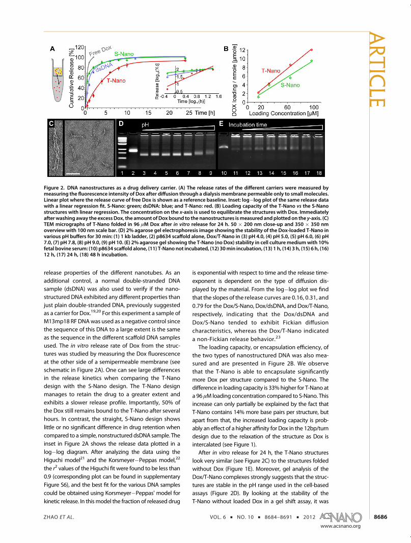
release properties of the different nanotubes. As anadditional control, a normal double-stranded DNAsample (dsDNA) was also used to verify if the nano-structured DNA exhibited any different properties thanjust plain double-stranded DNA, previously suggestedas a carrier for Dox.19,20 For this experiment a sample ofM13mp18 RF DNAwas used as a negative control sincethe sequence of this DNA to a large extent is the sameas the sequence in the different scaffold DNA samplesused. The in vitro release rate of Dox from the struc-tures was studied by measuring the Dox fluorescenceat the other side of a semipermeable membrane (seeschematic in Figure 2A). One can see large differencesin the release kinetics when comparing the T-Nanodesign with the S-Nano design. The T-Nano designmanages to retain the drug to a greater extent andexhibits a slower release profile. Importantly, 50% ofthe Dox still remains bound to the T-Nano after severalhours. In contrast, the straight, S-Nano design showslittle or no significant difference in drug retention whencompared to a simple, nonstructureddsDNA sample. Theinset in Figure 2A shows the release data plotted in alog�log diagram. After analyzing the data using theHiguchi model21 and the Korsmeyer�Peppas model,22
the r2 values of the Higuchi fit were found to be less than0.9 (corresponding plot can be found in supplementaryFigure S6), and the best fit for the various DNA samplescould be obtained using Korsmeyer�Peppas' model forkinetic release. In thismodel the fraction of released drug
is exponential with respect to time and the release time-exponent is dependent on the type of diffusion dis-played by the material. From the log�log plot we findthat the slopes of the release curves are 0.16, 0.31, and0.79 for the Dox/S-Nano, Dox/dsDNA, and Dox/T-Nano,respectively, indicating that the Dox/dsDNA andDox/S-Nano tended to exhibit Fickian diffusioncharacteristics, whereas the Dox/T-Nano indicateda non-Fickian release behavior.23
The loading capacity, or encapsulation efficiency, ofthe two types of nanostructured DNA was also mea-sured and are presented in Figure 2B. We observethat the T-Nano is able to encapsulate significantlymore Dox per structure compared to the S-Nano. Thedifference in loading capacity is 33%higher for T-Nano ata 96 μM loading concentration compared to S-Nano. Thisincrease can only partially be explained by the fact thatT-Nano contains 14% more base pairs per structure, butapart from that, the increased loading capacity is prob-ably an effect of a higher affinity for Dox in the 12bp/turndesign due to the relaxation of the structure as Dox isintercalated (see Figure 1).After in vitro release for 24 h, the T-Nano structures
look very similar (see Figure 2C) to the structures foldedwithout Dox (Figure 1E). Moreover, gel analysis of theDox/T-Nano complexes strongly suggests that the struc-tures are stable in the pH range used in the cell-basedassays (Figure 2D). By looking at the stability of theT-Nano without loaded Dox in a gel shift assay, it was
Figure 2. DNA nanostructures as a drug delivery carrier. (A) The release rates of the different carriers were measured bymeasuring the fluorescence intensity of Dox after diffusion through a dialysis membrane permeable only to small molecules.Linear plot where the release curve of free Dox is shown as a reference baseline. Inset: log�log plot of the same release datawith a linear regression fit. S-Nano: green; dsDNA: blue; and T-Nano: red. (B) Loading capacity of the T-Nano vs the S-Nanostructures with linear regression. The concentration on the x-axis is used to equilibrate the structures with Dox. Immediatelyafter washing away the excess Dox, the amount of Dox bound to the nanostructures ismeasured and plotted on the y-axis. (C)TEM micrographs of T-Nano folded in 96 μM Dox after in vitro release for 24 h. 50 � 200 nm close-up and 350 � 350 nmoverviewwith 100 nm scale bar. (D) 2% agarose gel electrophoresis image showing the stability of the Dox-loaded T-Nano invarious pH buffers for 30 min: (1) 1 kb ladder, (2) p8634 scaffold alone, Dox/T-Nano in (3) pH 4.0, (4) pH 5.0, (5) pH 6.0, (6) pH7.0, (7) pH 7.8, (8) pH 9.0, (9) pH 10. (E) 2% agarose gel showing the T-Nano (no Dox) stability in cell culturemediumwith 10%fetal bovine serum: (10) p8634 scaffold alone, (11) T-Nano not incubated, (12) 30min incubation, (13) 1 h, (14) 3 h, (15) 6 h, (16)12 h, (17) 24 h, (18) 48 h incubation.
ARTIC
LE
ZHAO ET AL . VOL. 6 ’ NO. 10 ’ 8684–8691 ’ 2012
www.acsnano.org
8687
possible to rule out gel shift and the implied struc-tural change attributable to Dox release. The T-Nanostructure seems to be stable over the time spansused in this report; see Figure 2E (see supplementaryFigure S7 for a gel of Dox-loaded T-Nano over thesame time span). Interestingly, the S-Nano structureappears not to reversibly return to its ground stateafter 24 h release as T-Nano does; see supplementaryFigure S3 for additional TEM images and agarosegels of the structures at various stages after releaseof Dox.Similarly to most controlled release systems24 and
as verified by our release rate mesurements, Dox willstart to diffuse out of the nanostructures immediatelyupon transfer to an environment with a low Doxconcentration, such as the cell culture. But becauseof the retention capabilities of the T-Nano, the Dox isreleased primarily at the cells due to cell binding anduptake of the nanostructures. The current opinion isthat DNA structures get degraded after endocytosisfollowed by the release of Dox inside endocytoticvesicles.14 Our data, although not sufficient to directlysupport this effect, are consistent with such aninterpretation.The cytotoxicity of Dox/T-Nano was examined by
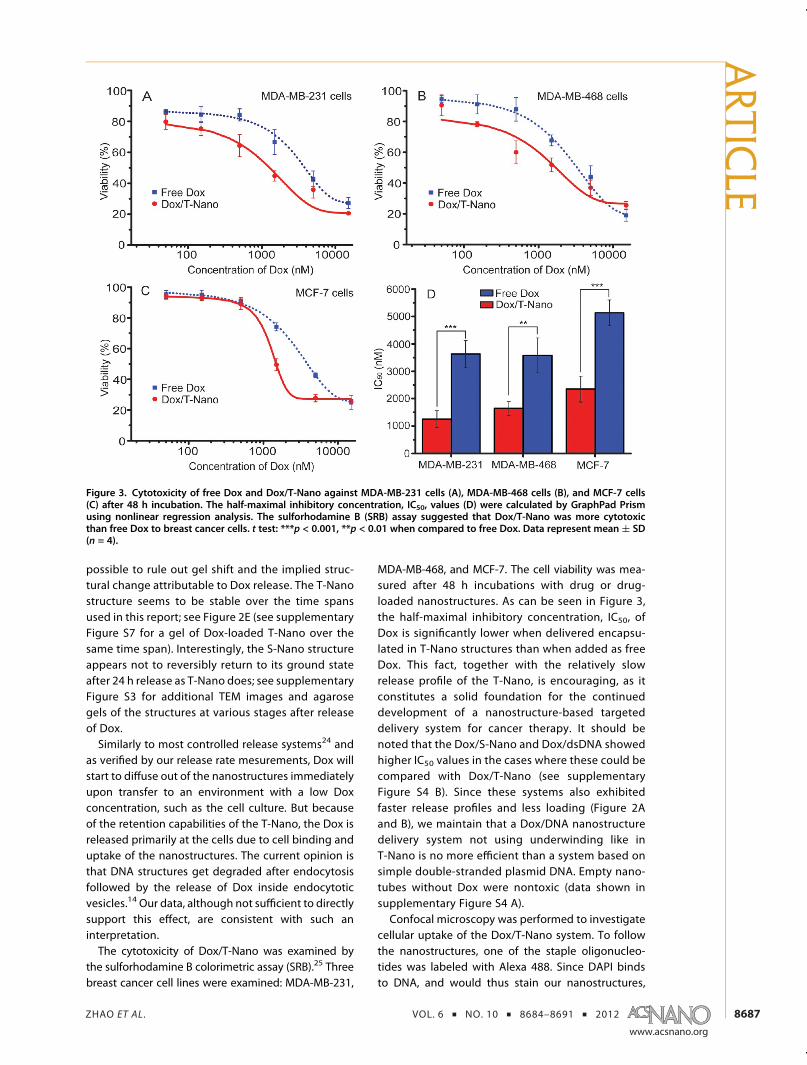
the sulforhodamine B colorimetric assay (SRB).25 Threebreast cancer cell lines were examined: MDA-MB-231,
MDA-MB-468, and MCF-7. The cell viability was mea-sured after 48 h incubations with drug or drug-loaded nanostructures. As can be seen in Figure 3,the half-maximal inhibitory concentration, IC50, ofDox is significantly lower when delivered encapsu-lated in T-Nano structures than when added as freeDox. This fact, together with the relatively slowrelease profile of the T-Nano, is encouraging, as itconstitutes a solid foundation for the continueddevelopment of a nanostructure-based targeteddelivery system for cancer therapy. It should benoted that the Dox/S-Nano and Dox/dsDNA showedhigher IC50 values in the cases where these could becompared with Dox/T-Nano (see supplementaryFigure S4 B). Since these systems also exhibitedfaster release profiles and less loading (Figure 2Aand B), we maintain that a Dox/DNA nanostructuredelivery system not using underwinding like inT-Nano is no more efficient than a system based onsimple double-stranded plasmid DNA. Empty nano-tubes without Dox were nontoxic (data shown insupplementary Figure S4 A).Confocal microscopy was performed to investigate
cellular uptake of the Dox/T-Nano system. To followthe nanostructures, one of the staple oligonucleo-tides was labeled with Alexa 488. Since DAPI bindsto DNA, and would thus stain our nanostructures,
Figure 3. Cytotoxicity of free Dox and Dox/T-Nano against MDA-MB-231 cells (A), MDA-MB-468 cells (B), and MCF-7 cells(C) after 48 h incubation. The half-maximal inhibitory concentration, IC50, values (D) were calculated by GraphPad Prismusing nonlinear regression analysis. The sulforhodamine B (SRB) assay suggested that Dox/T-Nano was more cytotoxicthan free Dox to breast cancer cells. t test: ***p < 0.001, **p < 0.01 when compared to free Dox. Data represent mean ( SD(n = 4).
ARTIC
LE
ZHAO ET AL . VOL. 6 ’ NO. 10 ’ 8684–8691 ’ 2012
www.acsnano.org
8688
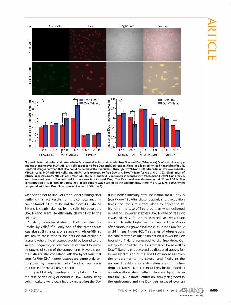
we decided not to use DAPI for nuclear staining afterverifying this fact. Results from the confocal imagingcan be found in Figure 4A, and the Alexa 488-labeledT-Nano is clearly taken up by the cells. Moreover, theDox/T-Nano seems to efficiently deliver Dox to thecell nuclei.Similarly to earlier studies of DNA nanostructure
uptake by cells,11,26,27 only one of the componentswas labeled (in this case, one staple with Alexa 488), sosimilarly to these reports, the data do not exclude ascenario where the structures would be bound to thesurface, degraded, or otherwise destabilized followedby uptake of some of the components. Nevertheless,the data are also consistent with the hypothesis thatlarge (g7kb) DNA nanostructures are completely en-docytosed by mammalian cells,11 and our opinion isthat this is the most likely scenario.To quantitatively investigate the uptake of Dox in
the case of free drug or bound in Dox/T-Nano, livingcells in culture were examined by measuring the Dox
fluorescence intensity after incubation for 0.5 or 2 h(see Figure 4B). After these relatively short incubationtimes, the levels of intracellular Dox appear to behigher in the case of free drug than when deliveredin T-Nano. However, if excess Dox/T-Nano or free Doxis washed away after 2 h, the intracellular levels of Doxare significantly higher in the case of Dox/T-Nanoafter continued growth in fresh culturemedium for 12or 24 h (see Figure 4C). This series of observationsindicate that the cellular elimination is lower for Doxbound to T-Nano compared to the free drug. Ourinterpretation of the results is that free Dox as well asDox/T-Nano is endocytosed as discussed above, fol-lowed by diffusion of the small Dox molecules fromthe endosomes to the cytosol and finally to thenucleus. The difference in depletion rates for the freedrug and Dox/T-Nano can most likely be attributed toan intracellular depot effect. Here we hypothesizethat the DNA nanostructures are slowly degraded inthe endosomes and the Dox gets released over an
Figure 4. Internalization and intracellular Dox level after incubation with free Dox and Dox/T-Nano. (A) Confocal microscopyimages of monolayer MDA-MB-231 cells exposed to free Dox and Dox-loaded Alexa 488-labeled twisted nanotubes for 2 h.Confocal images certified that Dox could be delivered to the nucleus through Dox/T-Nano. (B) Intracellular Dox level in MDA-MB-231 cells, MDA-MB-468 cells, and MCF-7 cells exposed to free Dox and Dox/T-Nano for 0.5 and 2 h. (C) Elimination ofintracellular Dox.MDA-MB-231 cells,MDA-MB-468 cells, andMCF-7 cellswere incubatedwith freeDox andDox/T-Nano for 2 hand then continued to be cultured in fresh medium (absent Dox). The Dox level was determined at 12 and 24 h. Theconcentration of Dox (free or equivalent) in cell culture was 5 μM in all the experiments. t test: **p < 0.01, *p < 0.05 whencompared with free Dox. Data represent mean ( SD (n = 3).
ARTIC
LE
ZHAO ET AL . VOL. 6 ’ NO. 10 ’ 8684–8691 ’ 2012
www.acsnano.org
8689
extended period of time, with the endosomes actingas local depots. In contrast, free Dox would get trans-ported in quickly, but reversely it would also diffuseout of the cells at a higher rate. This is, however,speculative. The details of DNA-nanostructures up-take in cell culture remain to a large extent unknown,and further microscopy studies are needed to eluci-date the exact pathway.Induction of apoptosis is the desired mechanism
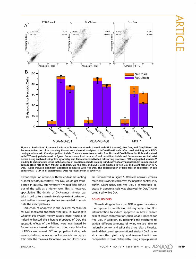
for Dox-mediated anticancer therapy. To investigatewhether this system merely caused more necrosis orindeed enhanced the inherent properties of Dox, theapoptotic effects of the T-Nano were investigated byfluorescence activated cell sorting. Using a combinationof FITC-labeled annexin V28 and propidium iodide, cellswere sorted into populations of live, necrotic, and apop-totic cells. The main results for free Dox and Dox/T-Nano
are summarized in Figure 5. Whereas necrosis remainsmore or less widespread across the negative control (PBSbuffer), Dox/T-Nano, and free Dox, a considerable in-crease in apoptotic cells was observed for Dox/T-Nanocompared to free Dox.
CONCLUSIONS
These findings indicate that DNA origami nanostruc-ture represents an efficient delivery system for Doxinternalization to induce apoptosis in breast cancercells at lower concentrations than what is needed forfree Dox. In addition, by designing the structures toexhibit different amounts of twist, we are able torationally control and tailor the drug release kinetics.We find that by using conventional, straight DNAnano-structures the cytotoxicity and release kinetics arecomparable to those obtained by using simple plasmid
Figure 5. Evaluation of the mechanisms of breast cancer cells treated with PBS (control), free Dox, and Dox/T-Nano. (A)Representative dot plots showing fluorescence channel analyses of MDA-MB-468 cells after dual staining with FITC-conjugated annexin V and propidium iodide. The cells were treated with free Dox and Dox/T-Nano for 48 h and stainedwith FITC-conjugated annexin V (green fluorescence, horizontal axis) and propidium iodide (red fluorescence, vertical axis)before being analyzed using flow cytometry and fluorescence-activated cell sorting protocols. FITC-conjugated annexin Vbinding to phosphatidylserine in the absence of propidium iodide staining is indicative of early apoptosis. (B) Comparison ofcell apoptosis rate of MDA-MB-231 cells, MDA-MB-468 cells, and MCF-7 cells exposed to free Dox and Dox/T-Nano for 48 h.Dox/T-Nano induced significant apoptosis compared with free Dox. The concentration of Dox (free or equivalent) in cellculture was 10 μM in all experiments. Data represent mean ( SD (n = 3).
ARTIC
LE
ZHAO ET AL . VOL. 6 ’ NO. 10 ’ 8684–8691 ’ 2012
www.acsnano.org
8690
DNA. In contrast, by using a twisted 12bp/turn design,a system with superior drug delivery characteristics isachieved. The promising release kinetics and cytotoxi-city of the Dox/T-Nano system, in combinationwith the
well-known flexibility of the DNA origami method todecorate the structures with targeting ligands,10 makethis a promising candidate platform for active target-ing of nanostructures intended for anticancer therapy.
MATERIALS AND METHODSFolding of DNA Origami Nanostructures. Each sample was pre-
pared by combining a 5 nM scaffold (p7560 or p8634, derivedfrom M13mp18), 25 nM of each staple oligonucleotide, buffer,and salts including 5 mM Tris, 1 mM EDTA (pH 7.8 at 20 �C), and10 mM MgCl2. Folding was carried out by rapid heat denatura-tion followed by slow cooling from 80 to 60 �C over 80min, then60 to 24 �C over 173 h. To remove the excess staples and/or freedoxorubicin, the DNA nanostructure suspensions were addedto VIVAspin 500 (molecular cutoff of 100 kD, Sartorius StedimBiotech GmbH) and centrifuged at 3000g for 30min followed bysuspension in Tris buffer (5 mM Tris, 1 mM EDTA, and 13 mMMgCl2). Samples were electrophoresed on 2% agarose gels(0.5� TBE, 11 mM MgCl2, 0.5 μg/mL ethidium bromide) at 70 Vfor 3 h in an ice�water bath.
Electron Microscopy. A 3 μL aliquot of Dox/T-NanoT samplewas spotted on a glow-discharged, carbon-coated Formvar grid(Electron Microscopy Sciences), incubated for 20 s, blotted offwith filter paper, and then stainedwith 2% (w/v) aqueous uranylformate solution. EM analysis was performed using a FEIMorgagni 268(D) transmission electron microscope at 80 kVwith nominal magnifications between 12 000 and 44 000.Images were recorded digitally by using the Advanced Micro-scopy Techniques Image Capture Engine 5.42.
Stability of Doxorubicin-Intercalated Nanotube. TheDox-intercalatednanotube was incubated with culture medium (including 10%fetal bovine serum) at 37 �C for 30min, 1, 3, 6, 12, 24 and 48 h orin various pH (4.0, 5.0, 6.0, 7.0, 7.8, 9.0, and 10.0) Tris buffer(5 mM Tris, 1 mM EDTA) for 30 min, and then the stability wasverified using agarose (2%) gel electrophoresis (0.5� TBE, 11mMMgCl2, 0.5 μg/mL ethidium bromide) at 70 V for 3 h in an ice�water bath.
Drug Intercalation Efficiency. To determine Dox intercalactionefficiency, 60 μL of Dox-intercalated nanotube was added intoVIVaspin 500 (molecular cutoff of 100 kD, Biotech) and centri-fuged at 3000g for 30 min. Dox content in the filtrate as freedrug was measured using a microplate reader. The drug inter-calaction efficiency (%) was calculated by the following equa-tion: Intercalaction efficiency (%) = (Dtotal � Dfree)/Dtotal � 100.Dtotal is the Dox content in Dox-intercalated nanotube solution;Dfree is the Dox content in the filtrate.
In Vitro Drug Release Behaviors of Doxorubicin-Intercalated Nanotube.The dialysis units with molecular weight cutoff of 20 000 Dawere used to carry out the drug release experiments. Phosphate-buffered saline (PBS, pH = 7.4) was used as the drug releasemedia. The sample Dox-intercalated nanotube or Dox solution(30 μL) was put into the dialysis unit. The sealed dialysis unitswere put into a polystyrene flotation device, floating on asurface of 1000 mL of release media in a beaker. The releasemedium was stirred at a speed of 200 rpm at 37 �C protectedfrom light. At certain time intervals, the dialysis unit was takenout and 10 μL of Dox-intercalated nanotube was taken out fromthe dialysis unit for measuring the nonreleased drug concen-trations by the microplate reader.
In Vitro Cytotoxicity Assay. The cytotoxicity of empty nano-tubes, free Dox, Dox/S-Nano, Dox/T-Nano, and Dox/dsDNA wastested against breast cancer cell lines MCF-7, MDA-MB-231, andMDA-MB-468 (MCF-7 human breast adenocarcinoma cell lineER pos., 231 adenocarcinoma ER neg., 468 adenocarcinoma ERneg.). The cells were obtained from ATCC (American TissueCulture Collection, Manassas, VA, USA) and maintained accord-ing to instructions. The cytotoxicity was evaluated by thesulforhodamine B cell cytotoxicity assay (G-Biosciences, StLouis, MO, USA). Cells were cultured in Dulbecco's modifiedEagle medium supplemented with 10% fetal bovine serum,
100 units/mL penicillin, and 100mg/mL streptomycin under 5%CO2 at 37 �C and collected by trypsinization using 0.25% trypsinsolution. Cells were seeded in 96-well plates (Costa, CorningIncorporated) at a density of 1� 104 cells/well and incubated for24 h to allow for cell attachment. The cells were incubated withDox/T-Nano or Dox at equivalent drug concentrations rangingfrom 50 to 15 000 nM for 48 h. At the end of the experiment, theSRB assay was performed according to the manufacturer'sinstructions. The absorbance was measured at 565 nm using amicroplate reader. Cell viability rate was calculated by thefollowing equation:
Cell viability rate (%) ¼ (Adrug � Ablank)=(Acontrol � Ablank)� 100
Adrug is the absorbance of the cells incubated with Dox/T-Nanoor Dox; Acontrol is the absorbance of the cells incubated with theculturemedium only; and Ablank is the absorbance of the culturemedium.
Intracellular Uptake of Doxorubicin-Intercalated Twisted Nanotube.For the Dox/T-Nano uptake study, 12-well plates were seededwith breast cancer cells at 2 � 105 per well, and the cells wereallowed to attach for 24 h. The medium was replaced with 1 mLof medium containing Dox/T-Nano or Dox solution (final Doxconcentration 5 μM) and incubated for 0.5 and 2 h. Cells werewashed three times to remove the free Dox/T-Nano or Dox withPBS buffer and lysed in 100 μL of cell lysis buffer for 10 min.A 10 μL amount of cell lysate was used to quantitate proteinconcentration by the BCA assay. The remaining portion wasextracted by dissolving each sample in 0.2 mL of acidifiedmethanol (0.1 M HCL, 90% methanol) solution, and the super-natant was analyzed for Dox level using the microplate reader(excitation wavelength: 485 nm, emission wavelength: 591 nm)after centrifugation at 16800g for 10 min. The data werenormalized to per milligram cell protein.
Intracellular Elimination of Doxorubicin. For the Dox/T-Nanoelimination study, 12-well plates were seeded with breastcancer cells at 2 � 105 per well, and the cells were allowed toattach for 24 h. Themediumwas replaced with 1mL of mediumcontainingDox/T-Nano or Dox solution (final Dox concentration5 μM) and incubated for 2 h. Cells were washed three times toremove the free Dox or Dox/T-Nano with PBS buffer andcontinued to incubate with fresh medium. Intracellular Doxlevel was determined at 12 and 24 h using the same method asthat described above for intracellular uptake assay.
Internalization of Doxorubicin-Intercalated Twisted Nanotube. Con-focal laser scanningmicroscopy (CLSM, Olympus FV1000) furtherconfirmed the cellular uptake of Dox/T-Nano. MDA-MB-231 cellswere plated at a density of 1 � 105 cells/well containing 22 mmsterile coverslips for 24 h. Dox and the Dox/T-Nano with equiva-lent Dox concentration (5 μM) were incubated for 2 h, andthen the cells were washed three times with PBS and fixed with3.7% formaldehyde in PBS. Vectashield (Vector Laboratories, Inc.Burlingame, CA, USA) was dropped on the slides to seal the cellsamples after the cells were washed three times with PBS. Thestained coverslips were imaged using CLSM.
Cell Apoptosis Analysis. Apoptotic cells were determined bydual staining with an annexin V-FITC and propidium iodide kit(Invitrogen, CA, USA) according to the manufacturer's instruc-tions. In brief, the breast cancer cells were seeded into six-wellculture plates at a concentration of 1 � 106 cells/well andincubated for 24 h to allow cell attachment. The cells werepretreated with Dox, Dox/T-Nano at equivalent drug concen-trations (10 μM), or empty nanotubes for 48 h and collected bytrypsinization using a 0.125% trypsin solution. After that, thecells were washed twice with PBS (pH = 7.4) and resuspended in100 μL of binding buffer at a density of 1� 105/mL. Then 2 μL of
ARTIC
LE
ZHAO ET AL . VOL. 6 ’ NO. 10 ’ 8684–8691 ’ 2012
www.acsnano.org
8691
annexin V-FITC was added, and cells were incubated at roomtemperature for 20 min in darkness. Then 5 μL of propidiumiodide stock solution (100 μg/mL) was added after adding400 μL of PBS, and cells were incubated for another 5 min indarkness. The cells were analyzed by flow cytometry (BectonDickinson, Sunnyvale, CA, USA) with CellQuest software within1 h. The numbers of cells undergoing necrosis (positive forpropidium iodide), early apoptosis (positive for annexin V), andlate apoptosis (double-positive for annexin V and propidiumiodide) stages of apoptosiswere quantified using flow cytometry.
Statistical Analysis. All datawere expressed asmean( SD. IC50values were calculated by GraphPad Prism using nonlinearregression analysis. The statistical significance was determinedusing a t test. Ap value less than0.05 (i.e.,p<0.05)was consideredto indicate statistical significance for all comparisons.
Conflict of Interest: The authors declare no competingfinancial interest.
Supporting Information Available: Structure designs, DNAsequences, additional TEM images, cytotoxicity data, and flowcytometry data are available free of charge via the Internet athttp://pubs.acs.org.
Acknowledgment. The project was funded by the SwedishResearch Council (VR) through a repatriation grant and a projectgrant to B.H. (grants 2010-6296 and 2010-5060). B.H. and A.M.N.are recipients of assistant professorships with startup fundingfunded by Carl Bennet AB, Karolinska Institutet, and Vinnova. A.S. is partially funded by a Karolinska Institutet faculty grant (KID).Funding support to A.M.N. by the Royal Swedish Academy ofSciences, Falk Foundation, Jeanssons Foundation, Axel and EvaWallströms Foundation, and the Swedish Research Council (VR)under grants 2011-3720 and 2009-3259 is acknowledged. Y.X.Z.is funded by the China Scholarship Council (CSC). We acknowl-edge Klas Udekwu and Ana Teixeira for helpful discussions andXiaoyuan Ren for helping Y.X.Z. get started.
REFERENCES AND NOTES1. Seeman, N. C. Nucleic-Acid Junctions and Lattices. J. Theor.
Biol. 1982, 99, 237–247.2. Rothemund, P. W. K.; Papadakis, N.; Winfree, E. Algorithmic
Self-Assembly of DNA Sierpinski Triangles. PLoS Biol. 2004,6, 240–252.
3. Douglas, S. M.; Dietz, H.; Liedl, T.; Högberg, B.; Graf, F.;Shih, W. M. Self-Assembly of DNA into Nanoscale Three-Dimensional Shapes. Nature 2009, 459, 414–418.
4. Rothemund, P. W. K. Folding DNA to Create NanoscaleShapes and Patterns. Nature 2006, 440, 297–302.
5. Liedl, T.; Högberg, B.; Tytell, J.; Ingber, D. E.; Shih, W. M. Self-Assembly of Three-Dimensional Prestressed TensegrityStructures from DNA. Nat. Nanotechnol. 2010, 5, 520–524.
6. Dietz, H.; Douglas, S. M.; Shih, W. M. Folding DNA intoTwisted and Curved Nanoscale Shapes. Science 2009, 325,725–730.
7. Högberg, B.; Liedl, T.; Shih, W. M. Folding DNA Origamifrom a Double-Stranded Source of Scaffold. J. Am. Chem.Soc. 2009, 131, 9154–55.
8. Douglas, S. M.; Bachelet, I.; Church, G. M. A Logic-GatedNanorobot for Targeted Transport of Molecular Payloads.Science 2012, 335, 831–834.
9. Andersen, E. S.; Dong, M.; Nielsen, M. M.; Jahn, K.;Subramani, R.; Mamdouh, W.; Golas, M. M.; Sander, B.;Stark, H.; Oliveira, C. L. P.; et al. Self-Assembly of a Nano-scale DNA Box with a Controllable Lid. Nature 2009, 459,73–76.
10. Peer, D.; Karp, J. M.; Hong, S.; Farokhzad, O. C.; Margalit, R.;Langer, R. Nanocarriers as an Emerging Platform forCancer Therapy. Nat. Nanotechnol. 2007, 2, 751–760.
11. Schüller, V. J.; Heidegger, S.; Sandholzer, N.; Nickels, P. C.;Suhartha, N. A.; Endres, S.; Bourquin, C.; Liedl, T. CellularImmunostimulation by CpG-Sequence-Coated DNAOrigami Structures. ACS Nano 2011, 5, 9696–9702.
12. Arora, H. C.; Jensen, M. P.; Yuan, Y.; Wu, A.; Vogt, S.;Paunesku, T.; Woloschak, G. E. Nanocarriers Enhance
Doxorubicin Uptake in Drug-Resistant Ovarian CancerCells. Cancer Res. 2012, 72, 769–778.
13. Kellogg, G. E.; Scarsdale, J. N.; Fornari, F. A., Jr. Identificationand Hydropathic Characterization of Structural FeaturesAffecting Sequence Specificity for Doxorubicin Intercala-tion into DNA Double-Stranded Polynucleotides. NucleicAcids Res. 1998, 26, 4721–4732.
14. Chang, M; Yang, C.-S.; Huang, D.-M. Aptamer-ConjugatedDNA Icosahedral Nanoparticles As a Carrier of Doxorubicinfor Cancer Therapy. ACS Nano 2011, 8, 6156–6163.
15. Ke, Y.; Bellot, G.; Voigt, N. V.; Fradkov, E.; Shih, W. M. TwoDesign Strategies for Enhancement of Multilayer�DNA-Origami Folding: Underwinding for Specific IntercalatorRescue and Staple-Break Positioning. Chem. Sci. 2012, 3,2587–2597.
16. Douglas, S. M.; Marblestone, A. H.; Teerapittayanon, S.;Vazquez, A.; Church, G.M.; Shih,W.M. Rapid Prototyping of3D DNA-Origami Shapes with caDNAno. Nucleic Acids Res.2009, 37, 5001–5006.
17. Ortiz-Lombardia, M.; Gonzalez, A.; Eritja, R.; Aymami, J.;Azorin, F.; Coll, M. Crystal Structure of a DNA HollidayJunction. Nat. Struct. Biol. 1999, 6, 913–917.
18. Frederick, C. A.; Williams, L. D.; Ughetto, G.; van der Marel,G. A.; van Boom, J. H.; Rich, A.; Wang, A. H. StructuralComparison of Anticancer Drug-DNA Complexes: Adria-mycin and Daunomycin. Biochemistry 1990, 29, 2538–2549.
19. Bagalkot, V.; Lee, I.-H.; Yu, M. K.; Lee, E.; Park, S.; Lee, J.-H.;Jon, S. A Combined Chemoimmunotherapy ApproachUsing a Plasmid-Doxorubicin Complex.Mol. Pharmaceutics2009, 6, 1019–1028.
20. Bagalkot, V.; Farokhzad, O. C.; Langer, R.; Jon, S. AnAptamer�Doxorubicin Physical Conjugate as a NovelTargeted Drug-Delivery Platform. Angew. Chem., Int. Ed.2006, 45, 8149–8152.
21. Higuchi, T. Mechanism of Sustained-Action Medication:Theoretical Analysis of Rate of Release of Solid DrugsDispersed in Solid Matrices. J. Pharm. Sci. 1963, 52,1145–1149.
22. Korsmeyer, R. W.; Gurnya, R.; Doelkera, E.; Buria, P.; Peppas,N. A. Mechanisms of Solute Release from Porous Hydro-philic Polymers. Int. J. Pharm. 1983, 15, 25–35.
23. Lin, L. Y.; Lee, N. S.; Zhu, J.; Nyström, A. M.; Pochan, D. J.;Dorshow, R. B.; Wooley, K. L. Tuning Core vs. Shell Dimen-sions to Adjust the Performance of Nanoscopic Containersfor the Loading and Release of Doxorubicin. J. ControlledRelease 2011, 152, 37–48.
24. Charrois, J. R.; Allen, T. M. Drug Release Rate Influences thePharmacokinetics, Biodistribution, Therapeutic Activity,and Toxicity of Pegylated Liposomal Doxorubicin Formu-lations in Murine Breast Cancer. Biochim. Biophys. Acta2004, 1663, 167–177.
25. Skehan, P.; Storeng, R.; Scudiero, D.; Monks, A.; McMahon,J.; Vistica, D.; Warren, T. W.; Bokesch, H.; Kenney, S.; Boyd,M. R. New Colorimetric Cytotoxicity Assay for Anticancer-Drug Screening. J. Nat. Cancer Inst. 1990, 82, 1107–1112.
26. Hamblin, G. D.; Carneiro, K. M. M.; Fakhoury, J. F.; Bujold,K. E.; Sleiman, H. F. Rolling Circle Amplification-TemplatedDNA Nanotubes Show Increased Stability and Cell Pene-tration Ability. J. Am. Chem. Soc. 2012, 134, 2888–2891.
27. Ko, S.-H.; Liu, H.; Chen, Y.; Mao, C. DNA Nanotubes asCombinatorial Vehicles for Cellular Delivery. Biomacromo-lecules 2008, 9, 3039–3043.
28. Koopman, G.; Reutelingsperger, C. P.; Kuijten, G.A.; Keehnen,R. M.; Pals, S. T.; van Oers, M. H. Annexin V for FlowCytometric Detection of Phosphatidylserine Expression onB Cells Undergoing Apoptosis. Blood 1994, 84, 1415–1420.
29. www.molecularmovies.com.30. Castro, C. E.; Kilchherr, F; Kim, D.-N.; Shiao, E. L.; Wauer, T;
Wortmann, P.; Bathe, M.; Dietz, H. A Primer to ScaffoldedDNA Origami. Nat. Methods 2011, 8, 221–229.
ARTIC
LE