correspondence between the 17-segment model and...
TRANSCRIPT

J A C C : C A R D I O V A S C U L A R I M A G I N G V O L . 1 , N O . 3 , 2 0 0 8
© 2 0 0 8 B Y T H E A M E R I C A N C O L L E G E O F C A R D I O L O G Y F O U N D A T I O N I S S N 1 9 3 6 - 8 7 8 X / 0 8 / $ 3 4 . 0 0
P U B L I S H E D B Y E L S E V I E R I N C . D O I : 1 0 . 1 0 1 6 / j . j c m g . 2 0 0 8 . 0 1 . 0 1 4
Correspondence Between the 17-SegmentModel and Coronary Arterial Anatomy UsingContrast-Enhanced Cardiac MagneticResonance Imaging
José T. Ortiz-Pérez, MD, José Rodrı́guez, MD, Sheridan N. Meyers, MD, FACC,Daniel C. Lee, MD, Charles Davidson, MD, FACC, Edwin Wu, MD
Chicago, Illinois
O B J E C T I V E S The purpose of this study was to investigate the correspondence between the
coronary arterial anatomy and supplied myocardium based on the proposed American Heart Association
17-segment model.
B A C K G R O U N D Standardized assignment of coronary arteries to specific myocardial segments is
currently based on empirical assumptions.
M E T H O D S A cardiac magnetic resonance study was performed in 93 subjects following acute
myocardial infarction treated with primary percutaneous coronary intervention. Two observers blindly
reviewed all angiograms to examine the location of the culprit lesion and coronary dominancy. Two
additional observers scored for the presence of cardiac magnetic resonance hyperenhancement (HE) on
a 17-segment model. Segments were divided based on anatomical landmarks such as the interventric-
ular grooves and papillary muscles.
R E S U L T S In a per-segment analysis, 23% of HE segments were discordant with the empirically
assigned coronary distribution. Presence of HE in the basal anteroseptal, mid-anterior, mid-anteroseptal,
or apical anterior wall was 100% specific for left anterior descending artery occlusion. The left anterior
descending artery infarcts frequently involved the mid-anterolateral, apical lateral, and apical inferior
walls. No segment was 100% specific for right coronary artery or left circumflex artery (LCX) occlusion,
although HE in the basal anterolateral wall was highly specific (98%) for LCX occlusion. Combination of
HE in the anterolateral and inferolateral walls was 100% specific for a LCX occlusion, and when extended
to the inferior wall, was also 100% specific for a dominant or codominant LCX occlusion.
C O N C L U S I O N S Four segments were completely specific for left anterior descending artery
occlusion. No segment can be exclusively attributed to the right coronary artery or LCX occlusion.
However, analysis of adjacent segments increased the specificity for a given coronary occlusion. These
findings bring objective evidence in the appropriate segmentation of coronary arterial perfusion
territories and assist accurate assignment of the culprit vessel in various imaging modalities. (J Am Coll
Cardiol Img 2008;1:282–93) © 2008 by the American College of Cardiology Foundation
From the Feinberg Cardiovascular Research Institute and Division of Cardiology, Northwestern University Feinberg School ofMedicine, and the Bluhm Cardiovascular Institute, Northwestern Memorial Hospital, Chicago, Illinois. Dr. Wu received grantsfrom the GlaxoSmithKline Research and Education Foundation for Cardiovascular Disease, the American Heart AssociationScientist Development Grant, and the Feinberg Cardiovascular Research Institute and Department of Medicine. Drs.Ortiz-Pérez and Rodrı́guez were supported by grants from the Spanish Society of Cardiology.
Manuscript received October 5, 2007; revised manuscript received December 13, 2007, accepted January 3, 2008.

TnASivv(twamoncsm(hntals
el(rttgiewn
M
FrJicw�tdop
ntfCCi((soCssUCpsvgCpaaesllvtasosobiwtwtrcadaRnlffhp
rdial Infarction
J A C C : C A R D I O V A S C U L A R I M A G I N G , V O L . 1 , N O . 3 , 2 0 0 8
M A Y 2 0 0 8 : 2 8 2 – 9 3
Ortiz-Pérez et al.
17-Segment Model and Coronary Arterial Anatomy
283
o make appropriate clinical decisions con-cerning diagnosis and treatment, integrationof information between noninvasive func-tional imaging techniques and invasive coro-
ary angiography is essential. The American Heartssociation (AHA) Writing Group on Myocardialegmentation and Registration for Cardiac Imag-
ng recommends a 17-segment model of the leftentricle as an optimally weighted approach for theisual interpretation of regional left ventricularLV) abnormalities by multiple cardiac imagingechniques (1). Individual myocardial segmentsere assigned to coronary artery territories based on
vailable data with little correlation with delineatedyocardial segmentation (2). Cardiac magnetic res-
nance (CMR) imaging has emerged as a powerfuloninvasive technique for the evaluation of myo-ardial function, perfusion, and viability in a singletudy without the need of radioactive tracers ad-inistration, which is a distinct attribute of CMR
3,4). This imaging modality has been shown toave a higher spatial resolution when compared touclear perfusion imaging techniques (5). Addi-ionally, specific structures of the left ventricle, suchs the right ventricular insertion points and papil-ary muscles, can be readily identified to registeregments from one modality to another.
We have previously demonstrated that the sub-ndocardial extent of hyperenhancement (HE) fol-owing ST-segment elevation myocardial infarctionSTEMI) closely matches the myocardial bed atisk determined by angiography (6). The purpose ofhis study was to assess the correspondence betweenhe coronary arteries distribution by invasive an-iography and their supplied myocardium accord-ng to the 17-segment model by using contrast-nhanced CMR imaging in a cohort of patientsith first STEMI presenting for primary percuta-eous coronary intervention (PCI).
E T H O D S
rom a prospective study that investigated LVemodeling following STEMI carried out betweenanuary 2000 and June 2006, we retrospectivelydentified 115 subjects who met the followingriteria: 1) presence of chest pain for at least 30 minith electrocardiographic ST-segment elevation0.1 mV in at least 2 adjacent leads or suspicion of
rue posterior infarction; 2) attempted primary PCIuring the first 24 h of admission; 3) confirmationf acute myocardial infarction by creatine phos-
hokinase release above twice the upper limit of tormal; and 4) absence of clinical history or elec-rocardiographic evidence of prior myocardial in-arction, hemodynamic instability precluding theMR study, or other formal contraindication forMR imaging. We excluded 11 subjects with
nitial Thrombolysis In Myocardial InfarctionTIMI) flow grade �1 in the infarct-related arteryIRA), 2 subjects with previous coronary bypassurgery, and 1 subject with uncertain identificationf the IRA. A total of 101 eligible subjects had theirMR images reviewed for this study. All subjects
igned consent forms for their participation in thetudy, which was approved by the Northwesternniversity Institutional Review Board.oronary angiography. Selective coronary angiogra-hy was performed with a biplane system and atandard femoral approach. All studies were re-iewed by consensus of 2 experienced an-iographers who were masked to theMR data. The identification of the cul-rit lesion in the IRA was easily made inll cases based on the angiographic char-cteristics, the distal TIMI blood flow, thelectrocardiographic findings, and the re-ponse to treatment. The site of the culpritesion occlusion, as well as the severity andocation of other significant lesions wereisually assessed and registered accordingo classical landmarks. A proximal leftnterior descending (LAD) artery occlu-ion was considered when the site ofcclusion occurred proximally to the firsteptal or diagonal branch and a mid-LADcclusion from the first septal or diagonalranch to the next diagonal branch. Prox-mal left circumflex (LCX) occlusionsere considered those located previous to
he first relevant marginal branch, and mid-LCXas any occlusion between the first marginal and
he second marginal or posterolateral branch. Allight coronary artery (RCA) occlusions were lo-ated before the takeoff of the posterior descendingrtery. The coronary tree was considered rightominant when the posterior descending arterynd posterolateral branches originated from theCA and left dominant when they both origi-ated from the LCX. A balanced coronary circu-
ation was defined when the PDA originatedrom the RCA and all posterolateral branchesrom the LCX. All subjects received aspirin andeparin, and most were also treated with glyco-rotein IIb/IIIa inhibitors and clopidogrel. With
A B B
A N D
AHA �
Assoc
CMR �
reson
HE �
IRA �
LAD �
LCX �
LV �
PCI �
interv
RCA �
STEM
myoca
TIMI �
Myoca
he exception of 1 subject with a failed rev
R E V I A T I O N S
A C R O N YM S
American Heart
iation
cardiac magnetic
ance
hyperenhancement
infarct-related artery
left anterior descending
left circumflex
left ventricular
percutaneous coronary
ention
right coronary artery
I � ST-segment elevation
rdial infarction
Thrombolysis In
ascu-

l1C�oaiAiccaealepgamfit1Daawesitspaitiiapaitgatia
pemc
mttiacsLcp
pmots�wegcafgnfds
R
OintsTideadeS
HmSc7ta
J A C C : C A R D I O V A S C U L A R I M A G I N G , V O L . 1 , N O . 3 , 2 0 0 8
M A Y 2 0 0 8 : 2 8 2 – 9 3
Ortiz-Pérez et al.
17-Segment Model and Coronary Arterial Anatomy
284
arization procedure, all patients received at leaststent in the culprit lesion.MR. All studies were performed at a mean of 2.9
2 (range 1 to 7 days) after the admission. A totalf 70 patients returned for a second CMR study atmean of 5 � 3.1 months later. Subjects were
maged in supine position in a 1.5-T Sonata orvanto scanner (Siemens, Erlangen, Germany) and
maged with a dedicated cardiac phased-array re-eiver coil. Standard steady state free precessionines were taken during breath-holds in the 3 longxis views and contiguous 6-mm short axis slicesvery 10 mm from the mitral annulus to the LVpex. Ten to 15 min following intravenous gado-inium at a dose of 0.2 mmol/kg, delayed contrast-nhanced images were acquired in identical sliceositions using a inversion-recovery segmentedradient-echo sequence (7). Inversion times weredjusted to null normal myocardium (230 to 350s). Typical imaging parameters were as follows:
eld of view 360 to 400 mm, matrix size 256, echoime 4 ms, repetition time 40 ms, typical voxel size.4 � 1.4 � 6 mm.ata analysis. All images were de-identified for thenalysis. Representative basal, mid-ventricular, andpical slices as well as the 2-chamber long axis viewsere visually evaluated by a consensus of 2 experi-
nced observers to determine the presence or ab-ence of HE, defined as areas with a mean signalntensity more than 2 standard deviations abovehat of normal myocardium, in each 1 of the 17 LVegments. Segments were divided as previouslyroposed (1). Division between the anterior andnteroseptal segments was made at the anteriornterventricular groove and extended posteriorlyhrough the center of the left ventricle to divide thenferior and inferolateral segments. Likewise, thenferoseptal and inferior segments and the anteriornd anterolateral segments were divided by theosterior interventricular groove. The anteroseptalnd inferoseptal as well as the anterolateral andnferolateral segments were divided halfway be-ween the anterior and inferior interventricularrooves. In the apical segments, the anteroseptalnd inferoseptal segments were combined to formhe apical septum segment and the anterolateral andnferolateral segments were combined to form thepical lateral segment.
A segment was considered affected if HE wasresent in more than 50% of the circumferentialxtent for that segment, and segments with any wallotion abnormality were additionally noted. Adja-
ent short axis views to the representative basal, t
id, or apical views were used to confirm or rejecthe location and presence of any HE. Additionally,he endocardium and epicardium was planimeteredn sequential short axis contrast-enhanced images,nd the circumferential extent of the infarct wasomputed as the percentage of the endocardialurface presenting HE. Ejection fraction, indexedV volumes, and infarct size—expressed as per-entage of LV wall volume—were computed asreviously described (6).The specificity and predictive accuracy of the
resence of HE in each segment of the 17-segmentodel for the diagnosis of LAD, RCA, or LCX
cclusion was calculated irrespective of the loca-ion—proximal, mid, or distal—of the culprit le-ion. Quantitative variables are expressed as mean
standard deviation. One-way analysis of varianceas used to compare the infarct circumferentialxtent and CMR-derived parameters betweenroups, and Kruskal-Wallis test was performed toompare the mean number of segments with HEccording to the IRA. Chi-square test was appliedor comparison of qualitative variables betweenroups. Paired t test was used to compare theumber of segments with HE at baseline andollow-up and the number of segments with wallysfunction. A p value � 0.05 was consideredtatistically significant.
E S U L T S
f the 101 available subjects, 93 had a singlesolated area of HE. None had an additional coro-ary occlusion other than the IRA, although 20 ofhem were treated with an additional PCI of aignificant nonculprit lesion before the CMR study.able 1 depicts clinical and angiographic character-
stics of the study population. There were noifferences in risk factors profile, multivessel dis-ase, time-to-reperfusion, final TIMI flow grade,nd collateral grades according to the IRA. Figure 1emonstrates representative division of the contrast-nhanced CMR studies into 17 segments followingTEMI to the LAD, RCA, and LCX arteries.The remaining 8 subjects had 2 separate areas ofE clearly isolated and surrounded by normalyocardium and were excluded from the analysis.
even of them had a severe lesions—6 cases with ahronic total or subtotal occlusion and 1 case with a0% stenosis—in a vessel different than the IRAhat could explain the presence of these additionalreas of HE. For each of these cases, a representa-
ive single slice with its corresponding finding in the
In Myocardial Infarction.
J A C C : C A R D I O V A S C U L A R I M A G I N G , V O L . 1 , N O . 3 , 2 0 0 8
M A Y 2 0 0 8 : 2 8 2 – 9 3
Ortiz-Pérez et al.
17-Segment Model and Coronary Arterial Anatomy
285
Figure 1. Representative Segmentation and Pattern of Contrast HE Over 17-Segments of the LAD, RCA, and LCX Arteries
A single basal, mid, apical, and 2-chamber contrast-enhanced views of 3 subjects (left panels) following an acute myocardial infarctionwith occlusion to the left anterior descending (LAD) artery, right coronary artery (RCA), and left circumflex (LCX) artery. The dotted yel-low lines delineate and separate the 17-segments over the entire left ventricle. Bulls-eye diagrams (right) reveal the pattern of infarction
Table 1. Clinical and Angiographic Characteristics of the Study Population
Total (n � 93)
Culprit Artery
LAD (n � 44) RCA (n � 34) LCX (n � 15)
Clinical data
Age (yrs) 57 � 11 58 � 11 59 � 10 53 � 10
Male (%) 84 84 82 87
Diabetes (%) 15 11 18 20
Dyslipidemia (%) 54 52 48 67
Hypertension (%) 51 43 54 67
Smoker (%) 49 41 58 60
Multivessel disease (%) 59 50 79 40
Right dominance (%) 78 71 94 67
Final TIMI flow 3 (%) 90 89 91 93
Time-to-reperfusion (min)* 199 [135 to 345] 210 [148 to 390] 160 [104 to 283] 210 [165 to 450]
Good collaterals (%) 42 39 56 27
CMR data
Ejection fraction (%) 41.5 � 9.6 35.8 � 9.5 47.4 � 5.3 44.6 � 13.4
End-diastolic volume index (ml/m2) 78.1 � 16.0 79.0 � 17 75.4 � 15.5 82.1 � 14.3
End-systolic volume index (ml/m2) 45.6 � 14.5 50.0 � 16 39.9 � 10.2 46.1 � 13.4
Infarct size (% LV wall volume) 21.6 � 11.2 28.1 � 11.5 15.1 � 6.4 17.8 � 6.0
Data expressed as percentage or mean � standard deviation. *Data represents median [25th to 75th percentiles].CMR � cardiac magnetic resonance; LAD � left anterior descending; LCX � left circumflex; LV � left ventricular; RCA � right coronary artery; TIMI � Thrombolysis
for each example. CMR � cardiac magnetic resonance; HE � hyperenhancement.

cF
twaHoTi4tfwRrHra3amsovwL
sawimLmottcTii
ctbo6LHa1oo
J A C C : C A R D I O V A S C U L A R I M A G I N G , V O L . 1 , N O . 3 , 2 0 0 8
M A Y 2 0 0 8 : 2 8 2 – 9 3
Ortiz-Pérez et al.
17-Segment Model and Coronary Arterial Anatomy
286
ardiac catheterization laboratory is presented inigure 2.Subjects with LAD occlusion showed lower ejec-
ion fractions and greater infarct sizes than subjectsith either LCX or RCA occlusion (p � 0.01 for
ll). They also had higher number of segments withE (7.7 � 2.1) than subjects with LCX (5.1 � 2.4)
r RCA occlusion (4.3 � 1.5) (p � 0.001 for both).he infarct circumferential extent was also higher
n patients with LAD than LCX or RCA occlusion,0.1 � 10.0, 26.9 � 7.3, and 23.1 � 5.8, respec-ively (p � 0.001 for both). No significant dif-erence was observed in the number of segmentsith HE and circumferential extent between theCA and LCX occlusion, p � 0.6 and p � 0.5,
espectively.E distribution according to the IRA. A bulls-eyeepresentation with specificities of HE distributionccording to the culprit artery is presented in FigureA. At baseline, the presence of HE in the basalnteroseptal (segment 2), mid-anterior (segment 7),id-anteroseptal (segment 8), or apical anterior
egment (segment 13) was 100% specific for LADcclusion. In subjects with anterior infarction, in-olvement of the basal anteroseptum (segment 2)as, in addition, highly specific (85%) for proximal
Figure 2. Patients With Unrecognized Previous Myocardial Infar
A single representative delayed enhanced image with the correspopresented. The yellow arrows point out the acute infarct, and the rartery; PLT � platelet; other abbreviations as in Figure 1.
AD occlusion. No individual segment was 100% t
pecific for RCA or LCX occlusion. However,mong 9 studies with HE in the basal anterolateralall (segment 6), 7 had an occlusion in the LCX, 1
n the LAD, and another in a ramus. Thus, involve-ent of this segment was highly specific (98%) forCX occlusion. The basal inferolateral wall (seg-ent 5) was also highly specific (95%) for LCX
cclusion. The basal inferior wall (segment 4) washe most specific (93%) for RCA occlusion, al-hough this segment was additionally involved in 4ases of dominant or codominant LCX occlusion.he positive predictive value of the presence of HE
n every segment for each coronary artery is shownn Table 2.
An additional analysis, taking into account adja-ent segments, was performed to distinguish be-ween RCA and LCX occlusions. Thus, any com-ination of HE in the inferolateral wall (segments 5r 11) with HE in the anterolateral wall (segmentsor 12) was 100% specific for an occlusion of theCX or ramus. Furthermore, the presence of anyE in the anterolateral wall in combination with
ny HE in the inferior wall (segments 4 or 10) was00% specific for dominant or codominant LCXcclusion. Figure 4 illustrates 3 examples of LCXcclusion with variable extent of HE depending on
n
g findings in the catheterization lab for each of the 8 subjects isarrows point to the remote infarct. OM1 � obtuse marginal
ctio
ndined
he dominance and the site of the occlusion.

ostCd1�Tfidcfiw2(0H2aSdapww
rad(
s in
J A C C : C A R D I O V A S C U L A R I M A G I N G , V O L . 1 , N O . 3 , 2 0 0 8
M A Y 2 0 0 8 : 2 8 2 – 9 3
Ortiz-Pérez et al.
17-Segment Model and Coronary Arterial Anatomy
287
The greatest variability in coronary distributionccurred at the inferoapical wall (segment 15), withpecificities of 57%, 45%, and 46% for occlusion ofhe LAD, LCX, and RCA, respectively.hange in ce-CMR distribution. Infarcted myocar-ium decreased from 20.3 � 10.3% to 16.2 �0.5%; p � 0.001, which represents a relative 21.5
3.7% reduction in the mean initial infarct size.he number of segments with any HE decreased
rom 6.0 � 2.5 to 5.3 � 2.6; p � 0.001. Figure 3Bs an additional diagram with specificities of HEistribution at follow-up. There were no majorhanges in HE distribution between baseline andollow-up studies. According to infarct location,nfarct size significantly decreased among patientsith LAD infarction (n � 34, from 26.1 � 9.8% to0.5 � 11.0%; p � 0.001) and non-LAD infarctionn � 36, from 15.4 � 7.6% to 12.6 � 8.4%; p �.001). Similarly, the number of segments with anyE decreased among patients with LAD (7.5 �
.1 vs. 6.7 � 2.6, p � 0.01) and non-LAD infarctionss well (4.7 � 2.0 vs. 4.2 � 2.0, p � 0.001).egmental wall motion according to the IRA. Figure 5epicts the specificities of segmental wall motionbnormalities according to the IRA. In a per-atient analysis, the number of segments with HEas not different from the number of segments with
LAD R
A
B
Scale: 0% 1-49%
95% 83%
44%
100%100%
92%83%
43%
98%
100%
100%
92%
57%
12%
29%
64%91%
92% 92%
58%
100%100%
89%
42%
97%
100%
100%
94%
55%
22%
36%
71%89%
Figure 3. Specificity of Segmental HE According to the IRA Acut
Bulls-eye diagrams showing the distribution and specificities of CMsegments were 100% specific for LAD occlusion and none for RCAwas 100% specific for LCX occlusion (B). No major changes were seItalicized numbers designate the segment number. Abbreviations a
all motion abnormalities (6.0 � 2.6 vs. 6.3 � 3.3,
espectively, p � 0.3). However, in a per-arterynalysis, subjects with LAD occlusion had moreysfunctional segments than segments showing HE8.5 � 3.1 vs. 7.7 � 2.2, p � 0.05), whereas
A LCX
-79% 80-94% 95-100%
76%78%
75%
49%
80%98%
65%
56%
42%
45%
58%
60%
92%95%
75%78%
81%
65%
89%100%
65%
80%
53%
47%
61%
63%
96%96%
and on Follow-Up
according to the infarct related artery (IRA). At baseline, only 4X occlusion (A). At follow-up, the basal anterolateral segmentn the HE distribution between baseline and follow-up studies.Figure 1.
Table 2. Positive Predictive Value of HE Presence According to
Segment Number LAD RCA
1. Basal anterior 95 (76–99) —
2. Basal anteroseptal 100 (88–100) —
3. Basal inferoseptal 7 (2–21) 83 (66–92)
4. Basal inferior — 89 (74–95)
5. Basal inferolateral — 23 (10–48)
6. Basal anterolateral 11 (2–44) —
7. Mid-anterior 100 (91–100) —
8. Mid-anteroseptal 100 (92–100) —
9. Mid-inferoseptal 39 (26–53) 52 (38–66)
10. Mid-inferior 2 (0–12) 73 (58–84)
11. Mid-inferolateral — 30 (15–52)
12. Mid-anterolateral 80 (58–92) —
13. Apical anterior 100 (92–100) —
14. Apical septal 86 (73–93) 14 (7–27)
15. Apical inferior 56 (42–69) 33 (22–47)
16. Apical lateral 70 (51–84) —
17. Apex 91 (80–96) 4 (1–14)
Data expressed as percentages (95% confidence interval).
C
50
35%
46%
79%
93%
40%
51%
82%
93%
ely
R HEor LCen i
the IRA
LCX
5 (1–24)
—
10 (4–26)
11 (5–26)
76 (52–90)
89 (55–97)
—
—
9 (4–21)
25 (15–39)
70 (48–85)
20 (8–42)
—
—
10 (5–22)
30 (16–49)
2 (0–11)
HE � hyperenhancement; IRA � infarct related artery; other abbreviations as in Table 1.

p2Anewn
tts((p(
J A C C : C A R D I O V A S C U L A R I M A G I N G , V O L . 1 , N O . 3 , 2 0 0 8
M A Y 2 0 0 8 : 2 8 2 – 9 3
Ortiz-Pérez et al.
17-Segment Model and Coronary Arterial Anatomy
288
atients with no anterior infarction did not (4.4 �.0 vs. 4.2 � 2.0, p � 0.3).ssociation with ECG ST-segment elevation. Theumber of leads with ST-segment elevation av-raged 3.8 � 1.5. The total number of segmentsith HE (6.0 � 2.6) loosely correlated with theumber of ECG leads with ST-segment eleva-
Figure 4. Distribution of HE Due to Occlusion of the LCX Artery
Three examples that illustrate the wide variability in HE locationanatomy by angiography before and after PCI (right panels). Yelateral infarction due to occlusion of a second well-developed obocclusion of a distal codominant LCX artery. Case 3 is an occlusinant LCX artery. As a result, HE extends from the basal anterolatFigure 1.
LAD R
94% 92%
54% 41%
100%100%
90%92%
47%
100%
100%
100%
94%
69%
10%
32%
59%80%
2
3
6
8
Scale: 0% 1-49%
Figure 5. Specificity of Regional Dysfunction According to the I
Bulls-eye diagrams showing the extent and specificities of regional100% specific for LAD occlusion, but none was 100% specific for RC
ber. Abbreviations as in Figures 1 and 3.ion (3.8 � 1.5; R � 0.32, p � 0.002). A STEMIo the LAD typically had a high percentage ofubjects with ST-segment elevation in leads I93%), aVL (90%), V1 (73%), V2 (91%), V392%), and V4 (76%). Similarly, RCA infarctsresented with ST-segment elevation in leads II76%), III (74%), and aVF (80%). No lead was
CMR images (left panels) and the corresponding coronaryarrows point out the site of the occlusion. Case 1 is a focale marginal branch. Case 2 is an inferior infarction followingf a large marginal branch and distal embolization of a domi-wall to the basal inferoseptal territory. Abbreviations as in
LCX
42%
68%83%
63%
70%96%
59%
41%
47%
32%
61%
71%91%
9% 80-94% 95-100%
motion abnormalities according to the IRA. Five segments werer LCX occlusion. Italicized numbers designate the segment num-
onllowtuson oeral
CA
4%
9%
1%
8%
50-7
RA
wallA o

afSsAsbc1fpc
mafaa2ateaur
J A C C : C A R D I O V A S C U L A R I M A G I N G , V O L . 1 , N O . 3 , 2 0 0 8
M A Y 2 0 0 8 : 2 8 2 – 9 3
Ortiz-Pérez et al.
17-Segment Model and Coronary Arterial Anatomy
289
ssociated with a high percentage for LCX in-arcts. The percentage of subjects presenting withT-segment elevation with corresponding HE ishown in Table 3.greement between HE location and the AHA 17-egment model. Figure 6 illustrates the agreementetween the observed HE distribution and theoronary arterial supply. In a per-segment analysis,32 (23.4%) out of 564 segments with HE wereound to differ from the coronary distribution em-irically assigned with the 17-segment model. Ac-ording to the IRA, 12 (4.4%) of 270 LAD seg-
LAD R
86%
100%100%
95%
100%
100%
91%
52%83%
3
7
8
Scale: 0% 1-49%
Figure 6. Percentage Agreement of Observed HE Within Each IR
Bulls-eye diagrams illustrating the agreement of the observed CMRonary arterial distribution. Percent numbers equate to the percenta
Table 3. Percentage of Patients With HE Presenting With ST-Seg
ECG Lea
I aVL II III a
1. Basal anterior 60 55 2 0
2. Basal anteroseptal 60 55 2 0
3. Basal inferoseptal 13 10 67 2 6
4. Basal inferior 0 0 74 79 7
5. Basal inferolateral 7 10 24 28 2
6. Basal anterolateral 13 15 5 5
7. Mid-anterior 93 90 5 2
8. Mid-anteroseptal 87 85 5 2
9. Mid-inferoseptal 53 40 62 58 6
10. Mid-inferior 13 15 81 84 8
11. Mid-inferolateral 7 10 29 33 2
12. Mid-anterolateral 60 65 2 2
13. Apical anterior 93 90 7 5
14. Apical septal 87 85 24 21 2
15. Apical inferior 73 60 48 47 4
16. Apical lateral 60 65 10 9
17. Apex 93 90 14 9 1
Data expressed as the number of patients with HE in a particular segment as alead. Bold values indicate percentages �50%.ECG � electrocardiogram; other abbreviations as in Table 2.
nary artery distribution proposed by the AHA. Italicized numbers desig
ents with HE, 46 (49.5%) of 93 LCX segments,nd 74 (36.8%) of 127 segments for the RCA wereound to be discordant. The agreement was remark-bly low for the mid-anterolateral (segment 12) andpical lateral (segment 16) segments, 20% and9.6%, respectively. These segments are empiricallyttributed to the LCX, but for all these segments,he culprit lesion was located in the LAD, with thexception of 1 ramus occlusion. The agreement waslso low in the apical inferior (segment 15), attrib-ted to the RCA, which was 66.6% of the timeelated to an LAD or LCX occlusion.
LCX
30%
20%89%
70%76%
9% 80-94% 95-100%
one as Proposed by the AHA Segmentation
confined to each American Heart Association (AHA)-proposed cor-f times that HE was appropriately assigned to the empirical coro-
nt Elevation
ith ST-Segment Elevation >1 mV
aVR V1 V2 V3 V4 V5 V6
29 27 44 44 41 31 10
43 58 60 62 46 31 5
29 12 5 8 19 27 48
29 15 2 3 19 31 57
29 4 2 5 14 27 43
14 8 7 5 11 15 14
43 62 81 82 70 46 19
57 77 91 92 73 46 19
0 31 42 46 59 62 67
43 23 9 8 27 42 62
29 8 5 5 16 31 48
29 19 37 36 35 31 14
43 73 91 92 76 50 19
43 73 86 90 76 50 29
14 42 60 67 70 65 52
29 31 40 38 46 46 38
43 69 86 87 78 58 29
entage of the total number of ECGs with ST-segment elevation in a particular
CA
3%
3%
8%
50-7
A Z
HEge o
me
ds W
VF
0
0
5
8
3
3
3
3
0
3
8
3
5
3
8
5
3
perc
nate the segment number. Abbreviations as in Figures 1 and 3.
ts(5mwt
D
TcttsSpiwnTvm1ditt
ocm
pduatnlrfdpocmAmtabthse
aoaopaimihoarcgttwtoatntttrstions as in
J A C C : C A R D I O V A S C U L A R I M A G I N G , V O L . 1 , N O . 3 , 2 0 0 8
M A Y 2 0 0 8 : 2 8 2 – 9 3
Ortiz-Pérez et al.
17-Segment Model and Coronary Arterial Anatomy
290
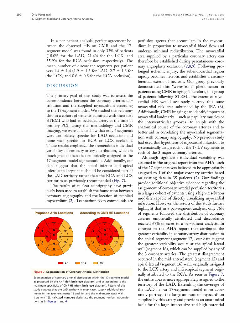
In a per-patient analysis, perfect agreement be-ween the observed HE on CMR and the 17-egment model was found in only 33% of patients18.0% for the LAD, 21.4% for the LCX, and5.9% for the RCA occlusion, respectively). Theean number of discordant segments per patientas 1.4 � 1.4 (1.9 � 1.3 for LAD, 2.7 � 1.8 for
he LCX, and 0.6 � 0.8 for the RCA occlusion).
I S C U S S I O N
he primary goal of this study was to assess theorrespondence between the coronary arteries dis-ribution and the supplied myocardium accordingo the 17-segment model. We studied this relation-hip in a cohort of patients admitted with their firstTEMI who had an occluded artery at the time ofrimary PCI. Using this methodology and CMRmaging, we were able to show that only 4 segmentsere completely specific for LAD occlusion andone was specific for RCA or LCX occlusion.hese results emphasize the tremendous individual
ariability of coronary artery distribution, which isuch greater than that empirically assigned to the
7-segment model segmentation. Additionally, ourata suggest that the apical inferior and apical
nferolateral segments should be considered part ofhe LAD territory rather than the RCA and LCXerritories as previously recommended (Fig. 7).
The results of nuclear scintigraphy have previ-usly been used to establish the foundation betweenoronary angiography and the location of suppliedyocardium (2). Technetium-99m compounds are
LCXRCALAD
d AHA Locations According to CMR HE Locations
egmentation of Coronary Arterial Distribution
ion of coronary arterial distribution within the 17-segment modeld by the AHA (left bulls-eye diagram) and as according to thespecificity of CMR HE (right bulls eye diagram). Results of thisest that the LAD territory in most cases supply additional seg-e apex (segments 15 and 16) and the mid-anterolateral wall2). Italicized numbers designate the segment number. Abbrevia-
bFigures 1 and 6.
erfusion agents that accumulate in the myocar-ium in proportion to myocardial blood flow andndergo minimal redistribution. The myocardialrea supplied by a particular coronary artery canherefore be established during percutaneous coro-ary angioplasty occlusion (2,8,9). Following pro-
onged ischemic injury, the subendocardial regionapidly becomes necrotic and establishes a circum-erential extent of necrosis. Our group previouslyemonstrated this “wave-front” phenomenon inatients using CMR imaging. Therefore, in a groupf patients following STEMI, the extent of myo-ardial HE would accurately portray this sameyocardial risk area subtended by the IRA (6).dditionally, CMR imaging can identify importantyocardial landmarks—such as papillary muscles or
he interventricular grooves—to couple with thenatomical course of the coronary arteries and toetter aid in correlating the myocardial segmenta-ion with coronary angiography. No previous studyad used this hypothesis of myocardial infarction toystematically assign each of the 17 LV segments toach of the 3 major coronary arteries.
Although significant individual variability wasssumed in the original report from the AHA, eachf the 17 segments was believed to be appropriatelyssigned to 1 of the major coronary arteries basedn existing data in 35 patients (2). Our findingsrovide additional objective evidence regarding thessignment of coronary arterial perfusion territoriesn a larger cohort of patients using a high-resolution
odality capable of directly visualizing myocardialnfarction. However, the results of this study furtherighlight that in a per-segment analysis, only 76%f segments followed the distribution of coronaryrteries empirically attributed and discordanceeached 67% of cases in a per-patient analysis. Inontrast to the AHA report that attributed thereatest variability in coronary artery distribution tohe apical segment (segment 17), our data suggesthe greatest variability occurs at the apical lateralall (segment 16), which can be supplied by any of
he 3 coronary arteries. The greatest disagreementccurred in the mid-anterolateral (segment 12) andpical lateral (segment 16) wall, originally assignedo the LCX artery and inferoapical segment origi-ally attributed to the RCA. As seen in Figure 7,he entire apex is more appropriately assigned to theerritory of the LAD. Extending the coverage ofhe LAD in our 17-segment model more accu-ately portrays the large amount of myocardiumupplied by this artery and provides an anatomical
Propose
Figure 7. S
Segmentatas proposemaximumstudy suggments in th(segment 1
asis for the large infarct size and high potential

fL
acnvCsdrwbLbI8o7Vs(HsaiRhdilastwtctpCgtifiitctsmsaD
mmswylHcnwan
idpbFvtss(iafatlopnnfcicwiwemoaawmcawTp
J A C C : C A R D I O V A S C U L A R I M A G I N G , V O L . 1 , N O . 3 , 2 0 0 8
M A Y 2 0 0 8 : 2 8 2 – 9 3
Ortiz-Pérez et al.
17-Segment Model and Coronary Arterial Anatomy
291
or adverse ventricular remodeling associated withAD infarctions.The correspondence between the coronary artery
natomy and the 17-segment model was also re-ently reported by Pereztol-Valdés et al. (9) usinguclear perfusion in a cohort of patients with singleessel disease population referred for elective PCI.onsistent with that previous report, our study also
hows that the greatest overlap in coronary arteryistribution occurs in the inferolateral region cor-esponding either to RCA or LCX territories, asell as the inferoseptal region that may be suppliedy the LAD artery, RCA, or even a left-dominantCX artery. However, there are some differencesetween the 2 studies that are worthy of mention.n our CMR study, only 4 segments (segments 2, 7,, and 13) were found to be 100% specific for LADcclusion as opposed to 8 segments (segments 1, 2,, 8, 13, 14, 16, and 17) reported by Pereztol-aldés. One patient with a proximal ramus occlu-
ion had HE located in the basal anterior segmentsegment 1), 7 subjects with RCA occlusion had
E in the apical septum (segment 14), and up to 8ubjects with LCX occlusion developed HE in thepical lateral wall (segment 16). We also found HEnvolving the apex (segment 17) in the setting ofCA and LCX occlusion. Several reasons mightave accounted for these differences. First, theseisparities may be simply explained because of the
ncreased number of subjects included in our study,eading to increased variability in coronary arterialnatomy and dominance. Second, the increasedpatial resolution of delayed enhanced CMR andhe absence of attenuation artifacts as comparedith nuclear perfusion techniques may contribute to
he increased detection of segments with subendo-ardial infarction in the periphery of the area at risk,hat otherwise could have been missed by singlehoton emission computed tomography. Third,MR imaging can identify the interventricular
rooves for better coronary angiography registra-ion. Finally, nuclear perfusion imaging looks atschemia, whereas contrast-enhanced CMR identi-es necrosis or fibrosis. Infarct appears as long as
schemia persists, a process that is influenced by theime of coronary occlusion and the presence ofollateral flow. In this regard, we have confirmed arend to a higher degree of collateral flow amongubjects presenting with RCA occlusion. This trendight have resulted in a smaller extent of HE in
uch cases and, consequently, a reduction in themount of myocardium allocated to the RCA.
iscrimination between RCA and LCX involve- pent based solely on the involvement of individualyocardial segments is difficult. Concomitant ob-
ervation of the anterolateral, lateral, and inferioralls may assist the evaluation. In our study, anal-sis of adjacent segments helped to clarify theocalization of disease. Thus, any combination of
E in the anterolateral wall and inferior wall wasompletely specific for a left dominant or codomi-ant occlusion. Presence of HE in the inferolateralall (segments 5 and 11) with or without HE in the
nterolateral segments was highly specific of aondominant LCX occlusion as well.Detection of multivessel disease is of crucial
mportance in the management of coronary arteryisease. Multiple studies have demonstrated im-roved outcomes and prognosis by reperfusion withypass surgery or PCI in this situation (10–12).unctional imaging techniques must therefore pro-ide precise knowledge of coronary artery distribu-ion to allow appropriate identification of multives-el disease. This capability has been extensivelytudied in nuclear perfusion imaging experimentally13,14) and clinically, using several methodologiesn patients with acute myocardial infarction (15)nd elective PCI (16–19). Several attempts forusing anatomic images from the coronary arteriesnd functional information from different modali-ies have been proposed in an effort to assist inocating and assessing the physiologic significancef multiple stenotic lesions. Faber et al. (20) pro-osed an algorithm based on the integration ofuclear perfusion imaging with conventional coro-ary angiography. The CMR is now widely used forunction, perfusion, and viability assessment. Be-ause of the paucity of studies correlating CMRmaging with coronary anatomy, most decisionsoncerning the involvement of coronary arterieshen interpreting CMR viability and perfusion
maging studies are extrapolated from previousork based on nuclear perfusion techniques. Setser
t al. (21) studied the correspondence between theyocardial 17-segment model and coronary anat-
my by coupling information from CMR imagesnd noninvasive computed tomography coronaryngiography in a small series of 26 patients. In lineith our findings, the authors showed that theaximal discordance of coronary distribution oc-
urred in the mid-anterolateral, apical lateral, andpical inferior wall (segments 12, 15, and 16),hich were supplied by the LAD in most cases.hey also found that the combined informationrovided by fusion of images was useful for the
lanning of surgery in those patients. However,
fmnostfpSjspitta
mcbfhsIHaIat
two
C
IilamrepOosHtL1fawvi
RE
R
J A C C : C A R D I O V A S C U L A R I M A G I N G , V O L . 1 , N O . 3 , 2 0 0 8
M A Y 2 0 0 8 : 2 8 2 – 9 3
Ortiz-Pérez et al.
17-Segment Model and Coronary Arterial Anatomy
292
usion of different modalities may be subject toisregistration or misalignment. Thus new tech-
ologies capable of obtaining both coronary anat-my and functional information in a single study—uch as positron emission tomography/computedomography scanners or coronary MR angiography/unctional CMR—would be better suited for thisurpose.tudy limitations. This study includes research sub-ects participating in a prospective observationaltudy investigating the impact of microvascularerfusion and infarct size on LV remodeling. Nonvestigational treatments were performed, but par-icipation in this study was voluntary and may nototally reflect findings for all patients followingcute myocardial infarction.
Additionally, the allocation of the IRA territoryight be underestimated in patients who develop
ongestive heart failure during hospital admission,ecause this clinical situation is a contraindicationor a CMR study. Almost two-thirds of the patientsad multivessel disease. It is unlikely, however, thatignificant coronary disease in vessels other than theRA could have affected the extent and location ofE. In this regard, 8 patients with an additional
rea of remote HE clearly not corresponding to theRA territory showed an occlusion in a coronaryrtery different than the IRA were excluded. Addi-ionally, we studied a population following an acute
ska A. Co-registration of cardiac MRI myocardial perfusio
he setting of chronic coronary artery disease, whereell-developed collaterals might reduce the extentf infarction.
O N C L U S I O N S
n patients presenting with STEMI, direct visual-zation of infarcted myocardium and anatomicalandmarks by high-resolution CMR imaging, en-bles registration between affected myocardial seg-ents and the occluded artery by invasive angiog-
aphy. The results of our findings bring objectivevidence to the assignment of coronary arterialerfusion territories within the 17-segment model.nly 4 segments were completely specific for LAD
cclusion, and no single segment could be exclu-ively attributed to a RCA or LCX occlusion.owever, analysis of adjacent segments increased
he specificity for a given coronary occlusion. TheAD territory is larger than the AHA-proposed7-segment model and is most often responsibleor myocardial infarctions involving the mid-nterolateral and all apical segments. These findingsill aid in the appropriate assignment of the culpritessel in CMR studies and may also influence thenterpretation of other noninvasive modalities.
eprint requests and correspondence: Dr. Edwin Wu, 201Huron, Galter 10-240, Chicago, Illinois 60611.
STEMI, and the results of this study may vary in E-mail: [email protected].
1
1
1
E F E R E N C E S
1. Cerqueira MD, Weissman NJ, Dilsi-zian V, et al. Standardized myocardialsegmentation and nomenclature fortomographic imaging of the heart: astatement for healthcare professionalsfrom the Cardiac Imaging Committeeof the Council on Clinical Cardiologyof the American Heart Association.Circulation 2002;105:539–42.
2. Gallik DM, Obermueller SD, SwarnaUS, Guidry GW, Mahmarian JJ, Ve-rani MS. Simultaneous assessment ofmyocardial perfusion and left ventric-ular function during transient coro-nary occlusion. J Am Coll Cardiol1995;25:1529–38.
3. Plein S, Ridgway JP, Jones TR,Bloomer TN, Sivananthan MU. Cor-onary artery disease: assessment with acomprehensive MR imaging proto-col—initial results. Radiology 2002;225:300–7.
4. Misko J, Dziuk M, Skrobowska E,Szalus N, Pietrzykowski J, Warczyn-
and rest gated SPECT in the assess-ment of myocardial perfusion, func-tion and viability. J Cardiovasc MagnReson 2006;8:389–97.
5. Wagner A, Mahrholdt H, Holly TA,et al. Contrast-enhanced MRI androutine single photon emission com-puted tomography (SPECT) perfu-sion imaging for detection of sub-endocardial myocardial infarcts: animaging study. Lancet 2003;361:374–9.
6. Ortiz-Perez JT, Meyers SN, Lee DC,et al. Angiographic estimates of myo-cardium at risk during acute myocar-dial infarction: validation study usingcardiac magnetic resonance imaging.Eur Heart J 2007;28:1750–8.
7. Simonetti OP, Kim RJ, Fieno DS, etal. An improved MR imaging tech-nique for the visualization of myocar-dial infarction. Radiology 2001;218:215–23.
8. Fram DB, Azar RR, Ahlberg AW, etal. Duration of abnormal SPECT
n imaging follow
ing resolution of acute ischemia: anangioplasty model. J Am Coll Cardiol2003;41:452–9.
9. Pereztol-Valdés O, Candell-Riera J,Santana-Boado C, et al. Correspon-dence between left ventricular 17myocardial segments and coronary ar-teries. Eur Heart J 2005;26:2637–43.
0. Hamm CW, Reimers J, Ischinger T,Rupprecht HJ, Berger J, Bleifeld W.A randomized study of coronary an-gioplasty compared with bypass sur-gery in patients with symptomaticmultivessel coronary disease. GermanAngioplasty Bypass Surgery Investiga-tion (GABI). N Engl J Med 1994;331:1037–43.
1. Hannan EL, Racz MJ, Walford G, etal. Long-term outcomes of coronary-artery bypass grafting versus stent im-plantation. N Engl J Med 2005;352:2174–83.
2. The final 10-year follow-up resultsfrom the BARI randomized trial.
J Am Coll Cardiol 2007;49:1600–6.
1
1
1
1
2
2
J A C C : C A R D I O V A S C U L A R I M A G I N G , V O L . 1 , N O . 3 , 2 0 0 8
M A Y 2 0 0 8 : 2 8 2 – 9 3
Ortiz-Pérez et al.
17-Segment Model and Coronary Arterial Anatomy
293
3. De Coster PM, Wijns W, Cauwe F,Robert A, Beckers C, Melin JA. Area-at-risk determination by technetium-99m-hexakis-2-methoxyisobutyl iso-nitrile in experimental reperfusedmyocardial infarction. Circulation1990;82:2152–62.
4. Sinusas AJ, Trautman KA, Bergin JD,et al. Quantification of area at risk dur-ing coronary occlusion and degree ofmyocardial salvage after reperfusionwith technetium-99m methoxyisobutylisonitrile. Circulation 1990;82:1424–37.
5. Gibbons RJ, Verani MS, BehrenbeckT, et al. Feasibility of tomographic99mTc-hexakis-2-methoxy-2-methyl-propyl-isonitrile imaging for the as-sessment of myocardial area at riskand the effect of treatment in acutemyocardial infarction. Circulation
1989;80:1277–86.16. Borges-Neto S, Puma J, Jones RH, etal. Myocardial perfusion and ventric-ular function measurements duringtotal coronary artery occlusion in hu-mans. A comparison with rest andexercise radionuclide studies. Circula-tion 1994;89:278–84.
17. Borges-Neto S, Watson JE, MillerMJ. Tc-99m sestamibi cardiacSPECT imaging during coronary ar-tery occlusion in humans: comparisonwith dipyridamole stress studies. Ra-diology 1996;198:751–4.
18. Ceriani L, Verna E, Giovanella L,Bianchi L, Roncari G, Tarolo GL.Assessment of myocardial area at riskby technetium-99m sestamibi duringcoronary artery occlusion: comparisonbetween three tomographic methodsof quantification. Eur J Nucl Med
1996;23:31–9.9. Pereztol-Valdés O, Candell-Riera J,Oller-Martinez G, et al. [Localiza-tion and quantification of myocar-dium at risk by myocardial perfusionSPECT during coronary artery oc-clusion]. Rev Esp Cardiol 2004;57:635– 43.
0. Faber TL, Santana CA, Garcia EV, etal. Three-dimensional fusion ofcoronary arteries with myocardialperfusion distributions: clinicalvalidation. J Nucl Med 2004;45:745–53.
1. Setser RM, O’Donnell TP, SmediraNG, et al. Coregistered MR imagingmyocardial viability maps and multi-detector row CT coronary angiogra-phy displays for surgical revasculari-zation planning: initial experience.
Radiology 2005;237:465–73.