ultrasound-mediated vascular gene transfection by...
TRANSCRIPT

J A C C : C A R D I O V A S C U L A R I M A G I N G V O L . 5 , N O . 1 2 , 2 0 1 2
© 2 0 1 2 B Y T H E A M E R I C A N C O L L E G E O F C A R D I O L O G Y F O U N D A T I O N I S S N 1 9 3 6 - 8 7 8 X / $ 3 6 . 0 0
P U B L I S H E D B Y E L S E V I E R I N C . h t t p : / / d x . d o i . o r g / 1 0 . 1 0 1 6 / j . j c m g . 2 0 1 2 . 0 5 . 0 1 7
Ultrasound-Mediated Vascular Gene Transfection byCavitation of Endothelial-Targeted Cationic Microbubbles
Aris Xie, BS,* Todd Belcik, RDCS,* Yue Qi, MD,* Terry K. Morgan, MD, PHD,†Shivam A. Champaneri, MD,‡ Sarah Taylor, PHD,‡ Brian P. Davidson, MD,*Yan Zhao, MS,* Alexander L. Klibanov, PHD,‡ Michael A. Kuliszewski, BS,§Howard Leong-Poi, MD,§ Azzdine Ammi, PHD,* Jonathan R. Lindner, MD*Portland, Oregon; Charlottesville, Virginia; and Toronto, Ontario, Canada
O B J E C T I V E S Ultrasound-mediated gene delivery can be amplified by acoustic disruption of
microbubble carriers that undergo cavitation. We hypothesized that endothelial targeting of micro-
bubbles bearing cDNA is feasible and, through optimizing proximity to the vessel wall, increases the
efficacy of gene transfection.
B A C K G R O U N D Contrast ultrasound-mediated gene delivery is a promising approach for site-
specific gene therapy, although there are concerns with the reproducibility of this technique and the
safety when using high-power ultrasound.
M E T H O D S Cationic lipid-shelled decafluorobutane microbubbles bearing a targeting moiety were
prepared and compared with nontargeted microbubbles. Microbubble targeting efficiency to endothe-
lial adhesion molecules (P-selectin or intercellular adhesion molecule [ICAM]-1) was tested using in vitro
flow chamber studies, intravital microscopy of tumor necrosis factor-alpha (TNF-�)–stimulated murine
cremaster muscle, and targeted contrast ultrasound imaging of P-selectin in a model of murine limb
ischemia. Ultrasound-mediated transfection of luciferase reporter plasmid charge coupled to micro-
bubbles in the post-ischemic hindlimb muscle was assessed by in vivo optical imaging.
R E S U L T S Charge coupling of cDNA to the microbubble surface was not influenced by the presence of
targeting ligand, and did not alter the cavitation properties of cationic microbubbles. In flow chamber studies,
surface conjugation of cDNA did not affect attachment of targeted microbubbles at microvascular shear
stresses (0.6 and 1.5 dyne/cm2). Attachment in vivo was also not affected by cDNA according to intravital
microscopy observations of venular adhesion of ICAM-1–targetedmicrobubbles and by ultrasoundmolecular
imaging of P-selectin–targeted microbubbles in the post-ischemic hindlimb in mice. Transfection at the site
of high acoustic pressures (1.0 and 1.8 MPa) was similar for control and P-selectin–targeted microbubbles but
was associated with vascular rupture and hemorrhage. At 0.6 MPa, there were no adverse bioeffects, and
transfection was 5-fold greater with P-selectin–targeted microbubbles.
C O N C L U S I O N S We conclude that ultrasound-mediated transfection at safe acoustic pressures
can be markedly augmented by endothelial juxtaposition. (J Am Coll Cardiol Img 2012;5:1253–62)
© 2012 by the American College of Cardiology Foundation
From the *Division of Cardiovascular Medicine, Oregon Health & Science University, Portland, Oregon; †Department ofPathology, Oregon Health & Science University, Portland, Oregon; ‡Cardiovascular Division, University of Virginia,Charlottesville, Virginia; and the §St. Michael’s Hospital, Toronto, Ontario, Canada. Dr. Davidson is supported by apost-doctoral fellowship grant (T32-HL094294) from the National Institutes of Health (NIH). Dr. Leong-Poi is supportedby the Clinician Scientist Award from the Heart and Stroke Foundation of Ontario. Dr. Lindner is supported by grantsR01-HL-078610, R01-DK-063508, R01-HL111969, and RC1-HL-100659 from the NIH. Dr. Klibanov is a cofounder andminority stockholder of Targeson, through which his laboratory receives an NIH subcontract, and he has received researchfunding from Philips Research North America. Dr. Lindner has received a grant from GE Medical Imaging. All other authorshave stated that they have no relationships relevant to the contents of this paper to disclose.
Manuscript received March 1, 2012; revised manuscript received May 1, 2012, accepted May 3, 2012.

I
mflmvsbs
u
C
m
I
pressure
J A C C : C A R D I O V A S C U L A R I M A G I N G , V O L . 5 , N O . 1 2 , 2 0 1 2
D E C E M B E R 2 0 1 2 : 1 2 5 3 – 6 2
Xie et al.
Targeted Ultrasound Gene Delivery
1254
ntegration of gene therapy into routine clinicalpractice patients is predicated on the ability tosafely and efficiently deliver genetic material totarget tissues in a manner that favorably alters cell
phenotype. Ultrasound energy can amplify transferof genetic material into cells of target tissues in vivo,the extent of which is further increased by thecoadministration of ultrasound contrast agentscomposed of protein- or lipid-stabilized gas micro-bubbles (1–6). Contrast ultrasound-mediated genedelivery (CUMGD) and transfection appears to bemost effective when genetic material is coupled tothe contrast agent surface (7,8).
See page 1263
The observation that CUMGD increases extrav-asation and cellular uptake of cDNA strongly sug-gests that local bioeffects produced by microbubblecavitation is responsible for increased transfection
efficiency (4,8,9). In an ultrasound field,microbubble contrast agents undergo ei-ther stable cavitation (volumetric oscilla-tion linearly related to acoustic pressure)or inertial cavitation (high-amplitudenonlinear oscillation and physical disrup-tion). Cavitation produces oscillatoryshear, high-pressure microstreaming, andphysical stretching of endothelial cells(10,11). These local effects can producephysical and functional changes in adjacentcells, such as microporation (2,8,9,12), en-hanced permeability through intercellularjunctions (13), and triggering of active cel-
lular uptake of macromolecules (14).Irrespective of the mechanism(s) responsible for
CUMGD, the proximity of the contrast agent tothe cell surface is likely to be a critical determinant.In vivo, this translates to proximity to the vascularwall since in most tissues microbubbles possess arheology similar to erythrocytes and remain entirelywithin the vascular compartment (15). Accordingly,endothelial targeting of ultrasound contrast agentsvia surface conjugation of ligands could amplifyCUMGD by promoting vascular juxtaposition (16).On the other hand, direct apposition of ultrasoundcontrast agents to the endothelial surface by target-ing may not necessarily convey benefit in tissuessuch as the heart, brain, or skeletal muscle wherethe majority of microbubbles during CUMGD areresident within capillaries and small noncapillarymicrovessels, where bubbles are already in proximity
nd-
n
y
stic
to the vessel boundaries. The aim of this study was e
to investigate whether it is feasible to target DNA-laden microbubbles to the endothelial surface andto determine the relative benefit of using a vascular-targeted approach in terms of both efficacy andsafety of transfection for CUMGD.
M E T H O D S
Microbubble preparation. Lipid-shelled decafluoro-butane microbubbles with a neutral charge were pre-pared by sonication of a gas-saturated aqueous sus-pension of distearoylphosphatidylcholine (2 mg/ml)and polyoxyethylene-40-stearate (1 mg/ml). Cationicbubbles were prepared by adding distearoyl-trimethylammoniumpropane (0.4 mg/ml) to the aque-ous suspension. For biotinylated cationic or neutralmicrobubbles, distearoylphosphatidyl-ethanolamine–polyethyleneglycol-2000 (DSPE-PEG2000) biotin (0.1mg/ml) was added to the aqueous suspension. Fluores-cent labeling for intravital microscopy was performed byadding dioctadecyl-tetramethylindocarbocyanineperchlorate (DiI) or dioctadecyloxacarbocyanineperchlorate (DiO). Targeted microbubbles wereprepared by conjugating biotinylated monoclonalantibodies (mAb) to the surface of cationic biotin-ylated microbubbles via a streptavidin link aspreviously described (16). Antibodies includedrat anti-mouse P-selectin (RB40.34), intercellularadhesion molecule-1 (ICAM-1) (3E2, BD Phar-mingen, San Jose, California), and isotype con-trol mAb (R3-34, BD Pharmingen). PlasmidcDNA was charge coupled to microbubbles byincubation of 50 �g per 108 microbubbles for 15min, which produces near saturation (8), and fortargeted microbubbles was performed after mAbconjugation. The mean diameter and concentra-tion of microbubbles was determined by electro-zone analysis (Multisizer III, Beckman Coulter,Fullerton, California). Electric surface potential(zeta potential) for cationic and neutral micro-bubbles was determined by measurement of theirelectrophoretic mobility (ZetaPALS, Brookhaven In-struments, Holtsville, New York) in 1 mmol/l KCl atpH 7.4.Microbubble–plasmid coupling. Quantitation of
icrobubble-associated cDNA was performed byuorescent labeling of plasmid (pCMV-GL3, Pro-ega, Madison, Wisconsin) with SYBR-Gold (In-
itrogen, Carlsbad, California). Microbubbles wereeparated from the aqueous suspension and washedy flotation centrifugation. After hyperbaric de-truction of microbubbles, the cDNA content was
A B B R E V I A T I O N S
A N D A C R O N YM S
CEU � contrast-enhanced
ltrasound
UMGD � contrast ultrasou
ediated gene delivery
CAM � intercellular adhesio
molecule
mAb � monoclonal antibod
MI � mechanical index
PNAP � peak negative acou
valuated by measuring fluorescent activity (Gemini

aobcbtmvicpaafip
vwa
f(sioab
eeeaiCrehasb
cpPThiw
J A C C : C A R D I O V A S C U L A R I M A G I N G , V O L . 5 , N O . 1 2 , 2 0 1 2
D E C E M B E R 2 0 1 2 : 1 2 5 3 – 6 2
Xie et al.
Targeted Ultrasound Gene Delivery
1255
XPS, Molecular Devices, Sunnyvale, California)compared with a standard.Characterization of microbubble cavitation. Salinealone or suspensions of cationic microbubbles (1 �107 ml�1) with and without cDNA were infusedthrough acoustically transparent tubing (2-mm in-ternal diameter, 0.9 ml/min) and were exposed toultrasound (1 MHz, 30-cycle pulses at a pulserepetition frequency of 50 Hz) at either 0.5- or1.0-MPa peak negative acoustic pressure (PNAP)using a single-element transducer (Olympus NDT,Olympus, Tokyo, Japan) and a random waveformgenerator (RAM-5000, Ritec, Warwick, RhodeIsland). A second broadband (10 KHz to 20 MHz)transducer was placed at a 60° angle as a passivecavitation detector and was interfaced with a re-ceiver (RAM-5000, Ritec) and an oscilloscope(44MXi-A, Teledyne LeCroy, Chestnut Ridge,New York). Acoustic response was quantified byboth initial signal amplitude and rate of signal decayindicating inertial cavitation.Flow chamber studies. Streptavidin (0.25 mg/ml) wasdsorbed on the surface of the disposable componentf an inverted flow chamber (GlycoTech, Gaithers-urg, Maryland) and blocked with casein. The flowhamber was mounted on a microscope, and micro-ubble adhesion to either streptavidin-coated or con-rol (casein only) plates was assessed using 8 differenticrobubble preparations according to the following
ariables: biotinylated versus nonbiotinylated, cat-onic versus neutral, with versus without plasmidDNA (pCMV-GL3, Promega). Microbubble sus-ensions (1 � 106 ml�1) were infused for 5 min atshear rate of 0.6 or 1.5 dyne/cm2, after which
dhesion density was averaged from �10 opticalelds. Each of the 32 experimental conditions waserformed in triplicate.
Intravital microscopy. To test binding efficiency inivo, intravital microscopy of the cremaster muscleas performed in 5 C57BL/6 mice 8 to 12 weeks of
ge 1 h after intrascrotal injection of TNF-� (0.5�g in 100 �l). The cremaster muscle was preparedor intravital microscopy as previously described16,17). Microbubble adhesion was tested for 4eparate cationic microbubble preparations accord-ng to the following 2 variables: ICAM-1–targetedr nontargeted control microbubbles, presence orbsence of plasmid cDNA on the surface. Micro-ubbles (1 � 107) were injected intravenously, and
adhesion was assessed 5 min later by fluorescentmicroscopy in 20 optical fields (total area: 1 mm2).
Differential labeling of microbubbles with DiI and mDiO allowed simultaneous injection of 2 agents at atime.Contrast ultrasound molecular imaging. Contrast-nhanced ultrasound (CEU) molecular imaging ofndothelial activation was performed to furthervaluate the effect of gene loading on microbubbledhesion. Unilateral hindlimb ischemia (20-minliac ligation) and reperfusion was performed in 1057BL/6 mice anesthetized with inhaled isoflu-
ane. CEU perfusion imaging was performed tovaluate microvascular blood flow in the proximalindlimb adductor muscles during arterial ligationnd approximately 45 min after reflow. For perfu-ion imaging, a continuous infusion of micro-ubbles (1 � 106 min�1) was used. Flow was
quantified from the product of microvascular bloodvolume and flux rate derived from post-destructiontime-intensity data acquired using a linear-arraytransducer and a contrast-specific multipulse tech-nique (Contrast-Pulse Sequencing, Sequoia, Sie-mens Medical Systems, Mountain View, Califor-nia) operating at a transmission frequency of 7.0MHz and a mechanical index (MI) of 0.16 (18).Absolute microvascular blood flow was calculatedby normalizing muscle microvascular blood vol-ume to the blood pool and dividing by tissuedensity (1.06 g/cm3). Thirty minutes after reflow,or after similar anesthesia time in nonischemiccontrols, molecular imaging of retained micro-bubbles was performed. Signal from retainedmicrobubbles was calculated from a high-MI(1.0) image obtained 8 min after intravenousinjection, from which signal from freely circulat-ing microbubbles was eliminated by digital sub-traction of frames subsequently obtained at apulsing interval of 10 s (16). P-selectin–targetedmicrobubbles with or without plasmid weretested in random order.Targeted gene transfection. Targeted CUMGD wasperformed in the model of post-ischemic hindlimbskeletal muscle in 42 C57BL/6 mice 10 to 12 weeksof age. For these experiments, both hindlimbsunderwent ischemia-reperfusion injury. Thirtyminutes after reflow, either nontargeted or P-selectin–targeted cationic microbubbles (2 � 108)ombined with a firefly luciferase (luc2) reporterlasmid with an SV40 promoter region (pGL4.13,romega) was infused intravenously over 1 min.he proximal adductor muscles of only the leftindlimb were exposed to ultrasound during the
njection and for an additional 9 min. Transducersere placed at a fixed distance (2 cm) from the
id-portion of the muscle using a transverse imag-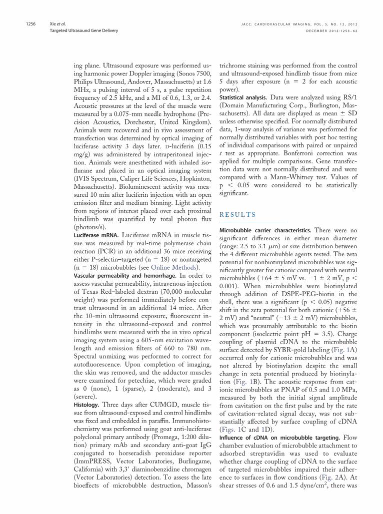
(
swcptc(C(b
ta5
s(tpnm0tss2wc
J A C C : C A R D I O V A S C U L A R I M A G I N G , V O L . 5 , N O . 1 2 , 2 0 1 2
D E C E M B E R 2 0 1 2 : 1 2 5 3 – 6 2
Xie et al.
Targeted Ultrasound Gene Delivery
1256
ing plane. Ultrasound exposure was performed us-ing harmonic power Doppler imaging (Sonos 7500,Philips Ultrasound, Andover, Massachusetts) at 1.6MHz, a pulsing interval of 5 s, a pulse repetitionfrequency of 2.5 kHz, and a MI of 0.6, 1.3, or 2.4.Acoustic pressures at the level of the muscle weremeasured by a 0.075-mm needle hydrophone (Pre-cision Acoustics, Dorchester, United Kingdom).Animals were recovered and in vivo assessment oftransfection was determined by optical imaging ofluciferase activity 3 days later. D-luciferin (0.15mg/g) was administered by intraperitoneal injec-tion. Animals were anesthetized with inhaled iso-flurane and placed in an optical imaging system(IVIS Spectrum, Caliper Life Sciences, Hopkinton,Massachusetts). Bioluminescent activity was mea-sured 10 min after luciferin injection with an openemission filter and medium binning. Light activityfrom regions of interest placed over each proximalhindlimb was quantified by total photon flux(photons/s).Luciferase mRNA. Luciferase mRNA in muscle tis-sue was measured by real-time polymerase chainreaction (PCR) in an additional 36 mice receivingeither P-selectin–targeted (n � 18) or nontargetedn � 18) microbubbles (see Online Methods).Vascular permeability and hemorrhage. In order toassess vascular permeability, intravenous injectionof Texas Red–labeled dextran (70,000 molecularweight) was performed immediately before con-trast ultrasound in an additional 14 mice. Afterthe 10-min ultrasound exposure, fluorescent in-tensity in the ultrasound-exposed and controlhindlimbs were measured with the in vivo opticalimaging system using a 605-nm excitation wave-length and emission filters of 660 to 780 nm.Spectral unmixing was performed to correct forautofluorescence. Upon completion of imaging,the skin was removed, and the adductor muscleswere examined for petechiae, which were gradedas 0 (none), 1 (sparse), 2 (moderate), and 3(severe).Histology. Three days after CUMGD, muscle tis-ue from ultrasound-exposed and control hindlimbsas fixed and embedded in paraffin. Immunohisto-
hemistry was performed using goat anti-luciferaseolyclonal primary antibody (Promega, 1:200 dilu-ion) primary mAb and secondary anti-goat IgGonjugated to horseradish peroxidase reporterImmPRESS, Vector Laboratories, Burlingame,alifornia) with 3,3= diaminobenzidine chromagen
Vector Laboratories) detection. To assess the late
ioeffects of microbubble destruction, Masson’srichrome staining was performed from the controlnd ultrasound-exposed hindlimb tissue from mice
days after exposure (n � 2 for each acousticpower).Statistical analysis. Data were analyzed using RS/1(Domain Manufacturing Corp., Burlington, Mas-sachusetts). All data are displayed as mean � SDunless otherwise specified. For normally distributeddata, 1-way analysis of variance was performed fornormally distributed variables with post hoc testingof individual comparisons with paired or unpairedt test as appropriate. Bonferroni correction wasapplied for multiple comparisons. Gene transfec-tion data were not normally distributed and werecompared with a Mann-Whitney test. Values ofp � 0.05 were considered to be statisticallysignificant.
R E S U L T S
Microbubble carrier characteristics. There were noignificant differences in either mean diameterrange: 2.5 to 3.1 �m) or size distribution betweenhe 4 different microbubble agents tested. The zetaotential for nonbiotinylated microbubbles was sig-ificantly greater for cationic compared with neutralicrobubbles (�64 � 5 mV vs. �1 � 2 mV, p �
.001). When microbubbles were biotinylatedhrough addition of DSPE-PEG-biotin in thehell, there was a significant (p � 0.05) negativehift in the zeta potential for both cationic (�56 �mV) and “neutral” (�13 � 2 mV) microbubbles,hich was presumably attributable to the biotin
omponent (isoelectric point pH � 3.5). Chargecoupling of plasmid cDNA to the microbubblesurface detected by SYBR-gold labeling (Fig. 1A)occurred only for cationic microbubbles and wasnot altered by biotinylation despite the smallchange in zeta potential produced by biotinyla-tion (Fig. 1B). The acoustic response from cat-ionic microbubbles at PNAP of 0.5 and 1.0 MPa,measured by both the initial signal amplitudefrom cavitation on the first pulse and by the rateof cavitation-related signal decay, was not sub-stantially affected by surface coupling of cDNA(Figs. 1C and 1D).Influence of cDNA on microbubble targeting. Flowchamber evaluation of microbubble attachment toadsorbed streptavidin was used to evaluatewhether charge coupling of cDNA to the surfaceof targeted microbubbles impaired their adher-ence to surfaces in flow conditions (Fig. 2A). At
shear stresses of 0.6 and 1.5 dyne/cm2, there was
J A C C : C A R D I O V A S C U L A R I M A G I N G , V O L . 5 , N O . 1 2 , 2 0 1 2
D E C E M B E R 2 0 1 2 : 1 2 5 3 – 6 2
Xie et al.
Targeted Ultrasound Gene Delivery
1257
minimal nonspecific attachment of nonbiotiny-lated microbubbles to streptavidin, irrespective ofmicrobubble surface charge or incubation withplasmid cDNA. Attachment density was high forneutral biotinylated microbubbles and was notaltered by incubation with plasmid cDNA, whichwas anticipated because of the minimal amountof cDNA that is charge coupled with neutralagent. In the absence of plasmid cDNA, attach-ment of biotinylated microbubbles was signifi-cantly less for cationic versus neutral micro-bubbles. Interestingly, the deficit in attachmentfor cationic biotinylated microbubbles was cor-rected by plasmid cDNA coupling to the micro-bubble surface. In both shear conditions, nonspe-cific attachment to plates without streptavidinwas minimal (�5 per optical field) for all 8microbubble preparations.
Intravital microscopy of TNF-�–treated cre-master muscle in mice was used to evaluate
Figure 1. Properties of Lipid Microbubble Carriers
(A) Fluorescent microscopy (excitation filter 530 to 560 nm) of SYBRbubbles (scale bar � 5 �m). (B) Mean � SD amount of cDNA chargBiot � presence of DSPE-PEG-biotin. *p � 0.01 versus both neutralby microbubble cavitation during ultrasound exposure at 1 MPa uspressure waveform for the second pulse is expanded. (D) Mean �
passive cavitation detector for cationic microbubbles with and withMPa peak negative acoustic pressure. The eventual stabilization aftobtained with saline alone (Online Fig. 1).
whether surface coupling of cDNA interferes
with the in vivo vascular adhesion of targetedmicrobubbles in an inflammatory disease–relatedmodel. Fluorescently labeled microbubbles re-mained entirely intravascular after their intrave-nous injection and were cleared from the circu-lation after 5 min. There was minimal attachmentof nontargeted cationic microbubbles (Fig. 2B).Targeting microbubbles to ICAM-1 by surfaceconjugation of anti-mouse ICAM-1 mAb in-creased microvascular retention, the extent ofwhich was not altered by the presence of plasmidcDNA. Almost all attachment was secondary tovenular endothelial attachment.
Noninvasive CEU molecular imaging of endo-thelial P-selectin in a model of murine hindlimbischemia-reperfusion injury was performed to fur-ther evaluate whether gene loading influences mi-crobubble adhesion. During iliac artery ligation,flow was reduced to 0.04 � 0.01 ml/min/g andincreased to 0.47 � 0.20 ml/min/g at the comple-
ld–labeled plasmid cDNA charge coupled to cationic micro-oupled to the surface of different microbubble preparations.parations. (C) Example of an acoustic amplitude response caused30-cycle ultrasound pulses every 20 ms (50 Hz). A portion of themplitude of microbubble response (peak-to-peak) measured by acDNA during sequential 1-MHz ultrasound pulses at 0.5- and 1.0-proximately pulse 10 was not significantly different from that
-goe cpreingSD aouter ap
tion of molecular imaging 45 min after reflow.

fiaFiiP0rll
ut1naMnrbgdfb2hal
J A C C : C A R D I O V A S C U L A R I M A G I N G , V O L . 5 , N O . 1 2 , 2 0 1 2
D E C E M B E R 2 0 1 2 : 1 2 5 3 – 6 2
Xie et al.
Targeted Ultrasound Gene Delivery
1258
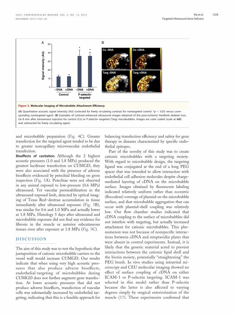
Retention of targeted microbubbles on CEU wasevaluated after intravenous injection of microbubblepreparations performed between 30 and 45 minafter release of the arterial ligature. Retention wasgreater for P-selectin–targeted versus nontargetedmicrobubbles and was not substantially affected bysurface coupling of plasmid cDNA (Fig. 3).Reporter gene transfection. Targeted CUMGD of
refly luciferase reporter plasmid (pGL4.13) wasssessed in post-ischemic hindlimb skeletal muscle.or these experiments, both hindlimbs underwent
schemia-reperfusion injury, and only the left prox-mal hindlimb was exposed to ultrasound. TheNAP for the 3 CUMGD ultrasound settings (MI.6, 1.3, and 2.4) were 0.6, 1.0, and 1.8 MPa,espectively. Gene transfection quantified 3 daysater by whole-body in vivo optical imaging of
Figure 2. Microbubble Adhesion Efficiency
(A) In vitro microbubble attachment density per optical field (OF) tofor shear stresses of 0.6 and 1.5 dyne/cm2. *p � 0.05 versus both tbiotinylated preparation with cDNA. (B) Illustrative image and quantargeted microbubbles observed by intravital microscopy in TNF-�–trelabeled ICAM-1–targeted microbubbles in a venule running vertically atargeted agent.
uciferase activity was detected only at the site of p
ltrasound exposure (Fig. 4). Luciferase activity athe 2 highest acoustic pressure conditions (1.0 and.8 MPa) was similar for P-selectin–targeted andontargeted microbubbles (Fig. 4A). Luciferasectivity was lower at an acoustic pressure of 0.6
Pa. At this lower pressure, transfection was sig-ificantly greater (approximately 5-fold) and moreeproducible for the P-selectin–targeted micro-ubbles. Similar results were found for luciferaseene expression by real-time PCR, which was noifferent for the 2 agents at 1.8 MPa but was higheror targeted compared with nontargeted micro-ubbles at 0.6 MPa (31,152 � 26,022% vs. 7,090 �,155% relative to GAPDH, p � 0.05). Immuno-istochemistry showed that the majority of lucifer-se transfection occurred in microvascular endothe-ial cells, smooth muscle cells, and surrounding
eptavidin-coated plates in a flow chamber. Data are displayedorresponding neutral biotinylated preparation and the cationicive data for in vivo attachment of cationic nontargeted or ICAM-1–cremasteric vessels. The image illustrates retention of DiI-s the image (scale bar: 20 �m); *p � 0.05 versus corresponding non-
strhe ctitatatedcros
erivascular cells irrespective of acoustic conditions
agwbiiuuiiwamfit
J A C C : C A R D I O V A S C U L A R I M A G I N G , V O L . 5 , N O . 1 2 , 2 0 1 2
D E C E M B E R 2 0 1 2 : 1 2 5 3 – 6 2
Xie et al.
Targeted Ultrasound Gene Delivery
1259
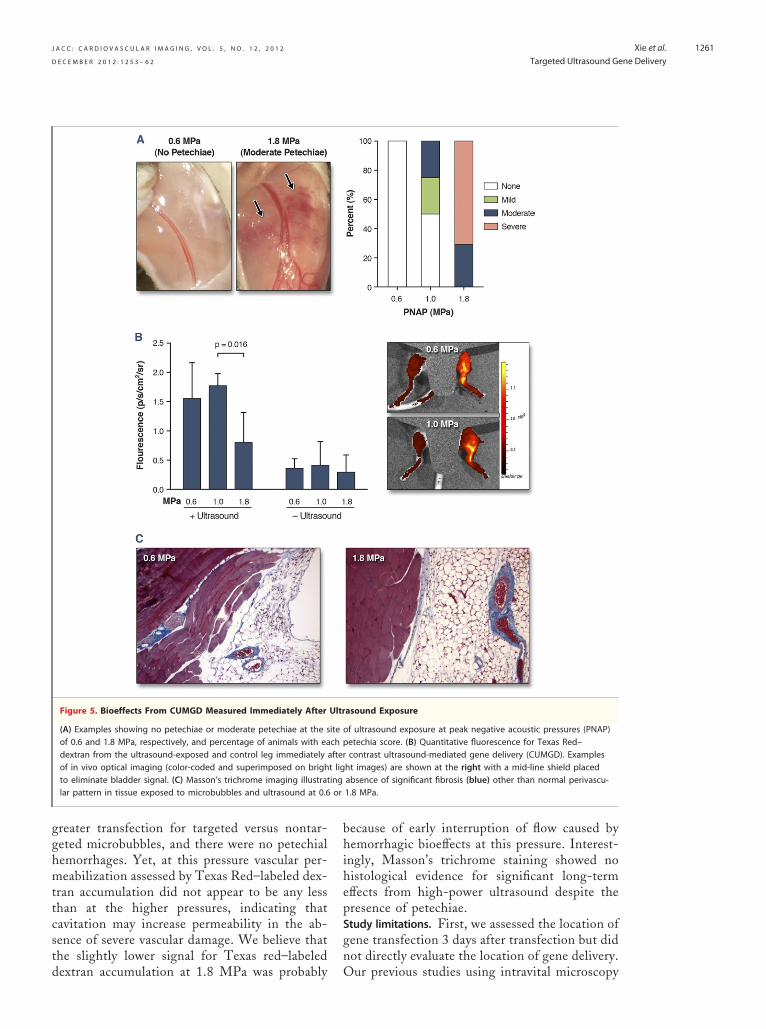
and microbubble preparation (Fig. 4C). Greatertransfection for the targeted agent tended to be dueto greater noncapillary microvascular endothelialtransfection.Bioeffects of cavitation. Although the 2 highestcoustic pressures (1.0 and 1.8 MPa) produced thereatest luciferase transfection on CUMGD, theyere also associated with the presence of adverseioeffects evidenced by petechial bleeding on grossnspection (Fig. 5A). Petechiae were not observedn any animal exposed to low-pressure (0.6 MPa)ltrasound. Yet vascular permeabilization in theltrasound-exposed limb, detected by optical imag-ng of Texas Red–dextran accumulation in tissuemmediately after ultrasound exposure (Fig. 5B),as similar for 0.6 and 1.0 MPa and actually lower
t 1.8 MPa. Histology 5 days after ultrasound andicrobubble exposure did not find any evidence for
brosis in the muscle or anterior subcutaneousissues even after exposure at 1.8 MPa (Fig. 5C).
D I S C U S S I O N
The aim of this study was to test the hypothesis thatjuxtaposition of cationic microbubble carriers to thevessel wall would increase CUMGD. Our resultsindicate that when using very high acoustic pres-sures that also produce adverse bioeffects,endothelial-targeting of microbubbles duringCUMGD does not further augment gene transfec-tion. At lower acoustic pressures that did notproduce adverse bioeffects, transfection of vascularcells was substantially increased by endothelial tar-
Figure 3. Molecular Imaging of Microbubble Attachment Efficie
(A) Quantitative acoustic signal intensity (AU) corrected for freely cisponding nontargeted agent. (B) Examples of contrast-enhanced ulcle 8 min after intravenous injection for control (Co) or P-selectin–tand subtracted for freely circulating agent.
geting, indicating that this is a feasible approach for
balancing transfection efficiency and safety for genetherapy in diseases characterized by specific endo-thelial epitopes.
Part of the novelty of this study was to createcationic microbubbles with a targeting moiety.With regard to microbubble design, the targetingligand was conjugated at the end of a long PEGspacer that was intended to allow interaction withendothelial cell adhesion molecules despite charge-mediated layering of cDNA on the microbubblesurface. Images obtained by fluorescent labelingindicated relatively uniform rather than eccentric(flocculent) coverage of plasmid on the microbubblesurface, and that microbubble aggregation that canoccur with plasmid-shell coupling was relativelylow. Our flow chamber studies indicated thatcDNA coupling to the surface of microbubbles didnot interfere with targeting, but actually increasedattachment for cationic microbubbles. This phe-nomenon was not because of nonspecific interac-tions between cDNA and streptavidin plates thatwere absent in control experiments. Instead, it islikely that the genetic material acted to preventinteractions between the cationic lipid shell andthe biotin moiety, potentially “straightening” thePEG brush. In vivo studies using intravital mi-croscopy and CEU molecular imaging showed noeffect of surface coupling of cDNA on eitherICAM-1 or P-selectin targeting. ICAM-1 wasselected in this model rather than P-selectinbecause the latter is also affected to varyingdegrees simply by surgical exteriorization of the
ating contrast for nontargeted control. *p � 0.05 versus corre-und images obtained of the post-ischemic hindlimb skeletal mus-ted (Targ) microbubbles. Images are color coded (scale at left)
ncy
rcultrasoarge
muscle (17). These experiments confirmed that

J A C C : C A R D I O V A S C U L A R I M A G I N G , V O L . 5 , N O . 1 2 , 2 0 1 2
D E C E M B E R 2 0 1 2 : 1 2 5 3 – 6 2
Xie et al.
Targeted Ultrasound Gene Delivery
1260
the presence of cDNA on cationic microbubblecarriers does not interfere with ligand-mediatedendothelial attachment under microvascular shearconditions.
The ultrasound conditions during CUMGD areimportant when assessing the relative benefit oftargeting. In our experiments, we chose an ultra-sound pulsing interval of 5 s. This is approximatelyhalf the time required for total microvascular refillfor microbubbles in skeletal muscle after a destruc-tive pulse (18,19). This scheme allowed us todestroy all microbubbles transiting through themicrocirculation within the ultrasound beam and totest the effects of endothelial juxtaposition withoutany differences in the cumulative number of tar-geted versus nontargeted microbubbles undergoingcavitation.
There is clear evidence that vascular events
Figure 4. Targeted Transfection of Luciferase Reporter Plasmid
(A) In vivo optical imaging data of luciferase reporter gene transof ultrasound exposure 10 min after intraperitoneal injection ofwas significantly lower at 0.6 MPa compared with 1.0 and 1.8 Mcoded) 3 days after bilateral hindlimb ischemia and intravenouscoupled cationic microbubbles during ultrasound at 0.6 MPa. Ve(C) Immunohistochemistry for luciferase with peroxidase illustratperivascular cells. Scale bar � 50 �m.
other than overt vascular rupture are responsible
for gene transfection with CUMGD (11,14,20).Our aim was to safely amplify gene transfectionby targeting cationic microbubble carriers to thevascular endothelium. Our in vivo transfectionstudies demonstrated successful CUMGD at thesite of ultrasound in all animals. CUMGD withhigh pressure ultrasound (1.0 to 1.8 MPa) re-sulted in a high amount of transfection. However,there were also cavitation-related microvascularruptures that have been previously described withhigh-power ultrasound and large doses of micro-bubble contrast agents (21). There were also nodifferences in transfection for targeted versusnontargeted microbubbles at high power. It islikely that violent cavitation at high acousticpressures produced large-pressure microstreamsand vascular effects conducive to transfectionirrespective of microbubble position relative the
ion 3 days after CUMGD, quantified as photon flux at the siteerin. PNAP � peak negative acoustic pressure. Transfectionor both agents. (B) Examples illustrating luminescence (color-ction of either nontargeted or P-selectin–targeted cDNA-l surface depiliation was performed to reduce light attenuation.transfection (arrows) of venular and capillary endothelium and
fectlucifPa finjentraing
vascular wall. At 0.6 MPa, there was 5 times
gn
or
J A C C : C A R D I O V A S C U L A R I M A G I N G , V O L . 5 , N O . 1 2 , 2 0 1 2
D E C E M B E R 2 0 1 2 : 1 2 5 3 – 6 2
Xie et al.
Targeted Ultrasound Gene Delivery
1261
greater transfection for targeted versus nontar-geted microbubbles, and there were no petechialhemorrhages. Yet, at this pressure vascular per-meabilization assessed by Texas Red–labeled dex-tran accumulation did not appear to be any lessthan at the higher pressures, indicating thatcavitation may increase permeability in the ab-sence of severe vascular damage. We believe thatthe slightly lower signal for Texas red–labeled
Figure 5. Bioeffects From CUMGD Measured Immediately After
(A) Examples showing no petechiae or moderate petechiae at the sof 0.6 and 1.8 MPa, respectively, and percentage of animals with eadextran from the ultrasound-exposed and control leg immediatelyof in vivo optical imaging (color-coded and superimposed on brighto eliminate bladder signal. (C) Masson’s trichrome imaging illustratlar pattern in tissue exposed to microbubbles and ultrasound at 0.6
dextran accumulation at 1.8 MPa was probably O
because of early interruption of flow caused byhemorrhagic bioeffects at this pressure. Interest-ingly, Masson’s trichrome staining showed nohistological evidence for significant long-termeffects from high-power ultrasound despite thepresence of petechiae.Study limitations. First, we assessed the location ofene transfection 3 days after transfection but didot directly evaluate the location of gene delivery.
asound Exposure
of ultrasound exposure at peak negative acoustic pressures (PNAP)etechia score. (B) Quantitative fluorescence for Texas Red–contrast ultrasound-mediated gene delivery (CUMGD). Examplesht images) are shown at the right with a mid-line shield placedabsence of significant fibrosis (blue) other than normal perivascu-1.8 MPa.
Ultr
itech paftert liging
ur previous studies using intravital microscopy

J A C C : C A R D I O V A S C U L A R I M A G I N G , V O L . 5 , N O . 1 2 , 2 0 1 2
D E C E M B E R 2 0 1 2 : 1 2 5 3 – 6 2
Xie et al.
Targeted Ultrasound Gene Delivery
1262
have demonstrated mainly endothelial andperivascular deposition (8). Also, we did notevaluate the effects of different ultrasound pulsingintervals or frequencies. As mentioned previ-ously, we chose our pulsing interval in order toprovide equipoise for comparing our 2 agents.We also believe that the benefit of cationicmicrobubble targeting will need to be demon-
8. Christiansen JP, French BA, KlibanovAL, Kaul S, Lindner JR. Targeted
1
1
1
1
1
1
2002;15:396–403.
therapeutic target for pro-angiogenic genetherapy.
Reprint requests and correspondence: Dr. Jonathan R.Lindner, Cardiovascular Division, UHN-62, OregonHealth & Science University, 3181 SW Sam JacksonPark Road, Portland, Oregon 97239. E-mail:
strated in models of ischemic tissue that is a likely [email protected].
R E F E R E N C E S
1. Shohet RV, Chen S, Zhou YT, et al.Echocardiographic destruction of al-bumin microbubbles directs gene de-livery to the myocardium. Circulation2000;101:2554–6.
2. Taniyama Y, Tachibana K, HiraokaK, et al. Local delivery of plasmidDNA into rat carotid artery usingultrasound. Circulation 2002;105:1233–9.
3. Porter TR, Iversen PL, Li S, Xie F.Interaction of diagnostic ultrasoundwith synthetic oligonucleotide-labeledperfluorocarbon-exposed sonicateddextrose albumin microbubbles. J Ul-trasound Med 1996;15:577–84.
4. Suzuki J, Ogawa M, Takayama K, etal. Ultrasound-microbubble-mediatedintercellular adhesion molecule-1small interfering ribonucleic acidtransfection attenuates neointimal for-mation after arterial injury in mice.J Am Coll Cardiol 2010;55:904–13.
5. Leong-Poi H, Kuliszewski MA, LekasM, et al. Therapeutic arteriogenesis byultrasound-mediated VEGF165 plasmidgene delivery to chronically ischemicskeletal muscle. Circ Res 2007;101:295–303.
6. Bekeredjian R, Chen S, FrenkelPA, Grayburn PA, Shohet RV.Ultrasound-targeted microbubble de-struction can repeatedly direct highlyspecific plasmid expression to theheart. Circulation 2003;108:1022–6.
7. Frenkel PA, Chen S, Thai T, ShohetRV, Grayburn PA. DNA-loaded al-bumin microbubbles enhanceultrasound-mediated transfection invitro. Ultrasound Med Biol 2002;28:817–22.
tissue transfection with ultrasound de-struction of plasmid-bearing cationicmicrobubbles. Ultrasound Med Biol2003;29:1759–67.
9. Mehier-Humbert S, Yan F, FrinkingP, Schneider M, Guy RH, BettingerT. Ultrasound-mediated gene deliv-ery: influence of contrast agent ontransfection. Bioconjug Chem 2007;18:652–62.
0. Wu J, Nyborg WL. Ultrasound, cav-itation bubbles and their interactionwith cells. Adv Drug Deliv Rev 2008;60:1103–16.
1. van Wamel A, Kooiman K, EmmerM, ten Cate FJ, Versluis M, de JongN. Ultrasound microbubble inducedendothelial cell permeability. J Con-trol Release 2006;116:e100–2.
2. Duvshani-Eshet M, Baruch L, Kessel-man E, Shimoni E, Machluf M. Ther-apeutic ultrasound-mediated DNA tocell and nucleus: bioeffects revealed byconfocal and atomic force microscopy.Gene Ther 2006;13:163–72.
3. Sheikov N, McDannold N, Vyk-hodtseva N, Jolesz F, Hynynen K. Cel-lular mechanisms of the blood-brainbarrier opening induced by ultrasoundin presence of microbubbles. UltrasoundMed Biol 2004;30:979–89.
4. Meijering BD, Juffermans LJ, vanWamel A, et al. Ultrasound andmicrobubble-targeted delivery of mac-romolecules is regulated by inductionof endocytosis and pore formation.Circ Res 2009;104:679–87.
5. Lindner JR, Song J, Jayaweera AR,Sklenar J, Kaul S. Microvascular rhe-ology of Definity microbubbles afterintra-arterial and intravenous admin-istration. J Am Soc Echocardiogr
16. Lindner JR, Song J, Christiansen J,Klibanov AL, Xu F, Ley K. Ultra-sound assessment of inflammation andrenal tissue injury with microbubblestargeted to P-selectin. Circulation2001;104:2107–12.
17. Lindner JR, Kahn ML, Coughlin SR,et al. Delayed onset of inflammationin protease-activated receptor-2-deficient mice. J Immunol 2000;165:6504–10.
18. Behm CZ, Kaufmann BA, Carr C, etal. Molecular imaging of endothelialvascular cell adhesion molecule-1 ex-pression and inflammatory cell re-cruitment during vasculogenesis andischemia-mediated arteriogenesis.Circulation 2008;117:2902–11.
19. Dawson D, Vincent MA, Barrett EJ,et al. Vascular recruitment in skeletalmuscle during exercise and hyperinsu-linemia assessed by contrast ultra-sound. Am J Physiol EndocrinolMetab 2002;282:E714–20.
20. Teupe C, Richter S, Fisslthaler B, et al.Vascular gene transfer of phosphomi-metic endothelial nitric oxide synthase(S1177D) using ultrasound-enhanceddestruction of plasmid-loaded micro-bubbles improves vasoreactivity. Circu-lation 2002;105:1104–9.
21. Miller DL, Quddus J. Diagnostic ul-trasound activation of contrast agentgas bodies induces capillary rupture inmice. Proc Natl Acad Sci U S A2000;97:10179–84.
Key Words: contrast ultrasound ygene delivery y microbubbles.
‹ A P P E N D I X
For a supplementary figure and expandedmethods, please see theonline versionof this paper.