clinical monograph upper cervical lead placement

1
K AEMMERER G ROUP, LLC P ORTFOLIO E XAMPLE Kaemmerer Group, LLC www.kaemmerergroup.com [email protected] 612.293.5448 www.linkedin.com/in/carolkaemmerer Client: Medtronic, Inc. Project: Technical Concept Paper: “Cervical Spinal Cord Stimulation for Pain Control Using a Surgically Implanted Lead System,” by Charles Dean Ray, M.D., FACS Objectives: To provide educational material from a well-recognized specialist regarding clinical techniques for implantation of a stimulation lead in a particularly difficult area (the cervical spine) for control of pain in the upper body or arms. Process: This paper was developed through telephone interviews and the cross- editing of drafts with the physician author. Show and tell A picture efficiently communicates the pain locations that can be addressed with the implantation of a stimulating lead in the cervical spine (Figure 1) and the surgical approach to implantation in difficult region of the spine (Figure 2). Copy close-up Note that the language and tone are appropriate for a teaching physician to use when explaining the surgical procedure to other physicians who do have not performed this procedure. Use of the word “inferior” to mean lower on the spine is a tip-off that this is not breezy marketing copy. “The electrode portion of a typical surgically-placed lead with four electrodes is approximately 3 cm long and usually overlies at least two cervical spinal segments. The surgical exposure should traverse at least one segment inferior to the entry point of epidural insertion to permit anchoring the lead neck into the soft tissues. Therefore, the incision should begin approximately three segments below the anticipated final position of the uppermost (cephalad) tip of the lead (see Figure 1).” “…Palpating over the prominent C7 dorsal spinal process will ordinarily provide the surgeon with a good landmark. If this is not particularly palpable, it is wise to locate the level by fluoroscopy or a cross-table x-ray and mark the skin…” [End of excerpt.]

Upload: carol-kaemmerer

Post on 16-Jan-2017

18 views

Category:

Documents


3 download

TRANSCRIPT

K A E M M E R E R G R O U P, L L C P O RT F O L I O E X A M P L E

Kaemmerer Group, LLC • www.kaemmerergroup.com • [email protected] • 612.293.5448 • www.linkedin.com/in/carolkaemmerer

Client: Medtronic, Inc.

Project: Technical Concept Paper: “Cervical Spinal Cord Stimulation for Pain Control Using a Surgically Implanted Lead System,” by Charles Dean Ray, M.D., FACS

Objectives: To provide educational material from a well-recognized specialist regarding clinical techniques for implantation of a stimulation lead in a particularly difficult area (the cervical spine) for control of pain in the upper body or arms.

Process: This paper was developed through telephone interviews and the cross-editing of drafts with the physician author.
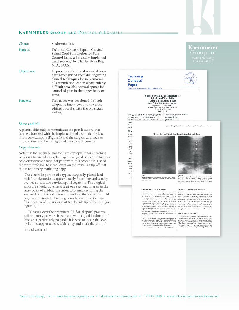
Show and tell

A picture efficiently communicates the pain locations that can be addressed with the implantation of a stimulating lead in the cervical spine (Figure 1) and the surgical approach to implantation in difficult region of the spine (Figure 2).

Copy close-up

Note that the language and tone are appropriate for a teaching physician to use when explaining the surgical procedure to other physicians who do have not performed this procedure. Use of the word “inferior” to mean lower on the spine is a tip-off that this is not breezy marketing copy.

“The electrode portion of a typical surgically-placed lead with four electrodes is approximately 3 cm long and usually overlies at least two cervical spinal segments. The surgical exposure should traverse at least one segment inferior to the entry point of epidural insertion to permit anchoring the lead neck into the soft tissues. Therefore, the incision should begin approximately three segments below the anticipated final position of the uppermost (cephalad) tip of the lead (see Figure 1).”

“…Palpating over the prominent C7 dorsal spinal process will ordinarily provide the surgeon with a good landmark. If this is not particularly palpable, it is wise to locate the level by fluoroscopy or a cross-table x-ray and mark the skin…”

[End of excerpt.]