cervical cord decompression using extended anterior cervical
TRANSCRIPT
114
But, it naturally advanced to spinal cord decompression with ex-tended technique (extended anterior cervical foraminotomy, EACF). Although many previous studies showed favorable clini-cal and radiological results, it has been used only for nerve root decompression. Here, we report the clinical results and effective-ness of this procedure for cervical cord decompression.
MATERIALS AND METHODS
PatientsBetween September 2008 and January 2013, we performed 53
EACF procedures. All our patients were operated on by a single surgeon via the unilateral approach. Twenty-two of the patients who showed cervical cord compression on their pre-operative magnetic resonance scan images with radicular and/or myelo-pathic symptoms were enrolled in this study. The preoperative
INTRODUCTION
Spondylotic lesions that cause cervical cord compression re-quire surgical decompression either through anterior cervical discectomy/corpectomy with fusion or laminectomy/lamino-plasty when they result in progressive neurological deficits2,3,13,20). These procedures have been regarded as a gold-standard tech-nique for decompression and stabilization. However, fusion-re-lated problems such as loss of the cervical range of motion, non-union, instrumentation failure, and graft extrusion are still the subject of debates7,20).
The Anterior cervical microforaminotomy (ACMF) technique involves not only the direct removal of the compressive abnor-mality but also the preservation of the motion segments without bone fusion or post-operative immobilization9,11,15).
ACMF was originally developed for cervical radiculopathy.
Cervical Cord Decompression Using Extended Anterior Cervical Foraminotomy Technique
Sung-Duk Kim, M.D.,1 Ho-Gyun Ha, M.D., Ph.D.,1,2 Cheol-Young Lee, M.D., Ph.D.,1 Hyun-Woo Kim, M.D., Ph.D.,1 Chul-Ku Jung, M.D., Ph.D.,1 Jong Hyun Kim, M.D., Ph.D.1
Department of Neurosurgery1, Konyang University Hospital, Daejon, Koera Department of Neurosurgery2, Teun Teun Hospital, Daejon, Korea
Objective : At present, gold-standard technique of cervical cord decompression is surgical decompression and fusion. But, many complications relat-ed cervical fusion have been reported. We adopted an extended anterior cervical foraminotomy (EACF) technique to decompress the anterolateral por-tion of cervical cord and report clinical results and effectiveness of this procedure.Methods : Fifty-three patients were operated consecutively using EACF from 2008 to 2013. All of them were operated by a single surgeon via the unilateral approach. Twenty-two patients who exhibited radicular and/or myelopathic symptoms were enrolled in this study. All of them showed cervi-cal cord compression in their preoperative magnetic resonance scan images.Results : In surgical outcomes, 14 patients (64%) were classified as excellent and six (27%), as good. The mean difference of cervical cord anterior-posterior diameter after surgery was 0.92 mm (p<0.01) and transverse area was 9.77 mm2 (p<0.01). The dynamic radiological study showed that the average post-operative translation (retrolisthesis) was 0.36 mm and the disc height loss at the operated level was 0.81 mm. The change in the Cobb angle decreased to 3.46, and showed slight kyphosis. The average vertebral body resection rate was 11.47%. No procedure-related complica-tions occurred. Only one patient who had two-level decompression needed anterior fusion at one level as a secondary surgery due to postoperative instability.Conclusions : Cervical cord decompression was successfully performed using EACF technique. This procedure will be an alternative surgical option for treating cord compressing lesions. Long-term follow-up and a further study in larger series will be needed.
Key Words : Cervical spondylosis · Anterior cervical foraminotomy · Spinal cord compression · Cervical myelopathy.
Clinical Article
• Received : March 7, 2014 • Revised : July 4, 2014 • Accepted : August 16, 2014• Address for reprints : Ho-Gyun Ha, M.D., Ph.D. Department of Neurosurgery, Teun Teun Hospital, 7 Mokjung-ro, Jung-gu, Daejon 301-808, Korea Tel : +82-42-600-9130, Fax : +82-42-600-8983, E-mail : [email protected]• This is an Open Access article distributed under the terms of the Creative Commons Attribution Non-Commercial License (http://creativecommons.org/licenses/by-nc/3.0) which permits unrestricted non-commercial use, distribution, and reproduction in any medium, provided the original work is properly cited.
J Korean Neurosurg Soc 56 (2) : 114-120, 2014
http://dx.doi.org/10.3340/jkns.2014.56.2.114
Copyright © 2014 The Korean Neurosurgical Society
Print ISSN 2005-3711 On-line ISSN 1598-7876www.jkns.or.kr
online © ML Comm
115
Cervical Cord Decompression Using EACF Technique | SD Kim, et al.
instability on cervical dynamic X-ray and already pre-existing narrowing of cervical spinal canal (that is, developmental steno-sis) excluded in this study.
Operative procedureThe detailed surgical technique of ACMF for cord compres-
sion has been reported by Jho8). A brief summary of our proce-dure from skin incision to exposure of the anterior column of the cervical spine is similar to other anterior approach to the cer-vical spine. An anterior cervical discectomy retractor system is applied to expose the ipsilateral longus colli muscle rather than the midline anterior disc surface. An operating microscope is es-sential for next stage. The lateral portion of the longus colli mus-cle is excised to expose the medial parts of the transverse process-es of the upper and lower vertebrae. Once the medial portion of the transverse processes of the upper and lower vertebrae is iden-tified, the ipsilateral uncovertebral joint between them can be seen; however, in advanced spondylosis, anatomical landmarks of the uncovertebral joint and transverse processes cannot be delineated. The uncovertebral joint is removed between the transverse processes using a high-speed microsurgical drill at-tached to an angled hand piece. To prevent injury to the verte-bral artery (VA), a thin layer of cortical bone is left attached to the ligamentous tissue covering the medial portion of this artery. Drilling continues down to the posterior longitudinal ligament. As drilling advances posteriorly, the direction of the drill is gen-tly inclined medially. When the posterior longitudinal ligament is exposed, a piece of thin cortical bone is left attached laterally to the periosteal and ligamentous tissue covering the VA.
This lateral remnant of the uncinate process is dissected from the ligamentous tissue and fractured at the base of the uncinate process. It is further dissected from the surrounding soft tissue and removed, which enables identification of the VA by its pul-sation between the transverse processes of the vertebrae. It is nec-essary to proceed cautiously with drilling at the base of the unci-nate process because the nerve root lies just adjacent to it. After the uncinate process becomes loosened at its base, it is safer to remove the thin layer of remaining bone of the uncinate process by fracturing it rather than by continued drilling. After remained piece of the uncinate process is removed, extended foraminoto-my procedure started. The posterior osteophytes, herniated part of disc, some parts of the upper and lower endplates are removed. The procedure crossed over the midline diagonally toward the opposite margin of the spinal dura mater.
The posterior longitudinal ligament is incised and resected to achieve adequate decompression of the ipsilateral nerve root and spinal cord. Sometimes the beginning of the contralateral nerve root is identified for adequate decompression of the spinal canal in the transverse axis. Multiple anterior foraminotomies are per-formed as needed. Using the holes of anterior foraminotomies, the spinal cord canal is enlarged in the longitudinal axis by re-moving the posterior portion of the vertebral bodies with Kerri-son rongeurs and a long-armed up-biting curette.
Radiological assessmentAll the patients took pre-operative plain radiography of cervi-
cal spine, computed tomography (CT), magnetic resonance im-aging (MRI), and dynamic plain radiography of cervical spine in six weeks after their operation. Analyses of the pre- and post-op-erative dynamic plain radiographs were done to evaluate postop-erative instability.
We measured five radiological data for evaluate to presence of postoperative instability and effectiveness of cervical cord decom-pression : the disc space height (DSH), sagittal plane displace-ment (translation), Cobb angle, vertebral body resection rate and degree of cervical cord decompression.
The disc space height was measured from the mid-point of the upper vertebral body to the mid-point of the lower vertebral body on the sagittal plane. To allow different magnification factors on different films, the measurements were compared with the height of the third cervical vertebrae (C3)18).
The sagittal plane displacement was measured on the cervical flexion-extension radiographs as the linear distance in millimeter scale from the posterior-inferior corner of the superior vertebra to the posterior-superior corner of the inferior vertebra body. A distance greater than 3.5 mm defined instability19).
The sagittal plane alignment was defined on the lateral neutral radiographs in relation to the line that joined the postero-inferi-or edge of C2 to the postero-inferior edge of C7. When all the in-tervening vertebral bodies were found anterior to this line, the alignment was defined as a lordotic curvature.
The sagittal angulation, which is the angle between a line on the superior end plate of the vertebral body above and a line on the inferior border of the body below the levels operated1), was measured on the neutral lateral view at the operated level, using Cobb’s method.
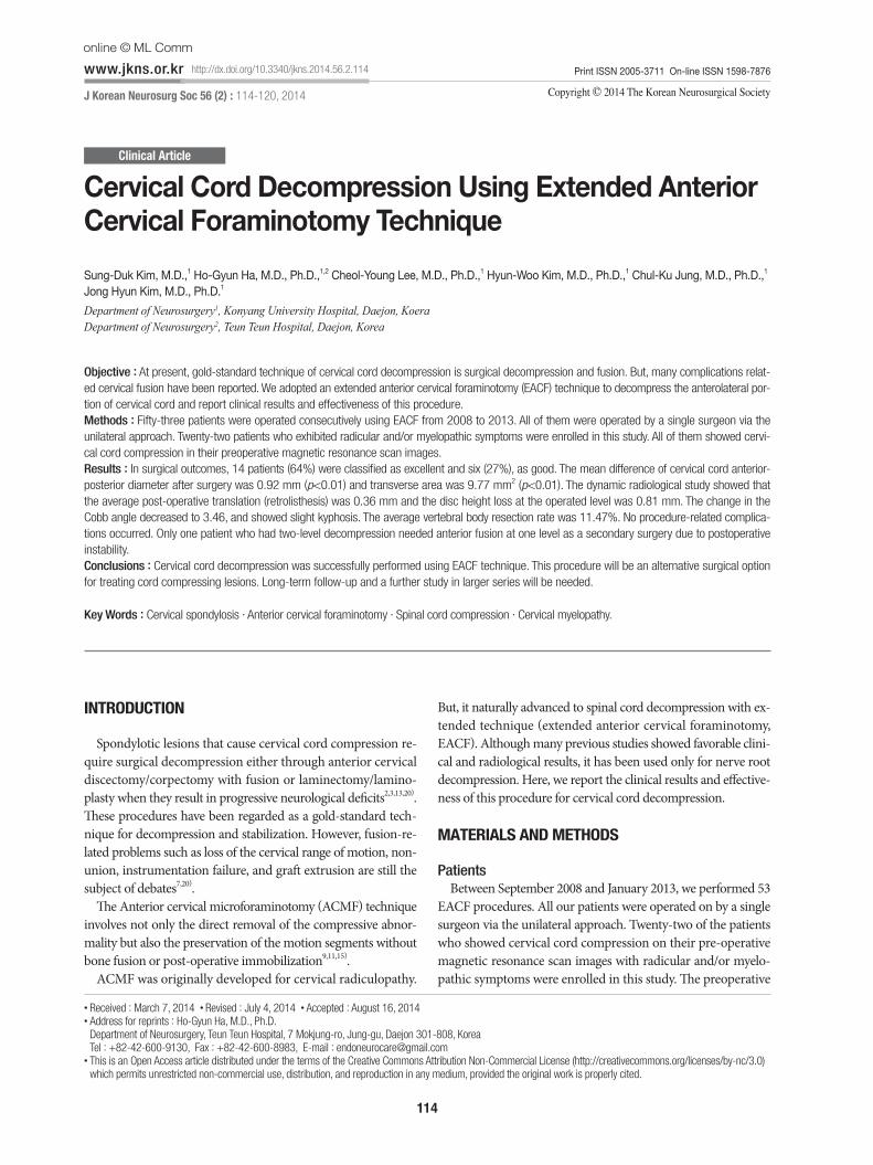
The vertebral body resection rate was measured on pre- and post-cervical 3-dimensional CT scan. All scans were performed on a 64-slice multi-detector computed tomography scanner (TOSHIBA Aquilion, Tokyo, Japan). The scanning parameters were as follows : voltage of 120 kV, current 150 mAs, field-of-view 147 mm and slice thickness of 3 mm (Fig. 1).
The degree of cervical cord decompression was measured an-tero-posterior diameter and transverse area of the spinal cord at the site of maximal compression. The data from preoperative and postoperative T2-weighted axial MRI analyzed using Aquarius iNutrition Edition ver4.4.6. (TeraRecon, Foster City, CA, USA) (Fig. 1).
Signal changes in the spinal cord on the T2 weighted images were also noted.
Clinical outcome assessmentThe post-operative pain improvement was measured using
the visual analogue score (VAS). The clinical outcome was grad-ed as “excellent” when the patient showed complete resolution of all symptoms, “good” when the patient experienced relief of ra-diculopathy but still experienced occasional minimal/mild resid-
116
J Korean Neurosurg Soc 56 | August 2014
ual non-radicular discomfort, “fair” when the patient showed mild residual symptoms of radiculopathy with or without mild/moderated residual non-radicular discomfort, and ”poor” when the patient continued to show significant radicular symptoms with or without non-radicular discomfort.
Statistical analysisThe statistical analysis was performed using PASW statistics
18.0 (SPSS Inc., Chicago, IL, USA). We were analyzed to the de-gree of cervical cord decompression, disc space height loss, trans-lation, Cobb angle and vertebral body resection rate using paired t-test. A p value of 0.05 was considered significant.
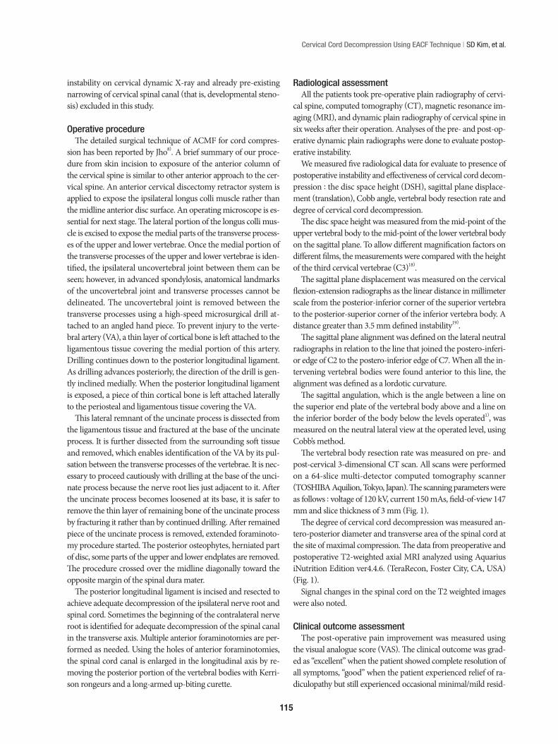
Illustrative casesA 52-year-old woman was referred to our hospital with a 6-
month history of posterior neck pain, right shoulder pain and progressive myelopathy that both upper and lower extremity numbness (right>left). On neurologic examination, she had Grade 4–/5 weakness of her right elbow flexion and extension. Her biceps and triceps reflexes were normal range. Spurling’s sign and Lhermitte’s sign were present bilaterally. CT and MRI scans demonstrated bulging disc C4–5, C5–6 with focal ossifica-tion posterior longitudinal ligament and showed cervical cord compression.
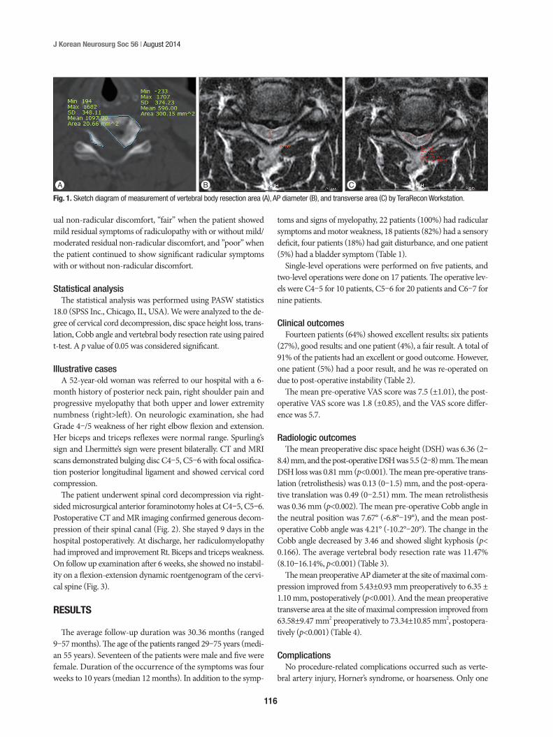
The patient underwent spinal cord decompression via right-sided microsurgical anterior foraminotomy holes at C4–5, C5–6. Postoperative CT and MR imaging confirmed generous decom-pression of their spinal canal (Fig. 2). She stayed 9 days in the hospital postoperatively. At discharge, her radiculomyelopathy had improved and improvement Rt. Biceps and triceps weakness. On follow up examination after 6 weeks, she showed no instabil-ity on a flexion-extension dynamic roentgenogram of the cervi-cal spine (Fig. 3).
RESULTS
The average follow-up duration was 30.36 months (ranged 9–57 months). The age of the patients ranged 29–75 years (medi-an 55 years). Seventeen of the patients were male and five were female. Duration of the occurrence of the symptoms was four weeks to 10 years (median 12 months). In addition to the symp-
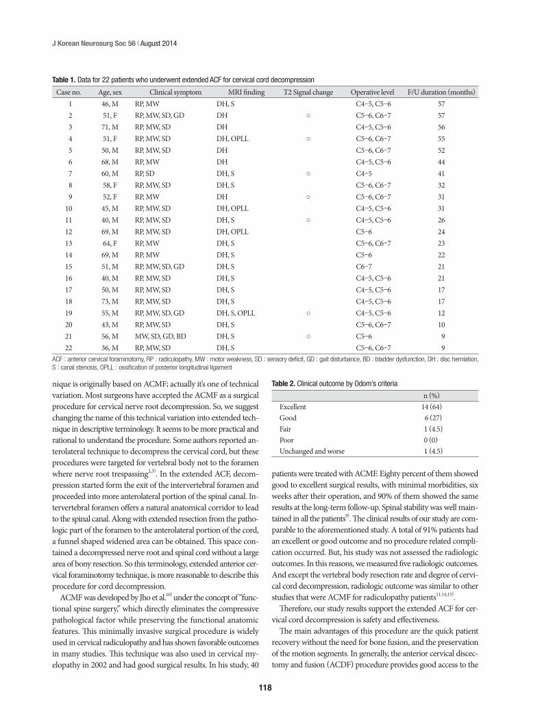
toms and signs of myelopathy, 22 patients (100%) had radicular symptoms and motor weakness, 18 patients (82%) had a sensory deficit, four patients (18%) had gait disturbance, and one patient (5%) had a bladder symptom (Table 1).
Single-level operations were performed on five patients, and two-level operations were done on 17 patients. The operative lev-els were C4–5 for 10 patients, C5–6 for 20 patients and C6–7 for nine patients.
Clinical outcomes Fourteen patients (64%) showed excellent results; six patients
(27%), good results; and one patient (4%), a fair result. A total of 91% of the patients had an excellent or good outcome. However, one patient (5%) had a poor result, and he was re-operated on due to post-operative instability (Table 2).
The mean pre-operative VAS score was 7.5 (±1.01), the post-operative VAS score was 1.8 (±0.85), and the VAS score differ-ence was 5.7.
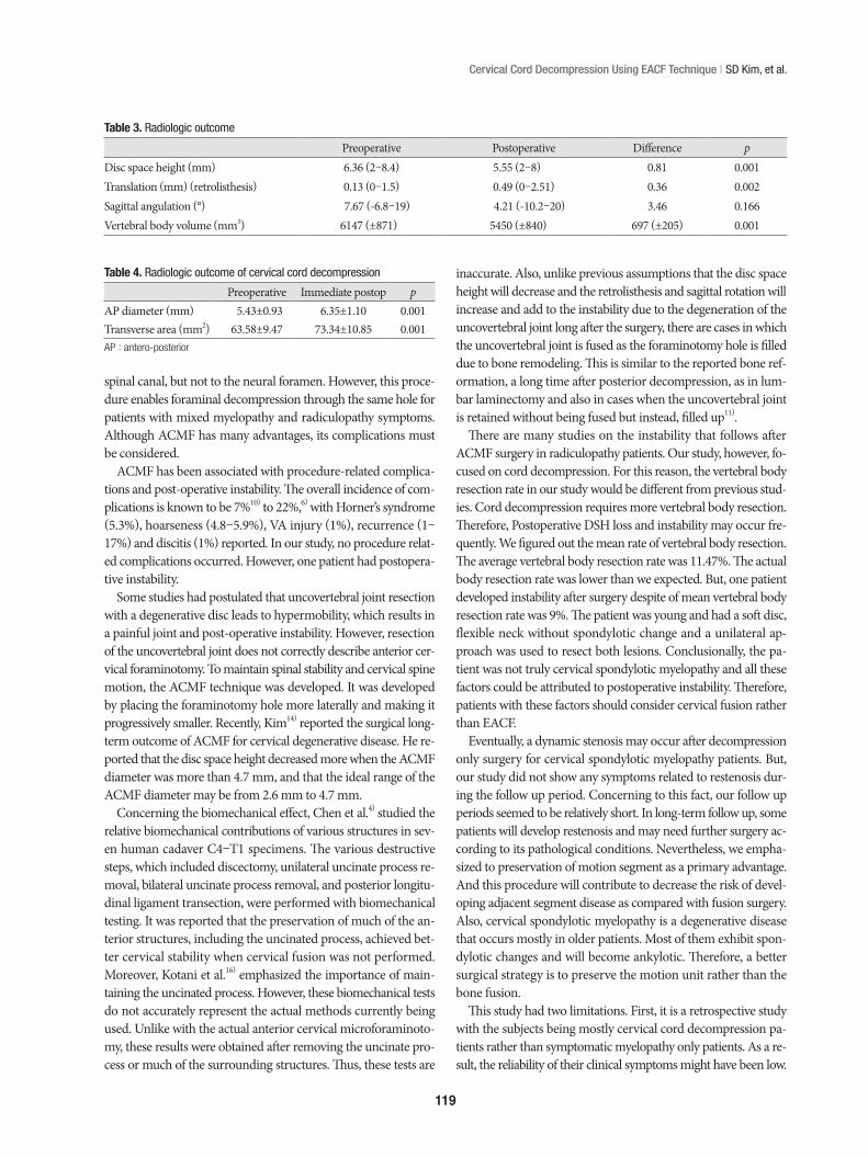
Radiologic outcomesThe mean preoperative disc space height (DSH) was 6.36 (2–
8.4) mm, and the post-operative DSH was 5.5 (2–8) mm. The mean DSH loss was 0.81 mm (p<0.001). The mean pre-operative trans-lation (retrolisthesis) was 0.13 (0–1.5) mm, and the post-opera-tive translation was 0.49 (0–2.51) mm. The mean retrolisthesis was 0.36 mm (p<0.002). The mean pre-operative Cobb angle in the neutral position was 7.67° (-6.8°–19°), and the mean post-operative Cobb angle was 4.21° (-10.2°–20°). The change in the Cobb angle decreased by 3.46 and showed slight kyphosis (p< 0.166). The average vertebral body resection rate was 11.47% (8.10–16.14%, p<0.001) (Table 3).
The mean preoperative AP diameter at the site of maximal com-pression improved from 5.43±0.93 mm preoperatively to 6.35 ± 1.10 mm, postoperatively (p<0.001). And the mean preoperative transverse area at the site of maximal compression improved from 63.58±9.47 mm2 preoperatively to 73.34±10.85 mm2, postopera-tively (p<0.001) (Table 4).
ComplicationsNo procedure-related complications occurred such as verte-
bral artery injury, Horner’s syndrome, or hoarseness. Only one
A B CFig. 1. Sketch diagram of measurement of vertebral body resection area (A), AP diameter (B), and transverse area (C) by TeraRecon Workstation.
117
Cervical Cord Decompression Using EACF Technique | SD Kim, et al.
patient needed anterior fusion as secondary surgery due to in-stability.
DISCUSSION
Cervical cord compressing lesions may eventually develop my-elopathic symptoms. But some patients presented only radicular symptoms as current clinical manifestations. Even in patients who had radicular symptoms, we frequently observed spinal cord com-pression in MRI and CT scan. In the literature, seventy-five per-cent of patients with myelopathy deteriorate in a stepwise fashion, 20% deteriorate slowly and steadily, and 5% have a rapid onset of symptoms with a stable plateau of dysfunction5). Most patients require surgical decompression when their condition results in progressive neurological deficit and functional declination. Tra-ditionally, surgical decompression has been performed to arrest neurological deterioration and prevent further disability. How-ever, recent studies have shown improved clinical outcomes.
At present, surgical treatment of cervical myelopathy can be broadly divided into anterior and posterior approaches. The an-terior approaches include anterior cervical discectomy and cor-pectomy, while the posterior approaches involve laminectomy with or without fusion, and laminoplasty12,13,17,21). The choice be-tween an anterior or posterior approach depends on the location, extent and type of the compressive pathology, the curvature of the spine, and the presence of instability. Like this, various tech-niques can be used presently, but the main goal of surgical inter-vention is spinal cord decompression. Optimal treatment of CSM is still controversial, and fusion-related complications such as loss of the range of motion, non-union, instrumentation failure and graft extrusion, and the adjacent-segment syndrome were ex-ist20,22). In our study, although all patients are not enough to crite-ria of cervical spondylotic myelopathy, main pathologic finding is cervical cord compression with degenerative change such as stenosis, OPLL. Through this study, we evaluate the effectiveness of EACF technique to CSM patients.
We propose EACF as an alternative minimally invasive non-fusion technique for cervical cord decompression. The ECAF tech-
A B
C
E
G H
F
D
Fig. 2. A and B : Axial, T2-weighted, MRI scans demonstrated C4–5, C5–6 disc herniation with cervical cord compression. C : Sagittal, T2-weighted MRI. D : Sagittal, cervical CT showed ossification of posterior longitudinal ligaments. E and F : Axial, T2-weighted, MRI scans showed excellent de-compression after surgery. G : Sagittal T2-weighted, MRI scans. H : Posto-perative cervical 3D CT showed microforaminotomy hole at C4-5, C5–6, Rt.
A BFig. 3. Flexion (A) and extension (B) dynamic roentgenograms of the cer-vical spine, obtained 6 weeks after surgery, confirming normal motion at the C4–5, C5–6 level.
118
J Korean Neurosurg Soc 56 | August 2014
nique is originally based on ACMF; actually it’s one of technical variation. Most surgeons have accepted the ACMF as a surgical procedure for cervical nerve root decompression. So, we suggest changing the name of this technical variation into extended tech-nique in descriptive terminology. It seems to be more practical and rational to understand the procedure. Some authors reported an-terolateral technique to decompress the cervical cord, but these procedures were targeted for vertebral body not to the foramen where nerve root trespassing2,3). In the extended ACF, decom-pression started form the exit of the intervertebral foramen and proceeded into more anterolateral portion of the spinal canal. In-tervertebral foramen offers a natural anatomical corridor to lead to the spinal canal. Along with extended resection from the patho-logic part of the foramen to the anterolateral portion of the cord, a funnel shaped widened area can be obtained. This space con-tained a decompressed nerve root and spinal cord without a large area of bony resection. So this terminology, extended anterior cer-vical foraminotomy technique, is more reasonable to describe this procedure for cord decompression.
ACMF was developed by Jho et al.10) under the concept of “func-tional spine surgery,” which directly eliminates the compressive pathological factor while preserving the functional anatomic features. This minimally invasive surgical procedure is widely used in cervical radiculopathy and has shown favorable outcomes in many studies. This technique was also used in cervical my-elopathy in 2002 and had good surgical results. In his study, 40
patients were treated with ACMF. Eighty percent of them showed good to excellent surgical results, with minimal morbidities, six weeks after their operation, and 90% of them showed the same results at the long-term follow-up. Spinal stability was well main-tained in all the patients9). The clinical results of our study are com-parable to the aforementioned study. A total of 91% patients had an excellent or good outcome and no procedure related compli-cation occurred. But, his study was not assessed the radiologic outcomes. In this reasons, we measured five radiologic outcomes. And except the vertebral body resection rate and degree of cervi-cal cord decompression, radiologic outcome was similar to other studies that were ACMF for radiculopathy patients11,14,15).
Therefore, our study results support the extended ACF for cer-vical cord decompression is safety and effectiveness.
The main advantages of this procedure are the quick patient recovery without the need for bone fusion, and the preservation of the motion segments. In generally, the anterior cervical discec-tomy and fusion (ACDF) procedure provides good access to the
Table 1. Data for 22 patients who underwent extended ACF for cervical cord decompression
Case no. Age, sex Clinical symptom MRI finding T2 Signal change Operative level F/U duration (months)1 46, M RP, MW DH, S C4–5, C5–6 572 51, F RP, MW, SD, GD DH ○ C5–6, C6–7 573 71, M RP, MW, SD DH C4–5, C5–6 564 51, F RP, MW, SD DH, OPLL ○ C5–6, C6–7 555 50, M RP, MW, SD DH C5–6, C6–7 526 68, M RP, MW DH C4–5, C5–6 447 60, M RP, SD DH, S ○ C4–5 418 58, F RP, MW, SD DH, S C5–6, C6–7 329 52, F RP, MW DH ○ C5–6, C6–7 31
10 45, M RP, MW, SD DH, OPLL C4–5, C5–6 3111 40, M RP, MW, SD DH, S ○ C4–5, C5–6 2612 69, M RP, MW, SD DH, OPLL C5–6 2413 64, F RP, MW DH, S C5–6, C6–7 2314 69, M RP, MW DH, S C5–6 2215 51, M RP, MW, SD, GD DH, S C6–7 2116 40, M RP, MW, SD DH, S C4–5, C5–6 2117 50, M RP, MW, SD DH, S C4–5, C5–6 1718 73, M RP, MW, SD DH, S C4–5, C5–6 1719 55, M RP, MW, SD, GD DH, S, OPLL ○ C4–5, C5–6 1220 43, M RP, MW, SD DH, S C5–6, C6–7 1021 56, M MW, SD, GD, BD DH, S ○ C5–6 922 36, M RP, MW, SD DH, S C5–6, C6–7 9
ACF : anterior cervical foraminotomy, RP : radiculopathy, MW : motor weakness, SD : sensory deficit, GD : gait disturbance, BD : bladder dysfunction, DH : disc herniation, S : canal stenosis, OPLL : ossification of posterior longitudinal ligament
Table 2. Clinical outcome by Odom’s criteria
n (%)Excellent 14 (64)Good 6 (27)Fair 1 (4.5)Poor 0 (0)Unchanged and worse 1 (4.5)
119
Cervical Cord Decompression Using EACF Technique | SD Kim, et al.
spinal canal, but not to the neural foramen. However, this proce-dure enables foraminal decompression through the same hole for patients with mixed myelopathy and radiculopathy symptoms. Although ACMF has many advantages, its complications must be considered.
ACMF has been associated with procedure-related complica-tions and post-operative instability. The overall incidence of com-plications is known to be 7%10) to 22%,6) with Horner’s syndrome (5.3%), hoarseness (4.8–5.9%), VA injury (1%), recurrence (1–17%) and discitis (1%) reported. In our study, no procedure relat-ed complications occurred. However, one patient had postopera-tive instability.
Some studies had postulated that uncovertebral joint resection with a degenerative disc leads to hypermobility, which results in a painful joint and post-operative instability. However, resection of the uncovertebral joint does not correctly describe anterior cer-vical foraminotomy. To maintain spinal stability and cervical spine motion, the ACMF technique was developed. It was developed by placing the foraminotomy hole more laterally and making it progressively smaller. Recently, Kim14) reported the surgical long-term outcome of ACMF for cervical degenerative disease. He re-ported that the disc space height decreased more when the ACMF diameter was more than 4.7 mm, and that the ideal range of the ACMF diameter may be from 2.6 mm to 4.7 mm.
Concerning the biomechanical effect, Chen et al.4) studied the relative biomechanical contributions of various structures in sev-en human cadaver C4–T1 specimens. The various destructive steps, which included discectomy, unilateral uncinate process re-moval, bilateral uncinate process removal, and posterior longitu-dinal ligament transection, were performed with biomechanical testing. It was reported that the preservation of much of the an-terior structures, including the uncinated process, achieved bet-ter cervical stability when cervical fusion was not performed. Moreover, Kotani et al.16) emphasized the importance of main-taining the uncinated process. However, these biomechanical tests do not accurately represent the actual methods currently being used. Unlike with the actual anterior cervical microforaminoto-my, these results were obtained after removing the uncinate pro-cess or much of the surrounding structures. Thus, these tests are
inaccurate. Also, unlike previous assumptions that the disc space height will decrease and the retrolisthesis and sagittal rotation will increase and add to the instability due to the degeneration of the uncovertebral joint long after the surgery, there are cases in which the uncovertebral joint is fused as the foraminotomy hole is filled due to bone remodeling. This is similar to the reported bone ref-ormation, a long time after posterior decompression, as in lum-bar laminectomy and also in cases when the uncovertebral joint is retained without being fused but instead, filled up11).
There are many studies on the instability that follows after ACMF surgery in radiculopathy patients. Our study, however, fo-cused on cord decompression. For this reason, the vertebral body resection rate in our study would be different from previous stud-ies. Cord decompression requires more vertebral body resection. Therefore, Postoperative DSH loss and instability may occur fre-quently. We figured out the mean rate of vertebral body resection. The average vertebral body resection rate was 11.47%. The actual body resection rate was lower than we expected. But, one patient developed instability after surgery despite of mean vertebral body resection rate was 9%. The patient was young and had a soft disc, flexible neck without spondylotic change and a unilateral ap-proach was used to resect both lesions. Conclusionally, the pa-tient was not truly cervical spondylotic myelopathy and all these factors could be attributed to postoperative instability. Therefore, patients with these factors should consider cervical fusion rather than EACF.
Eventually, a dynamic stenosis may occur after decompression only surgery for cervical spondylotic myelopathy patients. But, our study did not show any symptoms related to restenosis dur-ing the follow up period. Concerning to this fact, our follow up periods seemed to be relatively short. In long-term follow up, some patients will develop restenosis and may need further surgery ac-cording to its pathological conditions. Nevertheless, we empha-sized to preservation of motion segment as a primary advantage. And this procedure will contribute to decrease the risk of devel-oping adjacent segment disease as compared with fusion surgery. Also, cervical spondylotic myelopathy is a degenerative disease that occurs mostly in older patients. Most of them exhibit spon-dylotic changes and will become ankylotic. Therefore, a better surgical strategy is to preserve the motion unit rather than the bone fusion.
This study had two limitations. First, it is a retrospective study with the subjects being mostly cervical cord decompression pa-tients rather than symptomatic myelopathy only patients. As a re-sult, the reliability of their clinical symptoms might have been low.
Table 3. Radiologic outcome
Preoperative Postoperative Difference pDisc space height (mm) 6.36 (2–8.4) 5.55 (2–8) 0.81 0.001Translation (mm) (retrolisthesis) 0.13 (0–1.5) 0.49 (0–2.51) 0.36 0.002Sagittal angulation (°) 7.67 (-6.8–19) 4.21 (-10.2–20) 3.46 0.166Vertebral body volume (mm3) 6147 (±871) 5450 (±840) 697 (±205) 0.001
Table 4. Radiologic outcome of cervical cord decompression
Preoperative Immediate postop pAP diameter (mm) 5.43±0.93 6.35±1.10 0.001Transverse area (mm2) 63.58±9.47 73.34±10.85 0.001AP : antero-posterior
120
J Korean Neurosurg Soc 56 | August 2014
Second, the follow-up intervals were variable and relatively short.
CONCLUSION
A decompression and fusion surgery has been considered as a standard surgical treatment for cervical cord compressing lesions. But, in our study, a decompression without fusion procedure was successfully performed using EACF technique. This procedure achieves adequate anterior decompression of the spinal cord with preservation of the functional motion, and avoids fusion-related complications. With our clinical and radiological results, authors conclude that EACF is a possible and applicable surgical option for the spondylotic patients who need cervical cord decompres-sion.
In selecting patients, the pre-operative clinical status should be carefully considered to achieve an excellent outcome. Long-term follow-up and a further study in a larger series are needed.
References 1. Batzdorf U, Batzdorff A : Analysis of cervical spine curvature in patients
with cervical spondylosis. Neurosurgery 22 : 827-836, 19882. Chacko AG, Joseph M, Turel MK, Prabhu K, Daniel RT, Jacob KS : Mul-
tilevel oblique corpectomy for cervical spondylotic myelopathy preserves segmental motion. Eur Spine J 21 : 1360-1367, 2012
3. Chacko AG, Turel MK, Sarkar S, Prabhu K, Daniel RT : Clinical and ra-diological outcomes in 153 patients undergoing oblique corpectomy for cervical spondylotic myelopathy. Br J Neurosurg 28 : 49-55, 2014
4. Chen TY, Crawford NR, Sonntag VK, Dickman CA : Biomechanical ef-fects of progressive anterior cervical decompression. Spine (Phila Pa 1976) 26 : 6-13; discussion 14, 2001
5. Clarke E, Robinson PK : Cervical myelopathy : a complication of cervi-cal spondylosis. Brain 79 : 483-510, 1956
6. Hacker RJ, Miller CG : Failed anterior cervical foraminotomy. J Neuro-surg 98 (2 Suppl) : 126-130, 2003
7. Haden N, Latimer M, Seeley HM, Laing RJ : Loss of inter-vertebral disc height after anterior cervical discectomy. Br J Neurosurg 19 : 469-474, 2005
8. Jho HD : Decompression via microsurgical anterior foraminotomy for cervical spondylotic myelopathy. Technical note. J Neurosurg 86 : 297-302, 1997
9. Jho HD, Kim MH, Kim WK : Anterior cervical microforaminotomy for spondylotic cervical myelopathy : part 2. Neurosurgery 51 (5 Suppl) : S54-S59, 2002
10. Jho HD, Kim WK, Kim MH : Anterior microforaminotomy for treatment of cervical radiculopathy : part 1--disc-preserving “functional cervical disc surgery”. Neurosurgery 51 (5 Suppl) : S46-S53, 2002
11. Jung SS, Chung JC, Park KS, Chung SY, Park MS, Ha HG. : Long-term follow-up results of anterior cervical microforaminotomy. Korean J Spine 7 : 66-72, 2010
12. Kalsi-Ryan S, Karadimas SK, Fehlings MG : Cervical spondylotic myelop-athy : the clinical phenomenon and the current pathobiology of an in-creasingly prevalent and devastating disorder. Neuroscientist 19 : 409-421, 2013
13. Kim JG, Kim SW, Lee SM, Shin H : Surgical result of the combined an-terior and posterior approach in treatment of cervical spondylotic my-elopathy. J Korean Neurosurg Soc 3 : 188-191, 2006
14. Kim MH : Clinical and radiological long-term outcomes of anterior mi-croforaminotomy for cervical degenerative disease. Spine (Phila Pa 1976) 38 : 1812-1819, 2013
15. Kim YG, Lee JS, Park MS, Ha HG : Midterm follow-up results of anterior cervical microforaminotomy. J Korean Neurosurg Soc 35 : 250-255, 2004
16. Kotani Y, McNulty PS, Abumi K, Cunningham BW, Kaneda K, McAfee PC : The role of anteromedial foraminotomy and the uncovertebral joints in the stability of the cervical spine. A biomechanical study. Spine (Phila Pa 1976) 23 : 1559-1565, 1998
17. Lebl DR, Hughes A, Cammisa FP Jr, O’Leary PF : Cervical spondylotic myelopathy : pathophysiology, clinical presentation, and treatment. HSS J 7 : 170-178, 2011
18. Oh SH, Perin NI, Cooper PR : Quantitative three-dimensional anatomy of the subaxial cervical spine : implication for anterior spinal surgery. Neurosurgery 38 : 1139-1144, 1996
19. White AA 3rd, Johnson RM, Panjabi MM, Southwick WO : Biomechan-ical analysis of clinical stability in the cervical spine. Clin Orthop Relat Res (109) : 85-96, 1975
20. Wu XD, Yuan W, Chen HJ, Chen Y, Wang JX, Cao P, et al. : Neck motion following multilevel anterior cervical fusion : comparison of short-term and midterm results. J Neurosurg Spine 18 : 362-366, 2013
21. Yang HL, Chen GD, Zhang HT, Wang L, Luo ZP : Open-door lamino-plasty with plate fixation at alternating levels for treatment of multilevel degenerative cervical disease. J Spinal Disord Tech 26 : E13-E18, 2013
22. Zhu B, Xu Y, Liu X, Liu Z, Dang G : Anterior approach versus posterior approach for the treatment of multilevel cervical spondylotic myelopathy : a systemic review and meta-analysis. Eur Spine J 22 : 1583-1593, 2013