congenital cervical cysts, sinuses and fistulae cervical cysts, sinuses and fistulae ... and other...
TRANSCRIPT
Otolaryngol Clin N Am
40 (2007) 161–176
Congenital Cervical Cysts,Sinuses and Fistulae
Stephanie P. Acierno, MD, MPH,John H.T. Waldhausen, MD*
Department of Surgery, Children’s Hospital and Regional Medical Center,
University of Washington School of Medicine, G0035,
4800 Sand Point Way, NE, Seattle, WA 98105, USA
Congenital cervical cysts, sinuses, and fistulae must be considered in thediagnosis of head and neck masses in children and adults. These include, indescending order of frequency, thyroglossal duct cysts, branchial cleftanomalies, dermoid cysts, andmedian cervical clefts. A thorough understand-ing of the embryology and anatomy of each of these lesions is necessary toprovide accurate preoperative diagnosis and appropriate surgical therapy,which are essential to prevent recurrence. The following sections revieweach lesion, its embryology, anatomy, common presentation, evaluation,and the key points in surgical management.
Thyroglossal duct anomalies
Thyroglossal duct anomalies are the second most common pediatric neckmass, behind adenopathy in frequency [1]. Thyroglossal duct remnantsoccur in approximately 7% of the population, although only a minority ofthese is ever symptomatic [1].
Embryology
The thyroid gland forms from a diverticulum (median thyroid anlage) lo-cated between the anterior and posterior muscle complexes of the tongue atweek 3 of gestation. As the embryo grows, the diverticulum is displaced cau-dally into the neck and fuses with components from the fourth and fifth
* Corresponding author.
E-mail address: [email protected] (J.H.T. Waldhausen).
0030-6665/07/$ - see front matter � 2007 Elsevier Inc. All rights reserved.
doi:10.1016/j.otc.2006.10.009 oto.theclinics.com
162 ACIERNO & WALDHAUSEN
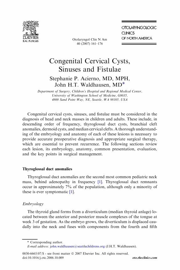
branchial pouches (lateral thyroid anlagen). The descent continues anteriorto or through the hyoid bone with the median anlage elongating into thethyroglossal duct (Fig. 1) [2]. By weeks 5 to 8 of gestation, the thyroglossalduct obliterates, leaving a proximal remnant, the foramen cecum, at the baseof the tongue and a distal remnant, the pyramidal lobe of the thyroid [1,2]. Ifthe duct fails to obliterate before the formation of the mesodermal anlage ofthe hyoid bone, it persists as a cyst [2].
Clinical presentation and diagnosis
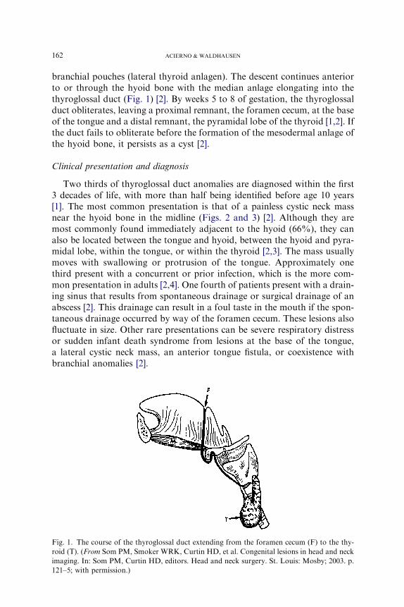
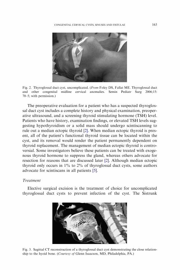
Two thirds of thyroglossal duct anomalies are diagnosed within the first3 decades of life, with more than half being identified before age 10 years[1]. The most common presentation is that of a painless cystic neck massnear the hyoid bone in the midline (Figs. 2 and 3) [2]. Although they aremost commonly found immediately adjacent to the hyoid (66%), they canalso be located between the tongue and hyoid, between the hyoid and pyra-midal lobe, within the tongue, or within the thyroid [2,3]. The mass usuallymoves with swallowing or protrusion of the tongue. Approximately onethird present with a concurrent or prior infection, which is the more com-mon presentation in adults [2,4]. One fourth of patients present with a drain-ing sinus that results from spontaneous drainage or surgical drainage of anabscess [2]. This drainage can result in a foul taste in the mouth if the spon-taneous drainage occurred by way of the foramen cecum. These lesions alsofluctuate in size. Other rare presentations can be severe respiratory distressor sudden infant death syndrome from lesions at the base of the tongue,a lateral cystic neck mass, an anterior tongue fistula, or coexistence withbranchial anomalies [2].
Fig. 1. The course of the thyroglossal duct extending from the foramen cecum (F) to the thy-
roid (T). (From Som PM, Smoker WRK, Curtin HD, et al. Congenital lesions in head and neck
imaging. In: Som PM, Curtin HD, editors. Head and neck surgery. St. Louis: Mosby; 2003. p.
121–5; with permission.)
163CONGENITAL CERVICAL CYSTS, SINUSES AND FISTULAE
The preoperative evaluation for a patient who has a suspected thyroglos-sal duct cyst includes a complete history and physical examination, preoper-ative ultrasound, and a screening thyroid stimulating hormone (TSH) level.Patients who have history, examination findings, or elevated TSH levels sug-gesting hypothyroidism or a solid mass should undergo scintiscanning torule out a median ectopic thyroid [2]. When median ectopic thyroid is pres-ent, all of the patient’s functional thyroid tissue can be located within thecyst, and its removal would render the patient permanently dependent onthyroid replacement. The management of median ectopic thyroid is contro-versial. Some investigators believe these patients can be treated with exoge-nous thyroid hormone to suppress the gland, whereas others advocate forresection for reasons that are discussed later [2]. Although median ectopicthyroid only occurs in 1% to 2% of thyroglossal duct cysts, some authorsadvocate for scintiscans in all patients [5].
Treatment
Elective surgical excision is the treatment of choice for uncomplicatedthyroglossal duct cysts to prevent infection of the cyst. The Sistrunk
Fig. 2. Thyroglossal duct cyst, uncomplicated. (From Foley DS, Fallat ME. Thyroglossal duct
and other congenital midline cervical anomalies. Semin Pediatr Surg 2006;15:
70–5; with permission.)
Fig. 3. Sagittal CT reconstruction of a thyroglossal duct cyst demonstrating the close relation-
ship to the hyoid bone. (Courtesy of Glenn Isaacson, MD, Philadelphia, PA.)
164 ACIERNO & WALDHAUSEN
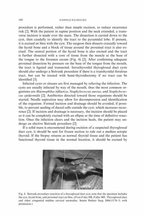
procedure is performed, rather than simple excision, to reduce recurrencerisk [2]. With the patient in supine position and the neck extended, a trans-verse incision is made over the mass. The dissection is carried down to thecyst, then caudally to identify the tract to the pyramidal lobe. If present,it is excised en bloc with the cyst. The surgeon then dissects cranially towardthe hyoid bone and a block of tissue around the proximal tract is also ex-cised. The central portion of the hyoid bone is also excised and the tractis further dissected with a core of tissue from the muscle at the base ofthe tongue to the foramen cecum (Fig. 4) [2]. After confirming adequateproximal dissection by pressure on the base of the tongue from the mouth,the tract is ligated and transected. Intrathyroidal thyroglossal duct cystsshould also undergo a Sistrunk procedure if there is a transhyoidal fistuloustract, but can be treated with hemi-thyroidectomy if no tract can beidentified [3].
Infected cysts or sinuses are first managed by relieving the infection. Thecysts are usually infected by way of the mouth, thus the most common or-ganisms are Haemophilus influenza, Staphylococcus aureus, and Staphylococ-cus epidermidis [2]. Antibiotics directed toward those organisms should bestarted. Needle aspiration may allow for decompression and identificationof the organism. Formal incision and drainage should be avoided, if possi-ble, to prevent seeding of ductal cells outside the cyst, which increases recur-rence [2]. If incision and drainage is necessary, the incision should be placedso it can be completely excised with an ellipse at the time of definitive resec-tion. Once the infection clears and the incision heals, the patient may un-dergo an elective Sistrunk procedure [2].
If a solid mass is encountered during excision of a suspected thyroglossalduct cyst, it should be sent for frozen section to rule out a median ectopicthyroid. If the biopsy returns as normal thyroid tissue and the patient hasfunctional thyroid tissue in the normal location, it should be excised by
Fig. 4. Sistrunk procedure resection of a thyroglossal duct cyst; note that the specimen includes
the cyst, hyoid bone, and proximal tract en bloc. (From Foley DS, Fallat ME. Thyroglossal duct
and other congenital midline cervical anomalies. Semin Pediatr Surg 2006;15:70–5; with
permission.)
165CONGENITAL CERVICAL CYSTS, SINUSES AND FISTULAE
the Sistrunk procedure [2]. If the mass is possibly the patient’s only func-tional thyroid tissue, the management becomes controversial. One option in-volves leaving the ectopic thyroid, either in situ or repositioning it laterallybelow the strap muscles or into the rectus abdominus or quadriceps muscles.This option aims to not render the patient permanently hypothyroid; how-ever, most patients still require long-term thyroid hormone therapy to treathypothyroidism or control the size of the ectopic thyroid tissue for cosmeticor functional reasons. This need for long-term therapy and the possibility ofmalignant degeneration have led some to recommend excision of the medianectopic thyroid regardless of the presence of additional thyroid tissue [2].
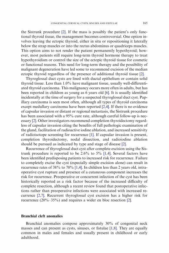
Thyroglossal duct cysts are lined with ductal epithelium or contain solidthyroid tissue. Less than 1.0% have malignant tissue, usually well-differenti-ated thyroid carcinoma. This malignancy occurs more often in adults, but hasbeen reported in children as young as 6 years old [6]. It is usually identifiedincidentally at the time of surgery for a suspected thyroglossal duct cyst. Pap-illary carcinoma is seen most often, although all types of thyroid carcinomaexcept medullary carcinoma have been reported [2,4]. If there is no evidenceof capsular invasion or distant or regional metastasis, the Sistrunk procedurehas been associated with a 95% cure rate, although careful follow-up is nec-essary [2]. Other investigators recommend completion thyroidectomy regard-less of capsular invasion citing the benefits of full pathologic examination ofthe gland, facilitation of radioactive iodine ablation, and increased sensitivityof radioisotope screening for recurrence [1]. If capsular invasion is present,completion thyroidectomy, nodal dissection, and radioiodine ablationshould be pursued as indicated by type and stage of disease [2].
Recurrence of thyroglossal duct cyst after complete excision using the Sis-trunk procedure is reported to be 2.6% to 5% [1,4]. Several factors havebeen identified predisposing patients to increased risk for recurrence. Failureto completely excise the cyst (especially simple excision alone) can result inrecurrence rates of 38% to 70% [1,4]. In children less than 2 years old, intra-operative cyst rupture and presence of a cutaneous component increases therisk for recurrence. Preoperative or concurrent infection of the cyst has beenhistorically reported as a risk factor because of the increased difficulty ofcomplete resection, although a recent review found that postoperative infec-tions rather than preoperative infections were associated with increased re-currence [2,7]. Recurrent thyroglossal cyst excision has a higher risk forrecurrence (20%–35%) and requires a wider en bloc resection [2].
Branchial cleft anomalies
Branchial anomalies compose approximately 30% of congenital neckmasses and can present as cysts, sinuses, or fistulae [1,8]. They are equallycommon in males and females and usually present in childhood or earlyadulthood.
166 ACIERNO & WALDHAUSEN
Embryology
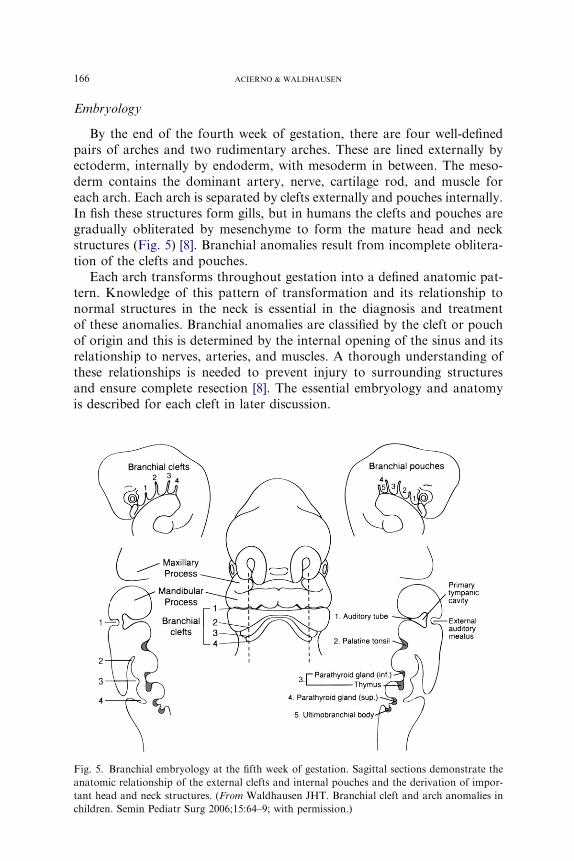
By the end of the fourth week of gestation, there are four well-definedpairs of arches and two rudimentary arches. These are lined externally byectoderm, internally by endoderm, with mesoderm in between. The meso-derm contains the dominant artery, nerve, cartilage rod, and muscle foreach arch. Each arch is separated by clefts externally and pouches internally.In fish these structures form gills, but in humans the clefts and pouches aregradually obliterated by mesenchyme to form the mature head and neckstructures (Fig. 5) [8]. Branchial anomalies result from incomplete oblitera-tion of the clefts and pouches.
Each arch transforms throughout gestation into a defined anatomic pat-tern. Knowledge of this pattern of transformation and its relationship tonormal structures in the neck is essential in the diagnosis and treatmentof these anomalies. Branchial anomalies are classified by the cleft or pouchof origin and this is determined by the internal opening of the sinus and itsrelationship to nerves, arteries, and muscles. A thorough understanding ofthese relationships is needed to prevent injury to surrounding structuresand ensure complete resection [8]. The essential embryology and anatomyis described for each cleft in later discussion.
Fig. 5. Branchial embryology at the fifth week of gestation. Sagittal sections demonstrate the
anatomic relationship of the external clefts and internal pouches and the derivation of impor-
tant head and neck structures. (From Waldhausen JHT. Branchial cleft and arch anomalies in
children. Semin Pediatr Surg 2006;15:64–9; with permission.)
167CONGENITAL CERVICAL CYSTS, SINUSES AND FISTULAE
Pathology
Branchial anomalies can be lined with either respiratory or squamous ep-ithelium. Cysts are often lined by squamous epithelium, whereas sinuses andfistulae are more likely to be lined with ciliated, columnar epithelium [8].Lymphoid tissue, sebaceous glands, salivary tissue, or cholesterol crystalsin mucoid fluid can also be seen. Squamous cell cancer can be found withinbranchial lesions in adults, although it is rare. It is difficult to distinguishbetween a primary lesion arising from within an anomaly and a metastaticlesion from an occult primary [1].
Diagnosis
Branchial anomalies can present as cysts, sinuses, or fistulae. Cysts areremnants of the cervical sinus without an external opening. Sinuses arethe persistence of the cervical sinus with its external opening, whereas a fis-tula also involves persistence of the branchial groove with breakdown of thebranchial membrane resulting in a pharyngocutaneous fistula [1]. The spe-cific presentation for each cleft is described in later discussion.
The evaluation of these lesions begins with a complete history and phys-ical, which may include upper airway endoscopy to locate the pharyngealopening. The pyriform sinus and the tonsillar fossa should be carefully ex-amined. In adults, fine needle aspiration should be performed to rule outmetastatic carcinoma or clarify the diagnosis [8]. This clarification is notnecessary in children and incisional biopsy should not be performed becausethis makes the resection more difficult. Ultrasound, CT, and MRI can beused to help define the lesion and its course, but CT is the current studyof choice. Current tomography is able to demonstrate the fistula in up to64% of cases [9]. Barium esophagram can also be helpful with a 50% to80% sensitivity for third and fourth branchial fistulae [10].
Treatment
The definitive treatment of all branchial anomalies is complete surgicalexcision. Unresected cysts and sinuses have a high risk for infection and in-complete resection results in high rates of recurrence [8]. Timing of resec-tion is controversial with some advocating for early resection to preventinfection whereas others support waiting until age 2 to 3 years [8,11,12].Twenty percent of lesions have been infected at least once before the timeof surgery [11]. As with thyroglossal duct cysts, acute infections should firstbe treated with antibiotics, needle aspiration, and, if necessary, incision anddrainage, followed by complete resection after resolution of the infection.Specific considerations for the resection of each type of anomaly are dis-cussed later.
168 ACIERNO & WALDHAUSEN
First cleft anomalies
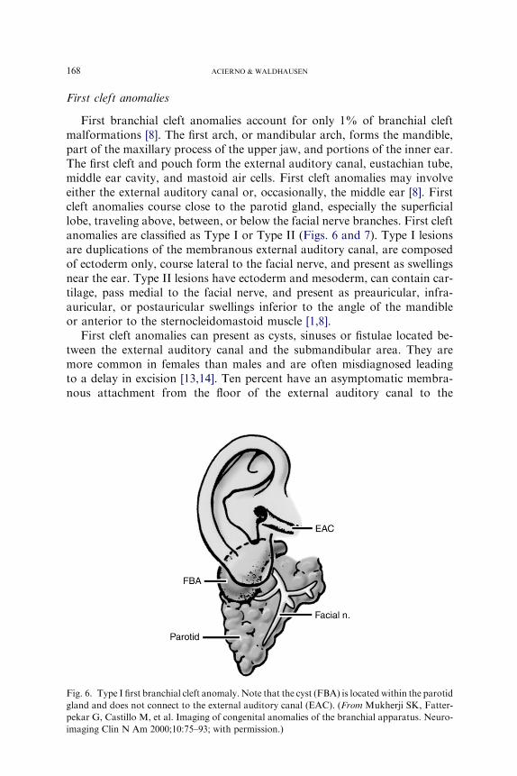
First branchial cleft anomalies account for only 1% of branchial cleftmalformations [8]. The first arch, or mandibular arch, forms the mandible,part of the maxillary process of the upper jaw, and portions of the inner ear.The first cleft and pouch form the external auditory canal, eustachian tube,middle ear cavity, and mastoid air cells. First cleft anomalies may involveeither the external auditory canal or, occasionally, the middle ear [8]. Firstcleft anomalies course close to the parotid gland, especially the superficiallobe, traveling above, between, or below the facial nerve branches. First cleftanomalies are classified as Type I or Type II (Figs. 6 and 7). Type I lesionsare duplications of the membranous external auditory canal, are composedof ectoderm only, course lateral to the facial nerve, and present as swellingsnear the ear. Type II lesions have ectoderm and mesoderm, can contain car-tilage, pass medial to the facial nerve, and present as preauricular, infra-auricular, or postauricular swellings inferior to the angle of the mandibleor anterior to the sternocleidomastoid muscle [1,8].
First cleft anomalies can present as cysts, sinuses or fistulae located be-tween the external auditory canal and the submandibular area. They aremore common in females than males and are often misdiagnosed leadingto a delay in excision [13,14]. Ten percent have an asymptomatic membra-nous attachment from the floor of the external auditory canal to the
Fig. 6. Type I first branchial cleft anomaly. Note that the cyst (FBA) is located within the parotid
gland and does not connect to the external auditory canal (EAC). (From Mukherji SK, Fatter-
pekar G, Castillo M, et al. Imaging of congenital anomalies of the branchial apparatus. Neuro-
imaging Clin N Am 2000;10:75–93; with permission.)
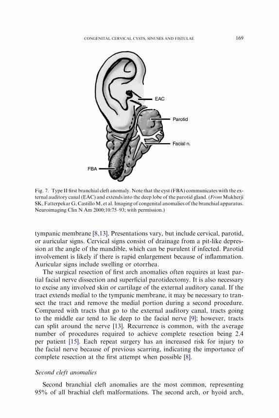
169CONGENITAL CERVICAL CYSTS, SINUSES AND FISTULAE
tympanic membrane [8,13]. Presentations vary, but include cervical, parotid,or auricular signs. Cervical signs consist of drainage from a pit-like depres-sion at the angle of the mandible, which can be purulent if infected. Parotidinvolvement is likely if there is rapid enlargement because of inflammation.Auricular signs include swelling or otorrhea.
The surgical resection of first arch anomalies often requires at least par-tial facial nerve dissection and superficial parotidectomy. It is also necessaryto excise any involved skin or cartilage of the external auditory canal. If thetract extends medial to the tympanic membrane, it may be necessary to tran-sect the tract and remove the medial portion during a second procedure.Compared with tracts that go to the external auditory canal, tracts goingto the middle ear tend to lie deep to the facial nerve [9]; however, tractscan split around the nerve [13]. Recurrence is common, with the averagenumber of procedures required to achieve complete resection being 2.4per patient [15]. Each repeat surgery has an increased risk for injury tothe facial nerve because of previous scarring, indicating the importance ofcomplete resection at the first attempt when possible [8].
Second cleft anomalies
Second branchial cleft anomalies are the most common, representing95% of all brachial cleft malformations. The second arch, or hyoid arch,
Fig. 7. Type II first branchial cleft anomaly. Note that the cyst (FBA) communicates with the ex-
ternal auditory canal (EAC) and extends into the deep lobe of the parotid gland. (FromMukherji
SK, FatterpekarG, CastilloM, et al. Imaging of congenital anomalies of the branchial apparatus.
Neuroimaging Clin N Am 2000;10:75–93; with permission.)
170 ACIERNO & WALDHAUSEN
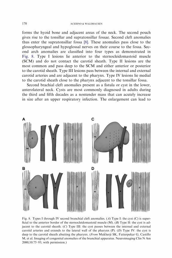
forms the hyoid bone and adjacent areas of the neck. The second pouchgives rise to the tonsillar and supratonsillar fossae. Second cleft anomaliesthus enter the supratonsillar fossa [8]. These anomalies pass close to theglossopharyngeal and hypoglossal nerves on their course to the fossa. Sec-ond arch anomalies are classified into four types as demonstrated inFig. 8. Type I lesions lie anterior to the sternocleidomastoid muscle(SCM) and do not contact the carotid sheath. Type II lesions are themost common and pass deep to the SCM and either anterior or posteriorto the carotid sheath. Type III lesions pass between the internal and externalcarotid arteries and are adjacent to the pharynx. Type IV lesions lie medialto the carotid sheath close to the pharynx adjacent to the tonsillar fossa.
Second brachial cleft anomalies present as a fistula or cyst in the lower,anterolateral neck. Cysts are most commonly diagnosed in adults duringthe third and fifth decades as a nontender mass that can acutely increasein size after an upper respiratory infection. The enlargement can lead to
Fig. 8. Types I through IV second branchial cleft anomalies. (A) Type I: the cyst (C) is super-
ficial to the anterior border of the sternocleidomastoid muscle (M). (B) Type II: the cyst is ad-
jacent to the carotid sheath. (C) Type III: the cyst passes between the internal and external
carotid arteries and extends to the lateral wall of the pharynx (P). (D) Type IV: the cyst is
deep to the carotid sheath abutting the pharynx. (From Mukherji SK, Fatterpekar G, Castillo
M, et al. Imaging of congenital anomalies of the branchial apparatus. Neuroimaging Clin N Am
2000;10:75–93; with permission.)
171CONGENITAL CERVICAL CYSTS, SINUSES AND FISTULAE
respiratory compromise, torticollis, or dysphagia. Fistulae, however, areusually diagnosed in infancy or childhood and present as chronic drainagefrom an opening along the anterior border of the SCM in the lower thirdof the neck [1,8].
Surgical resection of second cleft anomalies can be approached by way ofa transverse cervical incision placed within a natural skin fold. Cysts can belocated either superficially or deep to the cervical fascia. A careful explora-tion for an associated fistula tract must be performed with a complete exci-sion of the entire tract if one is found. Fistula excision can be facilitated bycannulating the tract with a 2-0 or 3-0 monofilament suture or probe. Thetract can also be injected with methylene blue; however this may stain thesurrounding tissues making dissection difficult [8]. As the tract is followed,the skin incision may have to be extended to allow adequate exposure, al-though step-ladder incisions may provide improved visualization of the tractnear the pharynx. The spinal accessory, hypoglossal, and vagus nerves mustbe protected from injury during the dissection. A finger or bougie in the oro-pharynx can help identify the opening in the tonsillar fossa. The thin tractmust be carefully ligated and divided at its entry into the fossa. If theexcision is complete, no drain is needed.
Third and fourth cleft anomalies
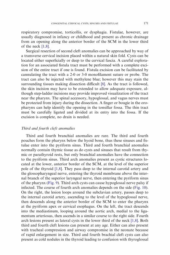
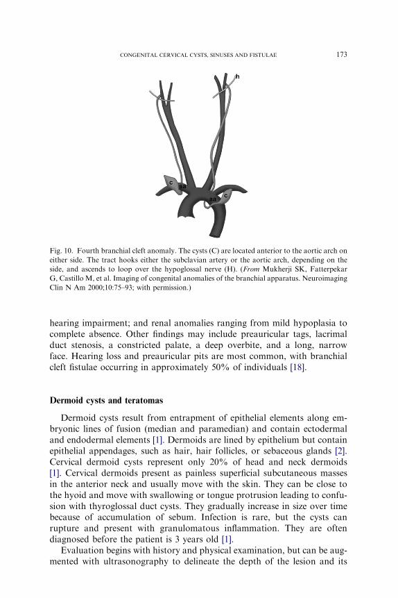
Third and fourth branchial anomalies are rare. The third and fourthpouches form the pharynx below the hyoid bone, thus these sinuses and fis-tulae enter into the pyriform sinus. Third and fourth branchial anomaliesnormally contain thymic tissue as do cysts and sinuses that result from thy-mic or parathyroid rests, but only branchial anomalies have the connectionto the pyriform sinus. Third arch anomalies present as cystic structures lo-cated at the lower, anterior border of the SCM, at the level of the superiorpole of the thyroid [1,8]. They pass deep to the internal carotid artery andthe glossopharyngeal nerve, entering the thyroid membrane above the inter-nal branch of the superior laryngeal nerve, then entering the pyriform sinusof the pharynx (Fig. 9). Third arch cysts can cause hypoglossal nerve palsy ifinfected. The course of fourth arch anomalies depends on the side (Fig. 10).On the right, the lesion loops around the subclavian artery, passes deep tothe internal carotid artery, ascending to the level of the hypoglossal nerve,then descends along the anterior border of the SCM to enter the pharynxat the pyriform apex or cervical esophagus. On the left, the tract descendsinto the mediastinum, looping around the aortic arch, medial to the liga-mentum arteriosus, then ascends in a similar course to the right side. Fourtharch lesions present as lateral cysts in the lower third of the neck [1,8]. Boththird and fourth cleft lesions can present at any age. Either can also presentwith tracheal compression and airway compromise in the neonate becauseof rapid enlargement in size. Third and fourth brachial cleft cysts can alsopresent as cold nodules in the thyroid leading to confusion with thyroglossal
172 ACIERNO & WALDHAUSEN
duct cysts. Other possible presentations include recurrent upper respiratorytract infections, neck or thyroid pain, or thyroid abscess.
Surgical therapy of third and fourth arch anomalies is similar to that ofsecond arch anomalies, with the following exceptions. Endoscopy should beused to identify the pyriform sinus entry point. This identification can allowcannulation or injection of the tract to aid with dissection. There are somereports of chemical cauterization of these tracts; however, there are no long-term results for this approach [16]. Fourth arch anomaly resections requireipsilateral hemithyroidectomy to completely excise the tract and possiblepartial resection of the thyroid cartilage to provide adequate exposure ofthe pyriform sinus [17].
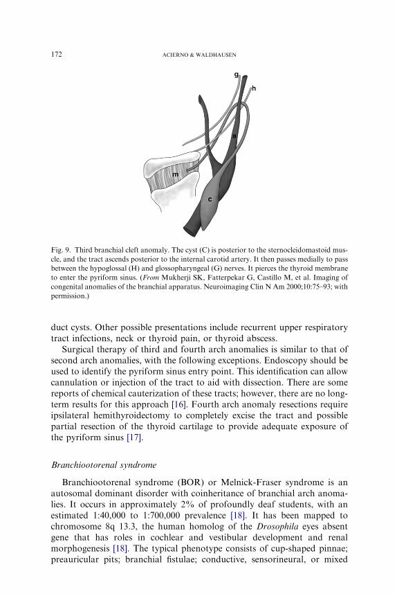
Branchiootorenal syndrome
Branchiootorenal syndrome (BOR) or Melnick-Fraser syndrome is anautosomal dominant disorder with coinheritance of branchial arch anoma-lies. It occurs in approximately 2% of profoundly deaf students, with anestimated 1:40,000 to 1:700,000 prevalence [18]. It has been mapped tochromosome 8q 13.3, the human homolog of the Drosophila eyes absentgene that has roles in cochlear and vestibular development and renalmorphogenesis [18]. The typical phenotype consists of cup-shaped pinnae;preauricular pits; branchial fistulae; conductive, sensorineural, or mixed
Fig. 9. Third branchial cleft anomaly. The cyst (C) is posterior to the sternocleidomastoid mus-
cle, and the tract ascends posterior to the internal carotid artery. It then passes medially to pass
between the hypoglossal (H) and glossopharyngeal (G) nerves. It pierces the thyroid membrane
to enter the pyriform sinus. (From Mukherji SK, Fatterpekar G, Castillo M, et al. Imaging of
congenital anomalies of the branchial apparatus. Neuroimaging Clin N Am 2000;10:75–93; with
permission.)
173CONGENITAL CERVICAL CYSTS, SINUSES AND FISTULAE
hearing impairment; and renal anomalies ranging from mild hypoplasia tocomplete absence. Other findings may include preauricular tags, lacrimalduct stenosis, a constricted palate, a deep overbite, and a long, narrowface. Hearing loss and preauricular pits are most common, with branchialcleft fistulae occurring in approximately 50% of individuals [18].
Dermoid cysts and teratomas
Dermoid cysts result from entrapment of epithelial elements along em-bryonic lines of fusion (median and paramedian) and contain ectodermaland endodermal elements [1]. Dermoids are lined by epithelium but containepithelial appendages, such as hair, hair follicles, or sebaceous glands [2].Cervical dermoid cysts represent only 20% of head and neck dermoids[1]. Cervical dermoids present as painless superficial subcutaneous massesin the anterior neck and usually move with the skin. They can be close tothe hyoid and move with swallowing or tongue protrusion leading to confu-sion with thyroglossal duct cysts. They gradually increase in size over timebecause of accumulation of sebum. Infection is rare, but the cysts canrupture and present with granulomatous inflammation. They are oftendiagnosed before the patient is 3 years old [1].
Evaluation begins with history and physical examination, but can be aug-mented with ultrasonography to delineate the depth of the lesion and its
Fig. 10. Fourth branchial cleft anomaly. The cysts (C) are located anterior to the aortic arch on
either side. The tract hooks either the subclavian artery or the aortic arch, depending on the
side, and ascends to loop over the hypoglossal nerve (H). (From Mukherji SK, Fatterpekar
G, Castillo M, et al. Imaging of congenital anomalies of the branchial apparatus. Neuroimaging
Clin N Am 2000;10:75–93; with permission.)
174 ACIERNO & WALDHAUSEN
relationship to the hyoid bone. If the lesion is inflamed, fine needle aspiratemay be helpful to distinguish between a ruptured dermoid cyst and an in-fected thyroglossal duct cyst. If the lesion is symptomatic, enlarging, orhas ruptured, surgical excision is recommended. Complete simple excisionis usually adequate, but if it is attached to the hyoid bone a Sistrunk proce-dure should be performed to prevent inadequate excision of an atypical thy-roglossal duct cyst [19]. Rate of recurrence is increased by incompleteresection or intraoperative rupture [2].
Teratomas differ from dermoid cysts in that they contain all three germlayers. Head and neck lesions compose less than 2% of teratomas, withthe most common sites being the nasopharynx and neck. They develop dur-ing the second trimester and present as rapidly expanding lateral or midlineneck masses. They may be diagnosed by prenatal ultrasonography, with30% accompanied by polyhydramnios because of esophageal obstruction[1]. If the diagnosis is known before delivery, cesarean section is recommen-ded. Although the lesions may initially be asymptomatic, rapid growth mayeventually lead to dysphagia and respiratory distress. Eighty percent of in-fants who have neonatal teratomas may die if untreated [1]. Ultrasound,CT, or MRI may be helpful in evaluating these lesions. Some neonatesmay require intubation or even extracorporeal membrane oxygenation ifthe lesion has caused pulmonary hypoplasia. Complete surgical excision isthe treatment of choice once the airway has been stabilized. Malignancyhas not been reported in pediatric cervical teratomas, so all critical struc-tures in the neck should be spared [1]. Malignant cervical teratomas have
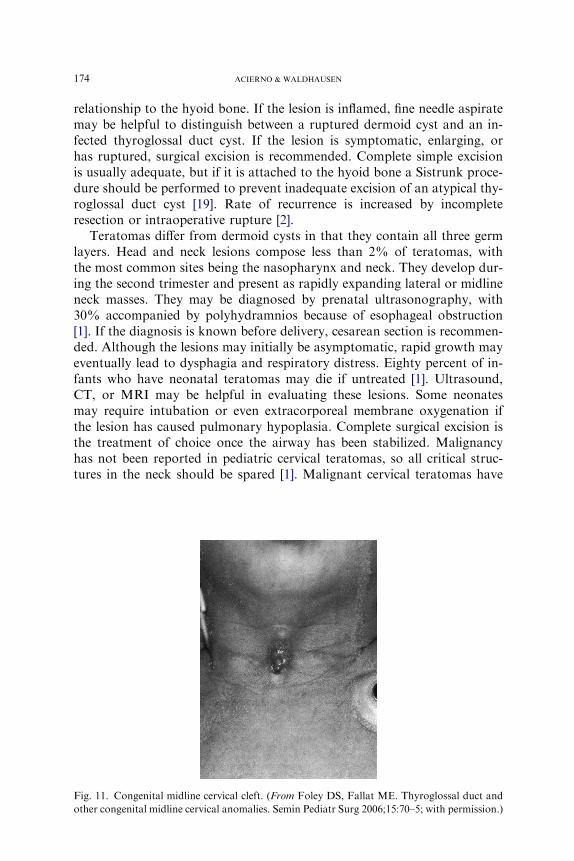
Fig. 11. Congenital midline cervical cleft. (From Foley DS, Fallat ME. Thyroglossal duct and
other congenital midline cervical anomalies. Semin Pediatr Surg 2006;15:70–5; with permission.)
175CONGENITAL CERVICAL CYSTS, SINUSES AND FISTULAE
been found in adults and require aggressive treatment because they canspread by hematogenous and lymphatic routes and carry a poor prognosis.
Midline cervical clefts
Midline cervical clefts are rare congenital cervical anomalies. They arepresent at birth as a cutaneous ulceration with overhanging skin or cartilag-inous tag in the anterior lower midline of the neck (Fig. 11). There is oftena sinus tract that extends downward from the skin and may connect to thesternum or mandible or end in a blind pouch. The embryologic origin is un-known but is believed to be a ‘‘mesodermal fusion abnormality involving thepaired branchial arches during gestational weeks 3 and 4’’ [2]. Fibrous tissuewith interwoven skeletal muscle is present. Most cases are sporadic, but canbe associated with other cleft abnormalities of the tongue, lower lip, or man-dible. If untreated, some clefts can result in neck contractures or growth de-formities of the mandible or sternum. Early surgical excision at the time ofdiagnosis is recommended, therefore, with complete excision of the skinlesion and the subcutaneous sinus to reduce the rate of recurrence. Thisexcision can usually be accomplished by stair-step incisions, but if morecomplicated may require a series of Z-plasty incisions to improve thecosmetic and functional result [2].
References
[1] Enepekides DJ. Management of congenital anomalies of the neck. Facial Plast Surg Clin
North Am 2001;9:131–45.
[2] Foley DS, Fallat ME. Thyroglossal duct and other congenital midline cervical anomalies.
Semin Pediatr Surg 2006;15:70–5.
[3] Perez-MartinezA, Bento-BravoL,Martinez-BermejoMA, et al. An intra-thyroid thyroglos-
sal duct cyst. Eur J Pediatr Surg 2005;15:428–30.
[4] Mohan PS, Chokshi RA,Moser RL, et al. Thyroglossal duct cysts: a consideration in adults.
Am Surg 2005;71(6):508–11.
[5] Pinczower E, Crockett DM, Atkinson JB, et al. Preoperative thyroid scanning in presumed
thyroglossal duct cysts. Arch Otolaryngol Head Neck Surg 1992;118:985–8.
[6] Peretz A, Lieberman E, Kapelsuhnik J, et al. Thyroglossal duct carcinoma in children: case
presentation and review of the literature. Thyroid 2004;14:777–85.
[7] Ostlie DJ, Burjonrappa SC, Synder CI, et al. Thyroglossal duct infections and surgical out-
comes. J Pediatr Surg 2004;39:396–9.
[8] Waldhausen JHT. Branchial cleft and arch anomalies in children. Semin Pediatr Surg 2006;
15:64–9.
[9] D’Souza AR, Uppal HS, De R, et al. Updating concepts of first brachial cleft defects: a
literature review. Int J Pediatr Otolaryngol 2002;62:103–9.
[10] Shrime M, Kacker A, Bent J, et al. Fourth branchial complex anomalies: a case series. Int J
Pediatr Otolaryngol 2003;67:1227–33.
[11] Roback SA, Telander RL. Thyroglossal duct cysts and branchial cleft anomalies. Semin
Pediatr Surg 1994;3:142–6.
[12] O’MaraW, Amedee R. Anomalies of the branchial apparatus. J La StateMed Soc 1998;150:
570–3.

176 ACIERNO & WALDHAUSEN
[13] Triglia JM, Nicollas R, Ducroz V, et al. First branchial cleft anomalies. Arch Otolaryngol
Head Neck Surg 1998;124:291–5.
[14] LibermanM,Kay S, Emil S, et al. Ten years experience with third and fourth branchial rem-
nants. J Pediatr Surg 2002;37:685–90.
[15] FordGR,BalakrishmanA, Evans JN, et al. Branchial cleft and pouch anomalies. J Laryngol
Otol 1992;106:137–43.
[16] Park SW, Han MH, Sung MH, et al. Neck infection associated with pyriform sinus fistula:
imaging findings. AJNR Am J Neuroradiol 2000;21:817–22.
[17] Nicollas R, Ducroz V, Garabedian EN, et al. Fourth branchial pouch anomalies: a study of
six cases and a review of the literature. Int J Pediatr Otorhinolaryngol 1998;44:5–10.
[18] Smith RJH, Schwartz C. Branchio-oto-renal syndrome. J CommunDisord 1998;31:411–21.
[19] Turkyilmaz Z, Sonmez K, Karabulut R, et al. Management of thyroglossal duct cysts in
children. Pediatr Int 2004;46:77–80.