a guide to cervical length screening, cervical cerclage
TRANSCRIPT
1
Management of Woman at high risk for Recurrent Second Trimester Miscarriage
or Preterm Birth in Singleton Pregnancies
A guide to cervical length screening, cervical cerclage and vaginal progesterone General comments:
• This guideline seeks to clarify the indications for cerclage and progesterone, within the context of
counselling and shared decision making, after appropriate evaluation of specific high-risk cases
at Tygerberg Hospital, namely:
o women with ≥ 2 mid-trimester losses, irrespective of cervical length in the current
pregnancy;
o women with 1 mid-trimester loss and a short cervix (≤ 25 mm, measured between 16w0d
and 24w0d) in the current pregnancy
o women with ≥ 1 previous preterm births before 34 weeks’ gestation and a short cervix
(as above) in the current pregnancy.
• Ensure all other risk factors for preterm labour are addressed as well (do Urine MCS, treat
symptomatic vaginal infections, inspect the cervix by speculum to rule out lateral tears, address
smoking, check for IUGR etc.).
• Underlying medical conditions, such as poorly controlled diabetes, hypertension, thyroid disease
and thrombophilia might have caused the adverse obstetric outcomes (rather than cervical
insufficiency).
• In most cases, patients with the above-mentioned problems do not present with the classic
combination of painful contractions and cervical changes. In the initial stage there is relatively
asymptomatic “ripening” or activation of the cervix leading to effacement (shortening) and
dilatation, with symptomatic contractions being a late sign. This emphasises the importance of
appropriate measurement of the cervix in high-risk cases.
• However, all patients with any of the risk factors below should be counselled about the signs and
symptoms of threatened preterm labour and encouraged to present ASAP if they occur. Patients
should also be advised about abstinence. [“In populations at increased risk for preterm labour,
there is no evidence to suggest a clear benefit from restricted sexual activity; however, this is a
simple intervention that causes no harm and may be a reasonable recommendation until better
evidence emerges”].
Cerclage or progesterone?
There are three broad categories of indications for transvaginal cerclage.
• History-indicated (prophylactic) cerclage.
This is typically performed at 12-14 weeks’ gestation, for women with two or three previous
second trimester losses, with a typical history (see below) of cervical incompetence/insufficiency,
TYGERBERG HOSPITAL Department of Obstetrics and Gynaecology: General Specialist
Services
2
regardless of the cervical length. Limited evidence suggests that the benefit is greatest for those
with three preterm deliveries. Progesterone may also be considered.
• Ultrasound-indicated cerclage.
There is general consensus that progesterone or cerclage be considered in women with a history of
spontaneous loss or preterm delivery (<34 weeks’ gestation) and a short cervix (≤ 25 mm, measured
between 16w0d and 24w0d). The cerclage is performed when indicated, but not beyond 23w6d. A
short cervix without the relevant history does not automatically meet the criterion.
• Rescue (Emergency / clinically-indicated) cerclage.
In such cases there is also dilation of the vaginal cervix (<4 cm, membranes not prolapsed beyond
the external os) without contractions, again before 24w. Rescue cerclage has been demonstrated to
have benefit, but cases must be selected carefully. Progesterone is not effective with advanced
effacement.
Progesterone may be offered for the first two categories above, starting at 16-24w until 34w0d.
The following high-risk women should undergo cervical screening and be offered a choice of cerclage (performed up to 23w6d) and/or progesterone, as appropriate, if cervical length is ≤25mm:
• A history of 2nd trimester miscarriage (between 16 and 26 weeks) suggestive of cervical
incompetence: Painless dilatation with a quick labour, and birth of a live baby or fresh stillbirth,
after excluding other causes of mid-trimester losses, e.g. intra-uterine death that required
induction, abruptio placentae, fetal abnormalities, polyhydramnios, and medical terminations.
Take note of specific indications for history-indicated cerclage above.
• Previous history of spontaneous preterm birth of a live baby from 27w0d to 34w0d (exclude
non-spontaneous causes e.g. iatrogenic delivery for pre-eclampsia, syphilis, intra-uterine
death etc.).
• Women with a previous LLETZ or Cone biopsy, or previous cervical trauma/tears or known
congenital uterine abnormality. Consider cerclage and/or progesterone.
• No need to workup previous late preterm deliveries (34-37 weeks).
Do not screen women at low-risk of preterm labour routinely, as it is not cost-effective.
Practical guide for cervical screening at HRC:
• Cervical length must be measured by a skilled operator using transvaginal ultrasound (see
technique - Appendix A).
• Cervical measurement by ultrasound can be done every second week between 16 and 26 weeks
(repeat in 1 week if significant shortening or borderline length not yet meeting criteria for
intervention).
• The lengths should be recorded on the gravidogram so that shortening over the gestation can be
appreciated.
• A cervical length of ≤25 mm indicates a higher risk for recurrent late miscarriage/preterm labour.
3
o Cervical length monitoring in high risk but asymptomatic women (with no signs of preterm
labour) should DISCONTINUE after 26 weeks as there are no preventative strategies to
treat a short cervix after this gestation (progesterone have been proven to be ineffective
when initiated after this time).
o However, knowledge of a very short or dilated cervix (cervical ripening) in certain high
risk women after 26 weeks may help with empiric management (altering activity level,
work and travel, increased surveillance, relocation close to Tygerberg, and ‘cooling down’
of the cervix with antibiotics, anti-inflammatories and corticosteroids). Individualise care
for women with specific risks (e.g. rural/farm workers, advanced age, very poor obstetric
history etc.). Discuss with a consultant to continue cervical surveillance after 26 weeks.
• The only effective therapeutic interventions after 26 weeks are tocolytics and corticosteroids to
advance fetal lung maturity in a patient with established preterm labour.
o If the cervical length is still >25mm at the 26 weeks visit, and there are no symptoms of
preterm labour, consider stepping down for routine care to a district hospital. The patient
must be well educated on the signs and symptoms of preterm labour and return
immediately if preterm labour is suspected. Discuss which labour ward to go to depending
on the gestation at which the symptoms occur (e.g. before 33 weeks: come to TBH, 33
and 34 weeks can deliver at district hospital, 35 or more can deliver at MOU).
o If a cervical cerclage was inserted, it is not necessary to perform serial ultrasound
evaluation of the cervix.
o If progesterone is being administered for a short cervix, continue with care at Tygerberg
HRC every two weeks, until 34 weeks, after which progesterone can be stopped and care
stepped down to MOU/BANC. It is generally not necessary to continue with cervical
surveillance after 26 weeks, but see note above about individualisation.
TREATMENT FOR SHORT CERVIX (≤25 mm) IN HIGH RISK WOMEN
There is no evidence comparing progesterone and cerclage therefore the choice of cerclage or
progesterone should be determined after discussion with the woman [NICE] and by the timing of the
previous loss, with cerclage less effective in preventing third trimester deliveries. Women can be
counselled that 17-20 cerclage procedures may prevent one preterm delivery (numbers needed to treat
NNT 17 to 20) and that progesterone is successful in 1 out of every 8 cases (NNT 6 to 8), to assist them
in making an informed decision (data from Cochrane review).
Placement and management of cerclage: see Appendix B.
Progesterone dose: 200 mg micronised progesterone vaginally at night, until 34 weeks.
A. Consider history-indicated cervical cerclage (MacDonald suture) or progesterone
• Typical history of ≥2 second trimester losses, even in absence of short cervix.
B. Consider prophylactic vaginal progesterone or cervical cerclage (MacDonald suture) for women
with:

4
• History of spontaneous preterm birth or PPROM (27-34 weeks) or a mid-trimester loss (16-26
weeks),
AND
• Cervical length ≤ 25 mm confirmed on ultrasound (16w0d-23w6d).
C. Rescue cerclage:
• If the cervix is already open (< 4cm) and the membranes not prolapsed or ruptured, consider
a rescue cervical cerclage (between 16-23w6d).
• Do not insert a rescue cerclage if there are contractions, active vaginal bleeding, prolapsed
membranes, dilatation > 4cm, no workable cervix or signs of infection.
• In women already on progesterone present with continued shortening of the cervix and
exposure of the membranes, consider a rescue cerclage as well (consultant decision).
Consideration of a trans-abdominal cerclage (TAC)
Women with a previous failed (the pregnancy was lost before viability) transvaginal cervical
cerclage must be discussed with the MFM/Obstetric Special Care team, prior to pregnancy, or
before 14w0d gestation (if already pregnant) for a possible TAC.
Criteria for consideration of transabdominal cerclage
Previous failed, appropriately inserted transvaginal cerclage
Significant damage to the cervix
Insufficient vaginal portion of the cervix
AUTHORISED BY S Gebhardt
COMPILED BY S Gebhardt, Z Momberg
COMMITTEE RESPONSIBLE S Gebhardt, L Geerts, D Hall, E Swart, L Muller
DATE EFFECTIVE 1 August 2020
REVIEW DATE 30 July 2022
EVIDENCE Based on the Standard Treatment Guidelines for SA, RCOG, NICE, Cochrane, SOGC and ISUOG guidelines.
Signed: GS Gebhardt
Head: General Specialist Services; Obstetrics and Gynaecology
5
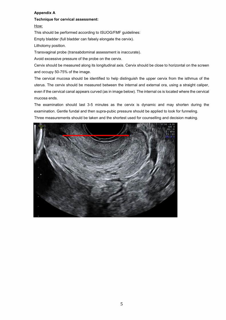
Appendix A Technique for cervical assessment: How:
This should be performed according to ISUOG/FMF guidelines:
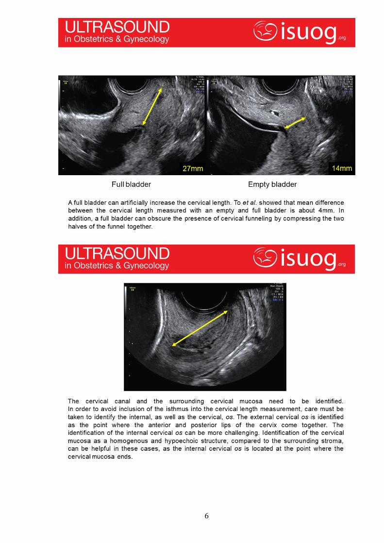
Empty bladder (full bladder can falsely elongate the cervix).
Lithotomy position.
Transvaginal probe (transabdominal assessment is inaccurate).
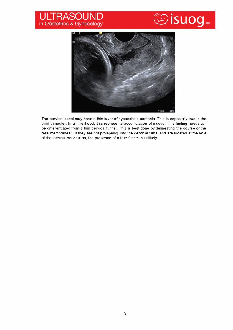
Avoid excessive pressure of the probe on the cervix.
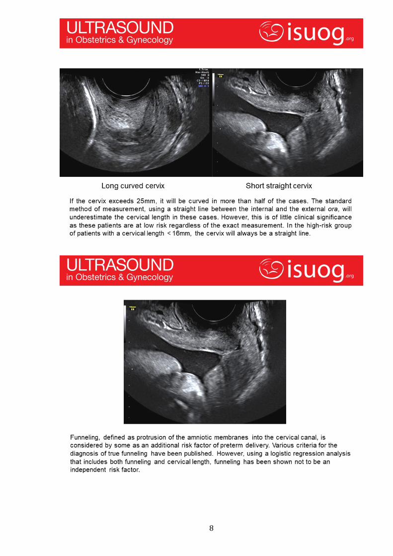
Cervix should be measured along its longitudinal axis. Cervix should be close to horizontal on the screen
and occupy 50-75% of the image.
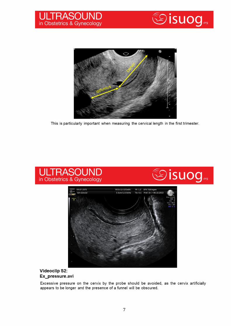
The cervical mucosa should be identified to help distinguish the upper cervix from the isthmus of the
uterus. The cervix should be measured between the internal and external ora, using a straight caliper,
even if the cervical canal appears curved (as in image below). The internal os is located where the cervical
mucosa ends.
The examination should last 3-5 minutes as the cervix is dynamic and may shorten during the
examination. Gentle fundal and then supra-pubic pressure should be applied to look for funneling.
Three measurements should be taken and the shortest used for counselling and decision making.
6
7
8
9

10
Appendix B Cervical Cerclage
Contraindications for cervical cerclage:
• Absolute contraindications:
Active labour
Active vaginal bleeding
Rupture of membranes
Fetal compromise or death, lethal fetal anomalies
Suspected or clinically confirmed abruptio placenta
Suspected or clinically confirmed chorioamnionitis
• Relative contraindications include the following:
Prolapsed membranes or dilatation more than 4cm
Vaginal spotting
Immunosuppression (i.e. complicated DM, HIV etc.)
Multifetal gestations
* If there is a valid indication for cerclage in the presence of a relative contraindication, a work
up as stipulated below should be done and the case discussed with the consultant in the
antenatal clinic or ward.
Clinical examination:
• Detailed obstetric and cervical evaluation
o Vaginal speculum to visually inspect the cervix for previous scarring, deformity
and length to ascertain the feasibility of placing a transvaginal cerclage.
o A wet mount smear must be performed on all patients to exclude bacterial
vaginosis (if present metronidazole 400 mg orally twice a day for 7 days or
clindamycin 300 mg orally twice daily for 7 days), trichomonas vaginalis (if
present single dose metronidazole 2g orally) or chronic cervicitis (individualize
management on suspected underlying cause).
o Urinary dipstix specifically to evaluate for the presence of proteinuria, glycosuria
and haematuria.
• Special investigations:
o Ensure that routine antenatal screening investigation results are noted on the
card and managed appropriately (syphilis, HIV, Rhesus). If HIV positive the
timing and results of the last CD4 and viral load and type and duration of current
treatment should be documented in the maternity case record.
o A midstream urine specimen for urine culture and ideally treat ASB prior to
cerclage insertion.
11
o Ultrasound to confirm a single intrauterine viable pregnancy and to exclude any
major anomalies – a formal anomaly scan can be arranged at a GA dependant
on the indication and urgency of the cerclage. In all surgical cases, fetal life
should be documented before and after the procedure.
• Counselling: Patients should be thoroughly counselled regarding the following risk
groups:
o Disease-related risks: premature rupture of membranes; pregnancy loss;
infection (chorioamnionitis); preterm labour requiring hospitalization; extreme
prematurity.
o Immediate procedure‐related risks: usual anaesthetic risks (minor with spinal
anaesthesia which is preferable); minor vaginal bleeding; maternal soft tissue
injury (rare); cervical trauma and injury during placement, rupture of
membranes.
o Ongoing procedure‐related risks: cervical injury from the suture in case of
uncontrollable preterm labour; chronic cervical irritation and inflammation with
possible intra‐amniotic infection, suture displacement/migration, increased
incidence of caesarean section (soft tissue dystocia from scar tissue).
• Preoperative Management
o Tocolytic drugs are not routinely given to women undergoing placement of a
cerclage however indomethacin has been reported to improve outcome in the
rescue cerclage where cervical activation has commenced.
o Use of antibiotic prior to or after cerclage is controversial. The following
conditions may warrant antibiotics prior to the cerclage placement:
Patients with evidence of chronic cervicitis (on speculum examination
and wet mount).
Patients with copious and malodorous vaginal discharge.
Emergency cerclage with exposure of the membranes to the vaginal
environment. Allow 48 hours of “cooling off” then re-evaluate cervix.
o If antibiotics are indicated then complete treatment prior to cerclage placement,
if possible.
• Placement of a transvaginal cervical cerclage
o The following description is applicable to a McDonald's technique with a good
vaginal portio (>1.5cm) and no old tears or trauma.
o Anaesthesia: Regional anaesthesia is preferred.
o Equipment needed:
5mm Mercilene tape suture on 48mm ½ circle taper-cut or cutting needle
12
o Aseptic syringe for irrigation
o Suction tubing with Yankauer suction tip
o Wertheim vaginal retractors
o Allis (soft tissue) forceps/clamps
o Doyen retractors
o Extra‐long blade weighted speculum
o Straight needle holders
• Positioning:
o Patient in a dorsal lithotomy position. The surgeon must protect vulnerable
neurologic, vascular, and bony points of the lower extremities and padding
should be used at all potential pressure points. The buttocks should be
positioned at the end of the table, with the table level. Hyperflexion of the hips
should be avoided, as this can cause femoral neuropathy. Final positioning of
the legs should be such to allow for assistants to be able to visualize the
operative field.
o The patient is then prepped and draped and the vagina should be gently
cleansed with a water‐based antiseptic solution.
• Procedure:
o The bladder must be emptied with aseptic technique and the cervix is visualised
by placing a long weighted speculum posteriorly, and curved or right angle
retractors anteriorly and laterally as needed.
o An Allis clamp is used to grasp the anterior and posterior lips of the cervix as high
in the vagina as possible to mobilise the cervix in a gentle downward and lateral
direction. The cervix can also be exposed and mobilized by Babcock forceps but
care should be taken not to traumatise the tissue.
o Ensure that only cervical stromal tissue is included in the suture and that the
cervical canal is not entered.
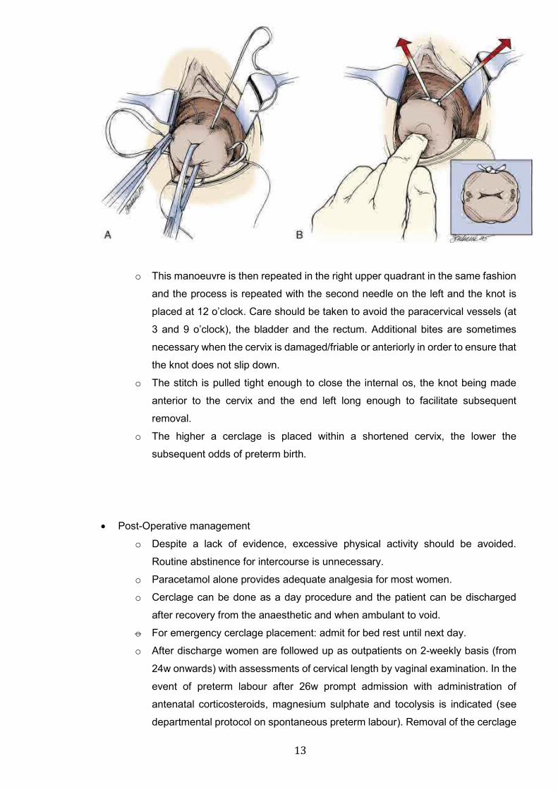
o The suture is placed above the Allis clamp, curving through the cervical stroma
of the right lower quadrant. In order to place the suture as high as possible; the
junction of the rugated vaginal epithelium and the smooth cervix just distal to the
cervical reflection is used as a landmark (note that the suture is placed too low
in the illustration below).
13
o This manoeuvre is then repeated in the right upper quadrant in the same fashion
and the process is repeated with the second needle on the left and the knot is
placed at 12 o’clock. Care should be taken to avoid the paracervical vessels (at
3 and 9 o’clock), the bladder and the rectum. Additional bites are sometimes
necessary when the cervix is damaged/friable or anteriorly in order to ensure that
the knot does not slip down.
o The stitch is pulled tight enough to close the internal os, the knot being made
anterior to the cervix and the end left long enough to facilitate subsequent
removal.
o The higher a cerclage is placed within a shortened cervix, the lower the
subsequent odds of preterm birth.
• Post-Operative management
o Despite a lack of evidence, excessive physical activity should be avoided.
Routine abstinence for intercourse is unnecessary.
o Paracetamol alone provides adequate analgesia for most women.
o Cerclage can be done as a day procedure and the patient can be discharged
after recovery from the anaesthetic and when ambulant to void.
o For emergency cerclage placement: admit for bed rest until next day.
o After discharge women are followed up as outpatients on 2-weekly basis (from
24w onwards) with assessments of cervical length by vaginal examination. In the
event of preterm labour after 26w prompt admission with administration of
antenatal corticosteroids, magnesium sulphate and tocolysis is indicated (see
departmental protocol on spontaneous preterm labour). Removal of the cerclage
14
upon suspected or confirmed preterm labour, or PPROM before 34w should be
discussed with the consultant on duty.
o Patients should be informed that they should present immediately to their health
care facility if they experience any of the following symptoms after cerclage
insertion: contractions or cramping, lower abdominal or back pain that comes and
goes like labour pain, any vaginal bleeding, fever or chills, nausea and vomiting,
foul‐smelling vaginal discharge, water breaking or leaking (rupture of
membranes).
• Cerclage removal:
o Elective cerclage removal is done at 36w gestation in the antenatal clinic during
a routine visit.
o Immediate removal if the women presents in advanced labour at any gestation.
o Immediate removal upon onset of preterm labour or PPROM after 34 weeks.