the subaxial cervical pedicle screw for cervical spine
TRANSCRIPT

The Subaxial Cervical Pedicle Screw for Cervical Spine Diseases: The Review of Technical Developments
and Complication Avoidance
Yoon Gyo JUNG,1 Sang Ku JUNG,2 Byung Jou LEE,3 Subum LEE,1 Seong Kyun JEONG,1 Myeongjong KIM,1 and Jin Hoon PARK1
1Department of Neurosurgery, Asan Medical Center, University of Ulsan College of Medicine, Seoul, Korea;
2Department of Emergency Medicine, Gangneung Asan Hospital, University of Ulsan College of Medicine, Gangneung, Korea;
3Department of Neurosurgery, Inje University Ilsan Paik Hospital, Neuroscience and Radiosurgery Hybrid Research Center, College of Medicine, Goyang, Korea
Abstract
This study aimed to review information on the subaxial cervical pedicle screw (CPS) including recent anatomical considerations, entry points, placement techniques, accuracy, learning curve, and complications. Relevant literatures were reviewed, and the authors’ experiences were summarized. The CPS is used for reconstruction of unstable cervical spine and achieves superior biomechanical stability compared to other fixation techniques. Various insertion and guidance techniques are established, among which, lateral fluoroscopy-assisted placement is the most common and cost-effective technique. Generally, placement under imaging guidance is more accurate than other techniques, and a three-dimensional template allows optimal trajectory for each pedicle regardless of intraoperative changes in spinal alignment. The free-hand technique using a curved pedicle probe without a funnel-like hole increases screw stability and reduces operation time, radiation exposure, and soft tissue injury. Compared to conventional lateral fluoroscopy-assisted placement, free-hand CPS placement by trained surgeons achieves superior accuracy comparable to that of image-guided navigation; in general, 30 training cases are sufficient for learning a safe and accurate technique for CPS placement. The complications of subaxial CPS are classified into three categories: complications due to screw misplacement, complications without screw misplacement, and others. Inexperienced surgeons may benefit from advanced techniques; however, the accuracy of CPS ultimately depends on the surgeon’s experience. Inexperienced surgeons should master the placement of the thoracolumbar pedicle screw in real practice and practice CPS insertion using cadavers. During the initial phase of the learning curve, careful preparation of surgery, reiterated identification, patterned safety steps, and supervision of the expert are necessary.
Key words: cervical spine, pedicle screw, internal fixation, learning curve
Received August 23, 2019; Accepted February 17, 2020
Copyright© 2020 by The Japan Neurosurgical Society This work is licensed under a Creative Commons Attribution-NonCommercial-NoDerivatives International License.
Online April 15, 2020
doi: 10.2176/nmc.ra.2019-0189
Neurol Med Chir (Tokyo) 60, 231–243, 2020
231
Review ARticle
cervical pedicle screw (CPS) placement yields the strongest biomechanical stability, which results in short segment fixation, preservation of the mobile segment, higher fusion rate, earlier mobilization and rehabilitation, and ultimately, a superior clinical outcome compared to other methods (Fig. 1).2,4,6–10)
Abumi et al.11,12) first reported subaxial CPS placement for traumatic lesions and subsequently expanded CPS application to non-traumatic lesions, reconstruction of the craniocervical junction, and correction of cervical kyphosis for stabilization of the unstable motion segment. CPS placement offers three-column fixation and has greater pullout strength
Introduction
Instrumented fusion surgery of the cervical spine is commonly performed for the treatment of cervical spine diseases. The instrumentation-based treatment approach in patients with cervical spine disorders involves pedicle screw, lateral mass screw, laminar screw and transfacet screw fixations.1–5) Subaxial

YG Jung et al.232
Neurol Med Chir (Tokyo) 60, May, 2020
than lateral mass screw placement.13–16) However, the small size of the mid-cervical pedicle, a large transverse angle of the cervical pedicle, and possible risk of vertebral artery (VA) and nerve root injury limit the routine application of subaxial CPS place-ment.13) In this study, we review current information on cervical CPS placement techniques for various applications and summarize the efficacies, technical guidelines, and potential adverse events. In addition, we provide an evidence-based recommendation to avoid neurovascular complications.
Selection of Reference Articles
A systemic literature review was performed through a search of PubMed using the following keywords: CPS, anatomy, entry point, accuracy, learning curve, and complication. Relevant articles were selected, and further sources were obtained from the references of selected articles. The authors’ experiences of patients with an unstable cervical spine who underwent treatment using CPS were described.
Fig. 1 Various cases of subaxial cervical pedicle screw (CPS) insertion. (A and B) A short segment fixation of a Hangman fracture patient. Preoperative computed tomography scan shows C2 hangman fracture (white arrow in A). Postoperative simple lateral radiograph shows a short segment fixation of CPS and reduced kyphosis of Hangman fracture (white arrowhead in B). (C and D) A reduction and fixation of a fractured bamboo spine with ankylosing spondylitis. Preoperative computed tomography scan shows a C5 fracture (white arrow in C). A simple lateral radiograph shows a well-aligned correction using short segment instrumentation (white arrowhead in D). (E and F) Reduction of traumatic spondyloptosis. Preoperative computed tomography scan shows a traumatic spondyloptosis on C7–T1 level (white arrow in E). This spondyloptosis was corrected and fused successfully by only posterior short segment instrumentation of CPS at the C6–T1 level (white arrowhead in F) in the postopera-tive computed tomography scan. (G–I) Correction of cervical kyphosis with infectious spondylodiscitis. Preopera-tive computed tomography scan shows a fixed cervical kyphosis with infectious spondylodiscitis at the C6 and C7 level (white arrow in G). After anterior C6 corpectomy with a grade IV osteotomy and support with mesh cage, the posterior short segment fixation of CPS from C5–C7 (white arrowhead in H) with a grade II osteotomy were performed. Postoperative computed tomography scan shows a well-aligned correction of kyphosis (white arrowhead in I).
A
E F G H I
B C D

Subaxial Cervical Pedicle Screw 233
Neurol Med Chir (Tokyo) 60, May, 2020
Anatomical considerations for the safety and accuracy of the cervical pedicle screw
For accurate and safe subaxial CPS placement, surgeons should understand the detailed three-dimensional (3D) morphology of the pedicle. With regard to reports on the morphology of the cervical pedicle,17–23) the measured parameters including pedicle transverse angle (PTA) and pedicle outer width (POW) are indicated in Fig. 2A. The smallest mean POW of 4.5 mm was obtained at C3, with gradual increases in the mean value caudally from C3 to C7.17,22) Karaikovic et al.24) reported that if the POW is sufficiently large, a canal of adequate size can be made with an appropriate tap, regardless of the pedicle inner width. Abumi et al.25) used 3.5–4.5-mm diameter screws and reported that screw insertion was difficult or impossible in cases with a POW of <4 mm; whereas, Park et al.26) used 3.5–4.5-mm diameter pedicle screws in cases with a POW of >3 mm on axial computed tomography (CT) scan.The pedicle axes at C3 and C4 are slightly elevatedcompared to the superior endplate of the vertebralbody, and those at C5–C7 are parallel and directedslightly downward (Fig. 2B).17)
Liu et al.18) reported that the cervical pedicles were smaller overall in Asians than in Europeans and Americans, and female individuals of both races had smaller pedicles than their male counterparts. However, the soft-tissue layer at the posterior aspect of the neck is thinner in Asians than in Europeans and Americans. Thickness of the soft tissue attrib-uted to the muscles and fat tissue contributes to a
muscle-pushing effect, which leads to screw malpo-sition and seems to be a more important affecting factor for a violation than the pedicle diameter. Based on these reasons, the CPS was initially developed and is frequently used in Asia. Therefore, planning of subaxial CPS placement should consider the factors of the patient’s sex, race, and importantly, individual neck morphology.
In general, the VA passes anterior to the lateral mass of C7 into the transverse foramen at C6 and courses upward to the transverse foramen at C1.8) A preoperative axial CT finding of thinning of the pedicle on one side indicates enlargement of the transverse foramen or invasion of the vertebral body, and magnetic resonance angiography or computed tomography angiography is needed to evaluate VA anomalies such as a tortuous course or unilateral-predominance. In patients with a VA anomaly or asymmetric unilateral dominance, surgeons should consider alternative safer techniques such as lateral mass screw insertion (Fig. 3).
Entry point and trajectory for subaxial cervical pedicle screw placement
The conventional entry point of CPS is 3 mm below the superior facet joint. The drill is angled 45° medially and advanced in a vertical line parallel to the endplate (Fig. 4A).13)
Abumi et al.25) recommended the locations of the entry point at the posterior surface of the lateral mass at the bisecting point of the width of each facet joint. In that study, funnel-shaped holes were made in the lateral mass to shorten the length of the cervical pedicle, which enhanced the safety by widening the zone of the screw trajectory angle. Those authors recommended a screw insertion angle at the transverse plane of 25–45° medial to the midline and at the sagittal plane parallel to the cranial endplate for the pedicles of C5 through C7 and in a slightly cephalad direction for those of C2 through C4 (Fig. 4B).
Many authors recommended that the entry point should be as lateral as possible in the articular mass and at 50° in the transverse plane for a safe corridor.23,27) In agreement with this recommendation, Hacker et al.28) reported that a line parallel to the contralateral lamina of approximately 50° in the transverse plane was a reliable intraoperative guide for accurate screw placement; however, the larger transverse angle of screw insertion required a wider midline dissection to avoid screw malposition due to the muscle-pushing effect and associated soft-tissue injury or bleeding.29)
To overcome this problem, Park et al.26,30) used a curved pedicle probe with an entry point at one-fourth the width medial to the lateral border of
Fig. 2 Anatomical considerations for a subaxial cervical pedicle screw. (A) The pedicle outer width (POW) ranges from 5.4 to 6.6 mm. The smallest mean POW of 4.5 mm is at C3, with gradual increases of the mean value caudally from C3 to C7. The overall mean pedicle transverse angle (PTA) ranges from 33.6° to 50.2°, approximately 45° from C3 to C6 and 33° at C7. (B) The pedicle axes at C3 and C4 are slightly elevated compared to the superior endplate of the vertebral body, and those at C5–C7 are parallel and directed slightly downward.
A B

YG Jung et al.234
Neurol Med Chir (Tokyo) 60, May, 2020
Fig. 3 The primary choice of a lateral mass screw for a vertebral artery (VA) anomaly and unilateral dominance. (A–E) The magnetic resonance imaging shows a C4–C7 cervical spondylosis and spinal cord compression (white arrow in A). The three dimensional computed tomography scan shows a unilaterally dominant VA to the right side (white arrow in B). A thinned right side pedicle of C3 and C4 (white arrow in C and D) indicates enlarge-ment of the transverse foramen and invasion of the unilaterally dominant VA to the vertebral body. At the right C3 and C4, lateral mass screws (white arrow in E) were primarily chosen instead of the cervical pedicle screw (CPS) because of a unilaterally dominant VA. (F–I) The simple lateral radiograph shows cervical subluxation of C3 and C4 (white arrow in F). The VA of the three dimensional computed tomography scan is dominant to the left side (white arrow in G). A right side VA is invisible (white arrowhead in G). Therefore, right C3 and C4 were fixed by CPS (white arrowhead in H) and left C3 and C4 were primarily fixed by lateral mass screw (white arrow in H) instead of CPS. The simple lateral radiograph shows correction of subluxation (white arrow in I).
A C
D
E
F G H I
B
the superior articular process in the axial plane at C3–C6 and at half width at C7 based on the notch level in the sagittal plane. During widening of the pilot hole and straightening of the cancellous pedicle track using curved and straight pedicle probes, the original entry point was shifted medi-ally with a concomitant decrease of the medial angle compared to the anatomical pedicle angle. This change resulted in a wider zone of the safe
angle without the need for a funnel-shaped hole, and consequently, a reduction in soft-tissue injury without unnecessary broad muscle dissection was achieved (Fig. 4C). In addition, CPS placement without a funnel-shaped hole allowed longer engagement between the bone and screw, which increased screw stability. They suggested that the sagittal trajectory should be perpendicular to the exposed lamina plane.

Subaxial Cervical Pedicle Screw 235
Neurol Med Chir (Tokyo) 60, May, 2020
Insertion techniques for the subaxial cervical pedicle screwFluoroscopy-guided insertion The conventional technique for subaxial CPS placement is comprised of the lateral fluoroscopy assisted procedure by Abumi et al.25) Fluoroscopy is considered the most cost-effective modality for accurate subaxial CPS placement and has gained popularity due to this advantage; however, it has a disadvantage of poor visualization of the lower cervical bony anatomy due to the overlying shoulders, and anterior– posterior fluoroscopic imaging is insufficient to guide the correct trajectory of the subaxial CPS placement. To overcome these disadvantages, Yukawa et al.31) introduced a fluoroscopic pedicle-axis view technique
that can simultaneously reveal the appropriate entry point and trajectory angle for each cervical vertebra. The inclined axis of the fluoroscopic image showed that the pedicle axis matched the insertion point, thereby reducing the risk of pedicle perforation.Image-guided navigation system Image-guided navigation systems have recently evolved substan-tially. Among the newest generation of navigation, intraoperative 3D image-based navigation systems such as the SIREMOBIL Iso-C3D system (Siemens AG, Medical Solutions, Erlangen, Germany) or the O-arm Surgical Imaging System (Medtronic Inc.,Littleton, MA, USA) are available. These systemsdo not require anatomical registration, and real-timeupdates of intraoperative anatomical changes canbe obtained. Recently, several studies have reportedpercutaneous CPS placement using 3D fluoroscopynavigation systems.32–34) Komatsubara et al.32) reportedthat 3D fluoroscopy-guided minimally invasive CPSplacement through a posterolateral approach helpedachieve a significant reduction in the surgery time,intraoperative bleeding, and screw deviation.
While these advanced navigation systems can improve the accuracy of CPS placement, they are not available on site at all hospitals due to the high cost, and surgeons who rely only on these systems may lose their surgical skill and experience in spinal instrument placement.26) In addition, movement of an adjacent segment of the spine or misalignment of the registration frame and optical array during surgery may lead to errors.Three-dimensional template-guided cervical pedicle screw placement The 3D template systems are custom navigation instruments for accurate CPS placement in individual patients. Lu et al.35) described that numerous commercial software packages for making 3D templates are available, but the manufacturing processes are similar. Preoperative planning and 3D simulation enables the surgeon to select the best trajectory and an appropriate screw for each pedicle, regardless of intraoperative changes in spinal align-ment. The simplicity of template application reduces the operation time and radiation exposure36); an average of approximately 80 seconds are is required from fixation of the template at the lamina to inser-tion of the pedicle screws. Fluoroscopy is used once only after screw insertion. Nevertheless, navigational templates have the disadvantages of high cost and long duration from the software application to the construction of 3D models, which requires about 1–7 days.35)
Direct exposure of the pedicle through lamino-foraminotomy Ludwig et al.37) reported that direct exposure of the pedicle through laminoforami-notomy provides supplemental visual and tactile
Fig. 4 Various entry points and trajectories for subaxial cervical pedicle screw (CPS) placement. (A) The conven-tional entry point of CPS is 3 mm below the superior facet joint. The drill is angled 45° medially and advanced in a vertical line parallel to the endplate. (B) Abumi et al.25) recommended the location of the entry point at the posterior surface of the lateral mass at the bisecting point of the width of each facet joint. They created funnel-shaped holes in the lateral mass to shorten the length of the cervical pedicle, which achieved safety through the widening of the zone of the screw trajec-tory angle. (C) Park et al.26,30) used a curved pedicle probe with an entry point at one-fourth width medial to the lateral border of the superior articular process in the axial plane; during widening of the pilot hole and straightening of the cancellous pedicle track using curved and straight pedicle probes, the original entry point was shifted medially, which resulted in a wider zone of the safe angle without the need of a funnel-shaped hole.
A
B
C

YG Jung et al.236
Neurol Med Chir (Tokyo) 60, May, 2020
cues to access the medial, superior, and inferior aspects of pedicle. Laminoforaminotomy requires no preoperative preparation or expensive equipment, and the majority of spine surgeons perform lamino-foraminotomy easily in a typical operating room environment. However, those authors conducted a cadaveric study and reported that laminoforaminotomy significantly decreased the risk of perforation at C7 alone, which indicates that laminoforaminotomy may not be useful for insertion of a CPS but can be used as an adjuvant technique for easy and direct identification of the pedicle.Free-hand technique Free-hand CPS placement is a technically demanding and difficult procedure. Several studies have focused on subaxial CPS placement, but few studies have assessed free-hand pedicle screw fixation in the subaxial cervical spine. Park et al.26,30) performed free-hand subaxial CPS placement with a curved pedicle probe without a funnel-like hole, and reported that the technique was effective, safe, and accurate; the free-hand technique reduced the surgical time, radiation exposure, soft tissue injury, and cost, and allowed conversion to lateral mass screw placement in case of insufficient ball-tip feedback. However, it is a difficult and unfamiliar technique, not only for inexperienced surgeons, but also for experienced surgeons.
Comparison of accuracy among different insertion techniques for CPS placement
The accuracy of subaxial CPS placement varies among studies.38) The studies reporting CPS perfora-tion and neurovascular injury associated with various surgical techniques are summarized in Table 1.
Abumi et al.39) and Yoshimoto et al.27) each used a conventional technique and reported a breach rate of the CPS of 6.7% and 11.1%, respectively.
Studies have demonstrated improved accuracy using image-guided navigation systems.40-44) Kotani et al.44) reported a breach rate of only 1.2% in the CT-based navigation group, which is significantly lower than the 6.7% breach rate in the lateral fluoroscopy group (P <0.01). Ito et al.43) evaluated 176 CPS cases and reported a perforation depth at the pedicle cortex of up to 2 mm in only 5 (2.8%) cases and no cortical perforation of >2 mm under 3D fluoroscopy-assistance (Iso-C3D) in all cases. Ishikawa et al.41) conducted a comparison study between lateral fluoroscopy techniques and a 3D fluoroscopy-assisted technique (Iso-C3D) and reported no difference in CPS malposition for all cortical perforations, but found superior accuracy of the Iso-C3D system for perforations ≥1 mm with statistical significance (7.3% vs. 17.5%, respectively; P <0.05). Ishikawa et al.42) additionally reported
that the O-arm-based navigation system facilitated more accurate and safe CPS placement; although CPS perforation was observed even in those cases in which the O-arm system was used, most of the violations were minor (<2 mm), accounting for 8.3% of the total (9/108) CPS cases, and no significant complications were observed. In contrast, three cases of major pedicle violations (≥2 and <4 mm) were observed, accounting for 2.8% of the total cases, and such violations may cause catastrophic complications. These findings suggest that, although navigation assisted technique usually is not associ-ated with major cortical violations and is relatively accurate, this equipment also may be related to major violations and serious complications.
Miller et al.45) conducted a comparison study between screw placement after partial laminec-tomy and blind screw placement in the cadaveric subaxial spine and reported a significantly lower incidence rate and severity of pedicle perforation in the partial laminectomy group versus the blind group (25% vs. 47.3%, respectively). Jo et al.46) performed 104 procedures involving subaxial CPS placement with laminoforaminotomy and reported pedicle perforation in 27.9% of the cases, of which, 8.7% of cases were >1 mm. No clinical complica-tions were observed in all cases.
Lu et al.36) performed 88 procedures of CPS place-ment using a 3D-template and reported deviation of <2 mm in 14 (15.9%) cases and that of 2–4 mm in 3 (3.4%) cases. Kaneyama et al.47) reported high accuracy of CPS placement using a 3D-template in 78 of 80 (97.5%) cases.
Park et al.26,30,48) performed CPS placement via a free-hand technique and reported perforation of the pedicle wall in 38 of 979 (3.8%) cases, and no associated neurovascular complications occurred in any case; among these cases, lateral directed and Grade 1 perforations were the most common find-ings, including 30 of 38 (3.1%) cases in the lateral direction and 25 of 38 (2.6%) cases in Grade 1.
For higher reliability and simple comparisons among the studies, only those with more than 50 patients and a breach rate of <10% were included (Table 2). Among these, the accuracy of the free-hand technique was higher than that of the lateral fluoroscopy-guided technique (3.8% vs. 6.7%, respectively). Although the free-hand technique was not superior to the 3D fluoroscopy (Iso-C3D)-assisted technique (2.8%), it achieved comparable performance considering the advantages of the free-hand technique, and no neurovascular complications were observed in both studies.
Although direct accuracy comparison among the studies was difficult to perform due to heterogeneity

Su
baxial Cervical P
edicle S
crew237
Neu
rol Med
Ch
ir (Tokyo) 60, May, 2020
Table 1 Review of the literature on screw perforation and neurovascular injury
Author Method of insertion
No. of screws (No. of patients)
No. of breached screws (rate)
Grading system of breached screws
Neurovascular injury
Vertebral artery injury
Nerve root injury
Abumi et al.39)
Lateral fluoroscopy 669 (180) 45 (6.7%) 1 2
Yoshimoto et al.27)
Lateral fluoroscopy 134 (26) 15 (11.1%) Partial (<1/2 of screw diameter)
Complete (>1/2 of screw diameter)
0 0
10 (7.4%) 5 (3.7%)
Yukawa et al.31)
Pedicle axis view by fluoroscopy
620 (144) 81 (13.1%) Screw exposure (<1/2 of screw diameter)
Pedicle perforation (>1/2 of screw diameter)
1 1
57 (9.2%) 24 (3.9%)
Kotani et al.44)
CT navigation 78 (17) 1 (1.2%) 0 0
Ito et al.43) 3D navigation (Iso-C3D)
176 (50) 5 (2.8%) Grade 1 (no perforation)
Grade 2 (≤2 mm)
Grade 3 (>2 mm)
0 0
171 (97.2%) 5 (2.8%) 0 (0%)
Ishikawa et al.41)
Lateral fluoroscopy 126 (30) 34 (27%) Grade 0 (no perforation)
Grade 1 (<1 mm)
Grade 2 (≥1 and <2 mm)
Grade 3 (≥2 mm)
1 0
92 (73.0%) 12 (9.5%) 6 (4.8%) 16 (12.7%)
3D navigation (Iso-C3D)
150 (32) 28 (18.7%) Grade 0 (no perforation)
Grade 1 (<1 mm)
Grade 2 (≥1 and <2 mm)
Grade 3 (≥2 mm)
1 0
122 (81.3%) 17 (11.3%) 6 (4%) 5 (3.3%)
Ishikawa et al.42)
3D navigation (O-arm) 108 (21) 12 (11.1%) Grade 0 (no perforation)
Grade 1 (<2 mm)
Grade 2 (≥2 and <4 mm)
Grade 3 (>4 mm)
96 (88.9%) 9 (8.3%) 3 (2.8%) 0 (0%)
Chachan et al.40)
3D navigation (O-arm) 241 (44) 17 (7.05%) Grade 0 (no perforation)
Grade 1 (<2 mm)
Grade 2 (≥2 and <4 mm)
Grade 3 (>4 mm)
0 0
224 (92.95%) 10 (4.15%) 7 (2.90%) 0 (0%)
(Continued )

YG Ju
ng et al.
238
Neu
rol Med
Ch
ir (Tokyo) 60, May, 2020
Table 1 (Continued)
Author Method of insertion
No. of screws (No. of patients)
No. of breached screws (rate)
Grading system of breached screws
Neurovascular injury
Vertebral artery injury
Nerve root injury
Jo et al.46) Laminoforaminotomy 104 (12) 29 (27.9%) Grade 0 (no perforation)
Grade 1 (<25% of screw diameter)
Grade 2 (25–50% of screw diameter)
Grade 3 (>50% of screw diameter)
0 0
75 (72.1%) 20 (19.2%) 6 (5.8%) 3 (2.9%)
Lu et al.36) 3D-template 88 (25) 17 (19.3%) Grade 0 (no perforation)
Grade 1 (<2 mm)
Grade 2 (≥2 and <4 mm)
Grade 3 (>4 mm)
0 0
71 (80.7%) 14 (15.9%) 3 (3.4%) 0 (0%)
Kaneyama et al.47)
3D-template 80 (20) 2 (2.5%) Grade 0 (no perforation)
Grade 1 (<50% of screw diameter)
Grade 2 (>50% of screw diameter)
Grade 3 (complete perforation)
0 0
78 (97.5%) 2 (2.5%) 0 (0%) 0 (0%)
Park et al.26,30,48)
Freehand 979 (162)
38 (3.8%) Grade 0 (no perforation)
Grade 1 (not violating the largest diameter of VA foramen)
Grade 2 (violating the largest diameter of VA foramen)
Grade 3 (complete occlusion of VA foramen)
0 0
941 (96.2%) 25 (2.6%) 5 (0.5%) 0
Subaxial Cervical Pedicle Screw 239
Neurol Med Chir (Tokyo) 60, May, 2020
of patients and different settings, image-guided navigation techniques seems to be most accurate for reducing cortical perforation, especially major perforation. However, the navigation system also resulted in major complications, even though the incidence was small. These findings support that the safety and accuracy of CPS placement depends more on the surgeon’s expertise and preoperative anatomical evaluation and less on the different insertion techniques. On this basis, surgeons may improve their accuracy with advanced insertion techniques, such as the navigation systems.
Learning curve for cervical pedicle screw placementYoshimoto et al.49) stratified patients into three
groups of Early, Middle, and Late according to the period of screw insertion, and they reported a reduction in the breach rate (12.0% [11/92], 7.0% [7/100], and 1.1% [1/88] in decreasing order) and no neurovascular injuries related to the CPS. The learning curve revealed significant improvement, especially in the late group, and the breach rate was comparable or superior to that of image-guided navigation. Those authors recom-mended that less-experienced surgeons should be assisted by experienced cervical spine surgeons until the technical skill for safe placement of the CPS is acquired. Heo et al.48) divided the surgical period into 1st, 2nd, 3rd, and 4th periods based on their previous results including 45, 30, 44, and 43 patients who underwent posterior cervical surgery, respectively. Out of 979 cases of planned CPS placement, perforation of the pedicle wall was observed in 38 (3.8%) cases, including 14 (5.93%) in the 1st period, seven (4.57%) cases in the 2nd period, nine (2.69%) cases in the 3rd period, and eight (3.73%) cases in the 4th period, which indicates increasing reduction of the breach rate with experience and a plateau at 3–4%. That study suggested that a minimum of 30 patients is required for safety and accuracy of CPS placement during the learning curve. In the initial learning period, the supervision of an expert and adhering to a protocol involving five steps (Fig. 5) will help to avoid complications.
Studies reporting the learning curves30,48,49) suggested that fully trained and experienced surgeons can achieve good outcomes even without special guid-ance equipment. For training purposes, inexperi-enced surgeons should master placement of the thoracolumbar pedicle screw in real practice and practice CPS insertion using cadavers.
Neurovascular complicationsComplications of subaxial CPS placement are
classified into three categories: complications due to screw misplacement, complications without screw misplacement, and others.
A perforation of the lateral cortex of the pedicle may cause VA injury, resulting in severe hemor-rhage and ischemia. Despite a higher frequency of minor cortical breaches, they are associated with high risk of VA injury because of the thin lateral cortex of pedicle.24,26,31)
An injury to the spinal cord or dural sac is another potential complication of screw misplacement in CPS procedures with perforation of the medial cortex, even though such cases are less frequent than perforation of the lateral cortex.50)
A nerve root injury is another potential complica-tion of screw misplacement, especially in cases with superior or inferior screws that violate the neural foramen. In the cervical spine, the nerves that are located 1.1–1.7 mm from the inferior aspect of the cranial pedicle occupy the inferior half of the neural foramen and exit at 45° to the coronal plane and 10° to the sagittal plane.51,52) Therefore, superior CPS place-ment is more likely to damage the nerve root than inferior CPS placement.39,53) In such cases, surgeons should remove the misplaced screw with or without conversion to a lateral mass screw or conduct patient follow-up without screw removal according to neuro-logic symptoms and postoperative CT images.
An iatrogenic foraminal stenosis is a representative complication without screw misplacement. Iatrogenic foraminal stenosis can be induced by excessive reduc-tion in spondylolisthesis and increase of tension in the spinal cord and nerve roots after correction of spinal alignment using instrumentation.54) Abumi et al.39) reported that distraction force to open the
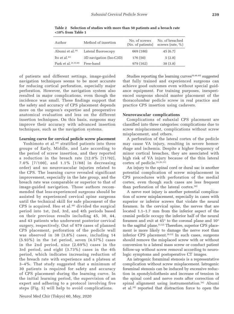
Table 2 Selection of studies with more than 50 patients and a breach rate <10% from Table 1
Author Method of insertion No. of screws (No. of patients)
No. of breached screws (rate, %)
Abumi et al.39) Lateral fluoroscopy 669 (180) 45 (6.7)
Ito et al.43) 3D navigation (Iso-C3D) 176 (50) 5 (2.8)
Park et al.26,30,48) Free-hand 979 (162) 38 (3.8)

YG Jung et al.240
Neurol Med Chir (Tokyo) 60, May, 2020
Fig. 5 Five safety steps for avoiding neurovascular complications in the initial surgical learning period are as follows: First, the screw entry points should be determined based on a preoperative computed tomography scan. Second, small-sized and curved pedicle probes can be used to ensure a proper medial angle for screw insertion. Third, a pedicle breach can be detected using a ball-tip probe. Fourth, the cervical pedicle screw should be changed to a lateral mass screw when a breach is detected. The final step is the ability to interpret intraopera-tive anterior-posterior radiographs after screw insertion.
narrowing of the foramen during a reduction maneuver could effectively prevent iatrogenic complications. For reduction of this unexpected complication, Yoshimoto et al.27) recommended prophylactic foraminotomy in patients with degenerative disorders.
Other potential complications include adjacent segment degeneration, pseudoarthrosis, screw loos-ening, screw fracture, and wound infection.39,54)
Conclusion
Subaxial CPS placement achieves superior biome-chanical stability compared to other cervical fixation techniques. Although there continues to be concern for neurovascular complications, the scope of CPS usage and efforts to decrease possible complications are increasing.
Although the accuracy of CPS placement using navigation was observed to be generally higher than other insertion methods, navigation could also be associated with complications; thus, the accuracy still depends on the surgeon’s experience and preop-erative planning using CT angiography.
A review of the literature revealed that two conflicting major surgical factors, namely, a reduction of muscle pushing effect and a laterally-located
entry point, could facilitate CPS placement accu-racy. Higher accuracy could be achieved by making a funnel-shaped hole into the lateral mass, using the posterolateral approach for navigation, or using a specially made highly curved and small-diameter pedicle probe, all of which will serve to overcome the two major surgical factors.25,30,43)
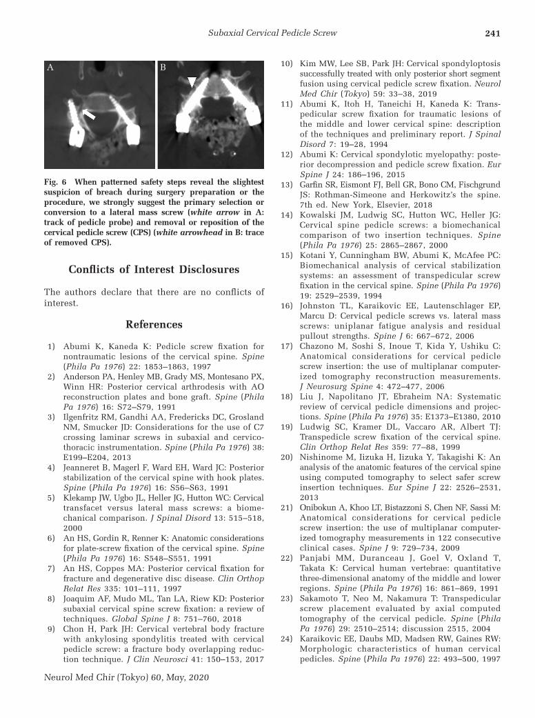
During the initial phase of the learning curve (before 30 patients), careful preparation of surgery, reiterated identification, patterned safety steps, and supervision by the expert during surgery are neces-sary. Using patterned safety steps for safety purposes, we suggest primary selection or conversion to the lateral mass screw and removal or repositioning of the CPS when there is even the slightest suspicion of breach during surgery preparation or the procedure (Figs. 3 and 6).26,28,30,55)
On the basis of enough training and experience with CPS placement, advanced insertion techniques, such as the navigation system, would be helpful for improving accuracy.
Acknowledgments
The authors thank all those who were involved in the previous research of CPS.
Subaxial Cervical Pedicle Screw 241
Neurol Med Chir (Tokyo) 60, May, 2020
Conflicts of Interest Disclosures
The authors declare that there are no conflicts of interest.
References
1) Abumi K, Kaneda K: Pedicle screw fixation fornontraumatic lesions of the cervical spine. Spine(Phila Pa 1976) 22: 1853–1863, 1997
2) Anderson PA, Henley MB, Grady MS, Montesano PX,Winn HR: Posterior cervical arthrodesis with AOreconstruction plates and bone graft. Spine (PhilaPa 1976) 16: S72–S79, 1991
3) Ilgenfritz RM, Gandhi AA, Fredericks DC, GroslandNM, Smucker JD: Considerations for the use of C7crossing laminar screws in subaxial and cervico-thoracic instrumentation. Spine (Phila Pa 1976) 38:E199–E204, 2013
4) Jeanneret B, Magerl F, Ward EH, Ward JC: Posteriorstabilization of the cervical spine with hook plates.Spine (Phila Pa 1976) 16: S56–S63, 1991
5) Klekamp JW, Ugbo JL, Heller JG, Hutton WC: Cervicaltransfacet versus lateral mass screws: a biome-chanical comparison. J Spinal Disord 13: 515–518,2000
6) An HS, Gordin R, Renner K: Anatomic considerationsfor plate-screw fixation of the cervical spine. Spine(Phila Pa 1976) 16: S548–S551, 1991
7) An HS, Coppes MA: Posterior cervical fixation forfracture and degenerative disc disease. Clin OrthopRelat Res 335: 101–111, 1997
8) Joaquim AF, Mudo ML, Tan LA, Riew KD: Posteriorsubaxial cervical spine screw fixation: a review oftechniques. Global Spine J 8: 751–760, 2018
9) Chon H, Park JH: Cervical vertebral body fracturewith ankylosing spondylitis treated with cervicalpedicle screw: a fracture body overlapping reduc-tion technique. J Clin Neurosci 41: 150–153, 2017
10) Kim MW, Lee SB, Park JH: Cervical spondyloptosissuccessfully treated with only posterior short segmentfusion using cervical pedicle screw fixation. NeurolMed Chir (Tokyo) 59: 33–38, 2019
11) Abumi K, Itoh H, Taneichi H, Kaneda K: Trans-pedicular screw fixation for traumatic lesions ofthe middle and lower cervical spine: descriptionof the techniques and preliminary report. J SpinalDisord 7: 19–28, 1994
12) Abumi K: Cervical spondylotic myelopathy: poste-rior decompression and pedicle screw fixation. EurSpine J 24: 186–196, 2015
13) Garfin SR, Eismont FJ, Bell GR, Bono CM, FischgrundJS: Rothman-Simeone and Herkowitz’s the spine.7th ed. New York, Elsevier, 2018
14) Kowalski JM, Ludwig SC, Hutton WC, Heller JG:Cervical spine pedicle screws: a biomechanicalcomparison of two insertion techniques. Spine(Phila Pa 1976) 25: 2865–2867, 2000
15) Kotani Y, Cunningham BW, Abumi K, McAfee PC:Biomechanical analysis of cervical stabilizationsystems: an assessment of transpedicular screwfixation in the cervical spine. Spine (Phila Pa 1976)19: 2529–2539, 1994
16) Johnston TL, Karaikovic EE, Lautenschlager EP,Marcu D: Cervical pedicle screws vs. lateral massscrews: uniplanar fatigue analysis and residualpullout strengths. Spine J 6: 667–672, 2006
17) Chazono M, Soshi S, Inoue T, Kida Y, Ushiku C:Anatomical considerations for cervical pediclescrew insertion: the use of multiplanar computer-ized tomography reconstruction measurements.J Neurosurg Spine 4: 472–477, 2006
18) Liu J, Napolitano JT, Ebraheim NA: Systematicreview of cervical pedicle dimensions and projec-tions. Spine (Phila Pa 1976) 35: E1373–E1380, 2010
19) Ludwig SC, Kramer DL, Vaccaro AR, Albert TJ:Transpedicle screw fixation of the cervical spine.Clin Orthop Relat Res 359: 77–88, 1999
20) Nishinome M, Iizuka H, Iizuka Y, Takagishi K: Ananalysis of the anatomic features of the cervical spineusing computed tomography to select safer screwinsertion techniques. Eur Spine J 22: 2526–2531,2013
21) Onibokun A, Khoo LT, Bistazzoni S, Chen NF, Sassi M:Anatomical considerations for cervical pediclescrew insertion: the use of multiplanar computer-ized tomography measurements in 122 consecutiveclinical cases. Spine J 9: 729–734, 2009
22) Panjabi MM, Duranceau J, Goel V, Oxland T,Takata K: Cervical human vertebrae: quantitative three-dimensional anatomy of the middle and lowerregions. Spine (Phila Pa 1976) 16: 861–869, 1991
23) Sakamoto T, Neo M, Nakamura T: Transpedicularscrew placement evaluated by axial computedtomography of the cervical pedicle. Spine (PhilaPa 1976) 29: 2510–2514; discussion 2515, 2004
24) Karaikovic EE, Daubs MD, Madsen RW, Gaines RW:Morphologic characteristics of human cervicalpedicles. Spine (Phila Pa 1976) 22: 493–500, 1997
Fig. 6 When patterned safety steps reveal the slightest suspicion of breach during surgery preparation or the procedure, we strongly suggest the primary selection or conversion to a lateral mass screw (white arrow in A: track of pedicle probe) and removal or reposition of the cervical pedicle screw (CPS) (white arrowhead in B: trace of removed CPS).
A B

YG Jung et al.242
Neurol Med Chir (Tokyo) 60, May, 2020
25) Abumi K, Ito M, Sudo H: Reconstruction of thesubaxial cervical spine using pedicle screw instru-mentation. Spine (Phila Pa 1976) 37: E349–E356,2012
26) Park JH, Jeon SR, Roh SW, Kim JH, Rhim SC: Thesafety and accuracy of freehand pedicle screwplacement in the subaxial cervical spine: a seriesof 45 consecutive patients. Spine (Phila Pa 1976)39: 280–285, 2014
27) Yoshimoto H, Sato S, Hyakumachi T, YanagibashiY, Masuda T: Spinal reconstruction using a cervicalpedicle screw system. Clin Orthop Relat Res 431:111–119, 2005
28) Hacker AG, Molloy S, Bernard J: The contralaterallamina: a reliable guide in subaxial, cervical pediclescrew placement. Eur Spine J 17: 1457–1461, 2008
29) Lee DH, Lee SW, Kang SJ, et al.: Optimal entrypoints and trajectories for cervical pedicle screwplacement into subaxial cervical vertebrae. EurSpine J 20: 905–911, 2011
30) Lee S, Seo J, Lee MK, et al.: Widening of the safetrajectory range during subaxial cervical pediclescrew placement: advantages of a curved pedicleprobe and laterally located starting point withoutcreating a funnel-shaped hole. J Neurosurg Spine27: 150–157, 2017
31) Yukawa Y, Kato F, Ito K, et al.: Placement and compli-cations of cervical pedicle screws in 144 cervicaltrauma patients using pedicle axis view techniquesby fluoroscope. Eur Spine J 18: 1293–1299, 2009
32) Komatsubara T, Tokioka T, Sugimoto Y, Ozaki T:Minimally invasive cervical pedicle screw fixationby a posterolateral approach for acute cervical injury.Clin Spine Surg 30: 466–469, 2017
33) Holly LT, Foley KT: Percutaneous placement ofposterior cervical screws using three-dimensionalfluoroscopy. Spine (Phila Pa 1976) 31: 536–540;discussion 541, 2006
34) Sugimoto Y, Ito Y, Shimokawa T, Shiozaki Y, MazakiT: Percutaneous screw fixation for traumatic spon-dylolisthesis of the axis using iso-C3D fluoroscopy-assisted navigation (case report). Minim InvasiveNeurosurg 53: 83–85, 2010
35) Lu T, Liu C, Dong J, Lu M, Li H, He X: Cervicalscrew placement using rapid prototyping drilltemplates for navigation: a literature review. Int JComput Assist Radiol Surg 11: 2231–2240, 2016
36) Lu S, Xu YQ, Lu WW, et al.: A novel patient-specificnavigational template for cervical pedicle screw place-ment. Spine (Phila Pa 1976) 34: E959–E966, 2009
37) Ludwig SC, Kramer DL, Balderston RA, Vaccaro AR,Foley KF, Albert TJ: Placement of pedicle screwsin the human cadaveric cervical spine: comparativeaccuracy of three techniques. Spine (Phila Pa 1976)25: 1655–1667, 2000
38) Theologis AA, Burch S: Safety and efficacy of recon-struction of complex cervical spine pathology usingpedicle screws inserted with stealth navigation and
3D image-guided (O-Arm) technology. Spine (Phila Pa 1976) 40: 1397–1406, 2015
39) Abumi K, Shono Y, Ito M, Taneichi H, Kotani Y,Kaneda K: Complications of pedicle screw fixationin reconstructive surgery of the cervical spine. Spine(Phila Pa 1976) 25: 962–969, 2000
40) Chachan S, Bin Abd Razak HR, Loo WL, Allen JC,Shree Kumar D: Cervical pedicle screw instrumenta-tion is more reliable with O-arm-based 3D navigation:analysis of cervical pedicle screw placement accu-racy with O-arm-based 3D navigation. Eur Spine J27: 2729–2736, 2018
41) Ishikawa Y, Kanemura T, Yoshida G, Ito Z, MuramotoA, Ohno S: Clinical accuracy of three-dimensionalfluoroscopy-based computer-assisted cervical pediclescrew placement: a retrospective comparative studyof conventional versus computer-assisted cervicalpedicle screw placement. J Neurosurg Spine 13:606–611, 2010
42) Ishikawa Y, Kanemura T, Yoshida G, et al.: Intra-operative, full-rotation, three-dimensional image(O-arm)-based navigation system for cervical pediclescrew insertion. J Neurosurg Spine 15: 472–478,2011
43) Ito Y, Sugimoto Y, Tomioka M, Hasegawa Y, NakagoK, Yagata Y: Clinical accuracy of 3D fluoroscopy-assisted cervical pedicle screw insertion. J NeurosurgSpine 9: 450–453, 2008
44) Kotani Y, Abumi K, Ito M, Minami A: Improvedaccuracy of computer-assisted cervical pedicle screwinsertion. J Neurosurg 99: 257–263, 2003
45) Miller RM, Ebraheim NA, Xu R, Yeasting RA:Anatomic consideration of transpedicular screwplacement in the cervical spine: an analysis of twoapproaches. Spine (Phila Pa 1976) 21: 2317–2322,1996
46) Jo DJ, Seo EM, Kim KT, Kim SM, Lee SH: Cervicalpedicle screw insertion using the technique withdirect exposure of the pedicle by laminoforami-notomy. J Korean Neurosurg Soc 52: 459–465,2012
47) Kaneyama S, Sugawara T, Sumi M: Safe and accu-rate midcervical pedicle screw insertion procedurewith the patient-specific screw guide templatesystem. Spine (Phila Pa 1976) 40: E341–E348,2015
48) Heo Y, Lee SB, Lee BJ, et al.: The learning curveof subaxial cervical pedicle screw placement: howcan we avoid neurovascular complications in theinitial period? Oper Neurosurg (Hagerstown) 17:603–607, 2019
49) Yoshimoto H, Sato S, Hyakumachi T, YanagibashiY, Kanno T, Masuda T: Clinical accuracy of cervicalpedicle screw insertion using lateral fluoroscopy:a radiographic analysis of the learning curve. EurSpine J 18: 1326–1334, 2009
50) Tomasino A, Parikh K, Koller H, et al.: The vertebralartery and the cervical pedicle: morphometric analysis
Subaxial Cervical Pedicle Screw 243
Neurol Med Chir (Tokyo) 60, May, 2020
of a critical neighborhood. J Neurosurg Spine 13: 52–60, 2010
51) Pech P, Daniels DL, Williams AL, Haughton VM: The cervical neural foramina: correlation of microtomy and CT anatomy. Radiology 155: 143–146, 1985
52) Xu R, Kang A, Ebraheim NA, Yeasting RA: Anatomic relation between the cervical pedicle and the adjacent neural structures. Spine (Phila Pa 1976) 24: 451–454, 1999
53) Ghori A, Le HV, Makanji H, Cha T: Posterior fixation techniques in the subaxial cervical spine. Cureus 7: e338, 2015
54) Nakashima H, Yukawa Y, Imagama S, et al.: Complications of cervical pedicle screw fixation
for nontraumatic lesions: a multicenter study of 84 patients. J Neurosurg Spine 16: 238–247, 2012
55) Park JH, Roh SW, Rhim SC: A single-stage posterior approach with open reduction and pedicle screw fixation in subaxial cervical facet dislocations. J Neurosurg Spine 23: 35–41, 2015
Address reprint requests to: Jin Hoon Park, MD, PhD, Department of Neurosurgery, Asan Medical Center, University of Ulsan College of Medicine, 88 Olympic-ro 43-gil, Songpa-gu, Seoul 05505, Korea.
e-mail: [email protected]