cervical fracture complicating ankylosing spondylitisdegreesofclarity.com/emsbasics/library/murray -...
TRANSCRIPT
Cervical Fracture Complicating Ankylosing Spondylitis
A Report of Eight Cases and Review of the Literature
GARVIN C. MURRAY, M.D. ROBERT H. PERSELLIN. M.D. Son Antonio, Texas
From the Division of Rheumatology, Depart- ment of Medicine, The University of Texas Health Science Center at San Antonio, San An- tonio, Texas. This study was supported in part by an Arthritis Clinical Research Center grant from the Arthritis Foundation and by grants from the South Central Texas Chapter of the Arthritis Foundation and the Ruth and Vernon Taylor Foundation. Requests for reprints should be addressed to Dr. Robert H. Persellin, Department of Medicine, The University of Texas Health Science Center at San Antonio, 7703 Floyd Curl Drive, San Antonio, TX 78284. Manuscript ac- cepted November 10,198O.
Fracture of the cervical spine is a serious and often fatal complication of ankylosing spondylitis. An evaluation of eight patients and a re- view of 75 additional cases from the literature are presented. Al- though this complication is relatively uncommon, it is clear that people with advanced disease and complete ankylosis of the cervical spine are at increased risk of sustaining cervical fracture. When fracture occurs it usually stems from minor trauma resulting most commonly in disruption of the lower cervical segments (iIfth through the seventh cervical vertebrae). Fracture is most likely the result of a hyperextension type injury, occurs through what was formerly an intervertebral space, and is unstable. Severe neurologic sequelae occur in 57 percent of the cases and the mortality rate (35 percent) is twice that observed with similar fracture involving normal spines. The majority of patients are best treated with closed reduction with halo traction together with body cast or jacket. Laminectomy is rarely indicated except in the event of an advancing neurologic deficit. With appropriate understanding and execution of management principles, the outcome in these patients can be favorable. Unfortunately, rec- ognition of cervical fracture in patients with ankylosing spondylitis is often needlessly delayed. Distortion of normal anatomy in spon- dylitics, predominant fracture location in lower cervical spine seg- ments and lack of obvious displacement make identification difficult. Thus, management is often inappropriate resulting in exessive neurologic injury and mortality.
Fracture of the cervical spine is a serious and potentially fatal com- plication of ankylosing spondylitis. It is associated with the develop- ment of severe neurologic deficits in 57 percent of the cases and results in a 35 percent mortality rate, twice that seen with fractures involving normal spines [l]. In over half the cases the precipitating trauma is of a minor nature, unlikely to result in fracture of a normal spine; in 7 percent no history of trauma is obtained. For this reason, and because neck pain is a common complaint of patients with ankylosing spon- dylitis, detection of these fractures is often delayed or overlooked, usually to the detriment of the patient.
This complication was considered to be uncommon. In an analysis of 2,506 cases of traumatic spinal injuries, only seven cases of ank- ylosing spondylitis were found [z]. Wilkinson and Bywaters [3] re- ported three cases of spinal fracture resulting in one death in their review of 212 patients with ankylosing spondylitis followed for as long as 20 years. Of 146 deaths among 836 patients with ankylosing spon- dylitis reported by Radford et al. [4], only three were attributable to vertebral fracture. From these data and from others in which long-term
May 1981 The American Journal of Medicine Volume 70 1033
CERVICAL FRACTURE COMPLICATING ANKYLOSING SPONDYLITIS-MURRAY ET AL.
follow-up was reported [5-y], it would appear that fracture of the ankylosed vertebral column is a rare event and is a minor contributor to mortality in ank- ylosing spondylitis.
Our experience has made us wary of this conclusion for we have had the opportunity to evaluate eight cases of cervical fracture complicating ankylosing spondylitis over the past five years. The diagnosis and management of cervical fracture in these patients presented unique and difficult problems, and the clinical course resulted in death or the development of severe neurologic se- quelae in half the cases. We present here our experience with these eight patients together with a review of 75 additional cases reported in the literature [1,2,8-381.
PATIENTS AND METHODS
of bradycardia and respiratory insufficiency developed when the patient turned to a prone position. Bradycardia and res- piratory insufficiency again developed with repositioning on postoperative days 9 and 10. Cervical fusion was considered; however, pneumonia and progressive respiratory insufficiency developed and the patient died on the 17th hospital day.
Comment: Failure to adequately evaluate the lower cervical region roentgenographically and to appro- priately analyze the symptoms of neurologic involve- ment resulted in quadriplegia and ultimately death in this case. The development of bradycardia and respi- ratory insufficiency when the patient turned to a prone position probably resulted from loss of alignment of the fracture segments and associated spinal cord com- pression [39].
Five patients with ankylosing spondylitis and cervical fracture were hospitalized at the Teaching Hospitals of the University of Texas Health Science Center at San Antonio between June 1973 and December 1978. Each patient was evaluated by one of the members of the Division of Rheumatology. In addition, we evaluated the medical records and roentgenograms in three other previously unreported cases, two from community hos- pitals in San Antonio (Cases 6 and 7) and one from the Uni- versity Hospital in San Diego (Case 8).
Case 2. A 66 year old man with a 40 year history of ankylosing spondylitis fell at night while walking to his bathroom. lie struck the posterior aspect of his head and neck, and experi- enced the immediate onset of neck pain. He was unable to move his extremities for approximately 15 minutes. Subse- quently, he was able to ambulate but experienced what he described as electrical shocks down his spine and into his extremities with head movement. Because of persistent neck pain, he was taken by automobile 250 miles to a Veterans’ Administration facility for hospitalization.
REPRESENTATIVE CASES
Case I. A 48 year old man with a 20 year history of ankylosing spondylitis was involved in a motor vehicle accident and sustained a fracture of the left tibia. While being evaluated in the emergency room, he complained of neck pain and an electric shock-like sensation radiating down his spine and into his extremities whenever he moved his head. Neurologic ex- amination reportedly disclosed no abnormalities. Films of the spine revealed complete ankylosis of the cervical segments associated with severe osteopenia. No fracture or dislocation was seen; however, the vertebral segments below the sixth cervical vertebra were not well visualized. The left leg was placed in a cast and the patient was admitted for further ob- servation. Repeat films of the entire spine were obtained. No fractures were detected, but again the lower cervical segments were not well seen. After transfer from a stretcher to a bed, the patient complained of increased neck pain and experienced the sudden onset of numbness and weakness in his legs. Neurologic evaluation revealed partial motor and sensory dysfunction in the lower extremities associated with absent reflexes. Over the next 12 hours an inability to urinate and progressive motor and sensory deficits to a sixth cervical ver- tebral level developed. Tomography of the cervical spine showed a fracture through the seventh cervical vertebra-first thoracic vertebra interspace with 90 percent anterior dis- placement of seventh cervical vertebra on the first thoracic vertebra.
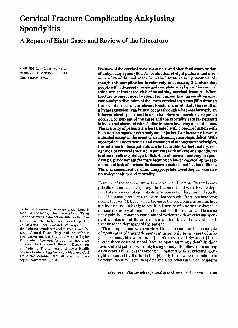
On examination he was noted to sit with a very rigid posture. The fifth and sixth cervical vertebrae region was tender pos- teriorly; palpation of the area caused radiation of pain into both upper extremities. Neurologic examination was otherwise within normal limits. Films of the cervical spine revealed complete ankylosis of the entire cervical region with advanced osteopenia. A compression fracture of the superior portion of the sixth cervical vertebral body was noted (Figure 1). The fracture line extended through the posterior elements at the fifth cervical vertebral level, and slight anterior displacement of the superior fracture segment was present.
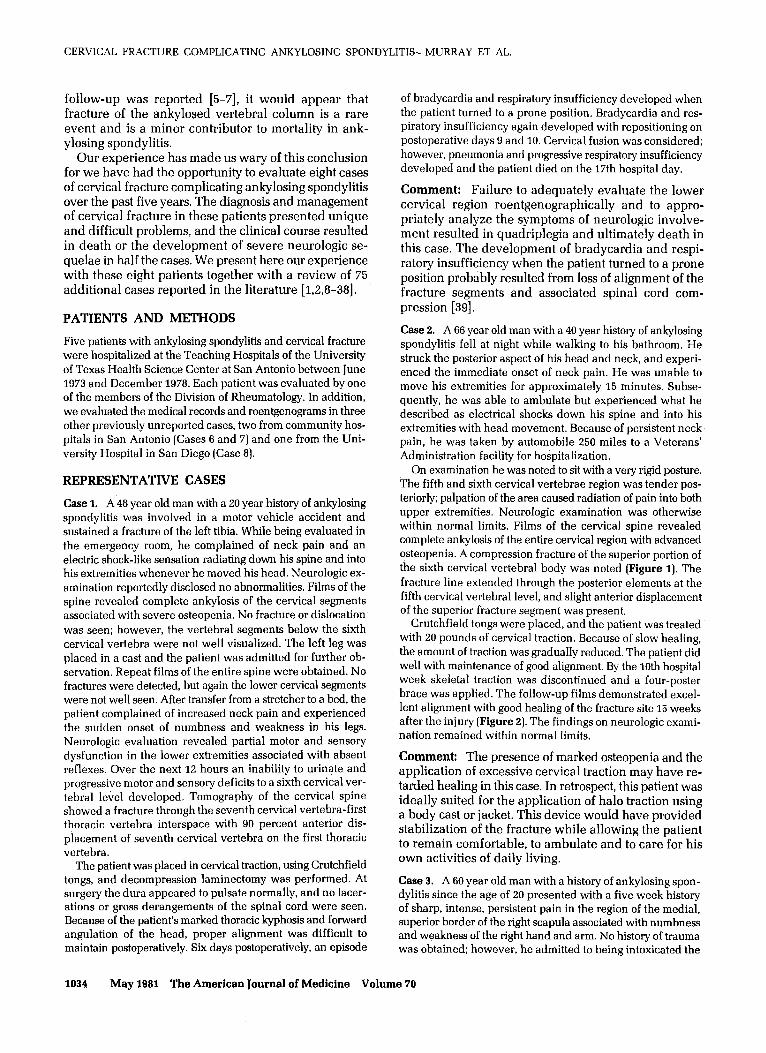
Crutchfield tongs were placed, and the patient was treated with 20 pounds of cervical traction. Because of slow healing, the amount of traction was gradually reduced. The patient did well with maintenance of good alignment. By the 10th hospital week skeletal traction was discontinued and a four-poster brace was applied. The follow-up films demonstrated excel- lent alignment with good healing of the fracture site I5 weeks after the injury [Figure 2). The findings on neurologic exami- nation remained within normal limits.
The patient was placed in cervical traction, using Crutchfield tongs, and decompression laminectomy was performed. At surgery the dura appeared to pulsate normally, and no lacer- ations or gross derangements of the spinal cord were seen. Because of the patient’s marked thoracic kyphosis and forward angulation of the head, proper alignment was difficult to maintain postoperatively. Six days postoperatively, an episode
Comment: The presence of marked osteopenia and the application of excessive cervical traction may have re- tarded healing in this case. In retrospect, this patient was ideally suited for the application of halo traction using a body cast or jacket. This device would have provided stabilization of the fracture while allowing the patient to remain comfortable, to ambulate and to care for his own activities of daily living.
Case 3. A 60 year old man with a history of ankylosing spon- dylitis since the age of 20 presented with a five week history of sharp, intense, persistent pain in the region of the medial, superior border of the right scapula associated with numbness and weakness of the right hand and arm. No history of trauma was obtained; however, he admitted to being intoxicated the
1034 May 1981 The American Journal of Medicine Volume 70
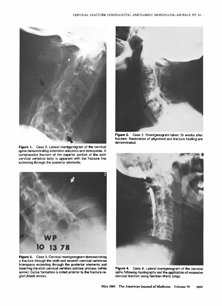
Figure 1. Case 2. Lateral roentgenogram of the cervical spine demonstrating extensive ankylosis and osteopenia. A compression fracture of the superior portion of the sixth cervical vertebral body is apparent with the fracture line extending through the posterior elements.
Case 3. Cervical roentgenogram demonstrating a fracture through the sixth and seventh cervical vertebrae interspace extending through the posterior elements and bisecting the sixth cervical vertebra spinous process (white arrow). Callus formation is noted anterior to the fracture re- gion (black arrow).
Figure 2. Case 2. Roentgenogram taken 15 weeks after fracture. Restoration of alignment and fracture healing are demonstrated.
CERVICAL FRACTURE COMPLICATING ANKYLOSING SPONDYLITIS-MURRAY ET AL.
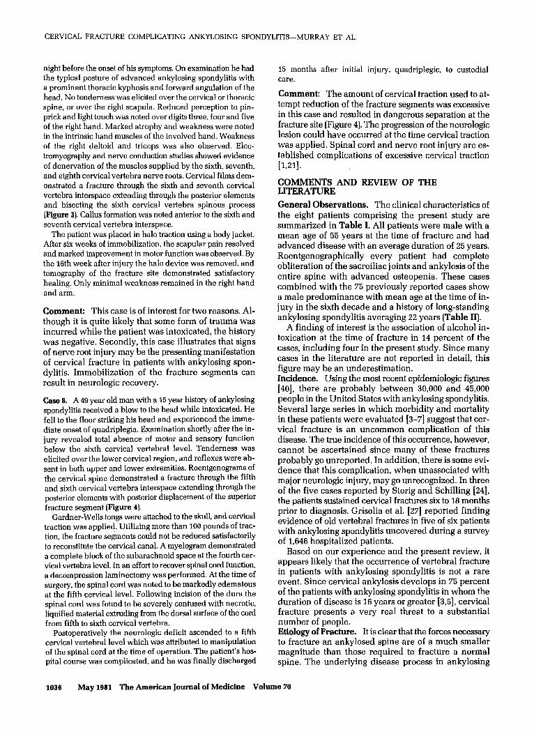
Figure 4. Case 8. Lateral’ roentgenogram of the cervical spine following myelography and the application of excessive cervical traction using Gardner-Wells tongs.
May 1981 The American Journal of Medicine Volume 70 1035
CERVICAL FRACTURE COMPLICATING ANKYLOSING SPONDYLITIS-MURRAY ET AL,
night before the onset of his symptoms. On examination he had the typical posture of advanced ankylosing spondylitis with a prominent thoracic kyphosis and forward angulation of the head. No tenderness was elicited over the cervical or thoracic spine, or over the right scapula. Reduced perception to pin- prick and light touch was noted over digits three, four and five of the right hand. Marked atrophy and weakness were noted in the intrinsic hand muscles of the involved hand. Weakness of the right deltoid and triceps was also observed. Elec- tromyography and nerve conduction studies showed evidence of denervation of the muscles supplied by the sixth, seventh, and eighth cervical vertebra nerve roots. Cervical films dem- onstrated a fracture through the sixth and seventh cervical vertebra interspace extending through the posterior elements and bisecting the sixth cervical vertebra spinous process (Figure 3). Callus formation was noted anterior to the sixth and seventh cervical vertebra interspace.
15 months after initial injury, quadriplegic, to custodial care.
Comment: The amount of cervical traction used to at- tempt reduction of the fracture segments was excessive in this case and resulted in dangerous separation at the fracture site [Figure 4). The progression of the neurologic lesion could have occurred at the time cervical traction was applied. Spinal cord and nerve root injury are es- tablished complications of excessive cervical traction [l,Zl].
COMMENTS AND REVIEW OF THE LITERATURE
The patient was placed in halo traction using a body jacket. After six weeks of immobilization, the scapular pain resolved and marked improvement in motor function was observed. By the 16th week after injury the halo device was removed, and tomography of the fracture site demonstrated satisfactory healing. Only minimal weakness remained in the right hand and arm.
Comment: This case is of interest for two reasons. Al- though it is quite likely that some form of trauma was incurred while the patient was intoxicated, the history was negative. Secondly, this case illustrates that signs of nerve root injury may be the presenting manifestation of cervical fracture in patients with ankylosing spon- dylitis. Immobilization of the fracture segments can result in neurologic recovery.
General Observations. The clinical characteristics of the eight patients comprising the present study are summarized in Table I. All patients were male with a mean age of 55 years at the time of fracture and had advanced disease with an average duration of 25 years, Roentgenographically every patient had complete obliteration of the sacroiliac joints and ankylosis of the entire spine with advanced osteopenia. These cases combined with the 75 previously reported cases show a male predominance with mean age at the time of in- jury in the sixth decade and a history of long-standing at&losing spondylitis averaging 2.2 years [Table II].
Case 6. A 49 year old man with a 15 year history of ankylosing spondylitis received a blow to the head while intoxicated. He fell to the floor striking his head and experienced the imme- diate onset of quadriplegia. Examination shortly after the in- jury revealed total absence of motor and sensory function below the sixth cervical vertebral level. Tenderness was elicited over the lower cervical region, and reflexes were ab- sent in both upper and lower extremities. Roentgenograms of the cervical spine demonstrated a fracture through the fifth and sixth cervical vertebra interspace extending through the posterior elements with posterior displacement of the superior fracture segment (Figure 4).
Gardner-Wells tongs were attached to the skull, and cervical traction was applied. Utilizing more than 100 pounds of trac- tion, the fracture segments could not be reduced satisfactorily to reconstitute the cervical canal. A myelogram demonstrated a complete block of the subarachnoid space at the fourth cer- vical vertebra level. In an effort to recover spinal cord function, a decompression laminectomy was performed. At the time of surgery, the spinal cord was noted to be markedly edematous at the fifth cervical level. Following incision of the dura the spinal cord was found to be severely contused with necrotic, liquified material extruding from the dorsal surface of the cord from fifth to sixth cervical vertebra.
A finding of interest is the association of alcohol in- toxication at the time of fracture in 14 percent of the cases, including four in the present study. Since many cases in the literature are not reported in detail, this figure may be an underestimation. Incidence. Using the most recent epidemiologic figures [40], there are probably between 80,666 and 45,660 people in the United States with ankylosing spondylitis. Several large series in which morbidity and mortality in these patients were evaluated [8-71 suggest that cer- vical fracture is an uncommon complication of this disease. The true incidence of this occurrence, however, cannot be ascertained since many of these fractures probably go unreported. In addition, there is some evi- dence that this complication, when unassociated with major neurologic injury, may go unrecognized. In three of the five cases reported by Storig and Schilling [24], the patients sustained cervical fractures six to 18 months prior to diagnosis. Grisolia et al. [27] reported finding evidence of old vertebral fractures in five of six patients with ankylosing spondylitis uncovered during a survey of 1,646 hospitalized patients.
Postoperatively the neurologic deficit ascended to a fifth cervical vertebral level which was attributed to manipulation of the spinal cord at the time of operation. The patient’s hos- pital course was complicated, and he was finally discharged
Based on our experience and the present review, it appears likely that the occurrence of vertebral fracture in patients with ankylosing spondylitis is not a rare event. Since cervical ankylosis develops in 75 percent of the patients with ankylosing spondylitis in whom the duration of disease is 16 years or greater [3,5], cervical fracture presents a very real threat to a substantial number of people. Etiology of Fracture. It is clear that the forces necessary to fracture an ankylosed spine are of a much smaller magnitude than those required to fracture a normal spine. The underlying disease process in ankylosing
1036 May 1961 The American Journal of Medicine Volume 70

CERVICAL FRACTURE COMPLICATING ANKYLOSING SPONDYLITIS-MURRAY ET AL.
TABLE I
Age (Y4
Case and No. Sex
Clinical Characterlstfcs of Eight Patlents with Ankylosing Spondylitis and Cervical Fracture
Rosntgen- lniliai Duration Type sf bgr@iC Nsuroiogic Type of Final
of AS lsjury Appearance D&ii Treatment Outcome Comment
48, M 20 yr Motor vehicle accident
66, M 40 yr Fail in bedroom
60, M 40 yr No history of trauma
56, M 23 yr Motor vehicle accident
45, M 23 yr Fail in bedroom
49, M 25 yr Fall to ground
65, M
49, M
from bulldozer extendrng right upper through post extremity elements with 3 mm ant displacement of sup fx segment
18 yr Fail on sidewalk C6 vert body None compression fx extending through C5-6 post elements with 40 % forward anguiation of sup fx segment
15 yr Blow to head and C5-6 IS fx Quadriparesis _ . . _.
C7-Tl IS fx with No neuroiogic Cervical traction Death from Intoxicated at time
90% ant deficit initially: with Crutchfield respiratory of injury displacement of quadriparesis tongs; failure 17 days C7 on Ti later developed iaminectomy 1 after injury
day after injury
C6 vet? body Transient Cervical traction No neuroiogic Excessive cervical compression fx quadriparesis with Crutchfield deficit traction may extending tongs; four- have retarded through C5 post poster brace healing elements with 0.5 cm ant displacement of sup fx segment
C6-7 IS fx Sensory and Halo traction and Minimal residual intoxicated night extending motor deficits in body jacket weakness prior to onset of through post right upper symptoms elements at C6 extremity in
C6-7 distribution
Nondisplaced fx Ouadriparesis Cervical traction Minimal Difficulty through C7 vert below C6 cord with Crutchfield neuroiogic maintaining body extending level tongs; four- improvement alignment may obliquely poster brace have retarded through C5-6 healing post elements
C6 vert body fx Quadriparesis Cervical traction Death from Intoxicated at time extending below C5 cord with Gardner- oneumonia and of injury through C5 post level Wells tongs sepsis 15 wk elements; 30 % after injury post angulation of sup fx segment; fx of c3, 4, 5 spinous processes
C6-7 IS fx Weakness in the Halo traction and Complete 3 wk delay in fracture recognition
body cast neuroiogic recovery
Halo traction and No neuroiogic body cast; deficit anterior fusion with strut graft 4 mo after injury
Cervical traction Quadriparesis
Pseudoarthrosis developed requiring anterior fusion
intoxicated at time tall to tloor extending below C5 cord with Gardner- below C4 cord of injury
through post level Wells tongs; level elements with 1 iaminectomy; cm post halo traction displacement of and body cast: sup fx segment posterior
cervical fusion; cervical collar
NOTE: C7 = seventh cervical vertebrae, Tl = first thoracic vertebrae, etc.; IS = interspace: fx = fracture: ant = anterior; vert = vertebral; sup = superior: post = posterior.
May 1961 The American Journal of Medicine Volume 70 1037
CERVICAL FRACTURE COMPLICATING ANKYLOSING SPONDYLITIS-MURRAY ET AL
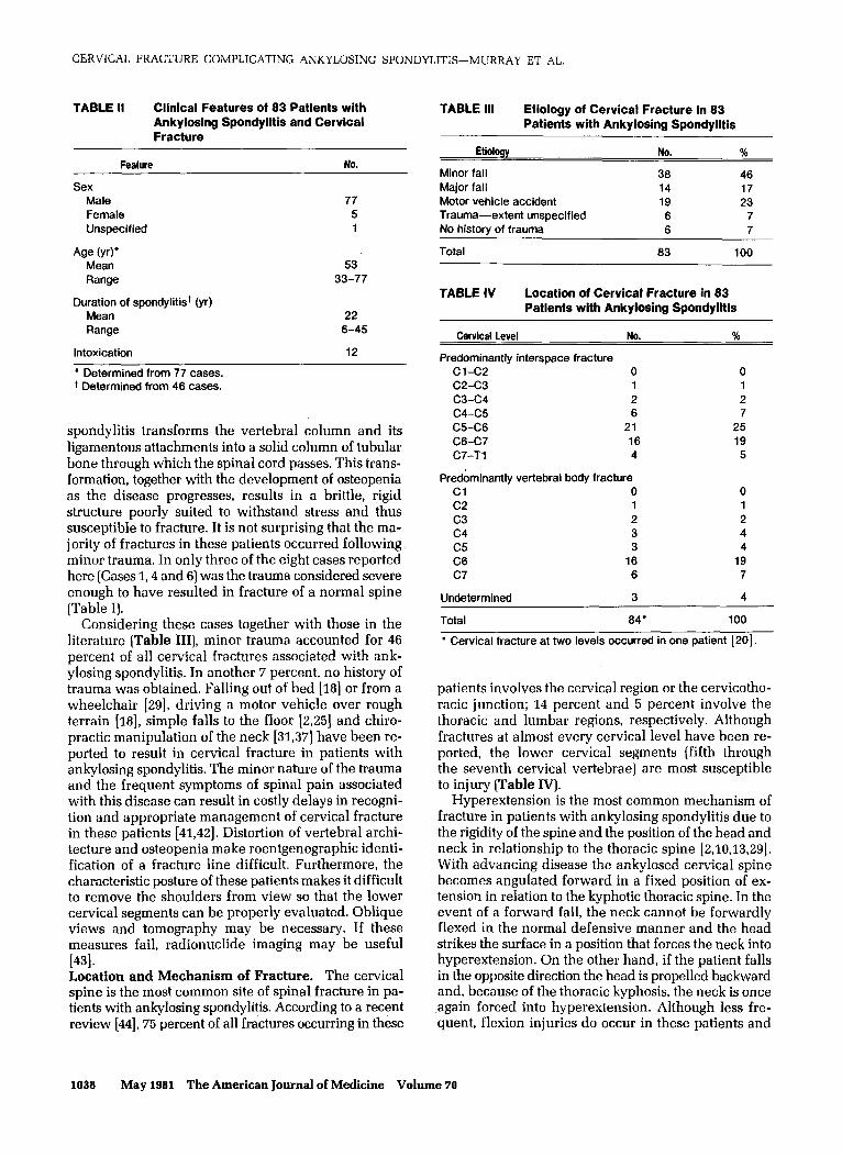
TABLE II Clinical Features of 83 Patients with Ankylosing Spondylltis and Cervical Fracture
TABLE Ill Etiology of Cervical Fracture in 83 Patients with Ankylosing Spondylitis
Fealure
Sex Male Female Unspecified
Age (~0’ Mean Range
Duration of spondylitis+ (yr) Mean Range
Intoxication
* Determined from 77 cases. t Determined from 46 cases.
No.
77 5 1
53 33-77
22 6-45
12
Etiology No. %
Minor fall 38 46 Major fall 14 17 Motor vehicle accident 19 23 Trauma-extent unspecified 6 7 No history of trauma 6 7
Total a3 100
TABLE IV Location of Cervical Fracture in 83 Patients with Ankylosing Spondylltis
spondylitis transforms the vertebral column and its ligamentous attachments into a solid column of tubular bone through which the spinal cord passes. This trans- formation, together with the development of osteopenia as the disease progresses, results in a brittle, rigid structure poorly suited to withstand stress and thus susceptible to fracture. It is not surprising that the ma- jority of fractures in these patients occurred following minor trauma. In only three of the eight cases reported here (Cases 1,4 and 6) was the trauma considered severe enough to have resulted in fracture of a normal spine (Table I).
Considering these cases together with those in the literature (Table III), minor trauma accounted for 46 percent of all cervical fractures associated with ank- ylosing spondylitis. In another 7 percent, no history of trauma was obtained. Falling out of bed [18] or from a wheelchair [Zt], driving a motor vehicle over rough terrain [18], simple falls to the floor [2,25] and chiro- practic manipulation of the neck [31,37] have been re- ported to result in cervical fracture in patients with a&losing spondylitis. The minor nature of the trauma and the frequent symptoms of spinal pain associated with this disease can result in costly delays in recogni- tion and appropriate management of cervical fracture in these patients [41,42]. Distortion of vertebral archi- tecture and osteopenia make roentgenographic identi- fication of a fracture line difficult. Furthermore, the characteristic posture of these patients makes it difficult to remove the shoulders from view so that the lower cervical segments can be properly evaluated. Oblique views and tomography may be necessary. If these measures fail, radionuclide imaging may be useful
[431. Location and Mechanism of Fracture. The cervical spine is the most common site of spinal fracture in pa- tients with ankylosing spondylitis. According to a recent review [@I, 75 percent of all fractures occurring in these
Cervical Level No. %
Predominantly interspace fracture Cl-C2 0 0 C2-C3 1 1 c3-c4 2 2 c4-c5 6 7 C5-C6 21 25 C6-C7 16 19 C7-Tl 4 5
Predominantly vertebral body fracture Cl 0 0 c2 1 1 c3 2 2 c4 3 4 c5 3 4 C6 16 19 c7 6 7
Undetermined 3 4
Total 84” 100
l Cervical fracture at two levels occurred in one patient [20].
patients involves the cervical region or the cervicotho- racic junction; 14 percent and 5 percent involve the thoracic and lumbar regions, respectively. Although fractures at almost every cervical level have been re- ported, the lower cervical segments (fifth through the seventh cervical vertebrae) are most susceptible to injury [Table IV].
Hyperextension is the most common mechanism of fracture in patients with ankylosing spondylitis due to the rigidity of the spine and the position of the head and neck in relationship to the thoracic spine [2,16,13,29]. With advancing disease the ankylosed cervical spine becomes angulated forward in a fixed position of ex- tension in relation to the kyphotic thoracic spine. In the event of a forward fall, the neck cannot be forwardly flexed in the normal defensive manner and the head strikes the surface in a position that forces the neck into hyperextension. On the other hand, if the patient falls in the opposite direction the head is propelled backward and, because of the thoracic kyphosis, the neck is once again forced into hyperextension. Although less fre- quent, flexion injuries do occur in these patients and
1038 May 1981 The American Journal of Medicine Volume 70

CERVICAL FRACTURE COMPLICATING ANKYLOSING SPONDYLITIS-MIfRRAY ET AL.
TABLE V Comparison of Initial and Final Neurologic Status and Mortality Rates Following Cervical Fracture of Ankylosed and Normal Spines
Initial neurologic deficit None Minor l Major+
Cervical Spine Ankylosed Normal 111
No. 46 No. %
25 30 27 35 12 15 34 45 46 55 15 20
Total 83 100 76 100
Final outcome Return to workz Neurologic deficit* Death7
36 43 54 71 18 22 8 11 29 35 14 18
Total 83 100 76 100
l Mild sensory of motor dysfunction or abnormal reflexes or symptoms of nerve root pressure. + Complete or incomplete spinal cord interruption. x Those who actually did return to work or had the neurologic po- tential. 5 Paraparesis, quadriparesis or residual neurologic deficit preventing return to work. n Within 13 months of injury.
TABLE VII Type of Management in 83 Patients with Ankylosing Spondylitis and Cervical Fracture
Deaths Type No. % No. %’
Conservative+ 48 58 14 29 Surgical 20 24 9 45
Laminectomy alone 9 . Cervical fusion alone 5 . 4 Both 6
Unspecified 15 ‘18 1 6 40
Total 83 100 29 35
l Refers to mortality rate within each treatment group. + Cervical traction with Crutchfield tongs, Gardner-Wells tongs, Vinke tongs, halo apparatus, Sayre halter, maxillary hooks; immo- bilization with Minerva jacket, cervical collar, four-poster brace; or bedrest alone.
generally result from forces directed anteriorly against the posterior aspect of the head or neck.
Most cervical fractures (70 percent) in these patients occur through what was formerly the intervertebral disc space. However, contrary to earlier reports [11,15,22],
fractures involving the vertebral bodies are not rare (Table IV). Some investigators have observed that hy- perextension injuries result in interspace fractures, whereas flexion trauma leads to fracture of the vertebral body [21,35].
Neurologic Sequelae and Mortality Rate. In the an- kylosed spine, fractures invariably extend through the entire width of the spine including the calcified liga-
TABLE VI Cause of Death in 29 Patients with Ankylosing Spondylltis and Cervical Fracture
CaUW No. %
Respiratory failure 10 35 Pulmonary infection 5 17 Perforated duodenal ulcer 2 7 Gastrointestinal hemorrhage 1 3 Pulmonary embolism 2 7 Unspecified 9 31
Total 29 100
mentous supporting structures. This was true in every case comprising the present series. Consequently, this type of fracture is extremely unstable and results in a high frequency of spinal cord and nerve root injury. Of our eight patients, cervical fracture resulted in the de- velopment of a neurologic deficit in seven. In one, quadriplegia was only transient and in two others the injury resulted in nerve root deficits which later re- solved. In the remaining four, however, severe perma- nent quadriplegia developed, culminating in death in two.
If the experience with cervical fractures of ankylosed spines is compared with a large series of similar frac- tures involving normal spines [l], some striking differ- ences become apparent. As shown in Table V, fracture of ankylosed cervical spines results in a higher fre- quency of neurologic deficits; severe neurologic se- quelae (paraparesis or quadriparesis) occur nearly three times more frequently. Death occurred nearly twice as often in patients with ankylosing spondylitis (35 versus 18 percent]. In general, delay in seeking medical at- tention [2,2], failure in fracture recognition [22], insta- bility of the fracture site, inappropriate treatment [31] and the reduced general state of health of patients with ankylosing spondylitis [a] resulted in a poorer out- come.
The causes of death complicating cervical fracture in 29 patients are listed in Table VI. Respiratory failure and pulmonary infection accounted for at least 52 per- cent of these deaths. The restrictive ventilatory defect from chest wall rigidity increases the risk of death from respiratory complications. In three cases death was due to either duodenal perforation or gastrointestinal hemorrhage [2,25,36]. In none of these cases was the therapeutic use of corticosteroids mentioned. Two pa- tients died following pulmonary embolization [27,36], and in nine the cause of death was not specified [9,12,18,25,26,29,32]. Management. The majority of patients in this review (58 percent) were managed conservatively, most com- monly with skeletal traction (Table VII). Owing to the extremely unstable nature of these fractures and the presence of distorted vertebral architecture, the appli- cation of skeletal traction and the maintenance of sat-
May 1981 The American Journal of Medicine Volume 79 1039
CERVICAL FRACTURE COMPLICATING ANKYLOSING SPONDYLITIS-MURRAY ET AL.
isfactory reduction was difficult. Care must be taken to prevent hyperextension of the superior fracture segment which can occur when traction is applied in the usual neutral position. Application of traction with the cervical spine in slight flexion is usually required. Loss of re- duction after attainment of satisfactory alignment re- sulted in progressive neurologic injury and/or sudden death in several cases [18,21,25]. This occurred following slight movements of the head, as with coughing or pa- tient repositioning during routine nursing care. In our experience the halo apparatus mounted on a body cast or jacket is the preferred method of immobilization since it provides rigid stabilization in all three planes and results in less patient discomfort [45,46].
cord compression responding to laminectomy have been reported [19,32].
The time required for healing does not appear ex- cessive in patients with ankylosing spondylitis. In fact, some investigators believe healing may be facilitated by the tendency toward bony ankylosis inherent in the underlying disease process [ll]. Prolongation in the healing process may result from excessive traction or inadequate immobilization. Pseudoarthrosis can occur [48, Case 71.
Twenty patients were treated surgically: nine with laminectomy alone, five with cervical fusion and six with both. Nine patients in this group died (45 percent) as compared to 14 (29 percent) in the conservatively managed group. The reason for the higher mortality rate in this group is unclear; the surgically treated patients may have had more severe initial neurologic injuries. The role of laminectomy in the management of cervical fractures is controversial [47]. However, one indication for this procedure appears clear-progression of a neurologic lesion or development of a neurologic deficit in a patient previously normal neurologically. Several instances of epidural hemorrhage and associated spinal
The principle objectives in the management of those who have incurred cervical fracture remain clear. These include reduction of the fracture and realignment of the spine to relieve or forestall spinal cord compression, immobilization of the fracture segments to prevent further neurologic injury and to facilitate healing, and anticipation and treatment of such complications as respiratory insufficiency and infection. The ultimate outcome is determined by the success or failure with which these objectives are met. Unfortunately, a recent critique of the early management of fracture-dislocation of the cervical spine by U.S. physicians was not favor- able [49].
ACKNOWLEDGMENT
We thank Dr. James Wild for providing information concerning Case 8.
REFERENCES
1. Rogers WA: Fractures and dislocations of the cervical spine. An end-result study. J Bone Joint Surg 1957; 39A: 341- 376.
2. Guttmann L: Traumatic paraplegia and tetraplegia in ank- ylosing spondylitis. Paraplegia 1966; 4: X38-201.
3. Wilkinson M, Bywaters EGL: Clinical features and course of ankylosing spondylitis. Ann Rheum Dis 1958; 17: 209- 228.
4. Radford EP, Doll R, Smith PG: Mortality among patients with ankylosing spondylitis not given x-ray therapy. N Engl J Med 1977; 297: 572-576.
5. Blumberg B. Ragan C: The natural history of rheumatoid spondylitis. Medicine (Baltimore] 1956; 35: I-31.
6. Brown WMC, Doll R: Mortality from cancer and other causes after radiotherapy for a&losing spondylitis. Br Med J 1965: 2: 1327-1332.
7. Kinsella TD, MacDonald FR, Johnson LG: Ankylosing suondvlitis: a late re-evaluation of 92 cases. Can Med Assoc J’IQSS; 95: l-9.
8. Stiasny H: Fractur der Halswirbelsaule bei Spondylarthritis ankylopoetica (Bechterew). Zentralbl Chir 1933; 60: 998- 1001.
9. Crooks F, Birkett AN: Fractures and dislocations of the cer- vical spine. Br J Surg 1944; 31: 252-265.
10. Barnes R: Paraplegia in cervical spine injuries. J Bone Joint Surg 1948: 30B: 234-244.
11. Bergmann EW: Fractures of the ankylosed spine. J Bone Joint Surg 1949: 3lA: 669-671.
12. Hathaway HR: Unusual fracture of cervical spine with Marie-Strumpell disease. Ohio State Med J 1950; 46: 236.
13. Tones DT. Corn 0: Fracture-dislocations of the cervical spine. Surg Ciin North Am 1953; 33: 1587-1595.
14. Schneider RC. Cherry G. Pantek H: The syndrome of acute central cervical spinal cord injury. J Neurosurg 1954; 11: 546-577.
15. Arosenius KE: Vertebral fracture in pelvo-spondylitis ossif-
icans. (Mb Bechterew]. Acta Sot Med Upsal 1954; 59: 238-240.
16. Lob A: Die Wirbelsaulenverletzuneen und ihre Ausheilune. Stuttgart: G Thieme, 1954; 217. v
17. Hinck VC: Cervical fracture dislocation in rheumatoid spondylitis: a case study. Am J Roentgen01 Radium Ther Nucl Med 1959; 82: 257.
18. Lemmen LJ, Laing PG: Fracture of the cervical spine in pa- tients with rheumatoid arthritis. 1 Neurosurg 1959; 16:
19.
20.
21.
22.
23.
24.
25.
26.
27.
28.
242-250. Pecker J, Javalet A, Le Menn G: Spondylarthrite ankylosante
et paraplegie par hematorachis extra-dural traumatique. Presse Med 1960; 68: 183-194.
Schafer H: Luxationsfraktur der Halswirbelsaule bei Mb. Bechterew. Rontgenfortschrift 1961; 95: 578-579.
Rand RW, Stern WE: Cervical fractures of the ankylosed rheumatoid spine. Neurochirurgia 1961; 4: 137-148.
Woodruff FP, Dewing SB: Fracture of the cervical spine in patients with a&losing spondylitis. Radiology 1963; 80: 17-21.
Lodge T: Radiology in the management of paraplegia. Clin Radiology 1963; 14: 365-380.
Storig E. Schilling F: Die fraktur der Halswirbelsaule bei Spondylarthritis ankylopoetica. Z Orthop 1963; 97: 492- 502.
Hollin SA. Gross SW, Levin P: Fracture of the cervical spine in patients with rheumatoid spondylitis. Am Surg 1965; 31: 532-536.
Rosenberg MA, Horowitz I: Fracture-dislocation of the cer- vical spine with rheumatoid spondylitis: case report and review of literature. J Can Assoc Radio1 1965; 16: 241- 243.
Grisolia A, Bell RL, Peltier LF: Fractures and dislocations of the spine complication a&dosing spondylitis. J Bone Joint Surg 1967; 49A: 339-344.
Janda WE, Kelly PJ, Rhoton AL, Layton DD: Fracture-dislo- cation of the cervical part of the spinal column in patients
1040 May 1981 The American Journal of Medicine Volume 70
CERVICAL FRACTURE COMPLICATING ANKYLOSING SPONDYLITIS-MURRAY ET AL.
29.
30.
31.
32.
33.
34.
35.
36.
37.
38.
with a&losing spondylitis. Mayo Clin Proc 1968; 43: 714-721.
Raine GET: Fractures of the cervical spine in ankylosing spondylitis. Proc R Sot Med 1970; 63: 657-658.
Leca A, Sicard A: Les fractures de la spondylarthrite ank- vlosante. A uronos de deux observations. Ann Chir 1970: 24: 883-894.= _
Vitas EB: Fractures de la colonne cervicale ankylosee par la maladie de Marie Strumnell. L’Union Med Can 1972: 101: 1818-1821.
Farhat SM, Schneider RC, Gray JM: Traumatic spinal ex- tradural hematoma associated with cervical fractures in rheumatoid spondylitis. J Trauma 1973; 13: 591-599.
Osgood CP, Abbasy M, Mathews T: Multiple spin fractures in ankylosing spondylitis. J Trauma 1975; 15: 163-166.
Lieberg OU, Spengler DM, Bailey RW: Two-level disruption of the ankylosed spine: A case report. J Trauma 1975; 15: 1064-1066.
Kewalramani LS, Taylor RG. Albrand OW: Cervical spine iniurv in natients with ankvlosina snondvhtis. 1 Trauma 1975;15: 931-934.
__ -
Wullenweber R, Bullerschen G, Fuchs E: Halswirbel- frakturen bei Soondviitis ancvloueoetica. Akt Traumat 1975; 5: 281-286: ” ” -
Rinsky LA, Reynolds GG, jameson RM, Hamilton RD: A cervical spinal cord injury following chiropractic manip- ulation. Paraplegia 1976; 13: 223-227.
Klems H: Halswirbelsaulenfraktur bei Spondylarthritis an- kylopoetica. Arch Orthop Unfali-Chir 1977; 203: 203- ,,“..
39.
40.
41.
42.
43.
44.
45.
46.
47.
48.
49.
Slabaugh PB, Nickel VL: Complications with use of the Stryker frame. J Bone Joint Surg 1978; 60A: 1111-1112.
Calin A, Fries JF: Striking prevalence of ankylosing spon- dylitis in “healthy” W27 positive males and females. A controlled study. N Engl J Med 1975; 293: 835-839.
Martel W, Page JW: Cervical vertebral erosions and sub- Iuxations in rheumatoid arthritis and ankylosing spon- dylitis. Arthritis Rheum 1960; 3: 546-556.
Sharu 1. Purser DW: Suontaneous atlanto-axial dislocation in -a&dosing spend-ylitis and rheumatoid arthritis. Ann Rheum Dis 1961; 20: 47-77.
Fordham EW, Ramachandran PC: Radionuclide imaging of osseous trauma. Semin Nucl Med 1974; 4: 411-429.
Hunter T. Dubo H: Spinal fractures complicating a&losing spondylitis. Ann Intern Med 1978; 88: 546-549.
Thomspon H: The “halo” traction apparatus. A method of external splinting of the cervical spine after injury. J Bone loint Sum 1962; 44B: 655-661.
Nickel VL,Perry J, Garrett A, Heppenstall M: The halo. A spinal skeletal traction fixation device. 1 Bone loint Sura 1968; 50A: 1400-1409.
Comarr AE, Kaufman AA: A survey of the neurological re- sults of 858 soinal cord iniuries. A comnarison of natients treated with and without jaminectomy: J Neurosurg 1956; 13: 95-106.
Martel W: Spinal pseudoarthrosis. A complication of ank- ylosing spondylitis. Arthritis Rheum 1978; 21: 485-490.
Sussman BJ: Fracture dislocation of the cervical spine. A critique of current management in the IJnited States. Paraplegia 1978-79; 16: 15-38.
May 1981 The American Journal of Medicine Volume 70 1041