the chemical nature of keratohyalin granules of the … fileintroduced by waldeyer (38) in 1882....
TRANSCRIPT
THE CHEMICAL NATURE OF KERATOHYALIN
GRANULES OF THE EPIDERMIS
A. GEDEON MATOLTSY and MARGIT N . MATOLTSY
From the Department of Dermatology, Boston University School of Medicine, Boston,Massachusetts 02118
ABSTRACT
Keratohyalin granules were isolated in the native form from the epidermis of newborn ratsby the use of citric acid and a detergent . The isolated granules revealed a fine granularsubstructure in the electron microscope similar to that seen in situ. Analyses of amino acidsby automated column-chromatography showed that proline and cystine are present inlarge proportions whereas histidine is present in a small amount . Accordingly, it was con-cluded that keratohyalin represents a sulfur-rich amorphous precursor of the horny cellcontent, rather than a sulfur-poor side product of the keratinization process, or a uniquehistidine-rich protein as proposed by in situ histochemical and radioautographic studies .
INTRODUCTION
Cytoplasmic granules were noted in differentiating Chévremont and Frédéric (10) observed a faintepidermal cells by Auffhammer (1) as early as reaction by the ferricyanide method, Barrnett and1869, and a granular layer was described in the Sognnaes (3) were unable to demonstrate -SH orepidermis by Langerhans in 1873 (20) . Since the -S-S- bonds with the dichlorodiphenyldi-granules appeared between the germinative and chloroethane (DDD) reagent. Since then, somecornified layers of the epidermis, they were gener- investigators have thought that keratohyalin rep-ally regarded as precursors of "keratin ." The resented a cytoplasmic debris, a side product ofgranules did not stain specifically ; thus it was keratinization (3), whereas others believed that itdebated as to whether they were composed of lipid was either a sulfur-poor (7, 24, 27, 35) or a sulfur-or protein . Ranvier (31) assumed lipid nature and rich precursor (8, 10) of the horny cell content .proposed to call them "eleidin" ; others postulated Recently, it was proposed that these granulesthat the granules were composed of protein such as contained histidine in large proportions . Reaven"hyalin." The popular name "keratohyalin" was and Cox (32, 33) and Nagy-Vezekényi (29)introduced by Waldeyer (38) in 1882 .
demonstrated a strong reaction with the PaulyHistochemistry contributed little to the better reagent in support of this view .
understanding of the chemical composition of On the basis of radioautographic studies, thekeratohyalin . After Giroud and Bulliard (16) view was advanced that keratohyalin granulesemphasized the importance of-SH and -S-S- were formed by a unique protein synthesized bybonds in keratinization, attempts were made to granular cells of the epidermis (4, 5, 15). This wasdemonstrate the presence of sulfur-containing based on the observation that tritiated glycine,amino acids in keratohyalin granules . While histidine, serine, arginine, and cystine were taken
THE JOURNAL OF CELL BIOLOGY . VOLUME 47, 1970 . pages 593-603
593
on Decem
ber 23, 2017jcb.rupress.org
Dow
nloaded from
up preferentially by granular cells whereas other born rat was selected as experimental material be-tritiated amino acids were incorporated preferen-tially by basal and spinous cells (11-15) . Fuku-yama and Epstein (13, 14) characterized theunique protein as being rich in histidine and poorin cystine since silver grains appeared concen-trated over keratohyalin granules after the use ofhistidine 3H, whereas after the use of cystine- 3Hthey appeared only at the "edge" of the granules .Fukuyama and Epstein's view on keratohyalinreceived support by biochemical studies of Bern-stein et al . (4, 5, 17, 18, 37) . These investigatorsextracted proteins from the horny and granularlayers of the epidermis, and found one fraction thatshowed a relatively high histidine content ; sulfur-containing amino acids were only demonstrable intraces .
Little effort has been made to study keratohyalingranules in vitro. Thus far, only two isolationmethods have been established, but neither hasproved adequate for collecting keratohyalin in asufficient amount for amino acid analysis . Re-cently, Ugel (36) used 1 .0 M phosphate buffer atpH 7 for the isolation of these granules from thehoof epidermis of the cow. In his view, the kerato-hyalin granules were solubilized by this reagent .After clearing the extracted material by ultracen-trifugation and dialysis against distilled water,Ugel noted aggregates which resembled kerato-hyalin granules in density and shape seen in thinsections of the hoof epidermis. Earlier, we (24)found that keratohyalin granules of the newbornrat epidermis were insoluble in the usual solventsof proteins, including buffers in the range of pH2.9-8 .6, 1-3 M urea solutions, and 0 .1-1 .0% solu-tions of trypsin. Consequently, we (22) used amixture of 2 M urea and 1 % trypsin to free thekeratohyalin granules from epidermal strips. Wenoted that this mixture did not attack the hornylayer, but dispersed the entire noncornified part ofthe epidermis . Keratohyalin granules obtainedafter repeated suspension and centrifugation of thedispersed epidermis revealed "sticky" surfaces ;most were seen in the electron microscope joinedtogether in clumps. The yield was very small be-cause most of the released granules adhered totissue fragments and were lost during the prepara-tive procedure .
During the past years our main effort has been toestablish a new method whereby keratohyalingranules can be obtained in large quantities foranalysis of amino acids . The epidermis of the new-
5 94 THE JOURNAL OF CELL BIOLOGY • VOLUME 47, 1970
cause electron microscopy indicated that kerato-hyalin granules did not incorporate filaments inthis tissue (6, 34) as in humans and other mammals(9, 26, 30) . According to Bonneville (6) the kerato-hyalin granules of the rat epidermis after birthconsisted of homogeneous dense material, andtonofilaments seemed to blend into this materialonly at the surface of the granule. Rhodin andReith (34) noted that keratohyalin granules of therat epidermis were frequently surrounded by ribo-somes, and filaments abutted their surface. In aneffort to find a new reagent for extraction of kerato-hyalin granules, we studied the solubilizing effectof a wide variety of reagents on the epidermis inthe light microscope, and examined structuralintegrity of the released keratohyalin granules inthe electron microscope . As a result, citric acidproved favorable because it disrupted the granularcells efficiently, and set free keratohyalin granulesin the native form from their environment. It wasalso found that clumping of isolated granules couldbe reduced by the use of a detergent such as Brij 35 .These observations formed the basis of a newmethod whereby keratohyalin granules can beisolated in large quantities for in vitro studies . Thenew method is described in this paper and data arepresented on the amino acid composition of theprotein contained by keratohyalin granules asdetermined by automated column chromatog-raphy .
MATERIALS AND METHODS
Experimental Tissue
Newborn white rats, not older than 24 hr, wereused. At this age, hair follicles have not yet fullydeveloped and the rat skin is virtually "hairless ."After decapitation, the entire skin was dissectedfrom the trunk area, placed with dermal surface ona filter paper, and stored at -20°C in the freezer .
Method of Isolation of Keratohyalin Granules
Preliminary studies showed that keratohyalingranules, about 10 mg in wet weight, can be obtainedfrom the skin of 15 newborn rats. Since this amountproved satisfactory for a single run in the amino acidanalyzer, the isolation procedure was worked out forepidermal samples obtained from 15 rats, and wasprepared as follows : To separate the epidermis fromthe dermis, the frozen rat skin with the filter paperwas immersed in veronal acetate buffer pH 7.2 ofMichaelis containing 0.5% trypsin, 0.4570 sucrose,
on Decem
ber 23, 2017jcb.rupress.org
Dow
nloaded from

and 0.3% Brij 35 (polyoxyethylene-lauryi-ether)preheated to 37 °C. After about I hr, the epidermiswas gently lifted in the peripheral region of the skinsample under the dissecting microscope with fineforceps and carefully pulled off in one sheet . Epider-mal sheets from 15 rats were rinsed three times insaline and then chopped into small fragments withscissors . After the addition of 15 ml 2% solution ofcitric acid containing 0.3% Brij 35, the epidermalfragments were homogenized by 20 strokes in a glasstissue grinder. The homogenate was diluted to 150ml with the same reagent and stirred with a mag-netic bar for 10 min . Tissue fragments were allowedto settle for about 5 min, and the supernatant wasfiltered through double paper S + S No . 588 . Theremaining tissue fragments were resuspended incitric acid, stirred, and the supernatant was filteredas described . The filtrates were pooled and centri-fuged for 30 min at 3000 rpm. The supernatant wasdiscarded, and the sediment was dispersed with amagnetic stirrer in 100 ml of veronal acetate bufferpH 7.2 of Michaelis containing 0.45% sucrose and0 .3 0/0 Brij 35. Stirring was continued until testsamples revealed keratohyalin granules singly or insmall clumps under the phase-contrast microscope .Following this, the isolated material was resuspendedand sedimented by the same procedure and thesediment was examined under the phase-contrastand electron microscopes for impurities .
In our estimate, about 90% of the material iso-lated by the above procedure consisted of keratohya-lin granules ; the rest was made up of cell debris(see Fig. 7) . Impurities markedly increased whenkeratohyalin was isolated from the epidermis of ratsolder than 1 day. From rats over I day old, thesamples were heavily contaminated by small hairfragments which could not be separated from kerato-hyalin granules as they settled with the same ve-locity. Furthermore, it was also observed that care-lessly prepared epidermal sheets yielded sampleswith abundant dermal contaminants such as collagenfibres, etc.
Amino Acid Analysis
The material obtained from 15 newborn rats wasprepared for amino acid analysis as follows : It waswashed twice in 100 ml of distilled water by repeatedsedimentation and suspension, and transferred to athick-walled test tube containing 6 N HCI. Afterreplacing air with nitrogen, the tube was sealed andplaced in an oven at 110 °C. Prior to hydrolysis, someof the samples were treated with performic acidaccording to Moore's technique (28) to convertcystine into cysteic acid. Hydrolyzed materials weretwice concentrated to dryness with a rotary evapora-tor, and the residue was dissolved in 0 .1 N HCI and
placed on top of the column of the Technicon auto-mated amino acid analyzer .
The analyses presented in this paper were obtainedfrom a pooled sample collected from 90 newbornrats. The sample was divided into three equal parts :one part was hydrolyzed for 24 hr, the second partfor 48 hr, and the third part for 72 hr. The hydrol-yzate of each part yielded sufficient material forduplicate runs in the amino acid analyzer . Theanalytical data were in good agreement with thoseobtained from individual samples prepared from theepidermis of 15 newborn rats . Cysteic acid values of48- and 72-hr hydrolyzates were close to being com-parable to those prepared from performic acidsamples, whereas the 24-hr hydrolyzates gave differ-ent values. The data, shown in Table I, were notcorrected .
Light and Electron Microscopy
For light microscopic studies, samples of the backskin of newborn rats were fixed in 4% neutral for-maldehyde solution and processed routinely forparaffin embedding . Sections were stained withhematoxylin and eosin .
For electron microscopic studies, small fragmentsof the back skin of the newborn rats, and pellets ofisolated keratohyalin granules embedded in agar,were fixed for I hr in cold, 1 % osmium tetroxidebuffered to 7.2 with veronal acetate. The sampleswere embedded in Epon 812 after washing in dis-tilled water, and dehydration in increasing ethanolseries. Sections were cut with a diamond knife inthe Porter-Blum microtome . Thick sections werestained with toluidine blue, and thin sections withuranyl acetate and lead hydroxide. An RCA EMU3F electron microscope was used to study structuresseen in thin'sections.
RESULTS
Light Microscopy
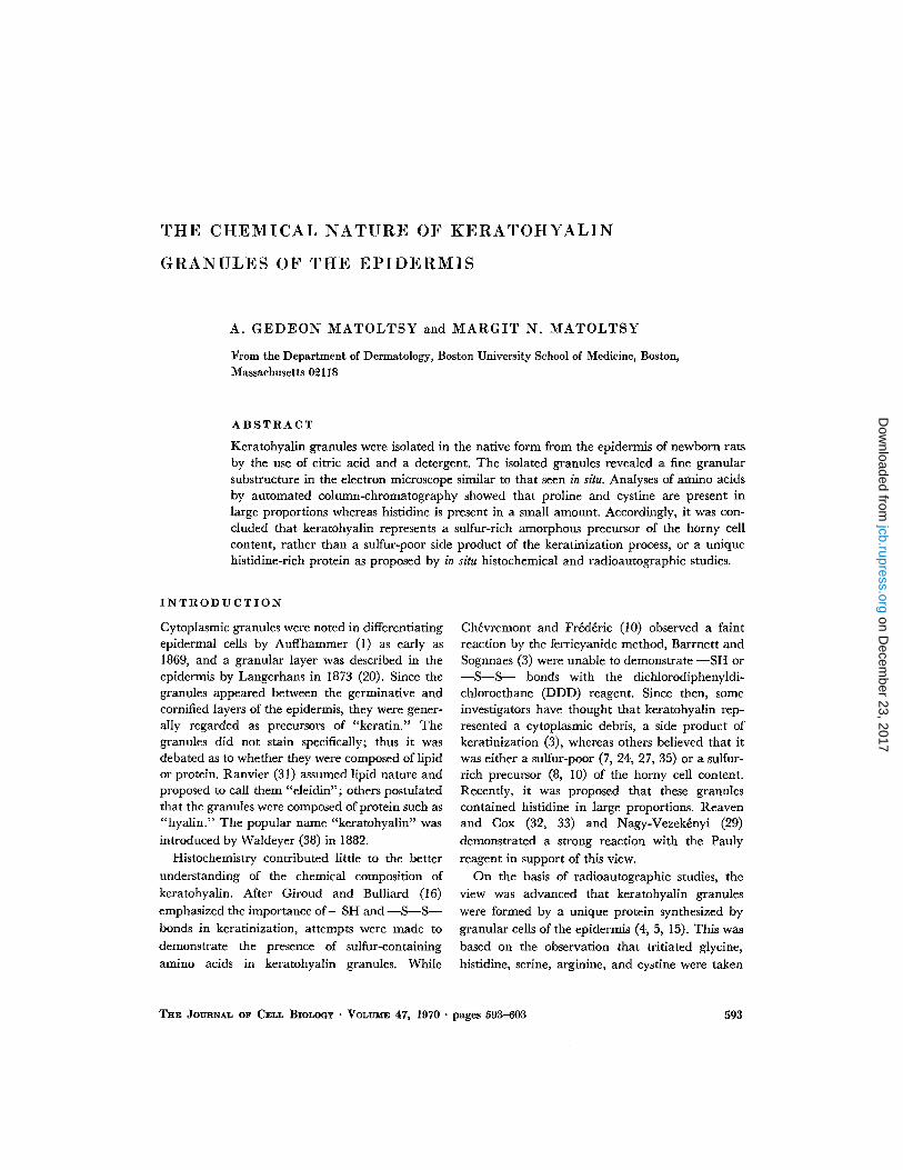
The epidermis of the newborn rat skin consists ofa single layer of basal cells, two layers of spinouscells, three to five layers of granular cells, and 10-12 layers of horny cells . Thus, about half or more ofthe nucleated cells are represented by granularcells in this epidermis (Fig. 1) . The lowermostgranular cells are relatively small and flat, andcontain many small and round keratohyalin gran-ules. The cells are somewhat larger in the midportion of the granular layer and comprise bothsmall and large granules. The upper granular cellsare filled with large granules and irregularlyshaped masses of keratohyalin .
A. GEDEON MATOLTSY AND MARGIT N. MATOLTSY Keratohyalin Granules of Epidermis
595
on Decem
ber 23, 2017jcb.rupress.org
Dow
nloaded from
FIGURE 1 Photomicrograph of the newborn rat epidermis . Note multiple layers of granular cells containingrelatively large and numerous keratohyalin granules . X 800.
FIGURE 2 The solubilizing effect of citric acid is demonstrated after 10 min treatment of the newborn rat skin .Note dissociation of the granular layer. X 800.
FIGURE 3 Remnants of the epidermis, including the stratum corneum and debris of the basal layer, are shownafter 1 hr treatment of the newborn rat skin with citric acid. X 800 .
FIGURE 4 Photomicrograph of a fresh preparation of isolated keratohyalin granules . Preparation was dispersedin saline under a cover glass. X 1600.
on Decem
ber 23, 2017jcb.rupress.org
Dow
nloaded from
Citric acid in 2% solution decomposes the non-cornified part of the epidermis as revealed by skinsections prepared from specimens after 10, 20, 30,and 60 min treatment. After 10 min, the hornylayer is separated from the rest of the skin at placeswhere citric acid has attacked the granular layer(Fig . 2) . After 10-60 min, large areas of the granu-lar layer, as well as portions of both basal andspinous layers, are removed whereas the hornylayer and the dermis are not affected (Fig . 3) .
Keratohyalin granules, isolated by the use ofcitric acid, are seen singly or in groups in smearpreparations (Fig. 4) . The granules are lightrefractile and appear isotropic in the polarizingmicroscope. When isolated granules are placed ina drop of 1.0 M phosphate buffer of pH 7, ethanol,pyridine, or dimethyl sulfoxide, they reveal nostructural changes . Dissolution, however, occursin strong acid and alkali solutions .
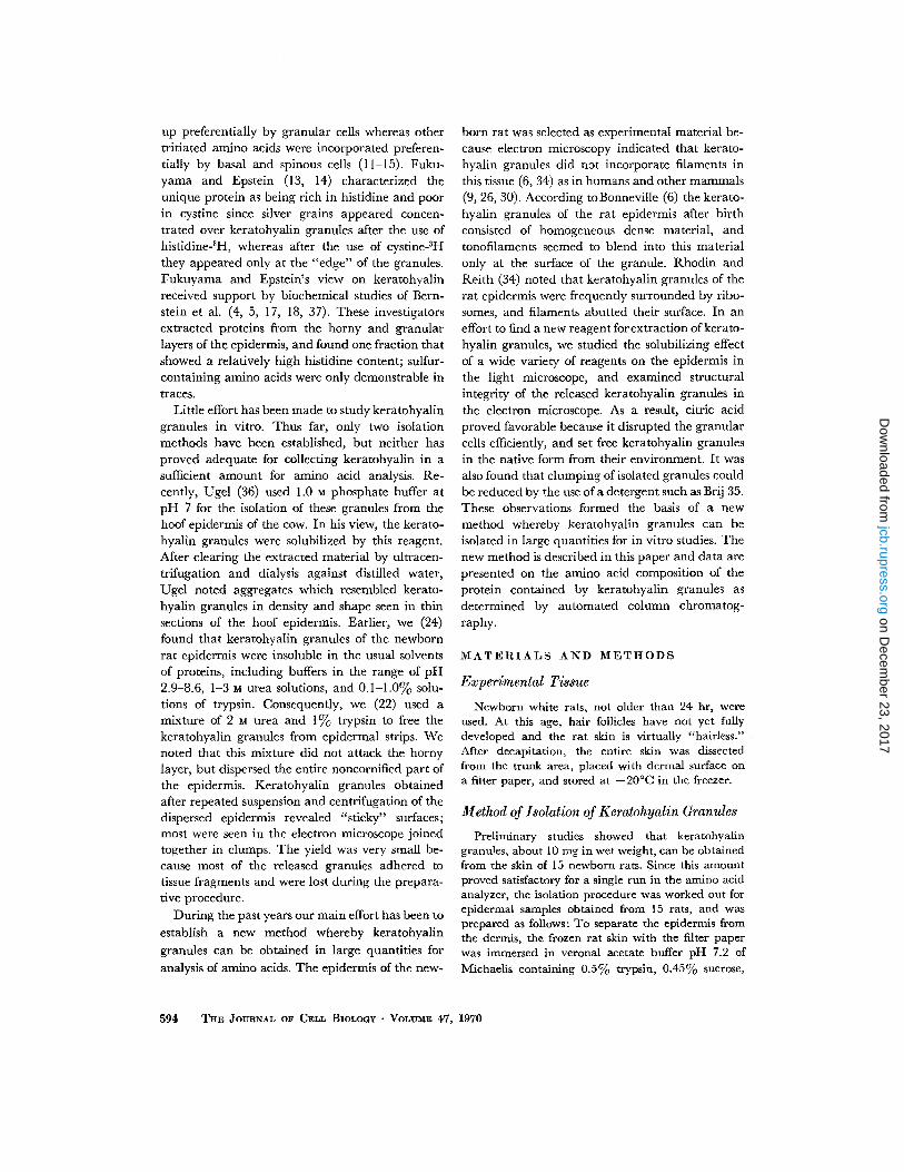
FIGURE 5 Part of the granular layer of the newborn rat epidermis. Note variable size of keratohyalin granulesand associations with ribosomes and filaments . X 12,200 .
FIGURE 6 Coalescing keratohyalin granules . Note granular substructure, close association with cytoplasmicfilaments and ribosomes, and the absence of a limiting membrane . X 66,600.
5 9 7
on Decem
ber 23, 2017jcb.rupress.org
Dow
nloaded from
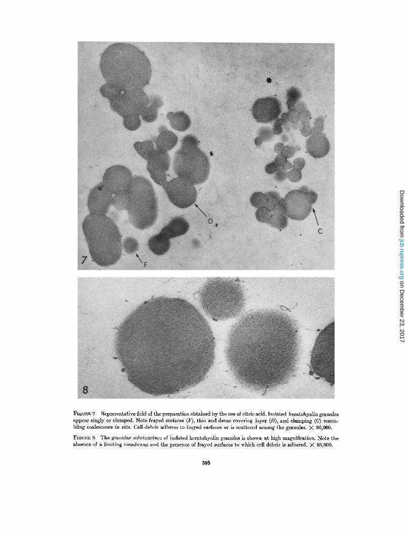
FIGURE 7 Representative field of the preparation obtained by the use of citric acid . Isolated keratohyalin granulesappear singly or clumped . Note frayed surfaces (F), thin and dense covering layer (D), and clumping (C) resem-bling coalescence in situ . Cell debris adheres to frayed surfaces or is scattered among the granules . X 36,000 .
FIGURE 8 The granular substructure of isolated keratohyalin granules is shown at high magnification . Note theabsence of a limiting membrane and the presence of frayed surfaces to which cell debris is adhered . X 83,300 .
59 8
on Decem
ber 23, 2017jcb.rupress.org
Dow
nloaded from
Electron Microscopy
In thin sections of the newborn rat epidermis,keratohyalin granules vary in size from about 100A to several micra (Fig . 5) . The small granules areround, the larger ones ovoid or irregular . Mostgranules are seen singly, some coalescing with oneanother (Fig. 6) . None of the keratohyalin granulesare limited by a membrane ; their surfaces are indirect contact with the cytoplasmic matrix . Ribo-somes and filaments often appear in their vicinityor attached to their surface . At low magnification,the keratohyalin granules reveal an amorphousstructure (Fig . 5), whereas at high magnification adensely packed granular substructure can beresolved (Fig. 6) .
A representative field of the preparation, ob-tained by the use of citric acid, is shown in Fig . 7 .It can be seen that isolated keratohyalin granulesappear singly or in clumps in thin sections ofpellets. Ribosomes or filaments are not seenattached or blended into the peripheral part of thegranules. The surface of most granules is frayed,revealing separation from filaments and othercytoplasmic components . Cell debris adheres tofrayed surfaces and may appear as a thin layeraround the granules. Neighboring granules either
TABLE IAmino Acid Composition of Isolated
Keratohyalin Granules
Residues per 1000 residues .
fuse or are separated by a thin layer of debris . Theisolated keratohyalin granules reveal a granularsubstructure at high magnification (Fig . 8) similarto that seen in situ (Fig. 6) .
Amino Acid AnalysisThe amino acid composition of the protein of
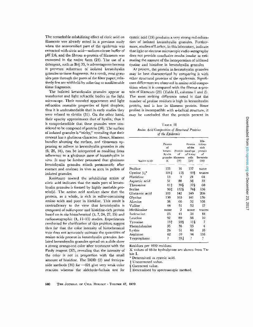
isolated keratohyalin granules is shown in Table I .The amino acids are listed in order of emergencefrom the column for 24-, 48-, and 72-hr hydroly-zates. It can be seen that threonine, serine, andtyrosine are degraded during longer periods ofhydrolysis whereas other amino acids are liberatedin increasing amounts as expected . The highestvalue is obtained for proline, corresponding to 133residues; half-cystine is represented by 108 resi-dues, i.e . a relatively large amount, and histidineby only 18 residues, i.e ., the smallest amount .
DISCUSSION
In the mammalian skin, keratohyalin granules arepresent in a small amount, and are not readilyaccessible for isolation, as they are located betweentwo highly insoluble layers such as the stratumcorneum and the dermis. The newborn rat ispreferable for the isolation of these granules be-cause keratohyalin is formed in this animal through-out the entire trunk epidermis in unusually largeamounts as revealed by multiple layers of granularcells and numerous large keratohyalin granules .Skin appendages, such as hair follicles and seba-ceous glands, have not yet fully developed, andsweat glands are absent. Consequently, the epi-dermis of newborn rats can be readily separatedfrom the dermis and obtained in "clean," kerato-hyalin-rich sheets. Reagents easily penetrate intoepidermal sheets through the base of the epidermis ;thus the keratohyalin granules are more accessiblefor isolation than from whole skin specimens . Al-though dissection of the skin is time consuming,and separation of the epidermis from the dermis istedious work, it is worth the effort because approxi-mately 100 mg in wet weight of keratohyalin gran-ules can be obtained from 150 newborn rats, anadequate amount for chemical analysis.
Citric acid is more suitable for isolation of ker-atohyalin granules than the urea-trypsin mixtureused in our previous studies (22) . This reagentrapidly sets free the granules and does not attacktheir structure. Release of the granules is primarilydue to effective solubilization of filament bundleswhich stabilize the cytoplasm of epidermal cells .
A. GEDEON MATOLTSY AND MARGIT N. MATOLTSY Keratohyalin Granules of Epidermis
599
Hydrolysis
Amino acids 24 hr 48 hr 72 hr
Cysteic acid 37.90 103 .51 108 .02Aspartic acid 48 .30 51 .12 55 .53Threonine 46 .25 40 .94 32 .49Serine 130 .02 90 .08 60 .39Glutamic acid 102 .03 103 .14 115 .95Proline 112 .62 132 .56 126 .46Glycine 123 .53 129 .90 125 .99Alanine 38 .41 38.17 40.08ValineCystineMethionine
40 .9423 .09none
43 .64nonenone
46 .58nonenone
Isoleucine 24 .47 25.45 26.89Leucine 88 .76 92.18 101 .01Tyrosine 50 .76 12 .91 6 .97Phenylalanine 33 .10 35.03 38.05Lysine 24 .98 26 .35 28 .37Histidine 12 .35 12 .57 17 .74Arginine 62 .49 62 .46 69.50
on Decem
ber 23, 2017jcb.rupress.org
Dow
nloaded from
The remarkable solubilizing effect of citric acid onfilaments was already noted in a previous studywhen the noncornified part of the epidermis wasextracted with citric acid-sodium-citrate buffer ofpH 2.6, and the fibrous a-protein of filaments wasrecovered in the native form (23) . The use of adetergent, such as Brij 35, is advantageous becauseit prevents adherence of isolated keratohyalingranules to tissue fragments . As a result, most gran-ules pass through the pores of the filter paper ; rela-tively few are withheld by adhering to nonfilterabletissue fragments .
The isolated keratohyalin granules appear astranslucent and light refractile bodies in the lightmicroscope . Their rounded appearance and lightrefraction resemble properties of lipid droplets ;thus it is understandable that in early studies theywere related to eleidin (31) . On the other hand,their opacity approximates that of hyalin ; thus itis comprehendible that these granules were con-sidered to be composed of protein (38) . The surfaceof isolated granules is "sticky," revealing that theircontent has a glutinous character . Hence, filamentbundles abutting the surface, and ribosomes ap-pearing to adhere to keratohyalin granules in situ(6, 26, 34), can be interpreted as resulting fromadherence to a glutinous mass of keratohyalin invivo . It may be further presumed that glutinouskeratohyalin granules attach permanently aftercontact and coalesce in vivo as seen in pellets ofisolated granules.
Resistance toward the solubilizing action ofcitric acid indicates that the main part of kerato-hyalin granules is formed by highly insoluble pro-tein(s) . The amino acid analyses show that theprotein, as a whole, is rich in sulfur-containingamino acids and poor in histidine . This result iscontradictory to the view that keratohyalin iscomposed of sulfur-poor and histidine-rich proteinbased on in situ histochemical (3, 7, 24, 27, 35) andradioautographic (4, 11-15) studies . Experimentsconducted for clarification of this problem suggestthus far that the color intensity of histochemicaltests does not accurately estimate the quantities ofamino acids present in keratohyalin granules . Iso-lated keratohyalin granules spread on a slide showa strong orange-red color after treatment with thePauly reagent (32), revealing that the intensity ofthe color is not in proportion with the smallamount of histidine. The DDD (2) and ferricya-nide methods (10) for -SH give very weak colorreaction whereas the aldehyde-fuchsin test for
600 THE JOURNAL OF CELL BIOLOGY . VOLUME 47, 1970
cysteic acid (19) produces a very strong red colora-tion of isolated keratohyalin granules . Further-more, studies of Lavker, in this laboratory, indicatethat light or electron microscopic radio autographydoes not provide conclusive results insofar as esti-mating the amount of the incorporation of tritiatedcystine and histidine in keratohyalin granules .
At present, the protein in keratohyalin granulesmay be best characterized by comparing it withother structural proteins of the epidermis . Signifi-cant differences are observed in amino acid compo-sition when it is compared with the fibrous a-pro-tein of filaments (23) (Table II, columns I and 2) .The most striking difference noted is that thenumber of proline residues is high in keratohyalinprotein, and is low in filament protein . Sinceproline is incompatible with a-helical structure, itmay be concluded that the protein present in
TABLE II
Amino Acid Composition of Structural Proteinsof the Epidermis
Residues per 1000 residues .X values of 48-hr hydrolyzate are shown from Ta-ble I .* Determined as cysteic acid .$ Uncorrected value .§ Corrected value .~~ Determined by spectroscopic method .
Amino acids
Proteinof
kerato-hyalin
granulesX
Proteinof
filaments(23)
Proteinof the
envelopeof horny
cells(25)
His-tidine-rich
proteinof
Bernstein(18)
Proline 133 16 137 noneCystine %* 104$ 1l§ 49§ tracesHistidine 13 9 21 64Aspartic acid 51 88 58 53Threonine 41$ 39§ 37§ 60Serine 90$ 112§ 74§ 156Glutamic acid 103 142 140 206Glycine 130 165 141 136Alanine 38 66 52 108Valine 44 51 52 12Methionine none 2 none tracesIsoleucine 25 41 33 18Leucine 92 89 58 10Tyrosine 13$ 22§ 11§ 7Phenylalanine 35 38 23 4Lysine 26 51 66 16Arginine 62 59 46 116Tryptophane ? 3311 ? ?
on Decem
ber 23, 2017jcb.rupress.org
Dow
nloaded from
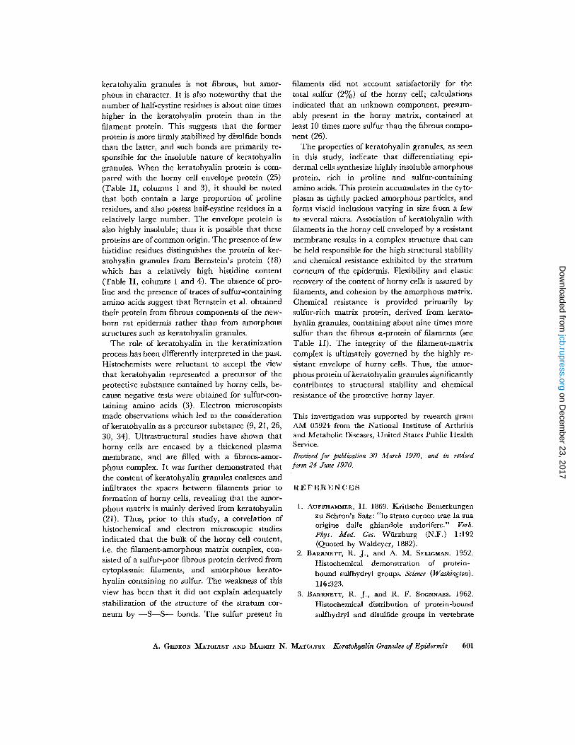
keratohyalin granules is not fibrous, but amor-phous in character. It is also noteworthy that thenumber of half-cystine residues is about nine timeshigher in the keratohyalin protein than in thefilament protein . This suggests that the formerprotein is more firmly stabilized by disulfide bondsthan the latter, and such bonds are primarily re-sponsible for the insoluble nature of keratohyalingranules . When the keratohyalin protein is com-pared with the horny cell envelope protein (25)(Table II, columns I and 3), it should be notedthat both contain a large proportion of prolineresidues, and also possess half-cystine residues in arelatively large number . The envelope protein isalso highly insoluble ; thus it is possible that theseproteins are of common origin . The presence of fewhistidine residues distinguishes the protein of ker-atohyalin granules from Bernstein's protein (18)which has a relatively high histidine content(Table II, columns 1 and 4) . The absence of pro-line and the presence of traces of sulfur-containingamino acids suggest that Bernstein et al . obtainedtheir protein from fibrous components of the new-born rat epidermis rather than from amorphousstructures such as keratohyalin granules .
The role of keratohyalin in the keratinizationprocess has been differently interpreted in the past .Histochemists were reluctant to accept the viewthat keratohyalin represented a precursor of theprotective substance contained by horny cells, be-cause negative tests were obtained for sulfur-con-taining amino acids (3) . Electron microscopistsmade observations which led to the considerationof keratohyalin as a precursor substance (9, 21, 26,30, 34) . Ultrastructural studies have shown thathorny cells are encased by a thickened plasmamembrane, and are filled with a fibrous-amor-phous complex. It was further demonstrated thatthe content of keratohyalin granules coalesces andinfiltrates the spaces between filaments prior toformation of horny cells, revealing that the amor-phous matrix is mainly derived from keratohyalin(21) . Thus, prior to this study, a correlation ofhistochemical and electron microscopic studiesindicated that the bulk of the horny cell content,i .e . the filament-amorphous matrix complex, con-sisted of a sulfur-poor fibrous protein derived fromcytoplasmic filaments, and amorphous kerato-hyalin containing no sulfur. The weakness of thisview has been that it did not explain adequatelystabilization of the structure of the stratum cor-neum by -S-S- bonds. The sulfur present in
filaments did not account satisfactorily for thetotal sulfur (2%) of the horny cell ; calculationsindicated that an unknown component, presum-ably present in the horny matrix, contained atleast 10 times more sulfur than the fibrous compo-nent (26) .
The properties of keratohyalin granules, as seenin this study, indicate that differentiating epi-dermal cells synthesize highly insoluble amorphousprotein, rich in proline and sulfur-containingamino acids. This protein accumulates in the cyto-plasm as tightly packed amorphous particles, andforms viscid inclusions varying in size from a fewto several micra . Association of keratohyalin withfilaments in the horny cell enveloped by a resistantmembrane results in a complex structure that canbe held responsible for the high structural stabilityand chemical resistance exhibited by the stratumcorneum of the epidermis . Flexibility and elasticrecovery of the content of horny cells is assured byfilaments, and cohesion by the amorphous matrix .Chemical resistance is provided primarily bysulfur-rich matrix protein, derived from kerato-hyalin granules, containing about nine times moresulfur than the fibrous a-protein of filaments (seeTable II). The integrity of the filament-matrixcomplex is ultimately governed by the highly re-sistant envelope of horny cells. Thus, the amor-phous protein of keratohyalin granules significantlycontributes to structural stability and chemicalresistance of the protective horny layer .
This investigation was supported by research grantAM 05924 from the National Institute of Arthritisand Metabolic Diseases, United States Public HealthService .Received for publication 30 March 1970, and in revisedform 24 June 1970 .
REFERENCES
1 . AUFFHAMMER, H. 1869 . Kritische Bemerkungenzu Schron's Satz : "lo strato corneo trae la suaorigine dalle ghiandole sudorifere ." Verh .Phys. Med . Ges . Würzburg (N.F .) 1 :192(Quoted by Waldeyer, 1882) .
2 . BARRNETT, R. J., and A. M . SELIGMAN . 1952 .Histochemical demonstration of protein-bound sulfhydryl groups . Science (Washington) .
116 :323 .3 . BARRNETT, R . J., and R. F . SOGNNAES . 1962 .
Histochemical distribution of protein-boundsulfhydryl and disulfide groups in vertebrate
A. GEDEON MATOLTSY AND MARGIT N. MATOLTSY Keratohyalin Granules of Epidermis
601
on Decem
ber 23, 2017jcb.rupress.org
Dow
nloaded from
keratin. In Fundamentals of Keratinization .E. O. Butcher and R . F. Sognnaes, editors .American Association for the Advancement ofScience, Washington, D .C . 27 .
4. BERNSTEIN, I. A. 1964. Relation of the nucleicacids to protein synthesis in the mammalianepidermis . In The Epidermis. W. Montagnaand W. C. Lobitz, editors . Academic PressInc., New York . 471 .
5 . BERNSTEIN, I. A. 1967 . Synthesis of unique pro-teins in epidermal keratinization. Proc . Int.Congr . Dermatol., 13th . 9 :1008 .
6 . BONNEVILLE, M. A. 1968 . Observations on epi-dermal differentiation in the fetal rat . Amer.J. Anal . 123 :147 .
7. BRAUN-FALCO, O . 1958 . The histochemistry ofpsoriasis . Ann. N. Y. Acad. Sci. 73 :936 .
8. BRODY, I . 1959 . The keratinization of epidermalcells of normal guinea pig skin as revealedby electron microscopy . J. Ultrastruct. Res .2:482.
9. BRODY, I . 1964. Different staining methods forthe electron microscopic elucidation of thetonofibrillar differentiation in normal epi-dermis. In The Epidermis. W. Montagna andW. C. Lobitz, editors. Academic Press Inc .,New York. 251 .
10. CHÈVREMONT, M ., and J . FRÉDÉRIC . 1943 . Unenouvelle méthode histochimique de mise enévidence des substances à fonction sulfhydrile .Application à l'épiderme, au poil et à lalevure . Arch . Biol. (Liège) . 54 :589 .
11 . Cox, A . J ., and E. P. REAVEN . 1967 . Histidineand keratohyalin granules . J. Invest. Dermatol.49 :31 .
12. FUKUYAMA, K., and W. L. EPSTEIN . 1966. Epi-dermal keratinization : Localization of iso-topically labeled amino acids . J. Invest. Der-matol. 47:551 .
13. FUKUYAMA, K., and W . L . EPSTEIN . 1967 . Ultra-structural autoradiographic studies of kerato-hyalin granule formation . J . Invest . Dermatol .49:595 .
14. FUKUYAMA, K., and W . L. EPSTEIN . 1969 . Sul-fur-containing proteins and epidermal kera-tinization . J. Cell Biol . 40 :830 .
15. FUKUYAMA, K., T. NAKAMURA, and I. A. BERN-STEIN . 1965 . Differentially localized incorpora-tion of amino acids in relation to epidermalkeratinization in the newborn rat. Anat. Rec .152 :525 .
16. GIROUD, A., and H . BULLIARD . 1930 . La kera-tinization de l'épiderme et des phanères .Arch. morphol . gen . exp. 29 :1 .
17. GUMUCIO, J., C . FELDKAMP, and I. A. BERN-
STEIN. 1967 . Studies on localization of "histi-
6 0 2
THE JOURNAL OF CELL BIOLOGY . VOLUME 47, 1970
dine-rich" peptide material present in epi-dermis of the newborn rat . J. Invest . Dermatol.49:545.
18. HOOBER, J . K., and I. A. BERNSTEIN . 1966 .Protein synthesis related to epidermal differ-entiation. Proc. Nat. Acad. Sci. U.S.A . 56 :594 .
19. LANDING, B . H., H. E. HALL, and C. D. WEST.1956. Aldehyde-fuchsin-positive material ofthe posterior pituitary . Lab. Invest . 5 :256 .
20. LANGERHANS, P . 1873. Ueber Tatkörperchenand rete Malpighii . Arch. Mikrosk . Anat . Ent-wicklungsmech. 9:730 .
21 . LAVKER, R. M., and A . G. MATOLTSY . 1970.Formation of horny cells. The fate of cellorganelles and differentiation products inruminai epithelium. J. Cell Biol . 44 : 501 .
22. MATOLTSY, A. G. 1962. Mechanism of kera-tinization. In Fundamentals of Keratinization.E. O. Butcher and R. F. Sognnaes, editors .American Association for the Advancement ofScience, Publ. No. 70, Washington, D.C .
23. MATOLTSY, A. G. 1965. Soluble prekeratin. InBiology of the Skin and Hair Growth . A. G .Lyne and B. F. Short, editors . Angus andRobertson Ltd ., Sydney, Australia. 291 .
24. MATOLTSY, A. G., and M . MATOLTSY. 1962. Astudy of morphological and chemical prop-erties of keratohyalin granules . J. Invest .Dermatol . 38:237 .
25. MATOLTSY, A. G., and M. MATOLTSY . 1966 .The membrane protein of horny cells. J.Invest . Dermatol. 46:127.
26. MATOLTSY, A. G., and P. F. PARAKKAL. 1967 .Keratinization . In Ultrastructure of Normaland Abnormal Skin. A. S . Zelickson, editor .Lea and Febiger, Philadelphia. 76 .
27. MONTAGNA, W., A. Z . EISEN, A. H. RADE-MACHER, and H. B. CHASE . 1954. Histologyand cytochemistry of human skin. V I. Thedistribution of sulfhydryl and disulfide groups .J. Invest . Dermatol . 23 :23 .
28 . MOORE, S. 1963 . On the determination of cys-tine as cysteic acid . J. Biol . Chem . 238 :235 .
29. NAGY-VEZEKÉNYI, C . 1969. On the histidinecontent of human epidermis . Brit. J. Derma-tol . 81 :685 .
30. ODLAND, G . F. 1964 . Tonofilaments and kera-tohyalin . In The Epidermis. W. Montagnaand W. C. Lobitz, editors. Academic PressInc., New York . 237 .
31 . RANVIER, L . 1884 . De l'éléidine et de la réparti-tion de cette substance dans la peau, la mu-queuse buccale et la muqueuse oesophagiennedes vertébrés. Arch. Physiol. Norm . Pathol., Ser .III. 3 :125 .
32. REAVEN, E. P., and A . J . Cox. 1963 . The histo-
on Decem
ber 23, 2017jcb.rupress.org
Dow
nloaded from
chemical localization of histidine in the hu-man epidermis and its relationship to zincbinding . J. Histochem. Cytochem. 11 :782 .
33 . REAVEN, E. P., and A . J . Cox. 1965 . Histidineand keratinization . J. Invest . Dermatol . 45 :422.
34. RHODIN, J . A . G., and E . J. REITH. 1962 . Ultra-structure of keratin in oral mucosa, skin,esophagus, claw, and hair. In Fundamentalsof Keratinization. E. O. Butcher and R . F .Sognnaes, editors . American Association forthe Advancement of Science, Washington,D.C. 61 .
35 . SZODORAY, L ., and C. NAGY-VEZEKfNYI . 1964 .
Histochemical studies of keratohyalin in hu-man epidermis . J. Invest . Dermatol. 42 :157 .
36 . UGEL, A. R . 1969 . Keratohyalin : Extractionand in vitro aggregation . Science (Washington) .166:250 .
37 . VOORHEES, J . J ., S . G . CHAKRABARTI, and I . A .BERNSTEIN. 1968. The metabolism of "histi-dine-rich" protein in normal and psoriatickeratinization . J. Invest . Dermatol . 51 :344.
38. WALDEYER, W . 1882 . Untersuchungen ueberdie Histogenese der Horngebilde, besondersder Haare und Federn . Beitr . Anat. Embryol.(Festschrift für Henle) . 141 .
A . GEDEON MATOLTSY AND MARGIT N . MATOLTSY Keratohyalin Granules of Epidermis 603
on Decem
ber 23, 2017jcb.rupress.org
Dow
nloaded from