seeing the invisible injurybraininjurycanada.ca/wp-content/uploads/2011/09/paulkelly_biac... ·...
TRANSCRIPT
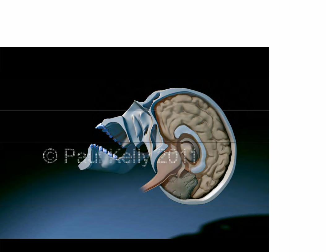
ConcussionsConcussionsSeeing the Invisible Injury
A three dimensional visualization of sport relatedA three-dimensional visualization of sport-related concussion.
Paul Kelly BSc Kinesiology MScBMCPaul Kelly, BSc Kinesiology, MScBMCBiomedical Communications
University of Torontoy
This presentation is copyright © Paul Kelly 2011. All rights reserved.

Today you will learn about:Today you will learn about:
The field of Biomedical CommunicationsThe field of Biomedical Communications
Published works referenced for the creation of aPublished works referenced for the creation of a 3D animation on concussions
Methods and tools used to create a biomedical 3D animation3 a at o
What is Biomedical Communications?What is Biomedical Communications?
What is Biomedical Communications?What is Biomedical Communications?
iomedical Communications is rooted iniomedical Communications is rooted in Medical Illustration, the study of art as
li d t di ipplied to medicine.
What is Biomedical Communications?What is Biomedical Communications?
he Biomedical Communications program at the p gniversity of Toronto is one of five accredited ograms in North America, the only such program in anada, for the study of communicating science and ealth messages through the use of visual media.

o see examples of student work from the Biomedical Communications program at the University of Toronto, please visit us at
http://www.bmc.med.utoronto.ca/bmc/index.php

What do Medical Illustrators and Bi di l C i t d ?Biomedical Communicators do?
Illustrations for print– Medical textbooks and scientific journals– Patient education resources– Medical-Legal
2D and 3D animations
Web-based learning modules
C t i d t i i i l tiComputerized training simulations
Art direction and project management

One organization that is developing cutting-edge interactive media is the erioperative Interactive Education team at Toronto General Hospital. Have a
look at http://pie.med.utoronto.ca
What are Medical Illustrators / Biomedical C i t ?Communicators?
rom the Association of Medical Illustrators website:
Professional artists with advanced education in both the life sciences and visual communications.
Visual problem solversVisual problem solvers
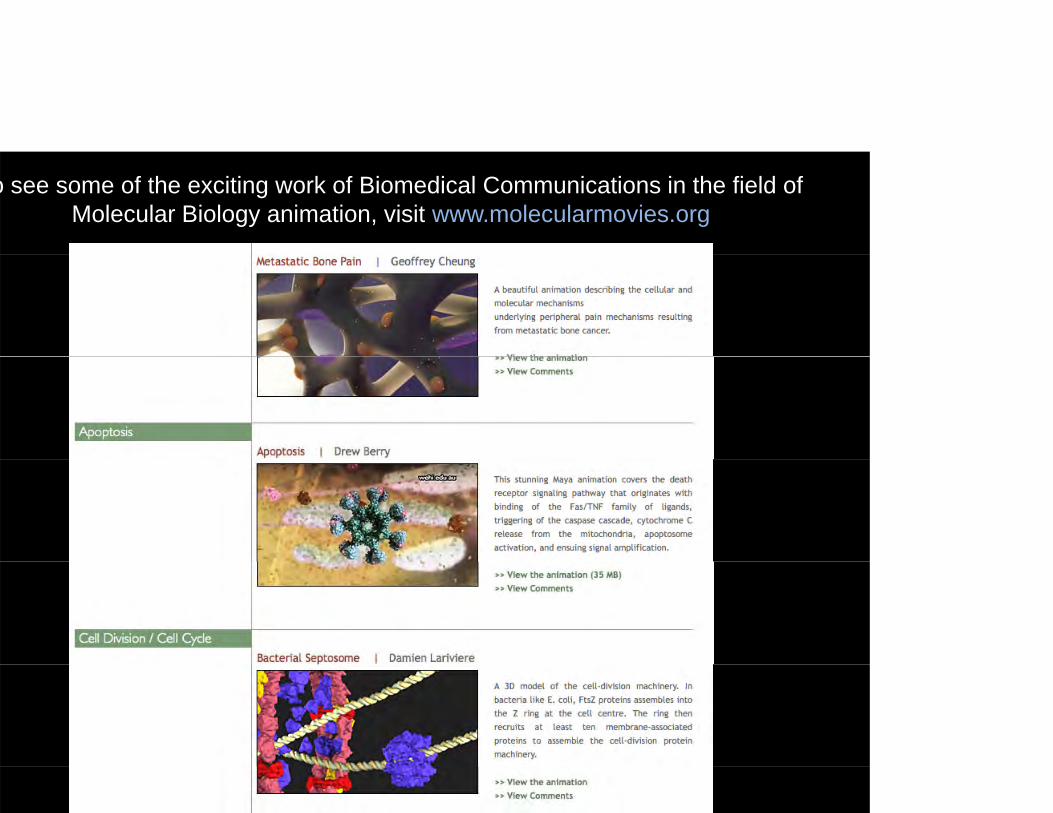
o see some of the exciting work of Biomedical Communications in the field of Molecular Biology animation, visit www.molecularmovies.org
Master’s Research ProjectMaster s Research Project
The Problem: Concussions in sportsThe Problem: Concussions in sports
I ll i t t i hi h t f ildIn collegiate sports, concussions, which are a type of mild Traumatic Brain Injury or mTBI, may constitute the most common form of injury. Many retired professional athletes in high-impact sports such as hockey and football have gone public about their p y g phealth problems suspected to be connected with concussions that took place during their careers, and some have gotten actively involved in head injury research.
What is a concussion?What is a concussion?
Concussion:Concussion:A concussion is a complex pathophysiologicalprocess affecting the brain induced by traumaticprocess affecting the brain, induced by traumatic biomechanical forces.
–International Conference on Concussion–International Conference on Concussion in Sport

Master’s Research ProjectMaster s Research Project
The Problem:The Problem:➡ Concussions in sports
➥ Attitude towards concussions
What much of literature in this area has acknowledged is that the ncidence of, reluctance in reporting, and challenges in assessment of concussions are all compounded by the inherent mentality ofof concussions are all compounded by the inherent mentality of sports culture that until very recently viewed concussions as an nsignificant injury, oftentimes not even as an injury at all.
Master’s Research ProjectMaster s Research Project
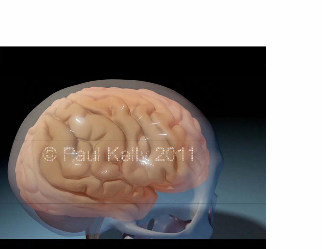
o challenge this attitude, we developed a gh-end 3D animation that would show ghat is not readily visible during a oncussion–what is happening to the brainoncussion what is happening to the brain side the skull during an impact, and how
his affects brain cells at the cellular levelhis affects brain cells at the cellular level.
Master’s Research ProjectMaster s Research Project
3-minute 3D animation depicting the3 minute 3D animation depicting the biomechanics and pathophysiology of concussions
Target audience: athletic coaches (lay public)g ( y p )
Project began in summer of 2010, final oject bega su e o 0 0, aanimation was completed in summer 2011. Roughly ~676 total work hours.
Master’s Research ProjectMaster s Research Project
A compelling interpretive visualizationA compelling interpretive visualization informed by empirical evidence but d i d t i tdesigned to communicate.
RESULTSRESULTS
THE ANIMATION…
At this point I'd like to depart slightly from the traditional format and go right to my results, which of course is the final animation itself. Then I'll describe how I made many of the visual decisions that went into designing this project and having seen this finished work you'll have a better understanding of what it is I'm describingdescribing.
Cli k h t t th i ti

MRP Advisory CommitteeMRP Advisory Committee
A multidisciplinary teamA multidisciplinary team

MRP Advisory CommitteeMRP Advisory CommitteeNicholas Woolridge, BFA, BScBMC, MScBMC, MSc,
CMICMI Director & Associate Professor, Biomedical Communications Department of Biology University of Toronto Mississaugay g
Doug Richards, MD, DipSportMedAssistant Professor, Faculty of Physical Education and Health Medical Director David L MacIntosh Sport Medicine ClinicMedical Director, David L. MacIntosh Sport Medicine ClinicUniversity of Toronto
Anne Agur, BScOT, MSc, PhDg , , ,Institute of Medical Science (IMS)Graduate Department Rehabilitation Sciences (GDRS)University of Toronto
Potential for visualizationPotential for visualization
s we began designing the content of thes we began designing the content of the nimation we identified three main
t f TBI th t b i li dspects of mTBIs that can be visualized.
Potential for visualizationPotential for visualization
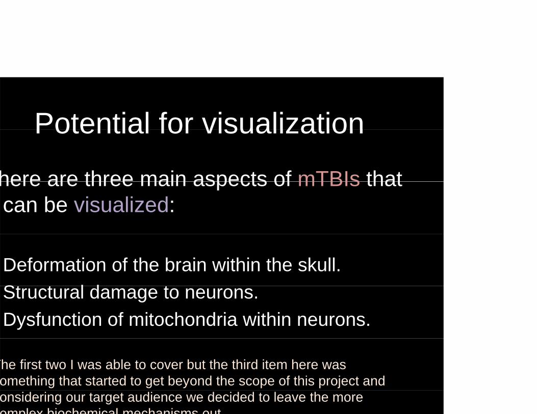
here are three main aspects of mTBIs thathere are three main aspects of mTBIs that can be visualized:
Deformation of the brain within the skull.St t l d tStructural damage to neurons.Dysfunction of mitochondria within neurons.
The first two I was able to cover but the third item here was omething that started to get beyond the scope of this project and onsidering our target audience we decided to leave the more omplex biochemical mechanisms out
Current Visualizations
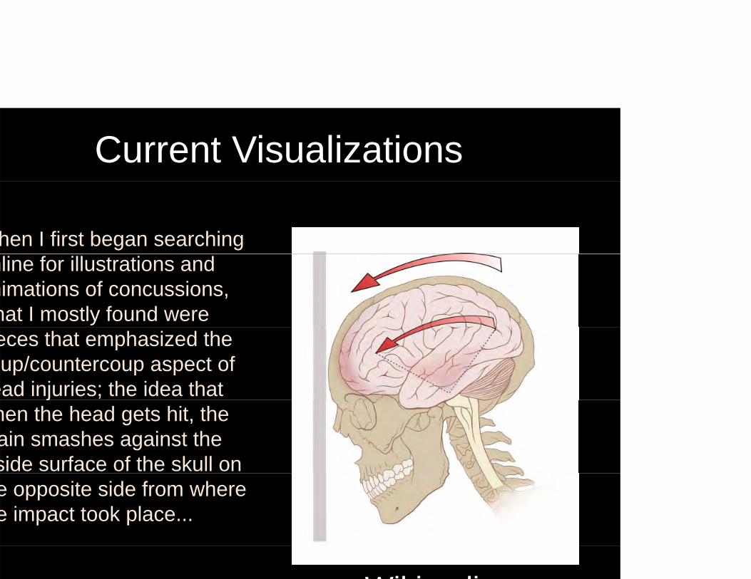
hen I first began searching nline for illustrations and nimations of concussions, hat I mostly found were eces that emphasized the up/countercoup aspect of
ead injuries; the idea that jhen the head gets hit, the ain smashes against the side surface of the skull on e opposite side from where e impact took place...
Wiki di
Limitations of current visualizationsLimitations of current visualizations
Response of brain tissue emphasize coup /Response of brain tissue emphasize coup / countercoup
ut through discussions with Dr. Richards and the prevailing research had come in contact with, I felt that this aspect should be ownplayed in comparison to the aspect of rotational acceleration andownplayed in comparison to the aspect of rotational acceleration and esulting tissue deformation.
Limitations of current visualizationsLimitations of current visualizations
Response of brain tissue emphasize coup /Response of brain tissue emphasize coup / countercoup
Context has been Med Legal / automotiveContext has been Med-Legal / automotive
Most of the visual resources available at the time of my initial literature i th d t f M d L l i th t d d k i threview were the products of Med-Legal companies that produced work in the
context of automotive crashes, and in these types of collisions we have a typical set of biomechanics involved. Typically either forward-backward (shaking head “yes”) or side-to-side (ears touching shoulders), not so much ( g y ) ( g ),twisting about the neck (shaking head “no”).
n sport-related concussions however we see a lot more of these axial head rotations and that's a visualization that I found both difficult to come acrossrotations and that s a visualization that I found both difficult to come across and one that is best demonstrated in 3D animation as opposed to 2D.
Cli k h t t thi id
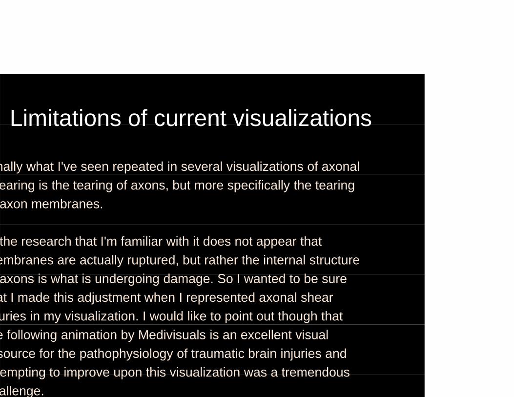
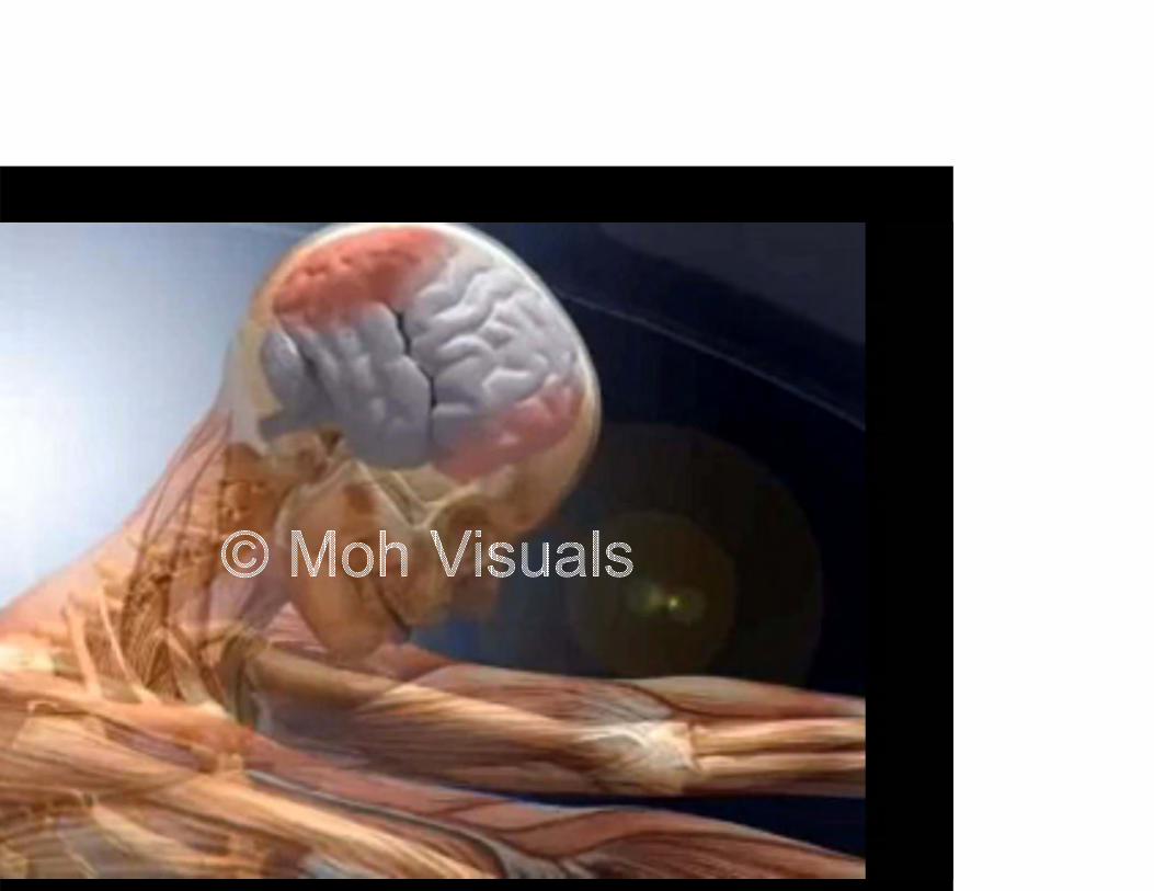
Limitations of current visualizationsLimitations of current visualizations
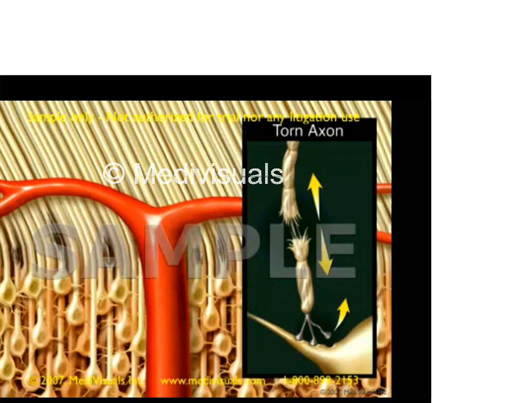
nally what I've seen repeated in several visualizations of axonal earing is the tearing of axons, but more specifically the tearing axon membranes.
the research that I'm familiar with it does not appear that embranes are actually ruptured, but rather the internal structure
i h t i d i d S I t d t baxons is what is undergoing damage. So I wanted to be sure at I made this adjustment when I represented axonal shear uries in my visualization. I would like to point out though that e following animation by Medivisuals is an excellent visual source for the pathophysiology of traumatic brain injuries and empting to improve upon this visualization was a tremendousempting to improve upon this visualization was a tremendous allenge.
Cli k h t t thi id

Limitations of current visualizationsLimitations of current visualizations
Response of brain tissue emphasize coup /Response of brain tissue emphasize coup / countercoup
Context has been Med Legal / automotiveContext has been Med-Legal / automotive
Damage to neurons suggests different fmechanism of injury
LITERATURE REVIEWLITERATURE REVIEW
Visual strategies to enhance learningVisual strategies to enhance learning
C i t & di iConcussion assessment & diagnosis
Brain response to impact
Axonal shear injuries
LITERATURE REVIEWLITERATURE REVIEW
Visual strategies to enhance learningVisual strategies to enhance learning
Visual strategies to enhance learning is the core of what is taught in the BMC curriculum, so this portion of my literature review was not as extensive as other areas because much of this materialnot as extensive as other areas because much of this material came to me through regular coursework. But I needed to look at some research that was specific to my project, so I looked for papers that dealt with using animation for teaching and p p g grepresenting anatomy in 3D.
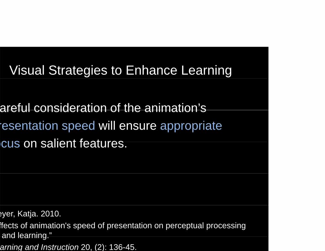
Visual Strategies to Enhance Learning
areful consideration of the animation’sareful consideration of the animation s resentation speed will ensure appropriate ocus on salient featuresocus on salient features.
eyer, Katja. 2010. ffects of animation's speed of presentation on perceptual processing and learning ”and learning. arning and Instruction 20, (2): 136-45.

Visual Strategies to Enhance Learning
n effective approach for learning spatialn effective approach for learning spatial elationships in anatomy involves a gradual onstruction of anatomical featuresonstruction of anatomical features.
ller, Robert. 2000. pproaches to learning spatial relationships in gross anatomy: Perspective from wider principles of learning ”Perspective from wider principles of learning.
inical Anatomy 13, (6): 439-43.


LITERATURE REVIEWLITERATURE REVIEW
Concussion assessment & diagnosisConcussion assessment & diagnosis
When I began my literature review I knew that there is a growing awareness e bega y e a u e e e e a e e s a g o g a a e essn professional sports about the dangers and risks associated with concussive head injuries.
Many organizations have responded to this by implementing return to playMany organizations have responded to this by implementing return-to-play guidelines intended to keep concussed players out of competition until symptoms resolve.
The criteria listed in these return-to-play guidelines are based on clinical research of the epidemiology of mild traumatic brain injuries and undergo continuous revision as research in this area advances.
Concussion Assessment & DiagnosisConcussion Assessment & Diagnosis
cCrea, Michael, Kevin Guskiewicz, Stephen Marshall, William Barr, Christopher Randolph, Robert Cantu, James Onate, Jingzhen Yang, and James Kelly. 2003.
cute effects and recovery time following concussion in collegiate football players: The NCAA concussion study.”
urnal of the American Medical Association (JAMA) 290, (19): 2556-63.
Concussion Assessment & DiagnosisConcussion Assessment & Diagnosis
Players with concussion exhibit morePlayers with concussion exhibit more evere symptoms, cognitive impairment,
d b l bl i di t l ftnd balance problems immediately after oncussion. On average, symptoms radually resolve by day 7.”
–McCrae et. al. 2003

Concussion Assessment & DiagnosisConcussion Assessment & Diagnosis
Standardized measurement ofStandardized measurement of ost-concussive symptoms, cognitive
ti i d t l t bilitunctioning, and postural stability may nhance clinical management of athletes ecovering from concussion.”
–McCrae et. al. 2003

Concussion Assessment & DiagnosisConcussion Assessment & Diagnosis
uskiewicz, Kevin M. Ph.D.; Mihalik, Jason P. M.S.; Shankar, Viswanathan M.Sc.; Marshall, Stephen W. Ph.D.; Crowell, Dean H. M.A.; Oliaro, Scott M. M.A.; Ciocca, Mario F. M.D.; Hooker, Daniel N. Ph.D., P.T.easurement of Head Impacts in Collegiate Football Players: Relationship Between Head Impact Biomechanics and Acute Clinical Outcome After Concussion”
eurosurgery. 2007 Dec;61(6):1244-52; discussion 1252-3.
Concussion Assessment & DiagnosisConcussion Assessment & Diagnosis
ifficulty in establishing a threshold for y goncussive injury:
Varying magnitude of impactVarying magnitude of impact
Varying location of impact
F f b i i tFrequency of sub-concussive impacts
Number of previous concussions
–Guskiewicz, et. al. 2007
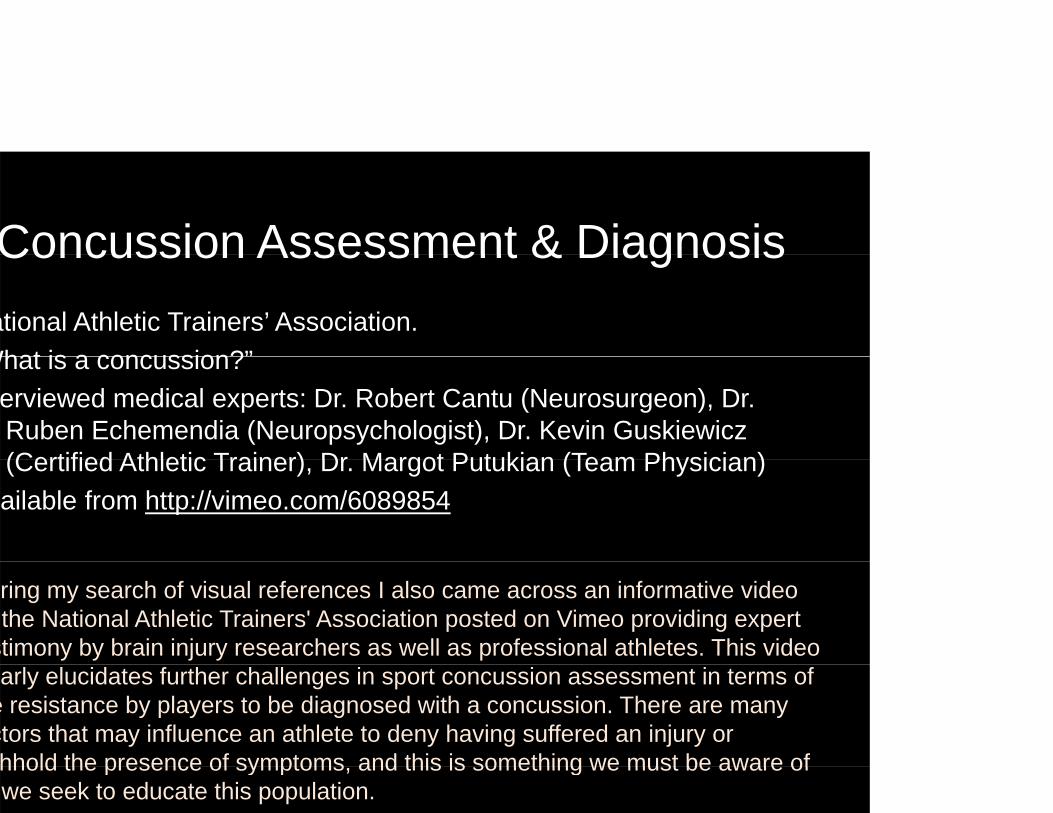
Concussion Assessment & DiagnosisConcussion Assessment & Diagnosisational Athletic Trainers’ Association.
What is a concussion?”What is a concussion?erviewed medical experts: Dr. Robert Cantu (Neurosurgeon), Dr. Ruben Echemendia (Neuropsychologist), Dr. Kevin Guskiewicz(Certified Athletic Trainer) Dr Margot Putukian (Team Physician)(Certified Athletic Trainer), Dr. Margot Putukian (Team Physician)ailable from http://vimeo.com/6089854
ring my search of visual references I also came across an informative video the National Athletic Trainers' Association posted on Vimeo providing expert
stimony by brain injury researchers as well as professional athletes. This video arly elucidates further challenges in sport concussion assessment in terms of
e resistance by players to be diagnosed with a concussion. There are many ctors that may influence an athlete to deny having suffered an injury or hhold the presence of symptoms and this is something we must be aware ofhhold the presence of symptoms, and this is something we must be aware of we seek to educate this population.
Click screenshot to see this ideo
© National Athletic Trainers’ AssociationClick screenshot to see this video

Concussion Assessment & DiagnosisConcussion Assessment & Diagnosis
eluctance/avoidance of reporting.p g
Maintaining status as tough and resilient
Competition for game time
Drive to earn scholarships/salary
Viewing injuries as a sign of weakness
Underestimating potential risks
–N.A.T.A.

Health Behavior ModelsHealth Behavior Models
ow perception of risk and lack of informationow perception of risk and lack of informationontributed to unhealthy behavior choices.
ermeni, E., C. Lionis, B. Davou, and E. Th Petridou. 2009. nderstanding Reasons for Non-Compliance in Motorcycle Helmet Use Among Adolescents in Greece.” g
ury Prevention 15, (1) (Feb): 19-23.

TARGET AUDIENCE: COACHES
ere was also a study done in 2010 by the CDC to evaluate the f i i f i k d i d f hi h h le of a concussion information packet designed for high school
hletic coaches, and this was where I determined what my target dience would be. As team leaders, coaches are responsible for e welfare of their athletes they are the main conduit ofe welfare of their athletes, they are the main conduit of ormation for players regarding safety and also serve as the ntral authority figure of the team. This study has indicated that aches are receptive to supplementary materials to educateaches are receptive to supplementary materials to educate hletes and parents about concussions. Providing visual evidence the rationale behind return-to-play guidelines may help provide aches with a resource to increase adherence and awareness and
stify taking an athlete out of play whenever that becomes cessary.
TARGET AUDIENCE: COACHES
oaches face many difficulties in theoaches face many difficulties in the management of mTBIs, and are receptive o materials that may assist them ino materials that may assist them in ommunicating risks and severity of mTBIso parents and players.
rmiento, Kelly, Jane Mitchko, Cynthia Klein, and Sharon Wong. 2010. aluation of the Centers for Disease Control and Prevention's Concussion Initiative for High School Coaches: “Heads Up: Concussion in High School S t "Sports".
e Journal of School Health 80 (3): 112-8
LITERATURE REVIEWLITERATURE REVIEW
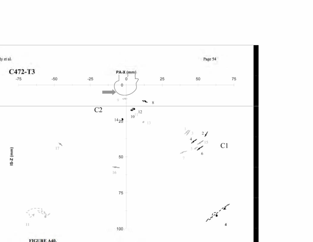
Brain response to head impactsBrain response to head impactshile attending the Annual Injury Biomechanics Symposium inhile attending the Annual Injury Biomechanics Symposium in olumbus Ohio, I saw an outstanding presentation by Dr. Warren ardy. His research involved using high-speed, biplane X-rays to
d th t f t l d it t t b dd d i thcord the movements of neutral-density targets embedded in the ains of cadaver heads.
hat Dr. Hardy and his colleagues attempted was to see inside the ain during a high-velocity impact. The results from this study ve me a great start on how I wanted my brain deformation shotsve me a great start on how I wanted my brain deformation shots look like.
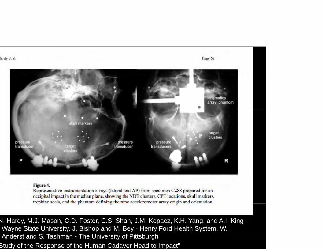
N. Hardy, M.J. Mason, C.D. Foster, C.S. Shah, J.M. Kopacz, K.H. Yang, and A.I. King -Wayne State University. J. Bishop and M. Bey - Henry Ford Health System. W. A d t d S T h Th U i it f Pitt b hAnderst and S. Tashman - The University of Pittsburgh
Study of the Response of the Human Cadaver Head to Impact”

Brain response to head impacts
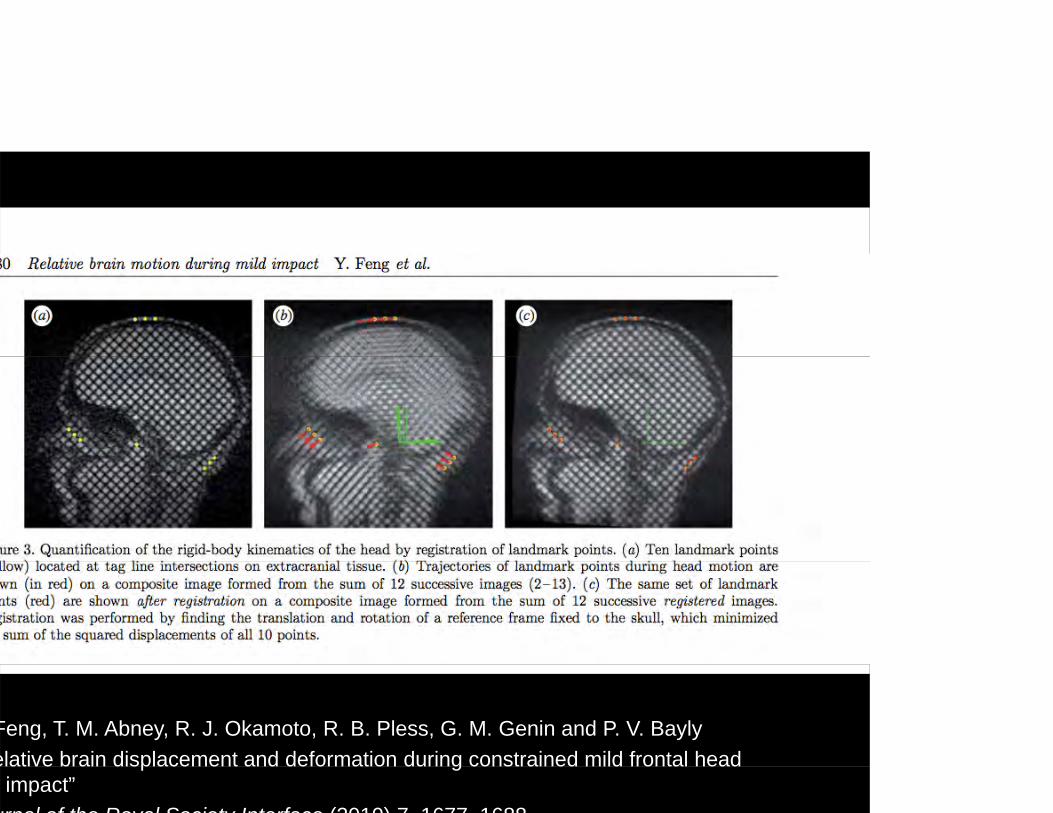
I continued my search in this niche area of research, I came ross a series of papers from the University of St. Louis. They ve conducted several studies involving subjects in MRIs. The bjects were placed in the MRIs with a specialized apparatus thatbjects were placed in the MRIs with a specialized apparatus that oduced sudden, quick head movements, but nothing too close the accelerations and decelerations associated with head
i Th t di bl t i t furies. These studies were able to acquire measurements of ain deformation in living human subjects and map these anges with tagged MRI images. The results from these studies
ere my reference for how I depicted the patterns of brain formation.
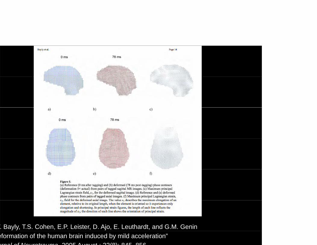
V. Bayly, T.S. Cohen, E.P. Leister, D. Ajo, E. Leuthardt, and G.M. Genineformation of the human brain induced by mild acceleration”urnal of Neurotrauma 2005 August ; 22(8): 845 856
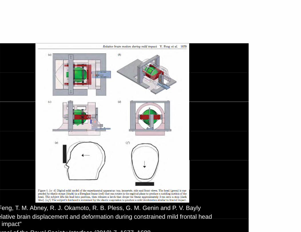
Feng, T. M. Abney, R. J. Okamoto, R. B. Pless, G. M. Genin and P. V. Baylyelative brain displacement and deformation during constrained mild frontal head p g
impact”urnal of the Royal Society Interface (2010) 7 1677 1688
Feng, T. M. Abney, R. J. Okamoto, R. B. Pless, G. M. Genin and P. V. Baylyelative brain displacement and deformation during constrained mild frontal head p g
impact”urnal of the Royal Society Interface (2010) 7 1677 1688
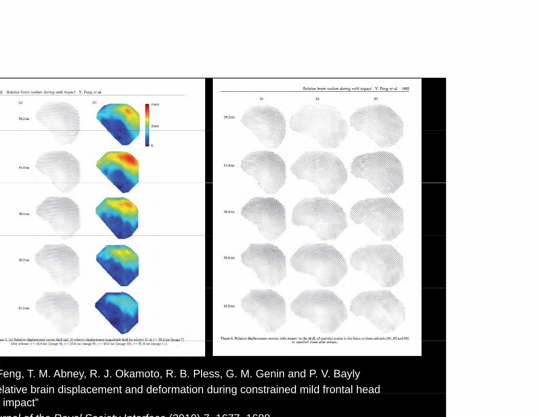
Feng, T. M. Abney, R. J. Okamoto, R. B. Pless, G. M. Genin and P. V. Baylyelative brain displacement and deformation during constrained mild frontal head p g
impact”urnal of the Royal Society Interface (2010) 7 1677 1688
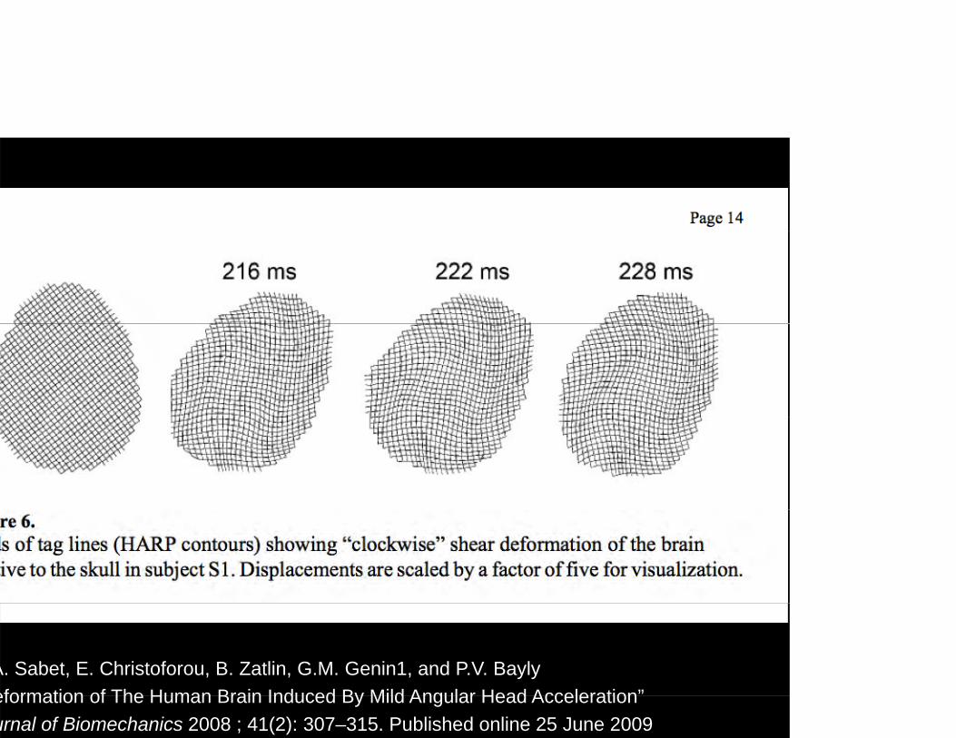
A. Sabet, E. Christoforou, B. Zatlin, G.M. Genin1, and P.V. Baylyeformation of The Human Brain Induced By Mild Angular Head Acceleration”eformation of The Human Brain Induced By Mild Angular Head Accelerationurnal of Biomechanics 2008 ; 41(2): 307–315. Published online 25 June 2009
LITERATURE REVIEWLITERATURE REVIEW
Axonal shear injuriesAxonal shear injuriesAs discussed in a 2003 paper by Dr. Douglas Smith of the University of Pennsylvania in Philadelphia, when the brain experiences high acceleration/deceleration forces, the myelinated axons of neurons become susceptible to breakage.
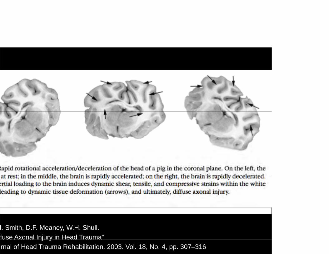
H. Smith, D.F. Meaney, W.H. Shull. ffuse Axonal Injury in Head Trauma”j yurnal of Head Trauma Rehabilitation. 2003. Vol. 18, No. 4, pp. 307–316

LITERATURE REVIEWLITERATURE REVIEW
Axonal shear injuriesAxonal shear injuriesOne of the challenges to concussion assessment and diagnosis lies in the difficulties with detecting a concussion through imaging techniques. But a new technique known as diffusion tensor imaging or DTI, which is a modified MRI imaging technique, has allowed researchers to visualize white matter tracts in the human brain.
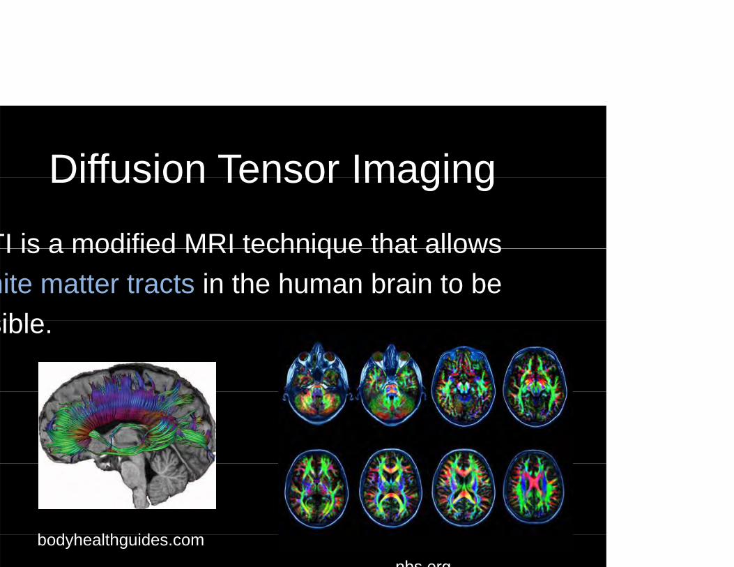
Diffusion Tensor ImagingDiffusion Tensor Imaging
TI is a modified MRI technique that allowsTI is a modified MRI technique that allows hite matter tracts in the human brain to be siblesible.
b d h lth idbodyhealthguides.compbs org
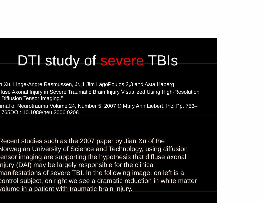
DTI study of severe TBIsDTI study of severe TBIsn Xu,1 Inge-Andre Rasmussen, Jr.,1 Jim LagoPoulos,2,3 and Asta Habergffuse Axonal Injury in Severe Traumatic Brain Injury Visualized Using High-Resolution Diffusion Tensor Imaging.”
urnal of Neurotrauma Volume 24, Number 5, 2007 © Mary Ann Liebert, Inc. Pp. 753–765DOI: 10.1089/neu.2006.0208
Recent studies such as the 2007 paper by Jian Xu of theRecent studies such as the 2007 paper by Jian Xu of the Norwegian University of Science and Technology, using diffusion tensor imaging are supporting the hypothesis that diffuse axonal njury (DAI) may be largely responsible for the clinicalnjury (DAI) may be largely responsible for the clinical manifestations of severe TBI. In the following image, on left is a control subject, on right we see a dramatic reduction in white matter volume in a patient with traumatic brain injury.p j y

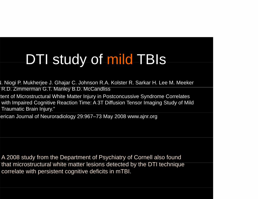
DTI study of mild TBIsDTI study of mild TBIsN. Niogi P. Mukherjee J. Ghajar C. Johnson R.A. Kolster R. Sarkar H. Lee M. Meeker
R D Zi G T M l B D M C dliR.D. Zimmerman G.T. Manley B.D. McCandlissxtent of Microstructural White Matter Injury in Postconcussive Syndrome Correlates
with Impaired Cognitive Reaction Time: A 3T Diffusion Tensor Imaging Study of Mild Traumatic Brain Injury.”
merican Journal of Neuroradiology 29:967–73 May 2008 www.ajnr.org
A 2008 study from the Department of Psychiatry of Cornell also found th t i t t l hit tt l i d t t d b th DTI t h ithat microstructural white matter lesions detected by the DTI technique correlate with persistent cognitive deficits in mTBI.

Axonal shear injuries
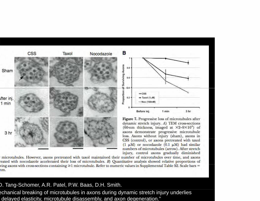
a paper from the University of Pennsylvania and p p y yrexel University in Philadelphia, Min Tang-Schomernd associates investigate dynamic stretch injuries of xons, and demonstrated that microtubule disruption strongly linked with axonal swelling and
egeneration.
he study suggests that dynamic stretch of axonshe study suggests that dynamic stretch of axons duces mechanical failure of microtubules.
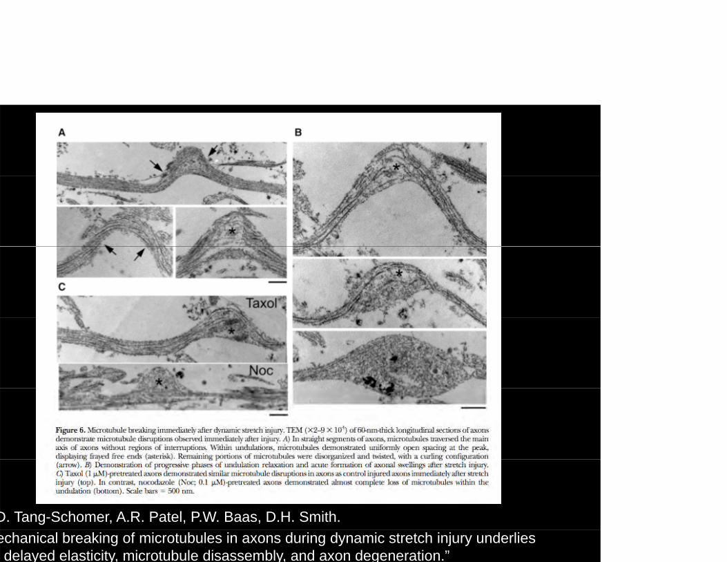
D. Tang-Schomer, A.R. Patel, P.W. Baas, D.H. Smith. echanical breaking of microtubules in axons during dynamic stretch injury underlies delayed elasticity, microtubule disassembly, and axon degeneration.”
D. Tang-Schomer, A.R. Patel, P.W. Baas, D.H. Smith. echanical breaking of microtubules in axons during dynamic stretch injury underlies delayed elasticity, microtubule disassembly, and axon degeneration.”


Mechanically-induced seizures?Mechanically induced seizures?here is one particular image in the animation I would like
o call your attention to, and that was the shot with the lectrical wave across the brain, and here we mention the omparison of concussions with seizures.
he vote is still out on this but Dr. Richards supports this heory as his first-hand experience with many concussed y p ythletes moments after their injuries has led him to agree hat the state of these individuals immediately post-injury trongly resembles that of someone coming to from a g y geizure. So this is why we wanted to include a visualization t some point in the animation that would indicate an lectrical disturbance in the brain.ect ca d stu ba ce t e b a
LITERATURE REVIEWLITERATURE REVIEWhe chief tenet of the convulsive theory is that since ye symptoms of concussion bear a strong semblance to those of a generalized epileptic
eizure, then it is a reasonable assumption that similar athobiological processes underlie them both.”
gel A. Shawhe Neurophysiology of Concussion”ogress in Neurobiology 2002 Jul;67(4):281 344ogress in Neurobiology. 2002 Jul;67(4):281-344.
LITERATURE REVIEWLITERATURE REVIEW…loss of consciousness is…due to functional eafferentation of the cortex as a consequence of ffuse mechanically-induced depolarization and ynchronized discharge of cortical neurons.”
gel A. Shawhe Neurophysiology of Concussion”rogress in Neurobiology 2002 Jul;67(4):281-344rogress in Neurobiology. 2002 Jul;67(4):281-344.
OBJECTIVESOBJECTIVESy objectives in this project were to communicate to a lay dience through a 3-minute 3D animation the physical response brain tissue to high acceleration and deceleration forces, depict e currently proposed mechanism of diffuse axonal injuries as ae currently proposed mechanism of diffuse axonal injuries as a ssible cellular-level component of injury, and show the related mage to the microtubules of axons. Now these ideas are still
ti l hil th i t id th t h f deoretical, while there is strong evidence that shear forces and onal injuries are involved with severe traumatic brain injuries, we ll don't know for sure what factors are at play in concussions,
hich again are a type of mild traumatic brain injury. e attempted to base my visualizations on what the most current search suggests might be happeningsearch suggests might be happening.
OBJECTIVESOBJECTIVES
Compelling interpretation of brain tissueCompelling interpretation of brain tissue deformation
Depiction of shearing force between Grey matter and White matter
Depict axonal injuries, i.e. microtubuleDepict axonal injuries, i.e. microtubule breakage
OBJECTIVESOBJECTIVES
gain this is a visualization informed bygain this is a visualization informed by mpirical evidence, but designed to
i tommunicate.
OBJECTIVESOBJECTIVESarrowing the focus and scope of this project became g p p jcreasingly important as we moved forward and nfortunately that means reducing the total content of hat I would be showing. I look forward to the pportunities to address other visualization needs lated to traumatic head injuries but in this particular oject I avoided going into surgical considerations, agnosis or the molecular level factorsagnosis, or the molecular-level factors.
OBJECTIVESOBJECTIVESocus is on education for the lay public, not:ocus is on education for the lay public, not:
Surgical interventions of TBIsSurgical interventions of TBIs
TBI detection measurement or assessmentTBI detection, measurement or assessment
Molecular pathways biochemical cascadesMolecular pathways, biochemical cascades
OBJECTIVESOBJECTIVES
END GOAL:END GOAL:Provide athletic coaches with visual
id f th ti l b hi d tevidence of the rationale behind return-to-play guidelines to help increase adherence and awareness.
METHODS & TOOLSMETHODS & TOOLSe production workflow involves integrating several assets together from ferent sources. Consultation, review, editing and revisions must also beferent sources. Consultation, review, editing and revisions must also be ken into account. The project itself is a dynamic process, and certain portions ay end up being repeated.
r animations begin in a ritten format We start ith a treatment hichr animations begin in a written format. We start with a treatment, which nctions like an abstract, it summarizes the viewer's experience, honing in on at sorts of emotional responses we wish to conjure. The treatment then velops into a written script, and at this point we start to become aware of the p p pce and timing of our animation. Every image that appears on screen is timed cording to the voice-over.
course our ultimate goal is to really focus in on what the visual elements ofcourse our ultimate goal is to really focus in on what the visual elements of r story will look like and what that overall story is. So alongside the script we velop our storyboard panels, which function as the foundation for our mation. The storyboard then develops into a 2D Animatic, and from there we
e ready to start working in 3D!
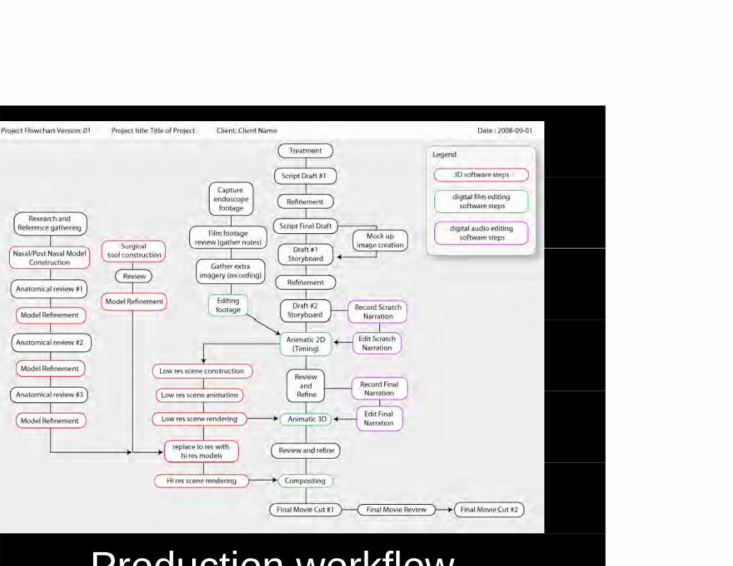
Production workflow
Treatment & ScriptTreatment & Script
reatment is like an abstract for a script.
summarizes the viewer's experience.
Treatment & ScriptTreatment & Script
he treatment becomes the script whichhe treatment becomes the script, which created alongside the storyboard. The
i t t icript contains:
Description of shots
Narration or “voice over”Narration or voice-over”
Sound effects and music

Storyboards...
i th i l i t

2D Animatic2D Animatic
Animated Storyboardy
The 2D animatic is basically an animated storyboard, and helps us work out all the timing and coordination for narration and camera moves. The following clip is an example:
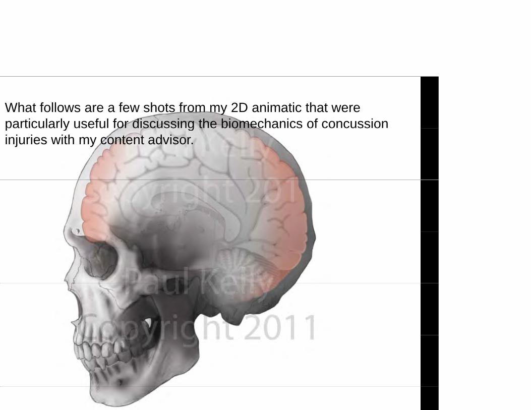
What follows are a few shots from my 2D animatic that were particularly useful for discussing the biomechanics of concussion pa t cu a y use u o d scuss g t e b o ec a cs o co cuss oinjuries with my content advisor.
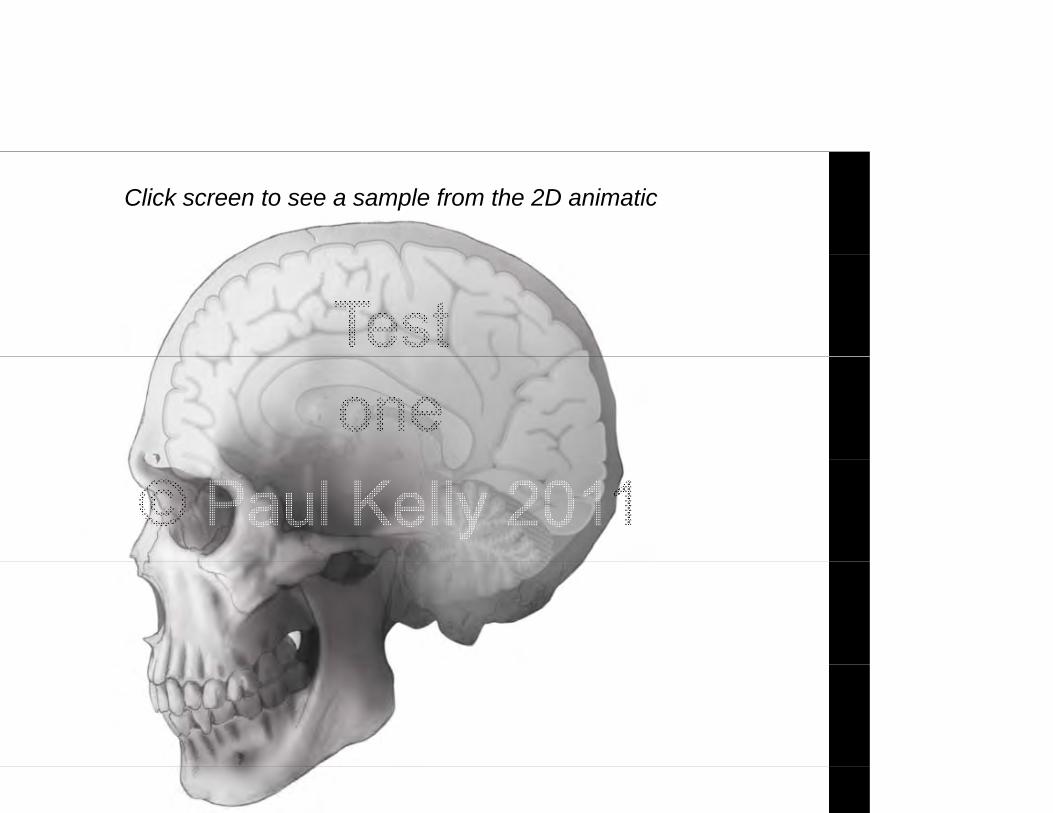
Click screen to see a sample from the 2D animatic
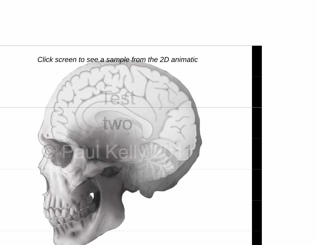
Click screen to see a sample from the 2D animatic
Click screen to see a sample from the 2D animatic

TOOLSTOOLS
OsirixOsirixA medical imaging enhancement t ltool
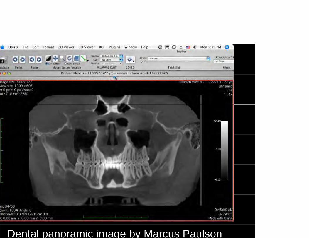
Dental panoramic image by Marcus Paulson
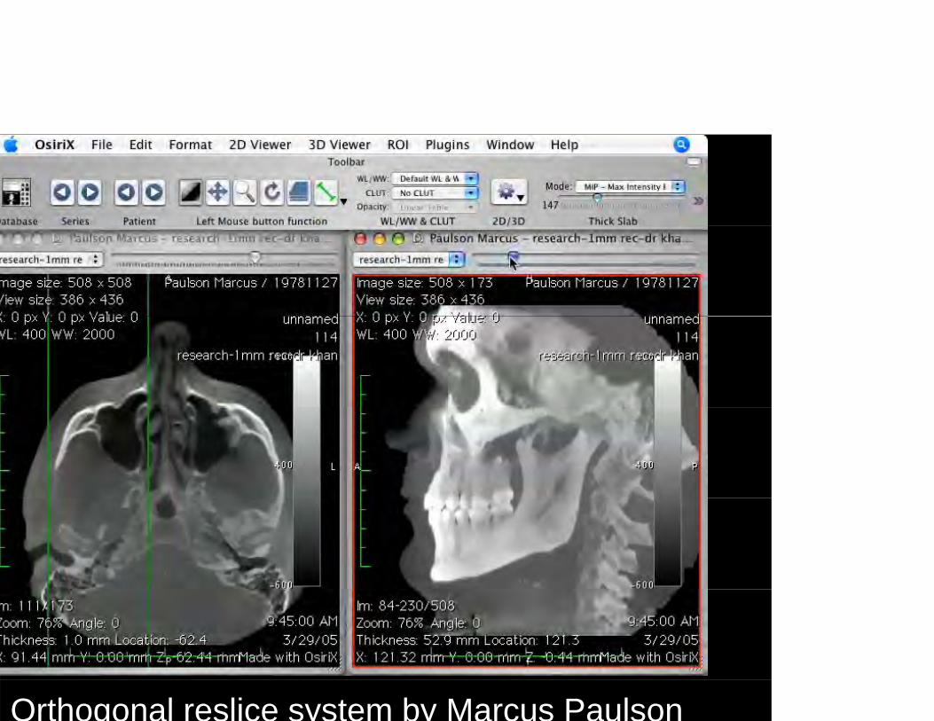
Orthogonal reslice system by Marcus Paulson
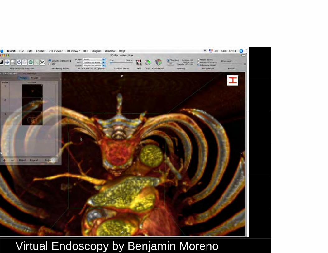
Virtual Endoscopy by Benjamin Moreno
TOOLSTOOLS
Pi l i Zb hPixologic ZbrushA digital sculpting tool
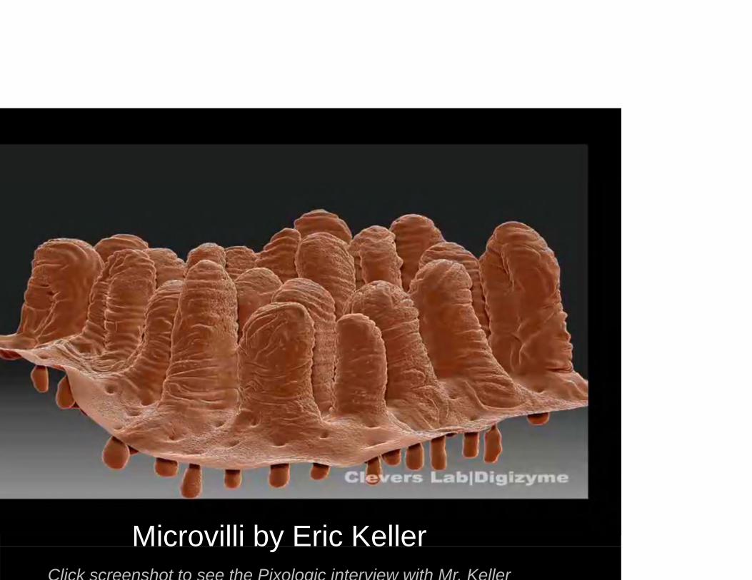
Microvilli by Eric KelleryClick screenshot to see the Pixologic interview with Mr. Keller
TOOLSTOOLS
Autodesk MayaAutodesk Maya The leading 3D animation software for character animation dynamicsfor character animation, dynamics, simulations, shading, texturing, and renderingand rendering.

Malaria animation by Drew Berry
TOOLSTOOLS
Adobe After EffectsAdobe After Effects A compositing software that integrates video audio stillintegrates video, audio, still images and special effects.

Blood flow animation by Andrew Kramer
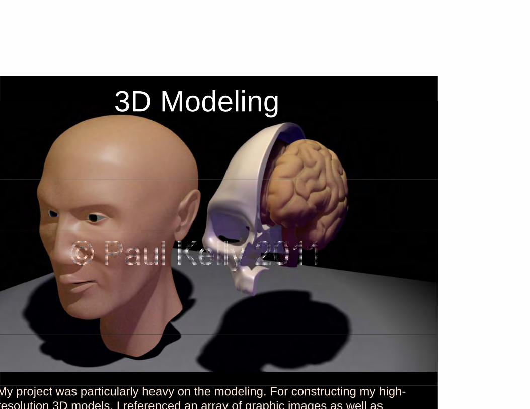
3D Modeling
My project was particularly heavy on the modeling. For constructing my high-resolution 3D models, I referenced an array of graphic images as well as
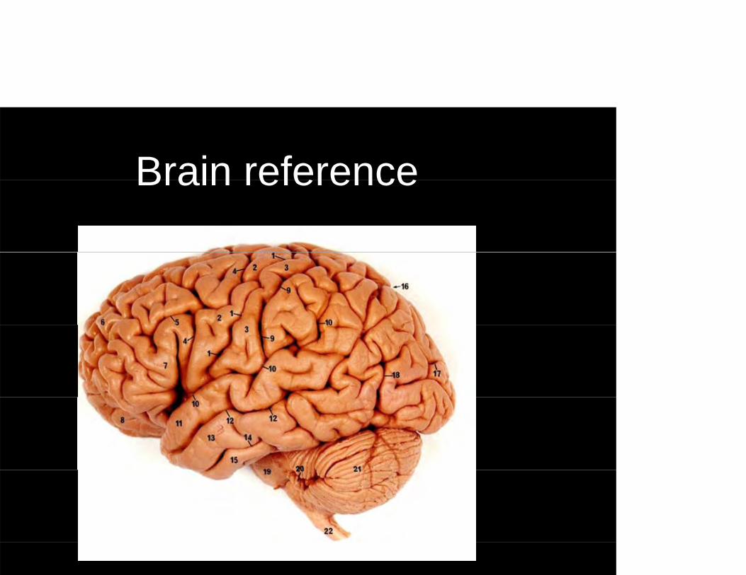
Brain referenceBrain reference
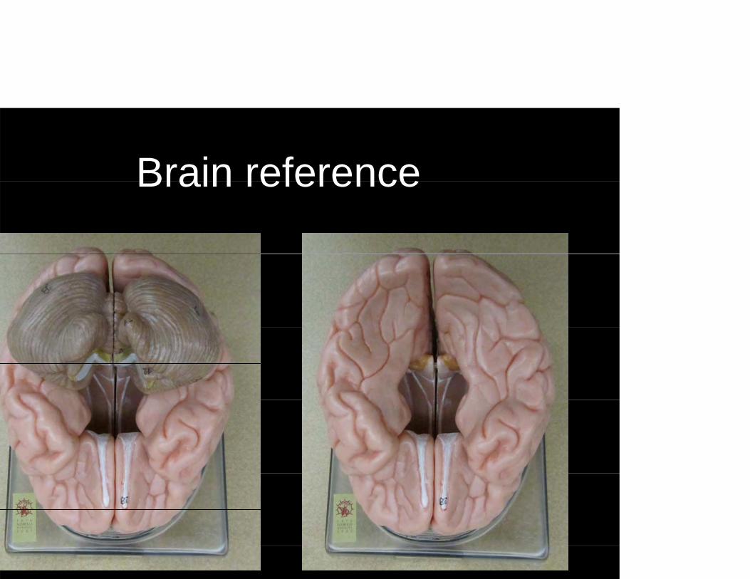
Brain referenceBrain reference
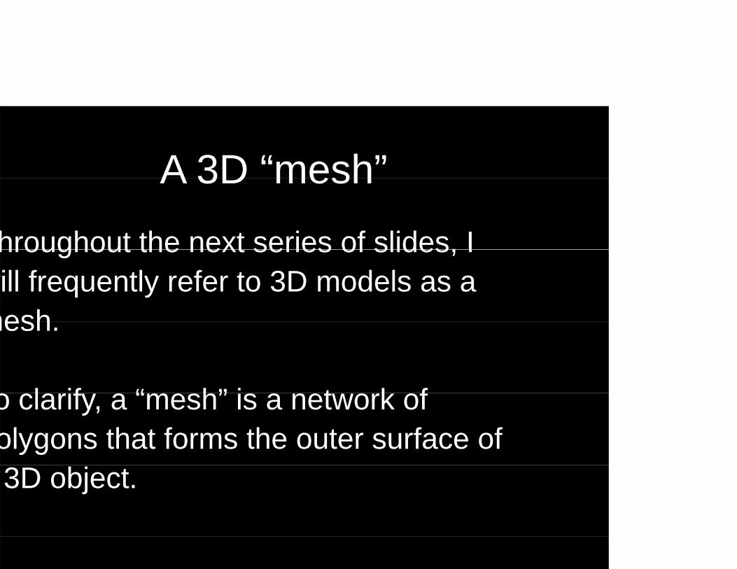
A 3D “mesh”A 3D mesh
hroughout the next series of slides, Ihroughout the next series of slides, I will frequently refer to 3D models as a meshmesh.
l if “ h” i t k fo clarify, a “mesh” is a network of olygons that forms the outer surface of 3D object.
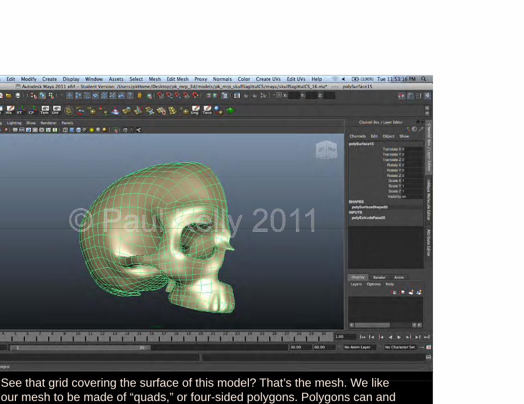
See that grid covering the surface of this model? That’s the mesh We likeSee that grid covering the surface of this model? That s the mesh. We like our mesh to be made of “quads,” or four-sided polygons. Polygons can and
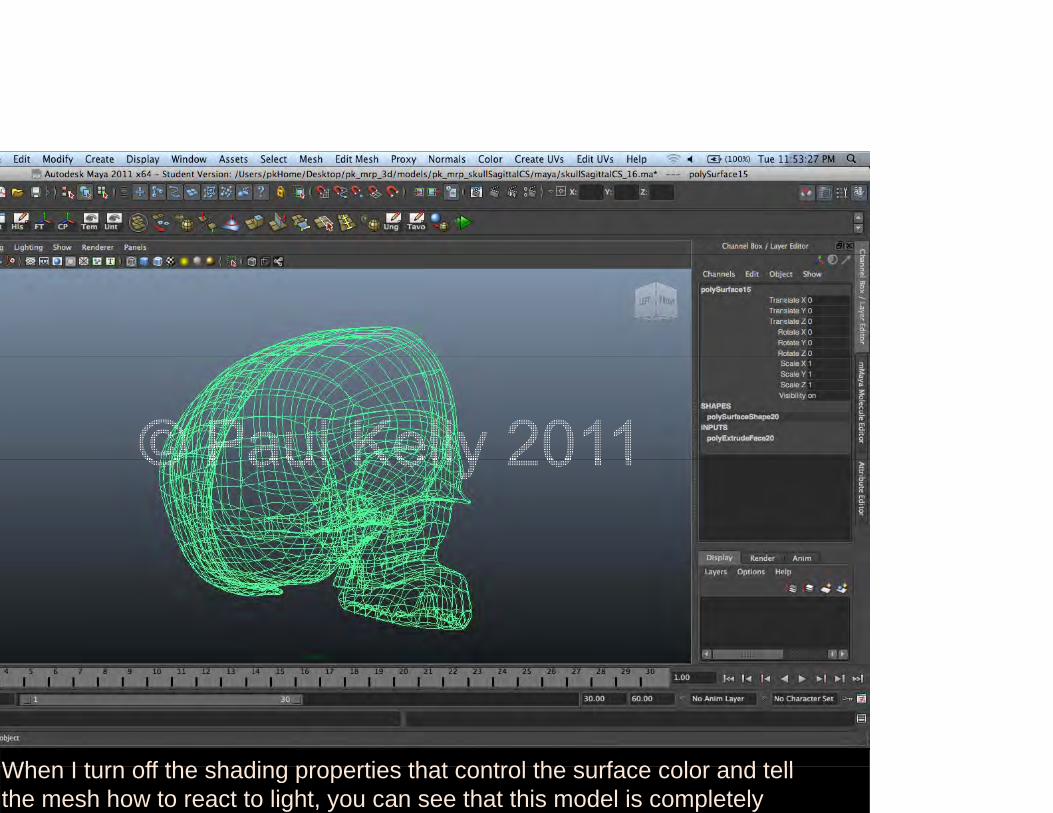
When I turn off the shading properties that control the surface color and tellWhen I turn off the shading properties that control the surface color and tell the mesh how to react to light, you can see that this model is completely

Modeling the brainModeling the brain…
Without a doubt the greatest modelingWithout a doubt, the greatest modeling challenge for me was the brain. The h b i i t i l diffi lthuman brain is a notoriously difficult structure to model in 3D, and I tried utilizing some unique tools in hopes of making my final product as accurate asmaking my final product as accurate as possible.

OSIRIX
Modeling the brainModeling the brain… attempted to use real data from an MRI pseries to build my brain model from. Luckily my advisor Nick Woolridge had had his brain scanned for a study he participated in and received a copy of the DICOM image set for his troubles. One feature of Osirix is that the program can
t 3D h th t th bcreate a 3D mesh that can then be exported to a 3D program.
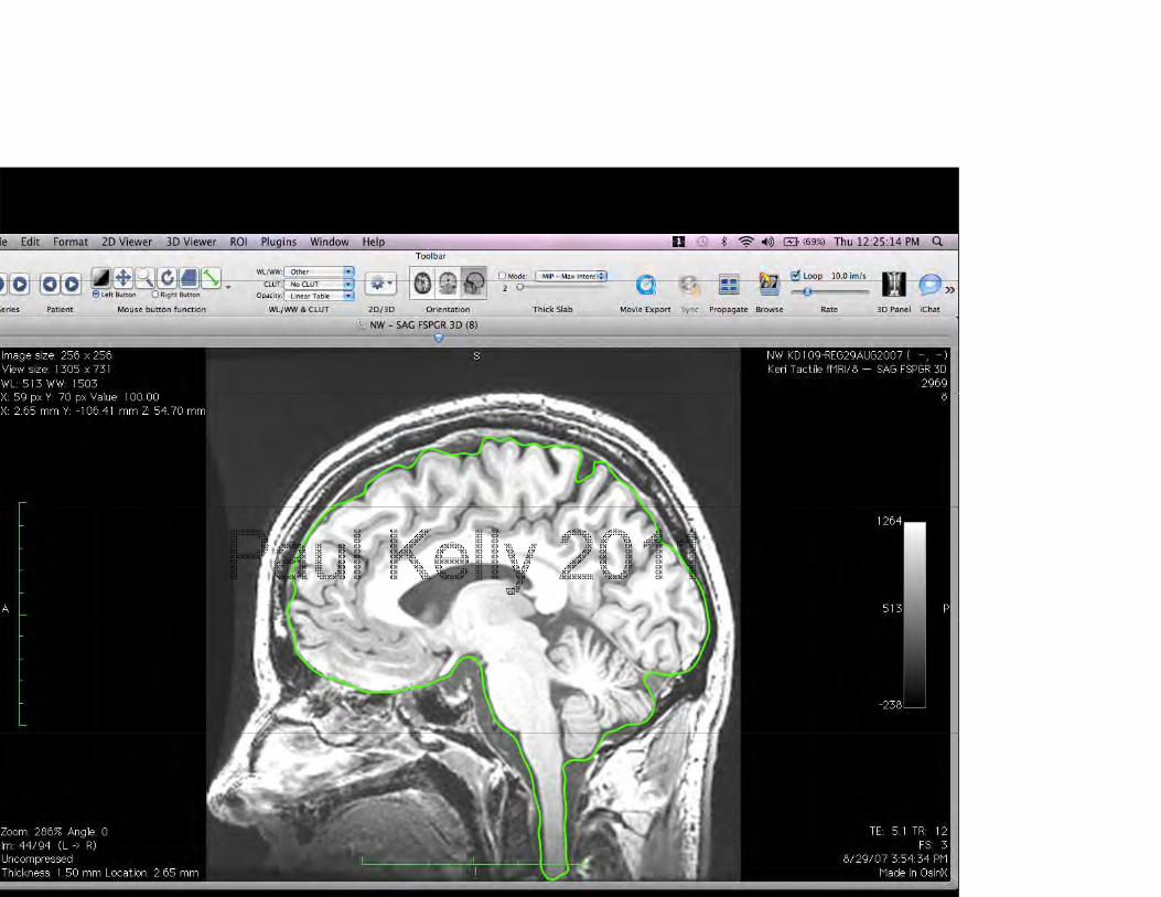

Modeling the brainModeling the brain…This algorithm will make a mesh out of gthe outermost layer detected in the scan series, so, in order to make a mesh of an nner structure, you have to isolate that region and Osirix does provide you with tools to do that. It's a bit tedious, but after carefully tracing the outline of the b i h I bl t i l tbrain on each scan, I was able to isolate the brain from the surrounding tissues.

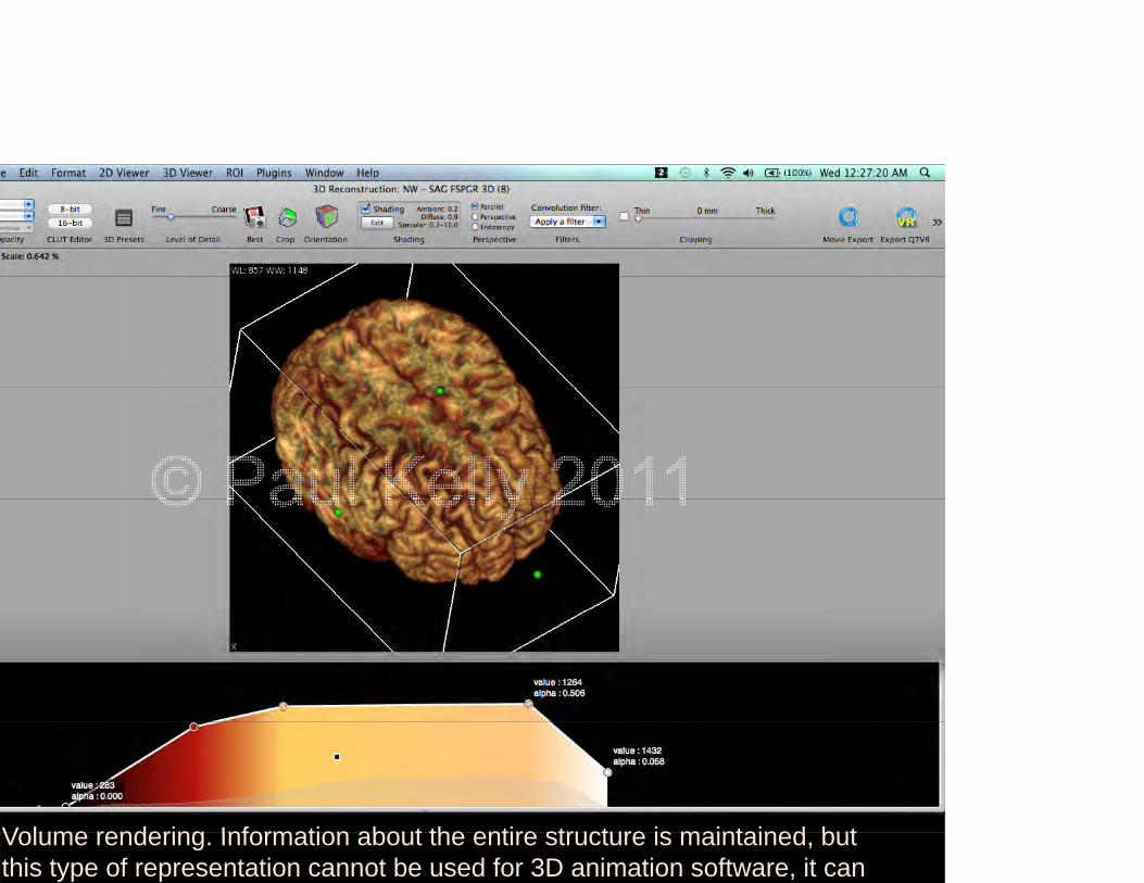
Volume rendering Information about the entire structure is maintained butVolume rendering. Information about the entire structure is maintained, but this type of representation cannot be used for 3D animation software, it can
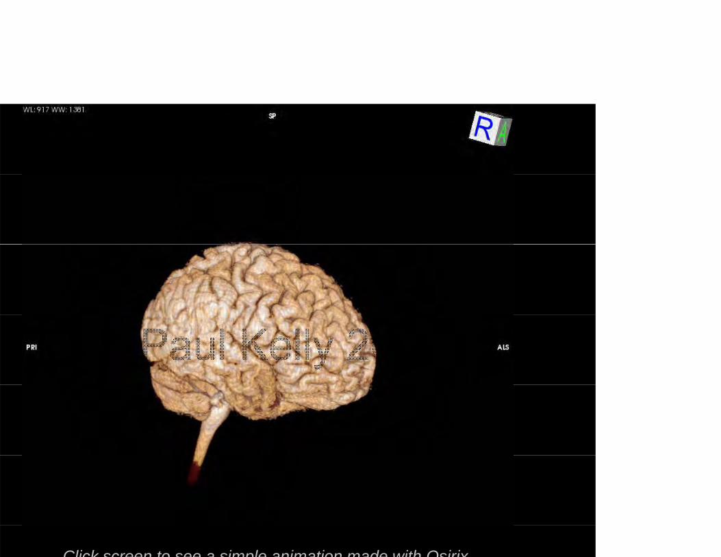
Click screen to see a simple animation made with Osirix

Modeling the brainModeling the brain…
While it appeared to be very clear inWhile it appeared to be very clear in the volumetric 3D reconstruction ( hi h i f ti b t h t i(which saves information about what is “inside” the mesh), unfortunately there were too many artifacts in the surface reconstruction to use that for a modelreconstruction to use that for a model.

For animating I would need just the outer shell such as this surfaceFor animating, I would need just the outer shell, such as this surface rendering. Unfortunately though this didn’t work out…

due to the unorganized and highly dense mesh that this algorithm…due to the unorganized and highly dense mesh that this algorithm produces. But this wasn't a total loss, because it did give me an accurate

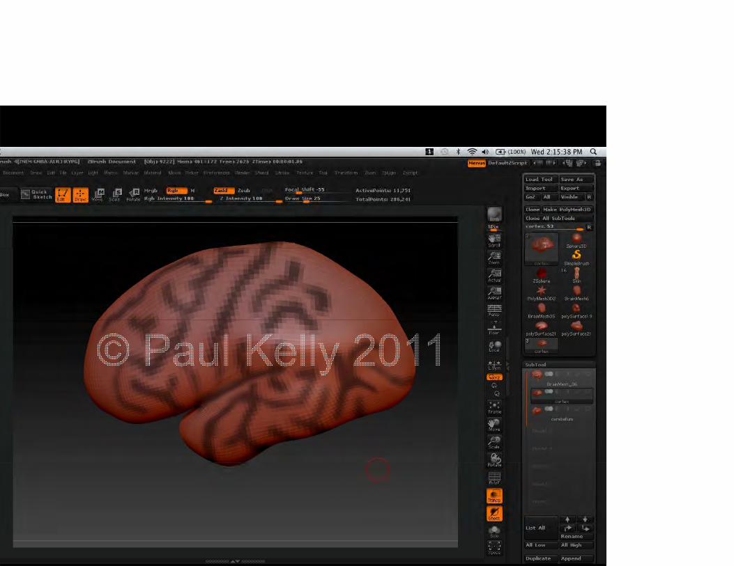
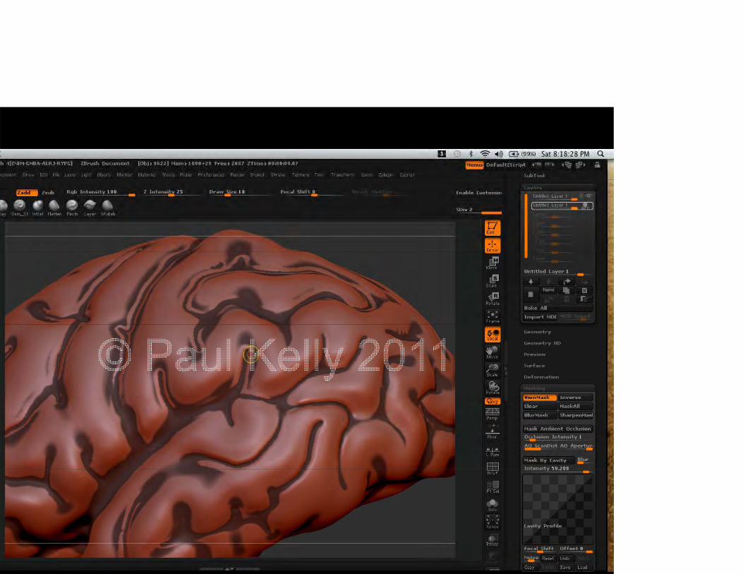
ZBRUSH
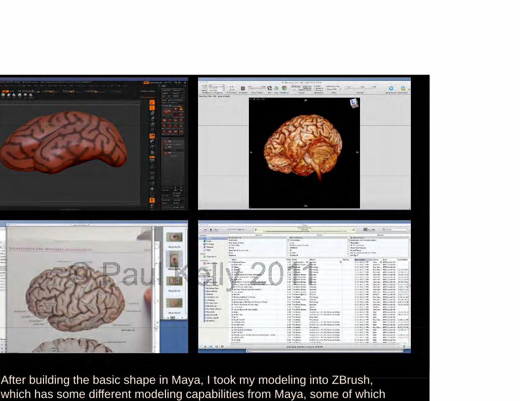
After building the basic shape in Maya I took my modeling into ZBrushAfter building the basic shape in Maya, I took my modeling into ZBrush, which has some different modeling capabilities from Maya, some of which


MAYA
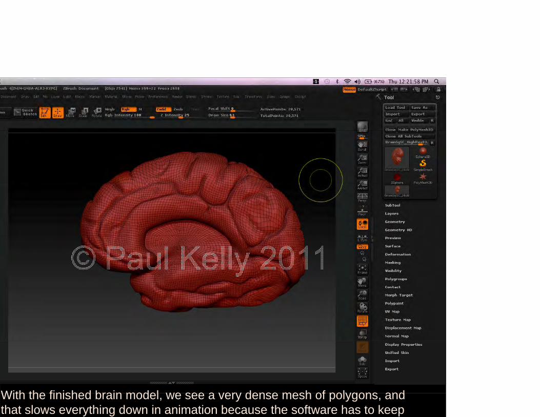
With the finished brain model we see a very dense mesh of polygons andWith the finished brain model, we see a very dense mesh of polygons, and that slows everything down in animation because the software has to keep
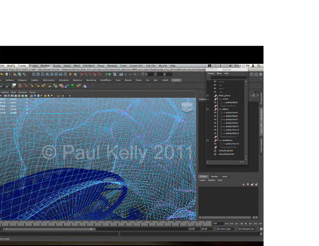
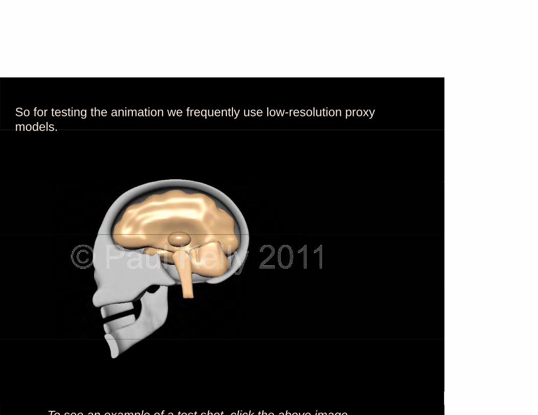
So for testing the animation we frequently use low-resolution proxy models.models.
To see an example of a test shot click the above image

BRAIN DEFORMATION

ed the results from the University of St Louis papers to make sure I wased the results from the University of St. Louis papers to make sure I was phasizing the brain tissue deformation in the right areas. One tool I used in
hi h i ll d l tti d f b ild t k f ti d 3D
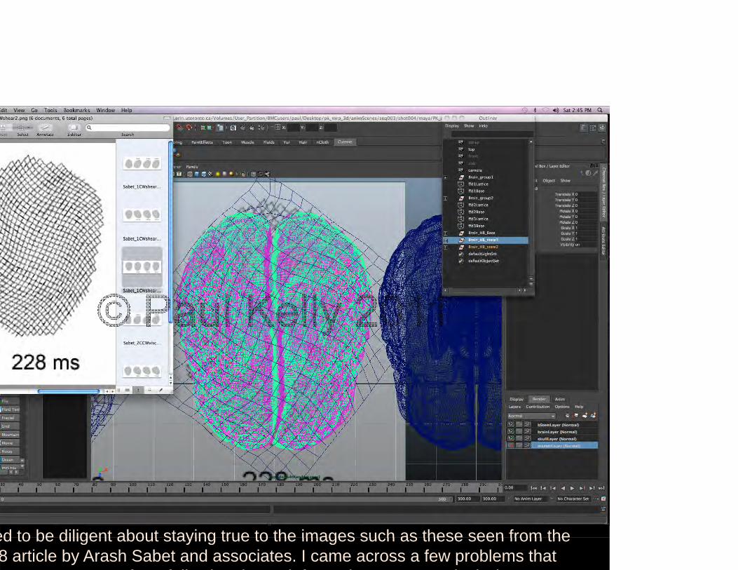
ed to be diligent about staying true to the images such as these seen from theed to be diligent about staying true to the images such as these seen from the 8 article by Arash Sabet and associates. I came across a few problems that
t t f f ll i th d f ti i l b t

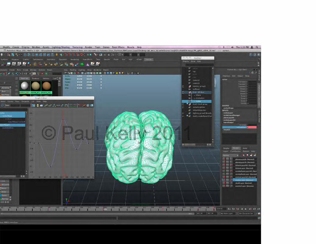
ce I had used the lattice tool to deform the brain model I then used ance I had used the lattice tool to deform the brain model, I then used an mation technique which basically allows me to key-frame between the normal, lt d b i d l d th d f d d l
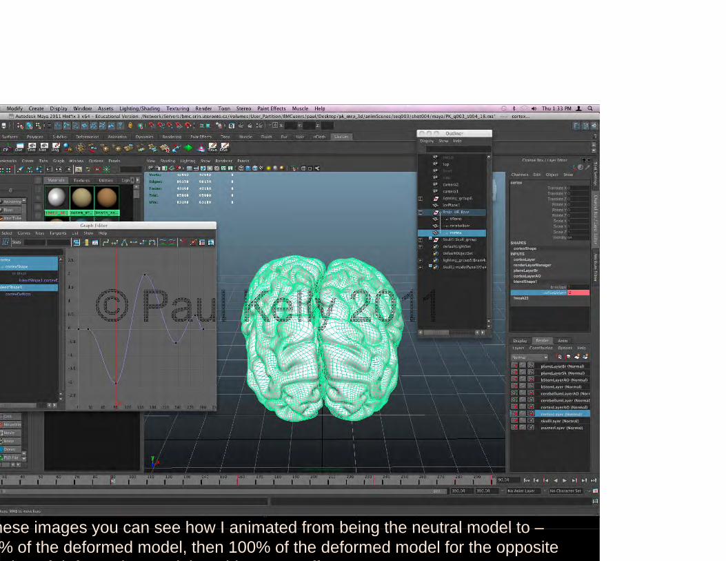
hese images you can see how I animated from being the neutral model to –hese images you can see how I animated from being the neutral model to % of the deformed model, then 100% of the deformed model for the opposite ti f d f ti d th thi t ff

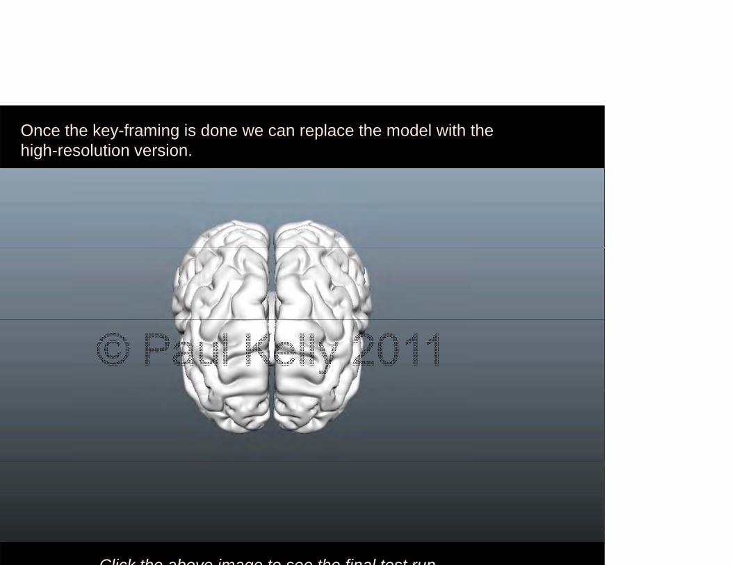
Once the key-framing is done we can replace the model with the high-resolution version.
Click the above image to see the final test run
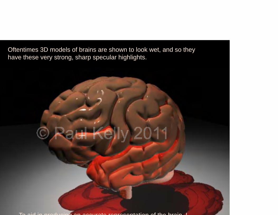
Oftentimes 3D models of brains are shown to look wet, and so they have these very strong, sharp specular highlights.
To aid in producing an accurate representation of the brain I
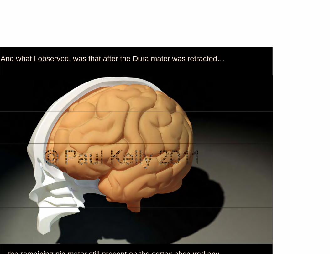
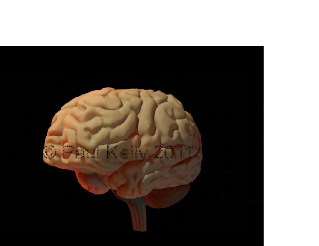
And what I observed, was that after the Dura mater was retracted…
the remaining pia mater still present on the cortex obscured any

AXON SHEARING
AXON SHEARINGAXON SHEARING
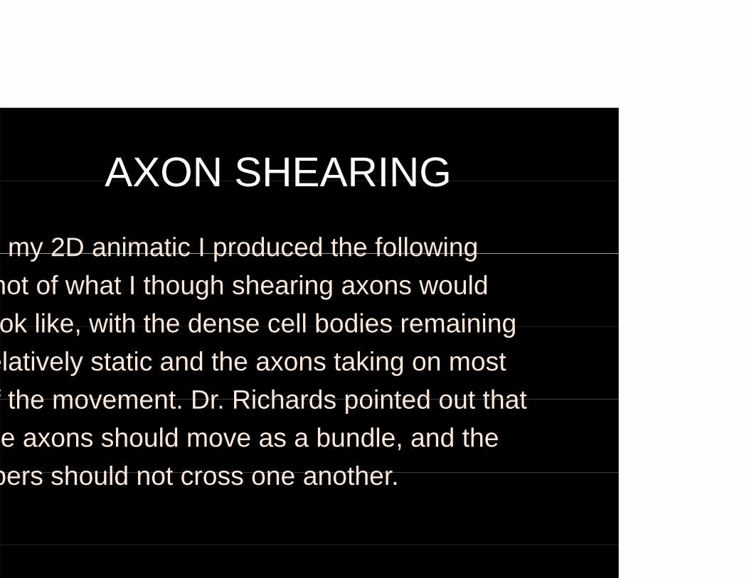
my 2D animatic I produced the following my 2D animatic I produced the following hot of what I though shearing axons would ok like with the dense cell bodies remainingok like, with the dense cell bodies remaining
elatively static and the axons taking on most f the movement Dr Richards pointed out thatf the movement. Dr. Richards pointed out that e axons should move as a bundle, and the
bers should not cross one anotherbers should not cross one another.
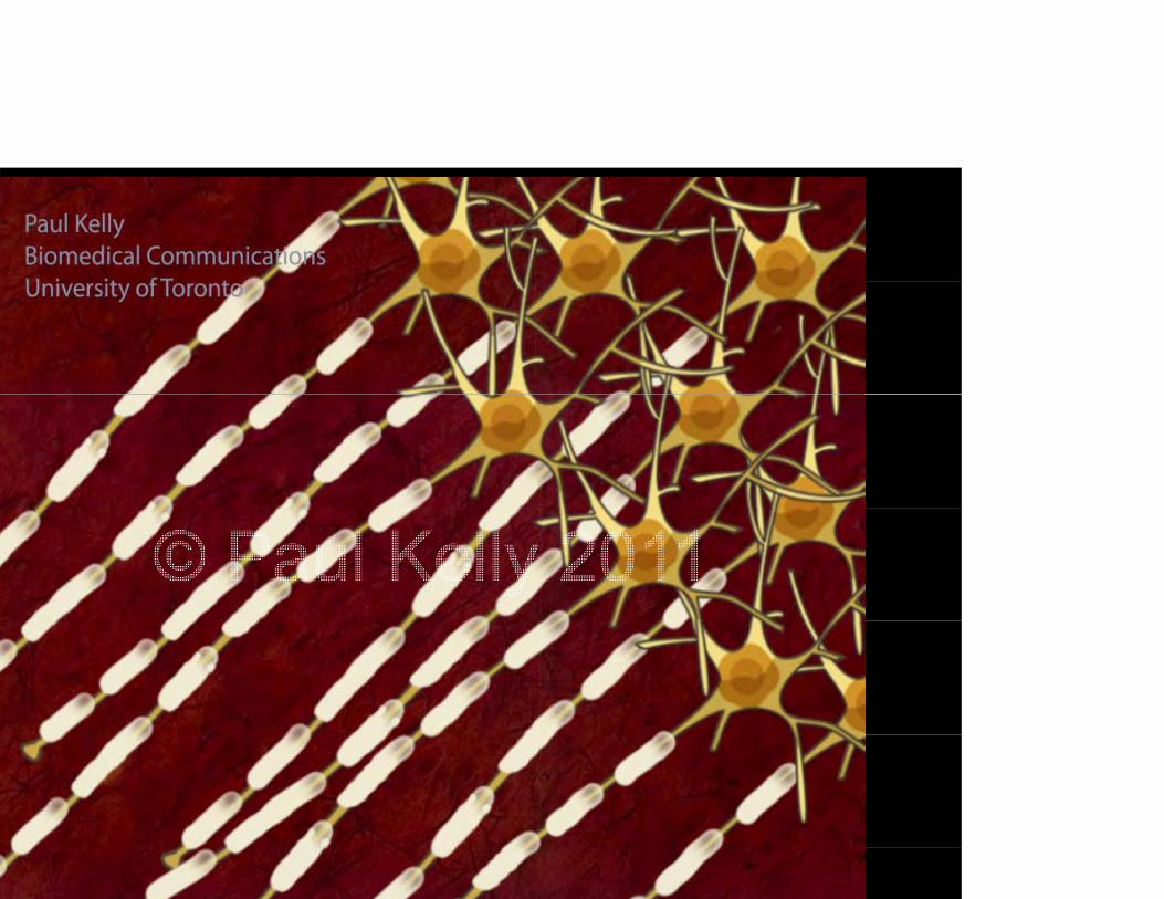
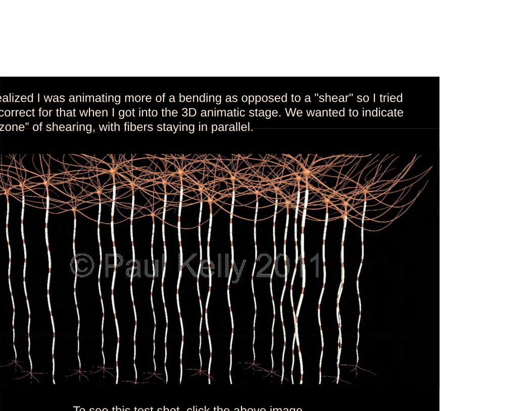
ealized I was animating more of a bending as opposed to a "shear" so I tried correct for that when I got into the 3D animatic stage. We wanted to indicate zone” of shearing with fibers staying in parallelzone of shearing, with fibers staying in parallel.
To see this test shot click the above image

e ended up going back to something closer to the original so that thee ended up going back to something closer to the original so that the earing zone would be indicated at the junction between the cell body and

MICROTUBULE BREAKAGE
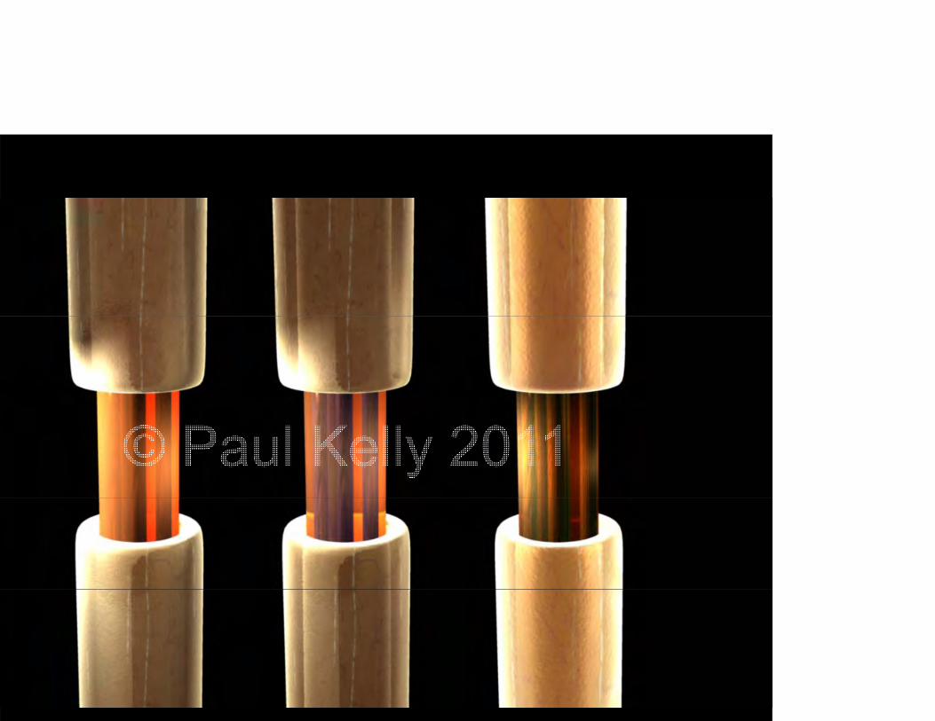
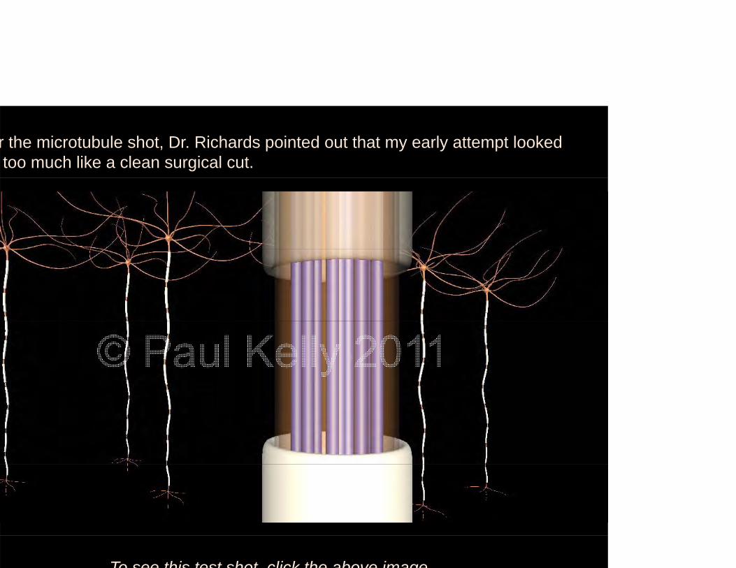
r the microtubule shot, Dr. Richards pointed out that my early attempt looked too much like a clean surgical cut.
To see this test shot click the above image
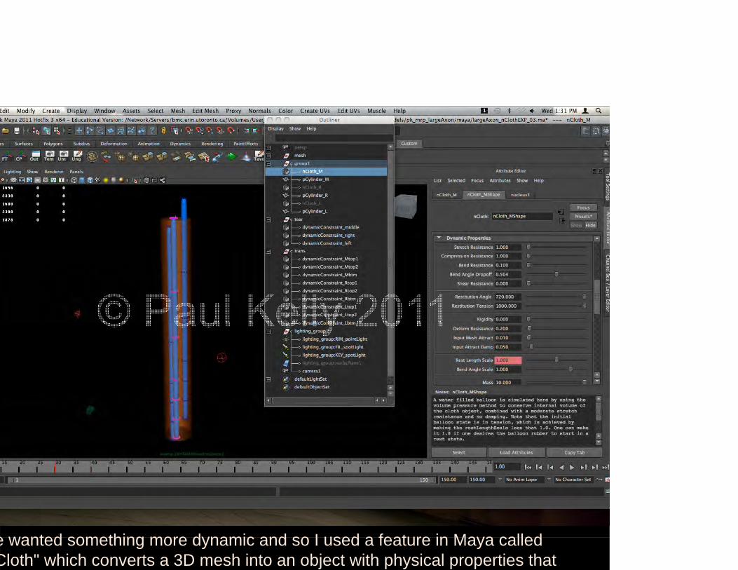
e wanted something more dynamic and so I used a feature in Maya callede wanted something more dynamic and so I used a feature in Maya called Cloth" which converts a 3D mesh into an object with physical properties that
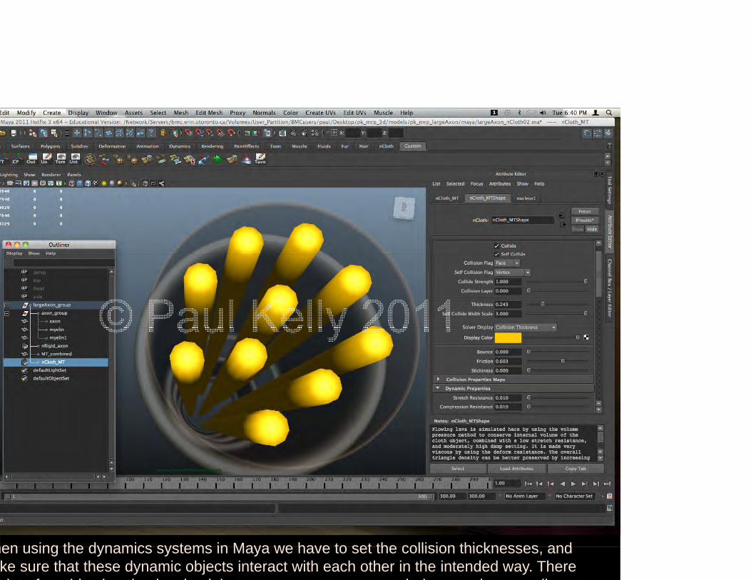
hen using the dynamics systems in Maya we have to set the collision thicknesses andhen using the dynamics systems in Maya we have to set the collision thicknesses, and ke sure that these dynamic objects interact with each other in the intended way. There l t f t bl h ti i l d b t it d d t ll

To see an example of a test shot click the above image
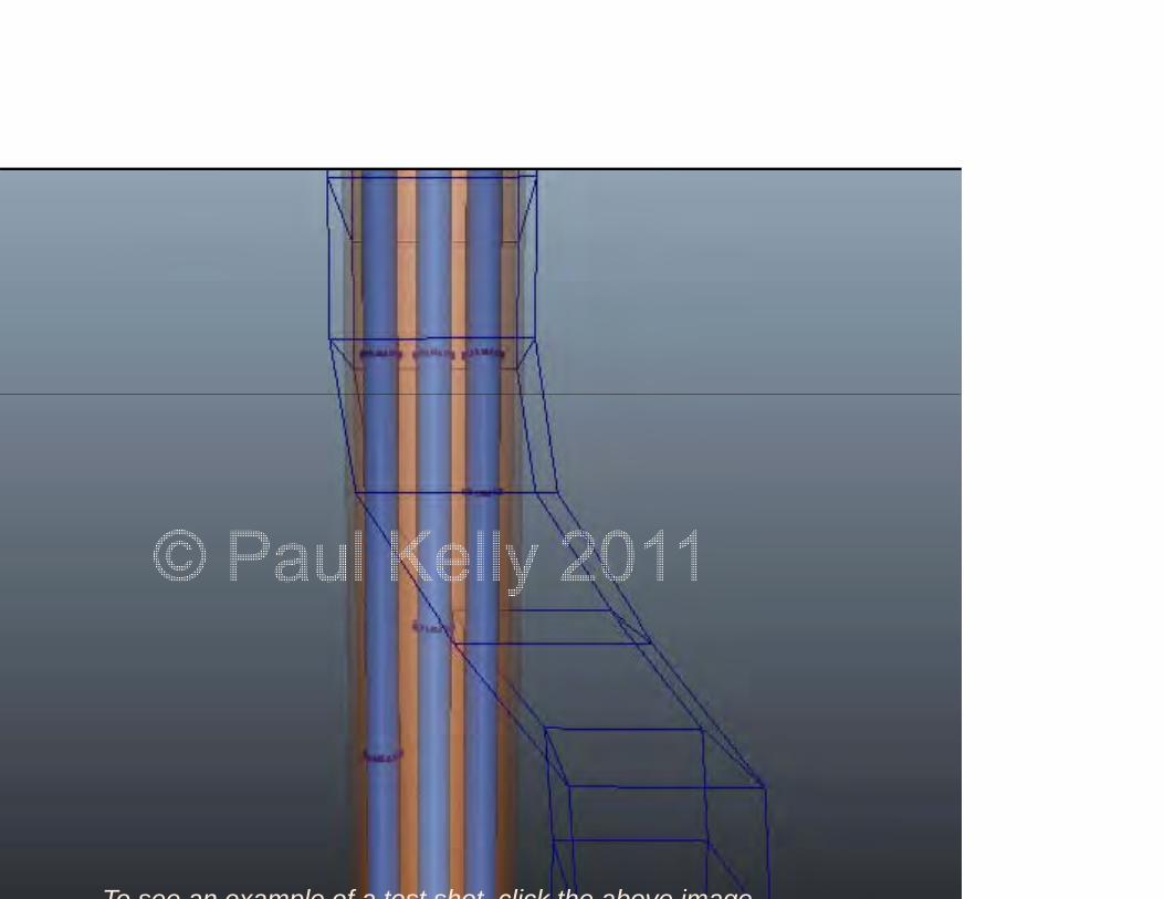
To see an example of a test shot click the above image
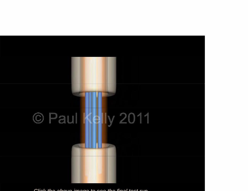
Click the above image to see the final test run
DISCUSSIONDISCUSSION
n order to reduce the incidence and cost of mTBIs, those t risk need to be educated about the potential severity of h i j d i j t Whil I t i lhe injury and proper injury management. While I certainly ave no right to tell another person they shouldn't play angerous contact sports, I do feel that they have a right g p y go know what they are subjecting themselves to.

DISCUSSIONDISCUSSION
3D visualization to assist both players and3D visualization to assist both players and coaches in understanding:
Importance of preventative strategies
Proper injury management
Strict adherence to return-to-play guidelines
Production TimeProduction TimeProject required a minimum of 676 work hours.Project required a minimum of 676 work hours.
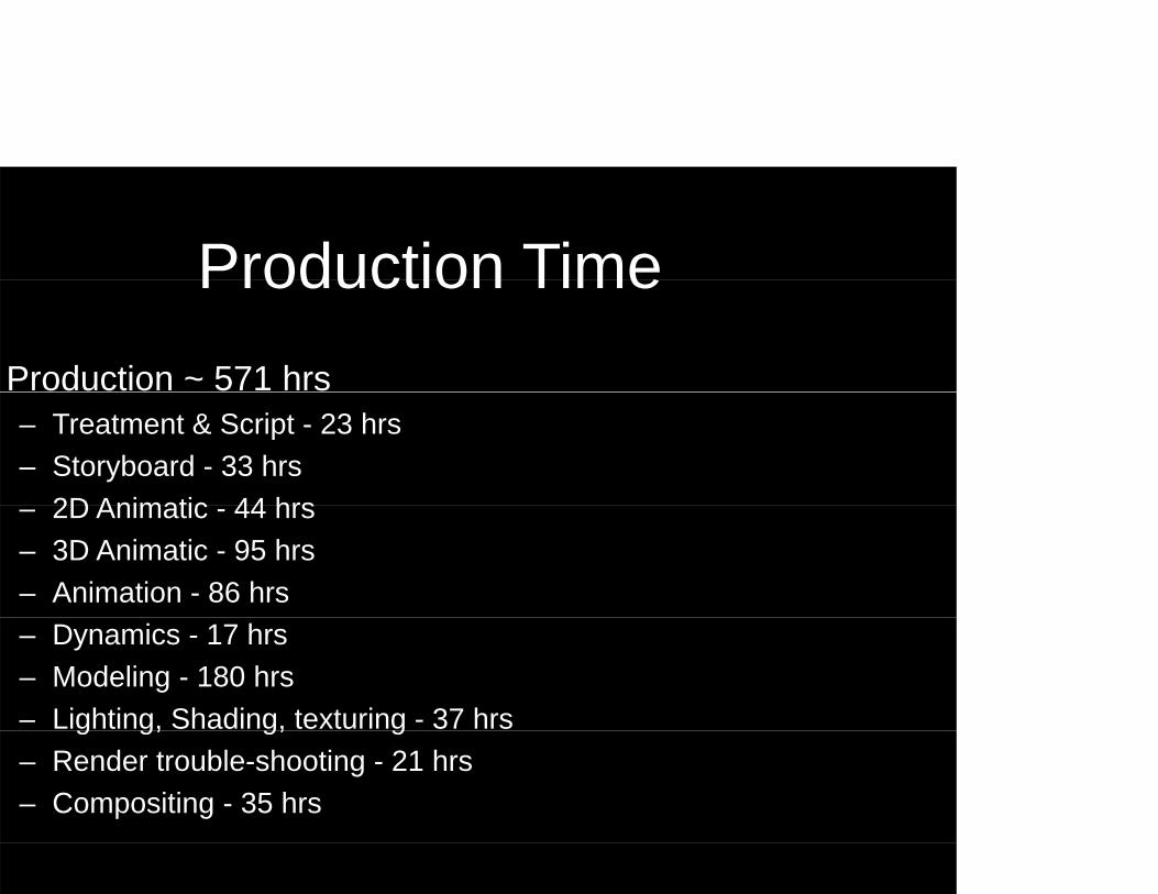
Production TimeProduction TimeProduction ~ 571 hrs– Treatment & Script - 23 hrs – Storyboard - 33 hrs
2D Animatic 44 hrs– 2D Animatic - 44 hrs – 3D Animatic - 95 hrs– Animation - 86 hrs – Dynamics - 17 hrs – Modeling - 180 hrs– Lighting, Shading, texturing - 37 hrsg g, g, g– Render trouble-shooting - 21 hrs– Compositing - 35 hrs
Limitations to the animationLimitations to the animation
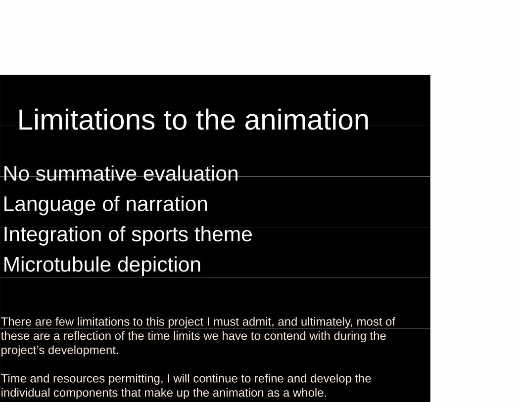
No summative evaluationNo summative evaluationLanguage of narrationI t ti f t thIntegration of sports themeMicrotubule depiction
There are few limitations to this project I must admit, and ultimately, most of these are a reflection of the time limits we have to contend with during the project’s development.
Time and resources permitting I will continue to refine and develop theTime and resources permitting, I will continue to refine and develop the individual components that make up the animation as a whole.
Potential future visualizationsPotential future visualizations
Working on this project has given me many ideas forWorking on this project has given me many ideas for future visualizations, and these could be handled in a variety of media, from simple illustrations to interactive
b it I f l thi f h i i t twebsites. I feel this area of research is very important, especially to the optimal health and well-being of young people involved in sports and I look forward to contributing to the further understanding of the underlying mechanisms of mild traumatic brain injuries.
Potential future visualizationsPotential future visualizationsIndirect impacts that transfer rotational accelerationp
Short- and long-term clinical manifestations
C f l ti b i hitConsequences of cumulative sub-concussive hits
Role of tau and beta-amyloid proteins
Related biochemical pathways
Mitochondrial dysfunctionMitochondrial dysfunction

Today we have looked at:Today we have looked at:
The field of Biomedical CommunicationsThe field of Biomedical Communications
Published works referenced for the creation of aPublished works referenced for the creation of a 3D animation on concussions
Methods and tools used to create a biomedical 3D animation3 a at o

SUMMARYSUMMARY
t is hoped that this visualization depicting physical eformation of cerebral tissue and associated injuries to
brain cell axons can assist both players and coaches inbrain cell axons can assist both players and coaches in nderstanding the importance of preventative strategies,
proper injury management and strict adherence to return-o pla g idelines I also hope that this is ali ation mao-play guidelines. I also hope that this visualization may be of use to bioengineers and sports medicine professionals conducting research into the epidemiology of mTBI.