saponin: properties, methods of evaluation and applications
TRANSCRIPT

_____________________________________________________________________________________________________ *Corresponding author: Email: [email protected];
Annual Research & Review in Biology 5(3): 207-220, 2015, Article no.ARRB.2015.022
ISSN: 2347-565X
SCIENCEDOMAIN international
www.sciencedomain.org
Saponin: Properties, Methods of Evaluation and Applications
Eskandar Moghimipour1 and Somayeh Handali2*
1Medicinal Plant Research Center, Ahvaz Jundishapur University of Medical Sciences, Ahvaz, Iran.
2Nanotechnology Research Center, Ahvaz Jundishapur University of Medical Sciences, Ahvaz, Iran.
Authors’ contributions
This work was carried out in collaboration between both authors. Author EM designed the study and
author SH wrote the first draft of the manuscript. Both authors read and approved the final manuscript.
Article Information
DOI: 10.9734/ARRB/2015/11674
Editor(s): (1) George Perry, Dean and Professor of Biology, University of Texas at San Antonio, USA.
Reviewers: (1) Antony de Paula Barbosa, Research Institute of Natural Products, Centro de Ciências da Saúde, Universidade Federal do
Rio de Janeiro, Brazil. (2) Anonymous, Ain Shams University, Egypt.
(3) Anonymous, Khon Kaen University, Thailand. (4) Nesrein M. Hashem, Animal production Dep., Agriculture Faculty, Alexandria University, Egypt.
Complete Peer review History: http://www.sciencedomain.org/review-history.php?iid=668&id=32&aid=6212
Received 29th
May 2014 Accepted 21
st August 2014
Published 24th
September 2014
ABSTRACT
Saponins are secondary metabolites with high molecular weight. They present in a wide range of plant species and are distributed throughout the bark, leaves, stems, roots and even flowers. Saponins are bitter in taste and in recent years, they have received considerable attention because of their various biological activities including hepatoprotective, anti-ulcer, anti-tumor, antimicrobial, adjuvant and anti-inflammatory activities. Saponins are composed of a lipid soluble aglycone consisting of either a sterol or more commonly a triterpenoid and water soluble sugar residues, due to their amphiphilic nature, they are highly surface active and their biological activities are related to their chemical structures. Both steroidal and triterpenoids saponins show detergent properties. The aim of the present article is to review the saponin and methods of evaluation and also, their application based on the recent studies.
Keywords: Saponin; separation; biological activity; steroids; triterpenoids.
Review Article
Moghimipour and Handali; ARRB, 5(3): 207-220, 2015; Article no.ARRB.2015.022
208
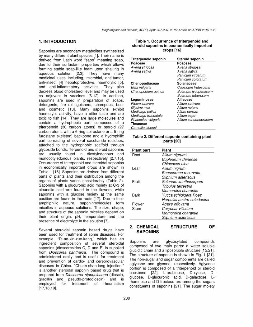
1. INTRODUCTION Saponins are secondary metabolites synthesized by many different plant species [1]. Their name is derived from Latin word “sapo” meaning soap, due to their surfactant properties which allows forming stable soap-like foam upon shaking in aqueous solution [2,3]. They have many medicinal uses including, microbial, anti-tumor, anti-insect [4] hepatoprotective, haemolytic [5], and anti-inflammatory activities. They also decrees blood cholesterol level and may be used as adjuvant in vaccines [6-12]. In addition, saponins are used in preparation of soaps, detergents, fire extinguishers, shampoos, beer and cosmetic [13]. Many saponins exhibit haemolytic activity, have a bitter taste and are toxic to fish [14]. They are large molecules and contain a hydrophobic part, composed of a triterpenoid (30 carbon atoms) or steroid (27 carbon atoms with a 6-ring spirostane or a 5-ring furostane skeleton) backbone and a hydrophilic part consisting of several saccharide residues, attached to the hydrophobic scaffold through glycoside bonds. Terpenoid and steroid saponins are usually found in dicotyledonous and monocotyledonous plants, respectively [2,7,15]. Occurrence of triterpenoid and steroidal saponins in economically important crops are shown in Table 1 [16]. Saponins are derived from different parts of plants and their distribution among the organs of plants varies considerably (Table 2). Saponins with a glucuronic acid moiety at C-3 of oleanolic acid are found in the flowers, while saponins with a glucose moiety at the same position are found in the roots [17]. Due to their amphiphilic nature, saponinmolecules form micelles in aqueous solutions. The size, shape, and structure of the saponin micelles depend on their plant origin, pH, temperature and the presence of electrolyte in the solution [7]. Several steroidal saponin based drugs have been used for treatment of some diseases. For example, “Di-ao-xin-xue-kang,” which has an ingredient composition of several steroidal saponins (dioscoresides C, D and E) is supplied from Dioscorea panthaica. The compound is administered orally and is useful for treatment and prevention of cardio- and cerebrovascular diseases in China. “Chuan-shan-long injection,” is another steroidal saponin based drug that is prepared from Dioscorea nipponicaand (dioscin, gracillin and pseudo-protodioscin) and is employed for treatment of rheumatism [17,18,19].
Table 1. Occurrence of triterpenoid and steroid saponins in economically important
crops [16]
Triterpenoid saponin Steroid saponin Poaceae Avena strigosa Avena sativa
Poaceae Avena strigosa Avena sativa Panicum virgatum Panicum coloratum
Chenopodiaceae Beta vulgaris Chenopodium quinoa
Solanaceae Capsicum frutescens Solanum lycopersicum Solanum tuberosum
Leguminosae Pisum sativum Glycine max Medicago sativa Medicago truncatula Phaseolus vulgaris
Alliaceae Allium sativum Allium nutans Alium porrum Allium cepa Allium schoenoprasum
Theaceae Camellia sinensi
-
Table 2. Different saponin containing plant
parts [20]
Plant part Plant
Root Allium nigrum L Bupleurum chinense Chiococca alba
Leaf Allium nigrum Beaucarnea recurvata Silphium asteriscus
Fruit Solanum xanthocarpum Tribulus terrestris Momordica charantia
Bark Yucca schidigera Roez Harpullia austro-caledonica
Flower Agave offoyana Stem Caryocar villosum
Momordica charantia Silphium asteriscus
2. CHEMICAL STRUCTURE OF SAPONINS
Saponins are glycosylated compounds composed of two main parts: a water soluble glucidic chain and a liposoluble structure [15,21]. The structure of saponin is shown in Fig. 1 [21]. The non-sugar and sugar components are called aglycone and glycone, respectively. Aglycone portion is composed of a triterpenoid or steroid backbone [22]. L-arabinose, D-xylose, D-glucose, D-glucuronic acid, D-galactose, L-rhamnose and D-fructose are among the sugars constituents of saponins [21]. The sugar moiety

Moghimipour and Handali; ARRB, 5(3): 207-220, 2015; Article no.ARRB.2015.022
209
is linked to the aglycone through an ester or ether glycosidic linkage at one or two glycosylation sites [23]. The aglycone may contain one or more unsaturated C–C bonds. When the oligosaccharide chain is attached at the C3 position the molecule is called monodesmosidic saponin, while saponins which have an additional sugar moiety at the C26 or C28 position, are named bidesmosidic [24]. The structure of saponins from different plant is depended on the types and amount of sugars, as well as the composition of steroid ring. It was observed that young plants have higher saponin contents than mature or old plants, although several factors such as physiological state and environmental factors affect the saponin contents [22]. Saponins are classified in two main groups according to the nature of their aglycone; saponosides with steroidic aglycone and saponosides with triterpenic aglycone. The steroidic aglycones have a skeleton with 27 carbon atoms. These molecules come from an intramolecular cetalisation which intervenes after oxidation in C16, C22 and C26 of a cholestanic precursor taking into account spiro-nature of C22; this hexacyclic skeleton is usually indicated by the spirostane term.In fresh plants, it is not rare that hydroxyl in C26 is engaged in a connection with a sugar. The structure may be pentacyclic; which is called
furostane. The triterpenic aglycones, come from the cyclization of the (3S)-2,3-epoxy-2,3-dihydrosqualene. This cyclization gives pentacyclic compounds like dammaranes, oleananes, ursanes, and hopanes. The majority of triterpenic sapogenins belong to these four basic skeletons. Different possible structures of saponins are shown in Fig. 2 [21].
2.1 Isolation and Identification of Saponins
The extraction techniques employed in saponin extraction include the conventional and the green technologies.The conventional extraction techniques are maceration, Soxhlet, and reflux extraction, where the green technologies are microwave-assisted, ultrasound-assisted and accelerated solvent extraction (Fig. 3). The conventional extraction is based on the solubility of solute from plant materials into solvent. So, it often utilizes a large quantity of solvent to extract the desired solute and sometimes is aided with elevated temperature by heating, and mechanical stirring or shaking. The green extraction method is involved less hazardous chemical synthesis, safer chemicals, energy efficiency and pollution prevention.
Fig. 1. Structure of saponin [21]

Moghimipour and Handali; ARRB, 5(3): 207-220, 2015; Article no.ARRB.2015.022
210
Fig. 2. Different possible structures of saponin aglycones [21]
Fig. 3. Current extraction techniques employed in extraction of saponins from plant materials [20]

Moghimipour and Handali; ARRB, 5(3): 207-220, 2015; Article no.ARRB.2015.022
211
3. CONVENTIONAL METHODS Maceration extraction: In the method, plant material is extracted by soaking the plant material in a specific solvent for a period of time. Ethanol and methanol are usually used as the extraction solvents to extract saponins from plant materials. Reflux and Soxhlet extractions: The only difference between reflux and Soxhlet is that Soxhlet apparatus consists of a thimble to house the plant material. This technique involves heating a solution to boiling and then returning the condensed vapors to the original flask. Subsequent extraction: The method is performed on plant materials using two extraction methods subsequently. Using this method may be lead to highly purify the extract before subjecting to HPLC analysis for isolation and identification of saponin.
3.1 Green Extraction Technologies Ultrasound-assisted extraction: The phenomenon of ultrasound creates cavitation bubbles in the solvent to denature the plant cell wall when the bubbles collapse at rare fraction resulted in a greater extraction yield of bioactive compounds. Microwave-assisted extraction: Microwaves are non-ionizing electromagnetic waves with a frequency range from 0.3 to 300 GHz. Microwaves are able to penetrate into biomaterials and generate heat by interacting with polar molecules such as water inside the materials. The water content of a plant material is responsible for the absorption of microwave energy which leads to internal superheating and cell structure disruption, and consequently, facilitates the diffusion of bioactive compound from the plant matrix. Accelerated solvent extraction: It is an automated rapid extraction technique that uses minimal solvent at elevated temperature and pressure. These processes are usually completed in 15–25 min using only 15–45 ml consumption of solvent. Using increased temperature enhances the solubility and mass transfer of solute to solvent, and elevated pressure keeps the solvent below its boiling point, enabling fast, safe, and efficient extraction of material from the plant [20].
There are several methods used for determination of saponins in plant material. Spectrophotometry, a simple and practical method, may be used to measure the amount of saponins [25]. Thin-layer chromatography (TLC) has been used successfully in the separation, purificationand determination of a large number of saponins in plant extracts [26,27,28]. Solvents which have been reported as suitable for developing thin layer plates are shown in Table 3 and different spray reagents that can be used which give characteristic colors with saponins are summarized in Table 4. Also, high performance liquid chromatography (HPLC) technique is widely employed for quantitative analysis of saponins (Table 5) [29]. C8 column (4.6×150 mm, 5 µm [30], C18 (25×0.4 cm, 5 µm) [31], C8 (250×4.6mm, 5mm I.D.) [32] and C18 column (250×10 mm, 5mm [10] are commonly used for HPLC detection. High-speed counter-current chromatography (HSCCC) is a continuous liquid–liquid partition chromatographic technique that in comparison with conventional column chromatography, eliminates the complications arising from the solid support matrix, such as stationary-phase deactivation, tailing of solute peaks, and contamination. It has been widely used to separate a variety of natural products including saponins [33]. Nuclear magnetic resonance (NMR) and fourier transform infra red (FTIR) are carried out to investigate unknown saponins in plant [27].
Table 3. Reported TLC solvents and their ratios for detection of saponins [34]
Solvent Type of saponin
Chloroform/methanol/water 65:20-30:10
non-polar
Chloroform/methanol/water 65:35:10
neutral and non-polar
Acetic acid/ethanol/water 70: 15:15
acid and neutral
n-Butanol/et hanoi/water 1:1:1
acid and neutral
n-Butanol/ethanol/1 M ammonia 60:13:30.5
polar and acid
n-Butanol/ethanol/l 5 M ammonia 7:2:5
polar and acid

Moghimipour and Handali; ARRB, 5(3): 207-220, 2015; Article no.ARRB.2015.022
212
Table 4. Spray agents suitable for detection saponins in TLC procedure [34]
Name Composition Condition Colors
Carr Price Saturated antimony trichloride in chloroform
Heat at 105°C for 15 min green-blue-grey
Liebermann-Burchard 30% Acetic anhydride in 50% osulphuric acid
Heat at 90 °C for 10 min green-blue
Vanillin-phosphoric acid
2% Solution of vanillin in phosphoric acid/ethanol (1:4)
Heat at 120 °C for 10-20 min
grey-blue-mauve
Ekkert 1% p-Anisaldehyde in acetic acid/sulphuric acid (98:2)
Heat at 90 °C for 10 min grey-lalue-mauv
Table 5. Different HPLC systems for analysis of saponins
Sample Column Mobile phase Detection (nm) Authors (Ref)
Codonopsis lanceolata C-18 acetonitrile: methanol: 0.1% aqueous formic acid (3: 2: 5, v/v/v)
207 Zhao et al. 2012 [35]
Moringa oleifera RP-C18 acetonitrile: water 250 -500 Sharma and Paliwal, 2013 [27]
Tribulus terrestris ODS-2 phosphoric acid buffer with pH 3
203 Ivanova et al., 2010 [36]
4. ORIGIN OF SAPONINS
Saponins are usually isolated from Agave attenuate, Cestrum parqui, Calliandra pulcherrima, Panax ginseng, Glycyrrhiza glabra, Allium sativum,A. nutans, A. minutiflorum [37], Saponaria officinalis, Quillaja saponaria, Gynostemma pentaphyllum [38], Achillea fragrantissima [39], Sesbania grandiflora [40], Sapindus mukorossi [41] and Medicago sativa[21,42]. The crude saponin extracts from flowering A. fragrantissima and vegetative part are 4 and 2.6%, respectively [39]. It has been reported that Platycodi Radix contains 1-4% triterpenoid saponins [43].In A. nutans, the concentration of saponins was determined to be 4% of total dry plant [16]. These compounds can be obtained from some marine organism [26] such as starfish, sponges and sea cucumbers [2,27,44]. Saponins are also found in defensive secretions of certain insects [21]. The two major commercial sources of saponins are Yucca schidigera, which grows in the arid Mexican desert, and Quillaja saponaria, a tree that grows in arid areas of Chile [45].
5. ACTIVITIES OF SAPONINS
5.1 Biological Action
5.1.1 Defensive role
It was shown that saponins protect plants from phytopathogenic microorganisms, insects and
phytophagous mammalian [42]. Their insecticidal activity may be related to the ability of producing alterations in the feeding behavior, in the molting process, interacting with hormones that regulate the growth and causing death in the different stages of development. Also, saponins can interact with the cell membranes and affect on the hydrophobic-lipophilic balance and permeability of these, because they are capable to form complex with cholesterol and reducing the rates of absorption [46].
Sea cucumbers are marine animals that are characterized by a slow motion and the absence of prominent structural defenses. So, they are vulnerable to predation. The body wall and viscera of these organisms contain saponins as defense systems to protect them [47]. Su et al. in 2008 investigated the bacterial resistance and immune response of white shrimp Litopenaeus vannamei against Vibrio alginolyticus while the shrimp were immersed in sea water containing different concentrations of saponin of Q. saponaria (0, 0.5, 1 and 2 mg L
-1) for 24, 48and
72 h. Their results showed that phagocytic activity was enhanced with increasing the saponin concentration. Phagocytic activity of shrimp immersed in 1 and 2 mg L
-1 of
saponinwas significantly higher than control group. Hyaline cells, the total haemocyte count, respiratory burst, superoxide dismutase activity, and glutathione peroxidase activity increased with enhancement of the saponin concentrations, whereas phenoloxidase activity was decreased.

Moghimipour and Handali; ARRB, 5(3): 207-220, 2015; Article no.ARRB.2015.022
213
They were concluded that L. vannamei immersed in water containing saponin could protect against V. alginolyticus infection [48].
5.2 Medicinal Applications 5.2.1 Antimicrobial activity Hazem et al. in 2012 reported that saponin extracted from flowering aerial part S of Achillea fragrantissima showed antifungal activity against Aspergillus, Fusarium and Rhizopus [39]. The saponin isolated from Capsicum frutescens, exhibited antifungal activity against Candida spp and Aspergillus fumigatus, with MICs ranging from 4.0 to 16 mg mL
-1 [49]. Yang et al. in 2006
investigated the antifungal activity of C-27 steroidal saponins against Candida albicans, C.glabrata, C. krusei, Cryptococcus neoformans, and Aspergillus fumigatus. These saponins showed significant activity against C. neoformans and A. fumigates that was comparable to the positive control amphotericin B [18]. Soetan et al. in 2006 studied the antimicrobial activity of saponins extract of Sorghum Bicolor against three pathogens, Escherichia coli, Staphylococcus aureus and C. albicans. The saponins inhibited the growth of the S. aureus, but not had inhibitory effect on E. coli and C.albicans. They demonstrated that
ineffectiveness of the saponins from S. bicolor on gram-negative bacteria and fungus may be as a result of the protective effect of the microbial membranes. The saponins may not be able to penetrate the cell membranes of the microorganisms [11]. Tsuzuki et al. in 2007 evaluated the antifungal activity of saponin extracted from Sapindus saponaria. The isolated saponins showed strong activity against C. parapsilosis [50]. Khanna et al. in 2008investigated the antimicrobial activity of saponin extracted from the leaves of Gymnema sylvestre and Eclipta prostrate. Their results revealed that the saponin fractions have significant antibacterial and antifungal activities [51]. Deshpande et al. in 2013 evaluated the antimicrobial activity of saponins isolated from roots of Cassia auriculata against Pseudomonas vesicularis, Streptococcus faecalis, Aeromonas hydrophilia, Salmonella typhae, Staphylococcus cohni, Serratia ficaria and E. coli at concentrations of 12.5, 25, 37.5 and 50 mg/ml. According to their results, saponins showed the best antimicrobial activity against P. vesicularis and least against E. coli at 50 mg/ml [52]. It has been previously reported that C. albicans, and C. tropicalic were sensitive to the saponins of G. glabra and Q. saponaria [53]. Other researches that evaluated the antimicrobial activity of saponins are summarized in Table 6.
Table 6. Some example of antimicrobial activity of saponins
Saponin Microorganisms Result Authors (Ref)
Quillja saponaria Pythium ultimum, Fusarium oxysporum, Alternaria solani, Colletotrichum coccodes, and Verticillium dahliae
The highest concentration (4%) of Q. saponaria showed moderate growth inhibition (35.9–59.1%) of all fungi except C. coccodes
Chapagain et al., [54]
Cyamopsis tetragonoloba
S. aureus, Salmonella Typhimurium, E. coli and Lactobacillus spp
100% MeOH fraction exhibited antibacterial activities against S. aureus, Salmonella Typhimurium and E. coli, but 20% and 60% MeOH fractions stimulated Lactobacillus spp. growth
Hassan et al., [55]
Solanum xanthocarpum and Centella asiatica
Klepsella pneumonia Saponin of S. xanthocarpumand C. asiatica inhibited the growth of K. pneumonia with diameter of zone of inhibition 19 and 21 mm, respectively.
Kannabiran et al., [56]
Anisopus mannii E. coli, K. pneumonia, Shigella dysentriae and Pseudomonas aeruginosa.
The saponin was more potent on E. coli (22.2 mm) and least on K. pneumonia (13.0 mm)
Aliyu et al., [57]

Moghimipour and Handali; ARRB, 5(3): 207-220, 2015; Article no.ARRB.2015.022
214
5.2.2 Anticancer activity
Yan et al. in 2009 evaluated the anticancer activity of steroid saponins isolated from the rhizome of Paris polyphylla var. yunnanensis. The saponins showed anticancer activity against lung adenocarcinoma cell line, both in vitro and in vivo. They demonstrated that the saponins could be regarded as promising drugs for cancer therapy [58]. Su et al. in 2011 investigated antitumor activity of polysaccharides and saponin extracted from sea cucumber. These results indicated that the in vitro anti-tumor effect of saponins is more potent than polysaccharides [44]. Several reports have been demonstrated that plants saponins can reduce the risk of colorectal cancer. Kim et al. in 2008 studied the apoptotic effect of crude saponins isolated from the roots of Platycodon grandiflorum in HT-29 human colon cancer cells. Their results showed saponins could inhibit HT-29 cell proliferation and induce apoptosis. The apoptosis was induced by DNA fragmentation and poly ADP-ribose polymerase (PARP) cleavage [43]. Rejinold et al. in 2011 prepared and evaluated saponin loaded chitosan nanoparticles as a cancer therapeutic agent for an enhanced and sustained release. They extracted saponin from Sapindus emarginatus and evaluated the cytotoxicity of the nanoparticles at different concentrations (0.1, 0.2, 0.4, 0.6, 0.8 and 1mg/ml) on mouse fibroblast cell line (L929), mouse embryonic fibroblast cell line (NIH-3T3), oral cancer cell line (KB) and prostate cancer cell line (PC3).The nanosaponin showed specific toxicity on prostate and oral cancer cells, while did not show any toxicity on normal L929 and NIH-3T3 cells. Many studies demonstrated that the induction of celldeath in cancer cells by anticancer saponins appeared in a dose and time-dependent manner. The results of Rejinold et al. study is in agreement with them. According to their results nanosaponin with higher concentrations of saponin could induce cancer cell death within 24h. They were concluded that the nanosaponin could be an efficient therapeutic agent for treatment of cancer [59]. Various mechanisms of growth inhibition of tumors cells are shown in Fig. 4. Some of saponins induce pore formation in mitochondrial membranes and induce apoptosis. In vitro studies in human glioma cells have showed that saponins may reduce protein expression which appears to be mediated through repressing the kinases MAPK1, MAPK3, MAPK8 and MAPK14. Saponins of Acacia victoriae promoted apoptosis by activation of
caspases and cytochrome C release. Another study reported that this saponin induces the expression of nuclear factor erythroid 2-related factor 2, a transcription factor, which mediates the expression of several detoxifying and antioxidant proteins. Apoptosis was observed in the cervix carcinoma cell line HeLa by induction of DNA fragmentation, upregulation of pro-apoptotic Bax, downregulation of anti-apoptotic Bcl-2 and caspase 3-activation. It was shown that soy saponin inhabited cell growth and reduced inflammatory responses by mediating increased inhibition of the transcription factor nuclear factor-kappa B (NFkB), which mediates expression of inflammatory proteins. These effects are the result of interference with degradation of the inhibitor of NFkB, IkB [60].
5.2.3 Anticardiovascular activity
It has been reported that ingestion of saponin containing food decrease cholesterol levels in the bloodstream and as a results decrease the risk of cardiovascular diseases [7, 61, 62]. It was also, reported that ginseng saponins decrease blood cholesterol levels in rabbits by increasing cholesterol excretion through bile acid formation [63].Elekofehinti et al. in 2012 showed that consumption of saponin from Solanum anguivifruit lead to reduction in the risk of hyperlipidemic symptoms and heart diseases [64]. It has been previously reported that the total saponins extracted from G. glabra and Q. saponaria were capable of forming complex with cholesterol. It was concluded that oral administration of total saponins of G. glabra and Q. saponaria may cause a reduction in cholesterol absorption through gastrointestinal system and as a result lowering the blood cholesterol [65].
5.2.4 Anti-inflammatory activity
Patel et al. in 2012 studied anti-inflammatory activity of saponin isolated from the Thespesia populnea (L.) leaves. According to their results, the saponin showed potent anti-inflammatory activity on acute and chronic inflammation models. They demonstrated that mechanisms for anti-inflammatory activity might be associated with the inhibition of prostaglandin and histamine [66]. Yassin et al. in 2013 investigated the anti-inflammatory activity of a saponin-containing fraction derived from methanolic extract of Gleditsia caspica fruits. They were observed that the saponin could significantly inhibit the progression of the inflammation in the treated

animals. They were demonstrated that the inhibitory effect of saponin could be due to inhibition of the enzyme cyclo-oxygenase and subsequent inhibition of prostaglandin synthe[67]. Different mechanisms are known for antiinflammatory activity of saponins. Saikosaponins induces anti-inflammatory effect by suppressing both the DNA binding activity and the nuclear translocation of nuclear factor of activated T cells (NF-AT) [68].Inflammation is managed by a large
Fig. 4. The schematic illustration depicts the different molecular pathways contributing to the anti-tumor properties of various saponins [60]
Fig. 5. Pharmacological targets of ginsenosides in inflammatory responses [69]NO: Nitric oxide, TNF-ɑ: Tumor necrosis factor alpha, PGE
adenosine monophosphate, ROS: phosphodiesterase, Akt: Protein Kinase B, P13K: Phosphatidylino
Moghimipour and Handali; ARRB, 5(3): 207-220, 2015; Article no.
215
animals. They were demonstrated that the inhibitory effect of saponin could be due to
oxygenase and subsequent inhibition of prostaglandin synthesis [67]. Different mechanisms are known for anti-inflammatory activity of saponins. Saikosaponins
inflammatory effect by suppressing both the DNA binding activity and the nuclear translocation of nuclear factor of activated T cells
8].Inflammation is managed by a large
amount of different pro-inflammatory mediators such as cytokines, nitric oxide (NO) and prostaglandin. Ginsenosides are biologically active saponin compounds found in ginseng. Some ginsenosides (e.g., GGRd and G-Rh2) can block TNFas well as the release of NO and PGE2, through repression of NF-kB activation signals. Pharmacological targets of ginsenosides in inflammatory responses are shown in Fig. 5 [69].
hematic illustration depicts the different molecular pathways contributing to the tumor properties of various saponins [60]
5. Pharmacological targets of ginsenosides in inflammatory responses [69]Tumor necrosis factor alpha, PGE2: Prostaglandin E2, cAMP:
adenosine monophosphate, ROS: Reactive oxygen species, PKA: Protein kinase AProtein Kinase B, P13K: Phosphatidylinositol-4,5-bisphosphate
3-kinase
; Article no.ARRB.2015.022
inflammatory mediators such as cytokines, nitric oxide (NO) and prostaglandin. Ginsenosides are biologically active saponin compounds found in Panax
. Some ginsenosides (e.g., G-Rb1, Rh2) can block TNF-a-production
as well as the release of NO and PGE2, kB activation signals.
Pharmacological targets of ginsenosides in inflammatory responses are shown in Fig. 5 [69].
hematic illustration depicts the different molecular pathways contributing to the
5. Pharmacological targets of ginsenosides in inflammatory responses [69]
, cAMP: Cyclic Protein kinase A, PDE:
bisphosphate

Moghimipour and Handali; ARRB, 5(3): 207-220, 2015; Article no.ARRB.2015.022
216
5.3 Saponins as Excipient
5.3.1. Adjuvants activity
The saponins are used as adjuvants in vaccines [7]. Q. saponaria saponins can stimulate both the humoral and the cellular immune responses against the pathogens. So, they can be used as adjuvants in vaccine formulations [42]. The mechanism of immune stimulatory action of saponins have not been clear, but it is believed that these compounds may induce production of cytokines such as interleukins and interferons in animal systems, that may be lead to stimulation of immune responses [22,24]. Kukhetpitakwong et al. in 2006 investigated immunological adjuvant activities of extracted saponin (methanolic fraction) from the pods of Acacia concinna on the cellular and humoral immune response of BALB/c mice against ovalbumin. Their results showed that saponin at concentrations of 40µg may activate T and B cells. Furthermore, ovalbumin specific IgG, IgG1 IgG2a and IgG2b antibody levels in serum were significantly enhanced by saponin as compared with ovalbumin control group. They were suggested that saponin might be effective on Th1 and Th2 helper T cells and at a dose of 40µg, could be used as vaccine adjuvant to increase immune responses [70].
5.3.2 Absorption enhancer Sajadi Tabassi et al. in 2007 evaluated the enhancing effect of total saponin extracted from Acanthophyllum squarrosum on intranasal insulin absorption in rat. According to their results, the saponin was able to improve insulin absorption through the nose and reduce blood glucose in rat [71].
5.4 Industrial Application
5.4.1 Cosmetics
Saponins are employed as stabilizers of cosmetic emulsions, and as foam intensification in shampoos and conditioners [7]. Alkanolamides are often used to prepare stable foam, but because of producing nitrosamines, they are potentially carcinogenic compounds. Aghel et al. in 2007 prepared an herbal shampoo using total saponins of Acanthophyllum squarrosum. Their results showed that the formulation containing 5% total saponin could produce stable foam in the absences of foam stabilizer. According to their results, alkanolamides can be substituted the saponins of A. squarrosum in shampoo formulation [72]. Also, saponins of Q. saponaria are used in cosmetics for preparation of lipstick and shampoo [73].
5.4.2 Food industries
The saponin of Q. saponaria has been exploited in food industries. It is used as foaming agents in beverages and confectionery [73]. Saponin of Chenopodium quinoa is used in preparation of beer [13]. Saponin of Quillaia is permitted to be used in food and “generally recognized as safe” (GRAS) in the USA. The usual saponin levels in use in the USA are shown in Table 7 [33].
The main known activates of saponins are summarized in Table 8 [24,50,57,60,67,74,75].
Table 7. Approximate average concentration of Quallaia saponin used in foodstuffs in the
USA
Foodstuff ppm
Beverages 95 Ice cream 0.12 Candy 18 Syrups 6.8
Table 8. A Summary of main known activities of saponins
Saponin Effect Ref.
Soyasaponin I reduction of lung metastases 60 Quillaja increases in immune-cell proliferation in vitro 24 Asparagus officinalis Antifungal properties in concentrations of 0·5 –8·0mg/ml depending
on the type of fungus 24
Vernonia amygdalina Anti-inflammatory activity 74 Maesa lanceolata Virucidal activity 24 Anabasis articulata Antibacterial activity 75 Gleditsia caspica anti-inflammatory activity 67 Acacia victoriae Anti-tumor activity 60 Anisopus mannii Antibacterial activity 57 Sapindus saponaria Antifungal activity 50

Moghimipour and Handali; ARRB, 5(3): 207-220, 2015; Article no.ARRB.2015.022
217
6. CONCLUSION Saponins are produced by plant, lower marine animals and some bacteria. They consist of a sugar moiety such as glucose, galactose, glucuronic acid and xylose that are linked to a hydrophobic aglycone which may be triterpenoid or steroid in nature. Several biological, pharmaceutical and industrial applications have been attributed to saponins including, immunostimulant, hypocholesterolaemic and anticarcinogenic properties. They have also many applications in food, agricultural and cosmetics industries. The wide spread incidence in plants and the potential pharmaceutical applications has led to extract of saponins and their identification in numerous species. Isolation and identification of the structure of saponins can be carried out using NMR, HPLC, GC and TLC.
COMPETING INTERESTS Authors have declared that no competing interests exist.
REFERENCES 1. Arif T, Bhosale JD, Kumar N, Mandal TK,
Bendre RS, Lavekar GS, Dabur R. Natural products – antifungal agents derived from plants. J Asian Nat Prod Res. 2009;11(7):621–638.
2. Faizal A, Geelen D. Saponins and their role in biological processes in plants. Phytochem Rev. 2013;12:877–893.
3. Hosamath PV. Evaluation of antibacterial activity of Litsea glutinosa. Int J Pharmaceut Appl. 2011;2(1):105-114.
4. Dixon RA, Sumner LW. Legume natural products: Understanding and manipulating complex pathways for human and animal health. Plant Physiol. 2003;131:878–885.
5. Bink A, Pellens K, Cammue BPA, Thevissen K. Anti-biofilm strategies: How to eradicate Candida biofilms? The Open Mycology J. 2011;5:29-38.
6. Hu X, Neil SJ, Cai W, Tang Z. Nitric oxide mediates elicitor-induced saponins synthesis in cell cultures of Panax ginseng. Funct Plant Biol. 2003;30:901-907.
7. Stanimirova R, Marinova K, Tcholakova S, Denkov ND, Stoyanov S, Pelan E. Surface rheology of saponin adsorption layers. Langmuir. 2011;27:12486–12498.
8. Oboh HA, Omofoma CO. The effects of heat treated lima beans (Phaseolus lunatus) on plasma lipids in hypercholesterolemic rats. Pak J Nutr. 2008;7(5):636-639.
9. Bhargava D, Shivapuri JN, Kar S, Pandit BR, Sidhiqie A, Upadhyay A, Thakur S, Mondal KC. Evaluation of antigonorrhoeal activity of saponins extract of Sapindus mukorossi Gaertn. Res J Pharm Biol Chem Sci. 2012;3(2):459-470.
10. Meesapyodsuk D, Balsevich J, Reed DW, Covello PS. Saponin biosynthesis in Saponaria vaccaria. cDNAs encodingb-amyrin synthase and a triterpene carboxylic acid glucosyltransferase. Plant Physiol. 2007;143:959–969.
11. Soetan KO, Oyekunle MA, Aiyelaagbe OO, Fafunso MA. Evaluation of the antimicrobial activity of saponins extract of Sorghum Bicolor L. Moench. Afr J Biotechnol. 2006;5:2405-2407.
12. Jyothi TC, Sindhu Kanya TC, Appu Rao AG. Influence of germination on saponins in soybean and recovery of soy sapogenol I. J Food Biochem. 2007;31:1–13.
13. Bhargava A, Shukla S, Ohri D. Chenopodium quinoa-an Indian perspective. Ind Crop Prod. 2006;23:73–87.
14. Ceyhun Sezgin AE, Aruk N. Determination of saponin content in Turkish Tahini Halvah by using HPLC. Adv J Food Sci Technol. 2010;2(2):109-115.
15. Mert-Turk F. Saponins versus plant fungal pathogens. J Cell Mol Biol. 2006;5:13-17.
16. De Geyter E, Lambert E, Geelen D, Smagghe G. Novel advances with plant saponins as natural insecticides to control pest insects. Pest Tech. 2007;1(2): 96-105.
17. Hostettmann K, Marston A. Chemistry and pharmacology of natural product: Saponins. University press.UK. 1995;18.
18. Yang CR, Zhang Y, Jacob MR, Khan SI, Zhang YJ, Li XC. Antifungal activity of C-27 steroidal saponins. Antimicrob Agents Chemother. 2006;50(5):1710–1714.
19. Qing LS, Xue Y, Zheng Y, Xiong J, Liao X, Ding LS, Li BG, Liu YM. Ligand fishing from Dioscorea nipponica extract using human serum albumin functionalized magnetic nanoparticles. J Chromatogr A. 2010;1217:4663–4668.
20. Cheok CY, Karim Salman HA, Sulaiman R. Extraction and quantification of saponins: A review.Food Res Int. 2014;59:16-40.

Moghimipour and Handali; ARRB, 5(3): 207-220, 2015; Article no.ARRB.2015.022
218
21. Chaieb I. Saponins as insecticides: A review. Tunis J Plant Prot. 2010;5:39-5.
22. Abid Ali Khan MM, Naqvi TS, Naqvi MS. Identification of phytosaponins as novel biodynamic agents: an updated overview. Asian J Exp .Biol. Sci. 2012;3(3):459-467.
23. Yücekutlu AN, Bildacı I. Determination of plant saponins and Some of Gypsophila species: A review of the literature. Hacettepe J Biol Chem. 2008;36(2):129-135.
24. Francis G, Kerem Z, Makkar HPS, Becker K. The biological action of saponins in animal system: A review. Br J Nutr. 2002;88:587–605.
25. Uematsu Y, Hirata K, Saito K. Spectrophotometric determination of saponin in Yucca extract used as food additive. J AOAC Int. 2000;83(6):1451-1454.
26. Oleszek W, Bialy Z. Chromatographic determination of plant saponins—an update (2002–2005). J Chromatogr A. 2006;1112:78–91.
27. Sharma V, Paliwal R. Isolation and characterization of saponin from Moringa oleifera (Moringaeceae) pods. Int J Pharm Pharm Sci. 2013;5(1):179-183.
28. Sharma V, Paliwal R. Isolation and characterization of saponins from Moringa oleifera (Moringaeceae) pods. Int J Pharm Pharm Sci. 2013;5(1):179-183.
29. Park IS, Kang EM, Kim N. High-performance liquid chromatographic analysis of saponin compounds in Bupleurum falcatum. J Chromatogr Sci. 2000;38:229-233.
30. Ahn MJ, Kim J. Identification and quantification of steroidal saponins in Polygonatum species by HPLC/ESI/MS. Arch Pharm Res. 2005;28(5):592-597.
31. Saxena D, Pul R, Dwivedi AK, Singh S. Characterization of sapindosides in Sapindus mukorosii saponins (reetha saponin) and quentitave determination of sapindoside B. J SciInd Res. 2004;63:181-186.
32. Combarieu E, Falzoni M, Fuzzati N, Gattesco F, Giori A, Lovati M, Pace R. Identification of Ruscus steroidal saponins by HPLC-MS analysis. Fitoterapia. 2002;73:583–596.
33. Zhao D, Yan M, Huang Y, Sun X. Efficient protocol for isolation and purification of different soyasaponins from soy
hypocotyls. J Sep Sci. 2012;35:3281–3292.
34. Oakenfull D. Saponin in food-a review. Food Chem. 1981;6:19-40.
35. Zhao B, Zhao W, Yuan Z. Optimization of extraction method for total saponins from Codonopsis lanceolata. Asian J of Traditional Medicines. 2012;7(1):14-17.
36. Ivanova A, Lazarova I, Mechkarova P, Tchorbanov B. HPLC method for screening of steroidal saponins and rutin as biological activity compounds in Tribulus terrestris. Biotechnol & Biotechnol. EQ. 2010;24:129-131.
37. Barile E, Bonanomi G, Antignani V, Zolfaghari B, Sajjadi SE, Scala F, Lanzotti V. Saponins from Allium minutiflorum with antifungal activity. Phytochemistry. 2007;68:596–603.
38. Utama-ang N, Chompreeda P, Haruthaithanasan V, Lerdvuthisopon N, Suwonsichon T, Wood K, Watkins BA. Identification of major saponins from Jiaogulan extract (Gynostemma pentaphyllum). Kasetsart J. (Nat. Sci.). 2006;40:59–66.
39. Hazem A, Fawzia AC, Dina G. Biochemical, antibacterial and antifungal activity of extracts from Achillea fragrantissima and evaluation of volatile oil composition. Der Pharmacia Sinica. 2012;3(3):349-356.
40. Goun E, Cunningham G, Chu D, Nguyen C, Miles D. Antibacterial and antifungal activity of Indonesian ethnomedical plants.Fitoterapia. 2003;76:592–596.
41. Suhagia BN, Rathod IS, Sindhu S. Sapindus mukorossi (Areetha): An overview. IJPSR. 2011;2(8):1905-1913.
42. Silva BP, Correa Soares JBR, Souza EP, Palatnikc M, Sousa CBP, Parente JP. Pulcherrimasaponin, from the leaves of Calliandra pulcherrima, as adjuvant for immunization in the murine model of visceral leishmaniasis. Vaccine. 2005;23:1061–1071.
43. Kim JY, Park KW, Moon KD, Lee MK, Choi J, Yee ST, Shim KH, Seo KI. Induction of apoptosis in HT-29 colon cancer cells by crude saponin from Platycodi Radix. Food Chem Toxicol. 2008;46:3753–3758.
44. Su X, Xue C, Li X, Gao X. Lou Y, Ding J. Antitumor activity of polysaccharides and saponin extracted from sea cucumber. Clinical and Cellular Imunology. 2011;2(1):1-5.

Moghimipour and Handali; ARRB, 5(3): 207-220, 2015; Article no.ARRB.2015.022
219
45. Cheeke PR. Actual and potential applications of Yucca schidigeraand Quillaja saponaria saponins in human and animal nutrition. J Anim Sci. 2000;77:1-10.
46. Pilla D’Incao1 M, Gosmann G, Machado V, Fiuza LM, Moreira GRP. Effect of saponin extracted from Passiflora alata Dryander (Passifloraceae) on development of the Spodoptera frugiperda (J.E. Smith) (Lepidoptera, Noctuidae).International J of Plant Research. 2012;2(5):151-159.
47. Dyck SV, Gerbaux P, Flammang P. Qualitative and quantitative saponin contents in five sea cucumbers from the Indian Ocean. Mar Drugs. 2010;8:173-189.
48. Su BK, Chen JC. Effect of saponin immersion on enhancement of the immune respose of withe shrimp Litopenaeus vannamei and its resistance against Vibrio alginolyticus. Fish & Shellfish Immunol. 2008;24:74-81.
49. Saleem M, Nazir M, Shaiq Ali M, Hussain H, Sup Lee Y, Riaz N, Jabbar A. Antimicrobial natural products: An update on future antibiotic drug candidates. Nat Prod Rep. 2010;27:238–254.
50. Tsuzuki JK, Svidzinski TIE, Shinobu CS, Silva LA, Rodrigues-Filho E, Cortez DAG, Ferreira ICP. Antifungal activity of the extracts and saponins from Sapindus saponaria L. An Acad Bras Cienc. 2007;79(4):577–583.
51. Khanna VG, Kannabiran K. Antimicrobial activity of saponin fractions of the leaves of Gymnema sylvestre and Eclipta prostrate. World J Microb Bio. 2008;24:2737-2740.
52. Deshpande S, Kewatkar S, Paithankar V. Antimicrobial activity of Saponins rich fraction of Cassia auriculata Linn against various microbial strains. IntCurr Pharm J. 2013;2(4):85-87.
53. Moghimipour E, Sadaghi-Nejad B, Handali S, Ameri A, Ramezani Z, Azemi ME. In vitro screening of anti-Candida activity of saponins extracted from Glycyrrhiza glabra and Quillaja saponaria. Asian J Pharm Clin Res. 2014;7(1):160-162.
54. Chapagain BP, Wiesman Z, Tsror (Lahkim) L. In vitro study of the antifungal activity of saponin-rich extracts against prevalent phytopathogenic fungi. Ind Crop Prod. 2007;26:109–115.
55. Hassan SM, Haq AU, Byrd JA, Berhow MA, Cartwright AL, Bailey CA. Haemolytic and antimicrobial activities of saponin-rich
extracts from guar meal. Food Chem. 2010;119:600–605.
56. Kannabiran K, Mohankumar T, Gunaseker V. Evaluation of antimicrobial activity of saponin isolated from Solanum xanthocarpum and Centella asiatica. Int. J Nat. Eng. Sci. 2009;3(1):25-28.
57. Aliyu AB, Musa AM, Abdullahi MS, Ibrahim MA, Tijjani MB, Aliyu MS, Oyewale AO. Activity of saponin fraction of Anisopus mannii against some pathogenic microorganisms. J med Plants Res. 2011;5(31):6709-6713.
58. Yan LL, Zhang YJ, Gao WY, Man SL, Wang Y. In vitro and in vivo anticancer activity of steroid saponins of Paris polyphylla var. yunnaensis. Exp Oncol. 2009:31(1):27–32.
59. Rejinolda NS, Muthunarayanan M, Muthuchelian K, Chennazhi KP, Nair SV, Jayakumar R.Saponin-loaded chitosan nanoparticles and their cytotoxicity to cancer cell lines in vitro. Carbohyd Polym. 2011;84:407–416.
60. Bachran C, Bachran S, Sutherland M, Bachran D, Fuchs H. Saponins in tumor therapy. Mini Rev Med Chem. 2008;8:575-584.
61. Afrose S, Sharoare Hossain Md, Salma U, Gaffar Miah A, Tsujii H. Dietary karaya saponin and Rhodobacter capsulatus exert hypocholesterolemic effects by suppression of hepatic cholesterol synthesis and promotion of bile acid synthesis in laying hens. Cholesterol. 2010;2010:1-7.
62. Lee MR, Chan Kim B, Kim R, Oh HI, Kyoung Kim H, Choi KJ, Sung CK. Anti-obesity effects of black ginseng extract in high fat diet-fed mice. J Ginseng Res. 2013;37(3):308-314.
63. EL-Farok DHM, EL-Denshry ES, Mahmou M, Asaaf N, Emam M. Lipid lowering effect of ginseng and alpha-lipoicacid in hypercholesterolemic patients. Global J of Pharmacology. 2013;7(3):298-306.
64. Elekofehinti OO, Adanlawo IG, Saliu JA, Sodehinde SA. Saponins from Solanum anguivi fruits exhibit hypolipidemic potential in Rattus novergicus. Der Pharmacia Lettre. 2012;4(3):811-814.
65. Moghimipour E, Kooshapour H, Rezaee S, Khalili S, Handali S. In vitro cholesterol binding affinity of total saponin extracted from Glycyrrhiza glabra. Asian J Pharm Clin Res. 2014;7(1):170-173.

Moghimipour and Handali; ARRB, 5(3): 207-220, 2015; Article no.ARRB.2015.022
220
66. Patel PP, Patil PH. Anti-inflammatory activity of saponin rich fraction isolated from the Thespesia populnea (L.) leaves. Intl J Biomed Pharma Sci. 2012;3(4):1526-1532.
67. Yassin NZ, Melek FR, Selim MA, Kassem IAA. Pharmacological activities of saponin-containing fraction derived from Gleditsia caspica Desf. methanolic fruit extract. Der Pharmacia Lettre. 2013;5(2):247-253.
68. Yadav VR, Prasad S, Sung B, Kannappan R, Aggarwal BB. Targeting inflammatory pathways by triterpenoids for prevention and treatment of cancer. Toxins. 2010;2:2428-2466.
69. Park J, Cho JY. Anti-inflammatory effects of ginsenosides from Panax ginseng and their structural analogs. Afr J Biotechnol. 2009;8(16):3682-3690.
70. Kukhetpitakwong R, Hahnvajanawong C, Homchampa P, Leelavatcharamas V, Satra J, Khunkitti W. Immunological adjuvant activities of saponin extracts from the pods of Acacia concinna. Int Immunopharmacol. 2006;6:1729–1735.
71. Sajadi Tabassi SA, Hosseinzadeh H, Ramezani M, Moghimipour E, Mohajeri SA. Isolation and characterization and
study of enhancing effect on nasal absorption of insulin in rat of the total saponin from Acanthophyllum squarrosum. Indian J Pharmacol. 2007;39(5):226-230.
72. Aghel N, Moghimipour E, Raies Dana A. Formulation of a herbal shampoo using total saponins of Acanthophyllum squarrosum. Iran J Pharm Res. 2007;6 (3):167-172.
73. Zengin ACA. Potential application Q. saponaria as an antimicrobial soaking agent in leather industry. J of Textile and Apparel. 2013;23(1):55-61.
74. Adiukwu PC, Kayanja FIB, Nambatya G, Adzu B, Twinomujuni S, Twikirize O, Ganiyu AA, Uwiduhaye E, Agwu E, Tanayen JK, Nuwagira P, Buzaare P. Anti-Inflammatory and anti-pyretic activity of the leaf, root and saponin fraction from Vernonia amygdalina. Br J Pharmacolo Toxicol. 2013;4(2):33-40.
75. Maatalah MB, Bouzidi NK, Bellahouel S, Merah B, Fortas Z, Soulimani R, Saidi S, Derdour A. Antimicrobial activity of the alkaloids and saponin extracts of Anabasis articulate. J Biotechnol. Pharm. Res. 2012;3(3):54-57.
________________________________________________________________________________ © 2015 Moghimipour and Handali; This is an Open Access article distributed under the terms of the Creative Commons Attribution License (http://creativecommons.org/licenses/by/4.0), which permits unrestricted use, distribution, and reproduction in any medium, provided the original work is properly cited.
Peer-review history: The peer review history for this paper can be accessed here:
http://www.sciencedomain.org/review-history.php?iid=668&id=32&aid=6212