s.-a. the temporomandibular pain -dysfunction syndrome
TRANSCRIPT

Junie 1974 S.-A. MEDIESE TYDSKRlf 1103
The Temporomandibular Pain -DysfunctionSyndrome
L. M. JONCK
SUMMARY
The temporomandibular pain-dysfunction syndrome is reviewed. The functional anatomy and the neurophysiologicalmechanism of the temporomandibular joint are discussed.The purpose of treatment is to reintroduce synchronisationof the moving parts of the joint.
S. Afr. Med. l., 48, 1103 (1974).
At academic level the aetiology of the temporomandibular(fM) pain-dysfunction syndrome is controversial becauseof confusion at clinical level, and this results in theappJjcation of principles and methods of therapy whichare often ineffective, excessive, or even damaging to thejoint.
Headache is man's most common complaint, and forthe clinician one of the most perplexing problems whichmay confront him. To pinpoint the source of the problemis difficult, because the symptoms are subjective and thedistribution of the pain can seldom be related to theactual structure involved.
Disturbances in the region of the TM joint give rise toa symptom complex where the pain distribution is suchthat more than one discipline of medicine may becomeinvolved: the ear, nose and throat specialist because of painin the ear and temporal area; the neurologist, orthopaedicsurgeon and specialist physician because of headaches,especially those involving the occipital and neck areas:dentists. oral surgeons and prosthodontists, because dentalproblems often cause pain referring to the face.
The temporomandibular pain-dysfunction syndrome isdue to a functional imbalance arising in the stomatognathiccomplex, involving the masticatory muscles, the temporomandibular joint itself and an incorrect jaw relationship.
REVIEW OF LITERATURE
Costen'·' in 1934 first focused attention on the dysfunctionsyndrome of the temporomandibular joint. and for manyyears the syndrome bore his name.
The mechanism of this syndrome was based on thesupposition that the bite of the jaws was too closed,leading to a posterior displacement of the condyle withconcomitant compression of the Eustachian tube andeven medial pressure on the chorda tympani nerve and
Faculty of Dentistry, University of PretoriaL. M. ]ONCK, M.SC., M.CH.D., D.Se.
Date received: 4 December 1973.
10
that retrusion of the mandible affected the auriculotemporal nerve. The syndrome included the followingsymptoms: pain in the region of the ear; a 'blocked'feeling in the ear and tinnitus; neuralgia in the distributionof the second and third divisions of the fifth cranialnerve; and headache, pain, or a burning sensation of thetongue.
Those who rejected Costen's anatomical explanation"·suggested a disturbance of the TM joint.
Since the time of Costen, many theories have beenadvanced. Granger' supported the theory of occlusal dysharmony, and suggested that tooth cusps are arranged ina manner which prevents free lateral and protrusivemovements of the jaw during mastication. It was alsosuggested that this dysharmony is the result of earlyloss of teeth. which leads to a backward displacement ofthe condyles with resultant compression of the highlyvascular and nerve-rich posterior part of the meniscus.The tooth-muscle theory, less mechanically orientated,postulated that, as a result of occlusal dysharmony. proprioceptive impulses arise which are responsible for'muscular imbalance and the associated symptoms.
Schwartz' is perhaps the one who has contributed mostto our knowledge of the TM joint and its functionaldisturbances, which he ascribes primarily to a masticatorymuscle imbalance.
In association with this theory is the psychophysiologicalconcept of Yemm9 and ewton,lO viz. that muscle spasm.as a result of tension, is the primary factor in the dysfunction syndrome. Pathological changes in the joint onlyarise at a much later stage. The muscular imbalance mayalso result in cuspal interference of the teeth. and maybe responsible for.a vicious cycle of muscular spasms.
Not one of the above theories is wholly accepted.The pathogenesis of the symptom complex remains toa great extent a mystery, but in the light of our cliaicalobservations and treatment, our knowledge is increasing.
THE STOMATOGNATIDC SYSTEM A!'.TJ) THEMECHANISM OF PAIN
This system represents an anatomical and physiologicalunit and includes the teeth and supporting tissues, theTM joints, ligaments, muscles and bones. as well as theafferent and efferent nerves and that part of the centralnervous system responsible for co-ordination and control.The teeth and the emotional state of the patient are themost variable components of the system.
During movement of the mandible, the mu c1es, ligaments and the meniscus are more important than the
1104 S.A. MEDICAL JOURNAL I June 1974
condyle and the glenoid fossa. Traditionally, only themasticatory muscles are associated with movements ofthe TM joints, but it is known that a tonic reactionoccurs in the whole of the muscular system, which extendsfrom the sternum to the hyoid bone, thence to themandible, the temporal, the occipital, the post- andprevertebral areas and the pectoral girdle. With everymovement of the mandible the tonus of this group ofmuscles is influenced.
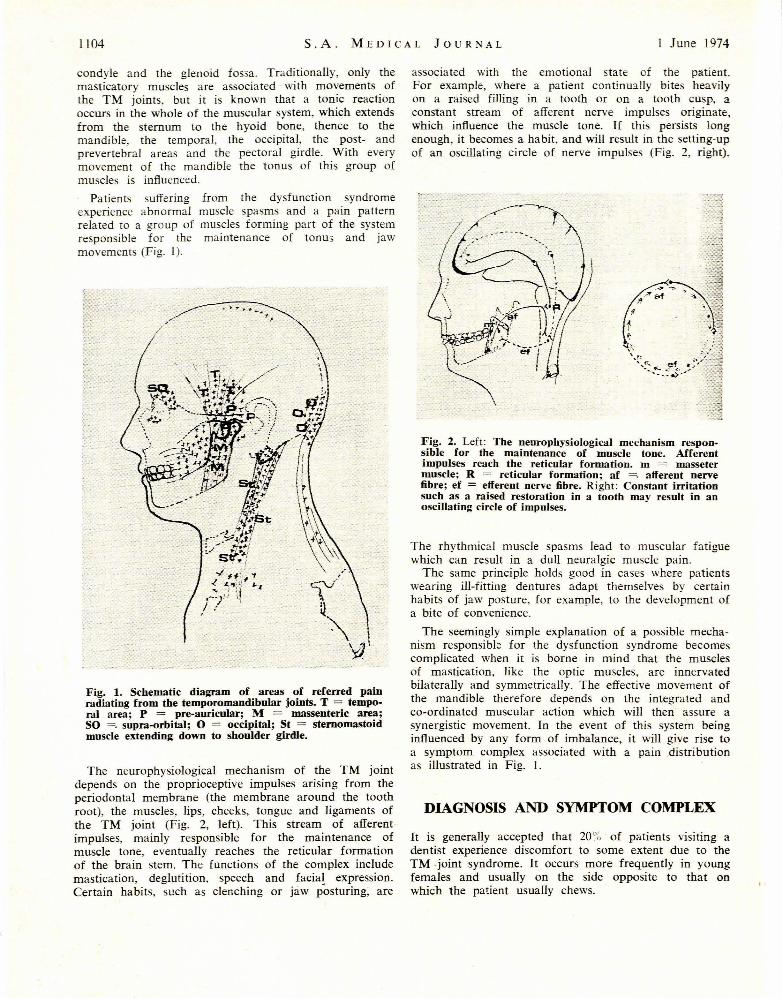
Patients suffering from the dysfunction syndromeexperience abnormal muscle spasms and a pain patternrelated to a group of muscles forming part of the systemresponsible for the maintenance of tonus and jawmovements (Fig. 1).
Fig. 1. Schematic diagram of areas of referred painradiating from the temporomandibular joints. T = temporal area; P = pre-auricular; M = massenteric area;SO =. supra-orbital; 0 = occipital; St = sternomastoidmuscle extending down to shoulder girdle.
The neurophysiological mechanism of the TM jointdepends on the proprioceptive impulses arising from theperiodontal membrane (the membrane around the toothroot), the muscles, lips, cheeks, tongue and ligaments ofthe TM joint (Fig. 2, left). This stream of afferentimpulses, mainly responsible for the maintenance ofmuscle tone, eventually reaches the reticular formationof the brain stem. The functions of the complex includemastication, deglutition, speech and facia! expression.Certain habits, such as clenching or jaw posturing; are
associated with the emotional state of the patient.For example, where a patient continmlly bites heavilyon a raised filling in a tooth or on a tooth cusp, aconstant stream of afferent nerve impulses originate,which influence the muscle tone. If this persists longenough, it becomes a habit, and will result in the setting-upof an oscillating circle of nerve impulses (Fig. 2, right).
Fig. 2. Left: The neurophysiological mechanism responsible for the maintenance of muscle tone. Afferentimpulses reach the reticular formation. m =. massetermuscle; R = reticular formatiou; af =. afferent nervefibre; ef = efferent nerve fibre. Right: Constant irritationsuch as a raised restoration in a tooth may result in anoscillating circle of impulses.
The rhythmical muscle spasms lead to muscular fatiguewhich can result in a dull neuralgic muscle pain.
The same principle holds good in cases where patientswearing ill-fitting dentures adapt themselves by certainhabits of jaw posture, for example, to the development ofa bite of convenience.
The seemingly simple explanation of a possible mechanism responsible for the dysfunction syndrome becomescomplicated when it is borne in mind that the musclesof mastication, like the optic muscles, are innervatedbilaterally and symmetrically. The effective movement ofthe mandible therefore depends on the integrated andco-ordinated muscular action which will then assure asynergistic movement. In the event of this system beinginfluenced by any form of imbalance, it will give rise toa symptom complex associated with a pain distributionas illustrated in Fig. 1.
DIAGNOSIS AND SYMPTOM COMPLEX
It is generally accepted that 20~~ of patients visiting adentist experience discomfort to some extent due to theTM .joint syndrome. It occurs more frequently in youngfemales and usually on the side opposite to that onwhich the patient usually chews.

I Junie 1974 S.-A. MEDIESE TYDSKRIF 1105
Two aspects determine the success of treatment; correctdiagnosis, and a profound knowledge of the aetiology.
The aetiological aspect of the syndrome covers a widefield, from the purely mechanical concept of an occlusalaetiology (it is where the teeth do not fit well) to thebroader concept of a functional disturbance of the wholestomatognathic complex, which is under the influence ofthe higher centres and where the psychological characteror make-up of the patient plays an important role.
The diagnosis of the syndrome is based on the followingsymptoms:
1. Pain occurs in the pre-auricular area, usually unilaterally, and is described as a dull pain. It may radiate tothe angle of the jaw, temporal and cervical areas, andmay be severe, especially on awakening, and may oftenincrease in severity during the day. Due to the closeassociation between the TM joint and external auditorymeatus, the patient is sometimes convinced that theorigin of the pain is in the ear.
2. There is a tenderness of the muscles in and aroundthe joint, especially the pterygoid, masseter and temporalmuscles.
3. The patient often complains of crepitus or clickingnoises in the affected joint. This clicking noise is often anindication of dysfunction of the muscles affecting thejoint.
4. Limited mandibular movement and, on opening themouth, deviation to the side where the symptoms originate.
5. Clinically and radiologically there are no pathologicalchanges to be seen in the TM joint, and palpation throughthe external auditory meatus elicits no pain.
From this can be deduced that the primary causativefactor of the TM joint pain-dysfunction syndrome isa functional one, which may well lead to organic changeon the articular surfaces at a later stage.
It would appear that the symptoms develop suddenlyor gradually, and the first complaint is often a vagueheadache or earache, or a stuffy, blocked feeling in the ear.Quite often the pain comes on after an innocent chewingmovement or an exaggerated yawn. In keeping with this,pain may develop after some innocent dental procedurerequiring a patient to maintain the mouth in an openposition for some time, or may follow tonsillectomy.
Clicking or crepitus in the joint is a common sign.The patient usually waits from 3 to 6 months beforeseeking medical opinion. There are sometimes complaintsof fibrositis and rheumatism.
At clinical examination, special note is taken of thepresence or absence of sufficient support in the posteriorsegments of the jaw; abnormal facets on the teeth whichare indications of detrimental jaw movements, especiallygrinding of teeth (bruxing); any deviation in the final3 mm of closure of the jaws; the free-way space; andthe jaw relationships. It is also very important to forman opinion of the emotional state of the patient. It isimperative to examine the mouth critically to eliminateany form of underlying pathology such as retained roots,impacted third molars etc., since these alone may give riseto abnormal muscular spasm as a result of chronicinfection.
11
TREATMENT
The patient must be assured that the problem is not seriousand that it can be treated conservatively. The mechanismof jaw movements is explained, and muscle exercisesprescribed to correct the imbalance and to normalise themovements of the mandible.
This involves changing the habits of the patient; myotherapeutic exercises and relief of acute episodes of painwith sedatives, analgesics or tranquillisers. Where tbepatient can be well-motivated, active treatment with aremovable appliance is usually unnecessary, since 50% ofpatients react favourably to conservative treatment. Thistreatment can be supplemented by physiotherapy.
Heat and cold, alternately applied, may have a calmingaction, which is analgesic and relieves pain resulting frommuscle spasm. Microwave and ultrasonic treatment isof greater value because it penetrates the tissues to adepth of 4 - 5 cm. They are applied for 3 - 10 minutesdaily for 10 days and are contra-indicated when theorbital area is affected, especially in young children.
Muscle exercises against resistance are encouraged.Retrusive relaxation exercises are of great value. Localanaesthetics and the spraying of ethyl chloride on the skinsometimes aid in relieving muscle spasm.
Nervous tension and anxiety sometimes play an important role in the pathogenesis and continuation of the paindysfunction syndrome. Tranquillisers combined with aspirincan be advantageously prescribed in these cases. Increasedmuscular activity is a common manifestation of subjugated emotional stress, fear, anxiety and frustration,and may give rise to abnormal jaw movements.
Concerning the active treatment, it is very important toremove irregular occlusal cuspal interferences and toreplace lost molars. Prevention of abnormal chewinghabits by means of a removable appliance is of value.Surgery and intra-articular injections are resorted to inspecial cases only.
Cortisone preparations sometimes give dramatic relief.The mechanism is unknown, since there is no proved inflammation present in the TM joint of the patient.
The benefit derived may lie in the fact that the limitedmovement in the joint after injection ensures a forcedperiod of rest. From a psychological point of view, thevery fact that something positive is being done, as wellas the patient's faith in injections, may also be beneficial.All surgical techniques are aimed at relieving musclespasm. Procedures which can be considered for suitablecases are a partial or complete myotomy of one of themuscles of mastication; or a high neck or intracapsularcondylectomy, with or without the removal of the disc.Even a coronoidectomy is successful in a large percentageof cases, probably because it brings about an alteredjaw relationship. In so doing, the overactivity of themuscles attached to the specific bone fragments is altered,with resultant immediate relief of symptoms.
Once again, it must be emphasised that the indicationsfor surgery in these cases of temporomandibular dysfunction syndrome are extremely limited, and should only

1106 S.A. MEDICAL JOURNAL 1 June 1974
be considered when all conservative measures have failedto restore the correct jaw relationship to within norma!physiological limits.
It is our belief that myofacial pain-joint dysfunctionsyndrome is basically a reversible physiological condition,and treatment should aim at reintroducing synchronisationof movements of the joint and other structures involved,thus restoring over-all functional harmony.
REFERENCES
I. Costen, l. B. (1934): Ann. Otol. Rhin. Laryng., 43, 12. Idem (1939): Ann. Otol., 48, 499.3. Hankey, G. T. (1954): Brit. Dent. l., 97, 249.4. Christensen, G. (1956): Ibid., I, 249.5. Schwartz, L. (1959): Disorders of the Temporomandibular Joint.
Philadelphia: W. B. Saunders.6. Idem (1955): J. Amer. Dent. Assoc., 51, 394.7. Granger, E. R. (1958): Ibid., 56, 659.8. Schwartz, L. (1955): Ibid., 51, 394.9. Yemm, R. (1969): Brit. Dent. l., 127, 508.
10. Newton, A. V. (1969): J. Prosth. Dentistry, 22, 647.
Amniotiese Bande by 'n Baba naAbdominale Swangerskap
'N GEVALBESPREKlNG
J. DU P. DE WIT,
SUMMARY
A baby, delivered by Caesarean section after a 36-weekextra-uterine pregnancy, with deformities due to amnioticbands, is described. The mechanism of amniotic banddeformities and the paediatric risks of extra-uterine pregnancy are briefly discussed.
S. A/r. Med. l., 48, 1106 (1974).
Daar is heelwat teoriee oor die ontstaan en gevolge vanamniotiese bande. Baie min inligting is egter gepubliseeri.v.m. die lot van die lewensvatbare baba na ekstra-uterineswangerskap. 'n Geval wat die kenmerke van albei toestande demonstreer, is onlangs teegekom.
'n Vyf-en-twintigjarige Bantoevrou met swangerskapsduur van ongeveer 36 weke is onlangs in die Kalafonghospitaal opgeneem. Ta kliniese ondersoek is 'n ekstrauterine swangerskap gediagnoseer. 'n Laparotomie is
Departement Kindergeneeskunde, Universiteit van Pretoriaen Kalafong-hospitaal, Pretoria
J. DU P. DE WIT, M.B. CH.B., D.A., M.MED. (PAED.)J. G. PRINSLOO, M.B. CH.B., M.MED. (PAED.), M.D.
Datum ontvang: 29 Januarie 1974.
J. G. PRINSLOO
gedoen en die fetus is in die vrve buikholte binne-in 'nintakte vrugsak gevind. Die plasenta was gesetel in diegebied van die linker FaIlopius-buis. 'n Manlike baba metmassa 2,08 kg is verlos.
Die baba het 'n Apgar-telling van 2 uit 10 een minuutna geboorte gehad en hy is geresussiteer. Hy het gouself begin asemhaal, maar het sianoties geword tensysuurstof toegedien is. Sy spiertonus was swak. Daar was'n Iitteken wat vanaf albei mondboeke oor die wangetot bo-oor die kop gestrek het, en stukkies vliese wasdaaraan geheg (Afb. 1). Dieselfde tipe letsel kon skuinsoor die rug gesien word (Afb. 2). Geen ander abnormaliteite kon klinies vasgestel word rue. R6ntgenfoto'svan die borskas het gedeeltelike kollaps van die linkerbokwab asook kollerige versluiering in die regter bokwabaangetoon. Die baba is met 'n binneaarse infuus beh~ndelen die suur-basisbalans is gekontroleer.
Die baba het hipotorues gebly en sy voedings swakgeneem en moes met 'n nasogastriese bills gevoed word.Die longbeeld het rontgenologies verbeter, maar sianotieseaanvalle het met tussenpose gedurende die eerste 7 lewensdae voorgekom. 'n Lumbaalpunksie is gedoen, maaronde-rsoek van die serebrospinaalvog het geen afwykingsaangetoon nie_ Nege dae na geboorte het die babad~arree ontwikkel. 'n Enteropatogene Escherichia coli