title: temporomandibular joint dysfunction (tmjd) testing
TRANSCRIPT

1
____________________________________________________________________________
Medical Policy
BCN Medical Policies are a source for BCN medical policy information only. These documents are not to be used to determine benefits or reimbursement. Please reference the
appropriate certificate or contract for benefit information. ____________________________________________________________________________
BCN Policy Effective Date: 7/15/21 (See policy history boxes for previous effective dates)
Title: Temporomandibular Joint Dysfunction (TMJD) Testing
and Treatment Description/Background Temporomandibular joint disorder refers to a group of disorders characterized by pain in the temporomandibular joint and surrounding tissues. Initial conservative therapy is generally recommended; there are also a variety of non-surgical and surgical treatment possibilities for patients whose symptoms persist. BACKGROUND Diagnosis of Temporomandibular Joint Disorder (TMJD) In the clinical setting, TMJD is often a diagnosis of exclusion and involves physical examination, patient interview, and dental record review. Diagnostic testing and radiologic imaging is generally only recommended for patients with severe and chronic symptoms. Diagnostic criteria for TMJD have been developed and validated for use in both clinical and research settings.(1-3) Symptoms attributed to TMJD vary and may include clicking sounds in the jaw, headaches, closing or locking of the jaw due to muscle spasms (trismus) or displaced disc, pain in the ears, neck, arms, and spine; tinnitus, and bruxism (clenching or grinding of the teeth). Treatment For many patients, symptoms of TMJD are short-term and self-limiting. Conservative treatments, such as eating soft foods, rest, heat, ice, avoiding extreme jaw movements, and anti-inflammatory medications are recommended prior to consideration of more invasive and/or permanent therapies (e.g. surgery).
2
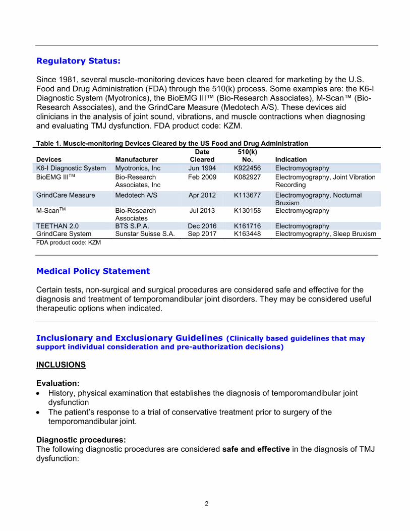
Regulatory Status: Since 1981, several muscle-monitoring devices have been cleared for marketing by the U.S. Food and Drug Administration (FDA) through the 510(k) process. Some examples are: the K6-I Diagnostic System (Myotronics), the BioEMG III™ (Bio-Research Associates), M-Scan™ (Bio-Research Associates), and the GrindCare Measure (Medotech A/S). These devices aid clinicians in the analysis of joint sound, vibrations, and muscle contractions when diagnosing and evaluating TMJ dysfunction. FDA product code: KZM. Table 1. Muscle-monitoring Devices Cleared by the US Food and Drug Administration Devices
Manufacturer
Date Cleared
510(k) No.
Indication
K6-I Diagnostic System Myotronics, Inc Jun 1994 K922456 Electromyography BioEMG IIITM Bio-Research
Associates, Inc Feb 2009 K082927 Electromyography, Joint Vibration
Recording GrindCare Measure Medotech A/S Apr 2012 K113677 Electromyography, Nocturnal
Bruxism M-ScanTM Bio-Research
Associates Jul 2013 K130158 Electromyography
TEETHAN 2.0 BTS S.P.A. Dec 2016 K161716 Electromyography GrindCare System Sunstar Suisse S.A. Sep 2017 K163448 Electromyography, Sleep Bruxism FDA product code: KZM Medical Policy Statement Certain tests, non-surgical and surgical procedures are considered safe and effective for the diagnosis and treatment of temporomandibular joint disorders. They may be considered useful therapeutic options when indicated. Inclusionary and Exclusionary Guidelines (Clinically based guidelines that may support individual consideration and pre-authorization decisions) INCLUSIONS Evaluation: • History, physical examination that establishes the diagnosis of temporomandibular joint
dysfunction • The patient’s response to a trial of conservative treatment prior to surgery of the
temporomandibular joint. Diagnostic procedures: The following diagnostic procedures are considered safe and effective in the diagnosis of TMJ dysfunction:

3
• Diagnostic X-ray, tomograms and arthrograms • CT scan or MRI (generally CT scans and MRIs are reserved for presurgical evaluations) • Cephalograms (x-rays of jaws and skull) • Pantograms (panoramic x-rays of maxilla and mandible) Non-surgical treatments: The following non-surgical treatments are considered safe and effective in the treatment of TMJ dysfunction: • Intraoral removable prosthetic devices/appliances (encompassing fabrication, insertion,
adjustment) • Pharmacologic treatment (such as anti-inflammatory, muscle relaxing and analgesic
medications). Surgical treatments: The following surgical treatments may be covered in the treatment of TMJ dysfunction: • Arthrocentesis, with or without ultrasound guidance • Manipulation for reduction of fracture or dislocation of the TMJ • Arthroscopic surgery in patients that objectively demonstrate (by physical examination or
imaging) internal derangements (displaced discs) or degenerative joint disease who have failed conservative treatment
• Open surgical procedures including, but not limited to, arthroplasties, condylectomies, meniscus or disc plication and disc removal, when TMJ dysfunction is the result of congenital anomalies, trauma or disease in patients who have failed conservative treatment
EXCLUSIONS Diagnostic procedures: The following diagnostic procedures are considered experimental/investigational in the diagnosis of TMJD: • Electromyography (EMG), including surface EMG • Kinesiography • Thermography • Neuromuscular junction testing • Somatosensory testing • Transcranial or lateral skull x-rays • Intra-oral tracing or gothic arch tracing (intended to demonstrate deviations in the positioning
of the jaws that are associated with TMJD) • Muscle testing • Standard dental radiographic procedures • Range of motion measurements • Computerized mandibular scan (this measures and records muscle activity related to
movement and positioning of the mandible and is intended to detect deviations in occlusion and muscle spasms related to TMJD)
• Ultrasound/sonogram (ultrasonic Doppler auscultation) • Joint vibration analysis

4
Non-surgical treatments*: The following non-surgical treatments are considered investigational in the treatment of TMJD: • Electrogalvanic stimulation • Iontophoresis • Biofeedback • Ultrasound • Devices promoted to maintain joint range of motion and to develop muscles involved in jaw
function • Orthodontic services/treatment • Dental restorations/prosthesis/treatment/appliances • TENS (transcutaneous electrical nerve stimulation) • PENS (percutaneous electrical nerve stimulation) • Acupuncture • Hyaluronic Acid • Platelet concentrates *Intra-oral reversible orthotic device (also known as occlusal orthotic, occlusal guard or bite splint), including fabrication, insertion and adjustment of the device is a certificate exclusion in most cases. Refer to current certificate. Surgical treatments: The following surgical procedure is considered experimental/investigational: • Arthroscopy of the TMJ for purely diagnostic purposes. CPT/HCPCS Level II Codes (Note: The inclusion of a code in this list is not a guarantee of coverage. Please refer to the medical policy statement to determine the status of a given procedure) Established codes: 20605 20606 21010 21050 21060 21070 21073 21116 21240 21242 21243 21480 21485 21490 29804 70328 70330 70332 70336 70350 70355 70486 70487 70488 97010 97024 Other codes (investigational, not medically necessary, not a benefit, etc.):
21089 21299 29800 J7321 J7323 J7324 J7325 J7326 E1399 97026 Any dental code

5
Rationale DIAGNOSIS OF TEMPOROMANDIBULAR DISORDER Clinical Context and Test Purpose TMJD (also known as temporomandibular joint syndrome) refers to a cluster of problems associated with the temporomandibular joint and musculoskeletal structures. The etiology of TMJD remains unclear and is believed to be multifactorial. TMJD is often divided into two main categories: articular disorders (eg, ankylosis, congenital or developmental disorders, disc derangement disorders, fractures, inflammatory disorders, osteoarthritis, joint dislocation) and masticatory muscle disorders (eg, myofascial pain, myofibrotic contracture, myospasm, neoplasia). The purpose of specific diagnostic tests in patients with suspected TMJD is to provide an option that is an alternative to or an improvement on existing diagnostic approaches, such as a comprehensive history and physical exam and alternative diagnostic tests. The question addressed in this evidence review is: do specific diagnostic tests improve the net health outcome for individuals with suspected TMJD? The following PICOs were used to select literature to inform this review. Populations The relevant population of interest are individuals with suspected TMJD. Interventions The diagnostic tests being considered are ultrasound, surface electromyography, and joint vibration analysis. Patients with suspected TMJD are managed by primary care providers, dentists, and otolaryngologists in an outpatient clinical setting. Comparators Comparators of interest include comprehensive history and physical exam and alternative diagnostic tests. Alternative diagnostic tests can include routine dental x-rays, panoramic radiographs, computed tomography, magnetic resonance imaging, and scintigraphy. Patients with suspected TMJD are managed by primary care providers, dentists, and otolaryngologists in an outpatient clinical setting. Outcomes The general outcomes of interest are test validity and other test performance measures. The existing literature evaluating ultrasound, surface electromyography, and joint vibration analysis as diagnostic tests for suspected TMJD has varying lengths of follow-up. While studies described below all reported at least one outcome of interest, longer follow-up was necessary to fully observe outcomes. Therefore, at least 1 year of follow-up is considered necessary to demonstrate efficacy.
6
Study Selection Criteria Below are selection criteria for studies to assess whether a test is clinically valid. a. The study population represents the population of interest. Eligibility and selection are
described. b. The test is compared with a credible reference standard. c. If the test is intended to replace or be an adjunct to an existing test; it should also be
compared with that test. d. Studies should report sensitivity, specificity, and predictive values. Studies that completely
report true- and false-positive results are ideal. Studies reporting other measures (eg, ROC, AUROC, c-statistic, likelihood ratios) may be included but are less informative.
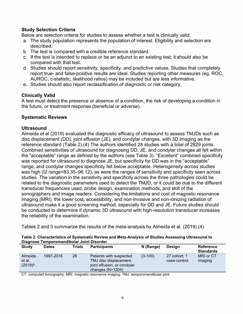
e. Studies should also report reclassification of diagnostic or risk category. Clinically Valid A test must detect the presence or absence of a condition, the risk of developing a condition in the future, or treatment response (beneficial or adverse). Systematic Reviews Ultrasound Almeida et al (2019) evaluated the diagnostic efficacy of ultrasound to assess TMJDs such as disc displacement (DD), joint effusion (JE), and condylar changes, with 3D imaging as the reference standard (Table 2).(4) The authors identified 28 studies with a total of 2829 joints. Combined sensitivities of ultrasound for diagnosing DD, JE, and condylar changes all fell within the “acceptable” range as defined by the authors (see Table 3). “Excellent” combined specificity was reported for ultrasound to diagnose JE, but specificity for DD was in the “acceptable” range, and condylar changes specificity fell below acceptable. Heterogeneity across studies was high (I2 range=83.35–96.12), as were the ranges of sensitivity and specificity seen across studies. The variation in the sensitivity and specificity across the three pathologies could be related to the diagnostic parameters used to detect the TMJD, or it could be due to the different transducer frequencies used, probe design, examination methods, and skill of the sonographers and image readers. Considering the limitations and cost of magnetic resonance imaging (MRI), the lower cost, accessibility, and non-invasive and non-ionizing radiation of ultrasound make it a good screening method, especially for DD and JE. Future studies should be conducted to determine if dynamic 3D ultrasound with high-resolution transducer increases the reliability of the examination. Tables 2 and 3 summarize the results of the meta-analysis by Almeida et al. (2019).(4) Table 2. Characteristics of Systematic Review and Meta-Analysis of Studies Assessing Ultrasound to Diagnose Temporomandibular Joint Disorder Study Dates Trials Participants N (Range) Design Reference
Standards Almeida et al. (2019)4,
1997-2016 28 Patients with suspected TMJ disc displacement, joint effusion, or condylar changes (N=1204)
(3-100) 27 cohort; 1 case-control
MRI or CT imaging
CT: computed tomography; MRI: magnetic resonance imaging; TMJ: temporomandibular joint
7
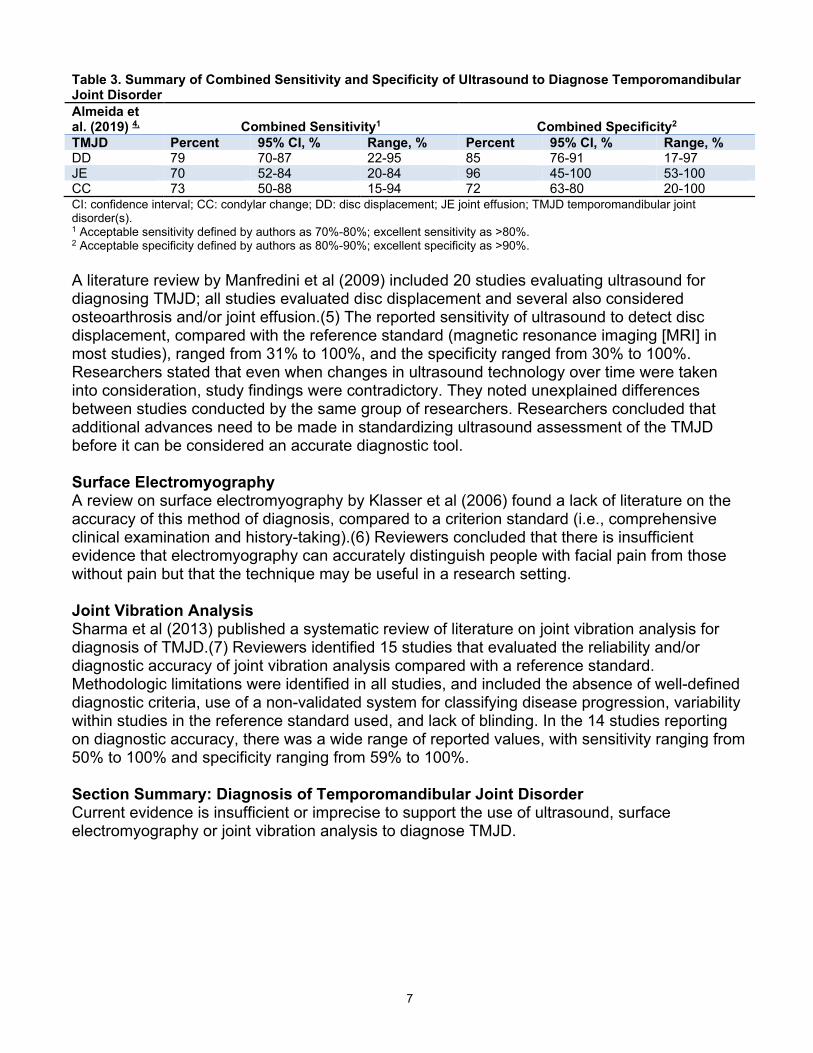
Table 3. Summary of Combined Sensitivity and Specificity of Ultrasound to Diagnose Temporomandibular Joint Disorder Almeida et al. (2019) 4,
Combined Sensitivity1
Combined Specificity2
TMJD Percent 95% CI, % Range, % Percent 95% CI, % Range, % DD 79 70-87 22-95 85 76-91 17-97 JE 70 52-84 20-84 96 45-100 53-100 CC 73 50-88 15-94 72 63-80 20-100 CI: confidence interval; CC: condylar change; DD: disc displacement; JE joint effusion; TMJD temporomandibular joint disorder(s). 1 Acceptable sensitivity defined by authors as 70%-80%; excellent sensitivity as >80%. 2 Acceptable specificity defined by authors as 80%-90%; excellent specificity as >90%. A literature review by Manfredini et al (2009) included 20 studies evaluating ultrasound for diagnosing TMJD; all studies evaluated disc displacement and several also considered osteoarthrosis and/or joint effusion.(5) The reported sensitivity of ultrasound to detect disc displacement, compared with the reference standard (magnetic resonance imaging [MRI] in most studies), ranged from 31% to 100%, and the specificity ranged from 30% to 100%. Researchers stated that even when changes in ultrasound technology over time were taken into consideration, study findings were contradictory. They noted unexplained differences between studies conducted by the same group of researchers. Researchers concluded that additional advances need to be made in standardizing ultrasound assessment of the TMJD before it can be considered an accurate diagnostic tool. Surface Electromyography A review on surface electromyography by Klasser et al (2006) found a lack of literature on the accuracy of this method of diagnosis, compared to a criterion standard (i.e., comprehensive clinical examination and history-taking).(6) Reviewers concluded that there is insufficient evidence that electromyography can accurately distinguish people with facial pain from those without pain but that the technique may be useful in a research setting. Joint Vibration Analysis Sharma et al (2013) published a systematic review of literature on joint vibration analysis for diagnosis of TMJD.(7) Reviewers identified 15 studies that evaluated the reliability and/or diagnostic accuracy of joint vibration analysis compared with a reference standard. Methodologic limitations were identified in all studies, and included the absence of well-defined diagnostic criteria, use of a non-validated system for classifying disease progression, variability within studies in the reference standard used, and lack of blinding. In the 14 studies reporting on diagnostic accuracy, there was a wide range of reported values, with sensitivity ranging from 50% to 100% and specificity ranging from 59% to 100%. Section Summary: Diagnosis of Temporomandibular Joint Disorder Current evidence is insufficient or imprecise to support the use of ultrasound, surface electromyography or joint vibration analysis to diagnose TMJD.

8
ORTHOTICS AND PHARMACOLOGIC TREATMENT OF TEMPOROMANDIBULAR DYSFUNCTION Clinical Context and Therapy Purpose The purpose of orthotics and pharmacologic treatment in patients with a confirmed diagnosis of TMJD is to provide a treatment option that is an alternative to or an improvement on existing therapies, such as alternative nonsurgical intervention. The question addressed in this evidence review is: do orthotics and pharmacologic treatment improve the net health outcome for individuals with a confirmed diagnosis of TMJD? The following PICOs were used to select literature to inform this review. Populations The relevant population of interest is individuals with confirmed TMJD. Interventions The therapies being considered are intraoral devices or appliances and pharmacological treatment. Intraoral devices and appliances are described in the Regulatory Status section above and can include stabilization splints. Pharmacological treatment can include nonsteroidal anti-inflammatory drugs, opioids, corticosteroids, muscle relaxants, antidepressants, anticonvulsants, and benzodiazepines. Patients with confirmed TMJD are actively managed by primary care providers, dentists, and otolaryngologists in an outpatient clinical setting. Comparators The main comparators of interest is alternative nonsurgical intervention, such as medications, physical therapy, and injections. Alternative medicine techniques can also be used, such as acupuncture, relation techniques, transcutaneous electric nerve stimulation (TENS), and biofeedback. Outcomes The general outcomes of interest are symptoms, functional outcomes, quality of life, and treatment related morbidity. Symptoms of TMJD may include, pain, tenderness, or aching in the jaw or one or both of the temporomandibular joints, difficulty or pain while chewing, and locking of the temporomandibular joint. The existing literature evaluating intraoral devices or appliances and pharmacologic treatment as a treatment for confirmed TMJD has varying lengths of follow-up, ranging from six weeks to one year of follow-up While the systematic reviews described below all reported at least one outcome of interest, longer follow-up was necessary to fully observe outcomes. Therefore, at least one year of follow-up is considered necessary to demonstrate efficacy. Study Selection Criteria Methodologically credible studies were selected using the following principles:

9
a. To assess efficacy outcomes, comparative controlled prospective trials were sought, with a preference for RCTs;
b. In the absence of such trials, comparative observational studies were sought, with a preference for prospective studies.
c. To assess longer term outcomes and adverse events, single-arm studies that capture longer periods of follow-up and/or larger populations were sought.
d. Studies with duplicative or overlapping populations were excluded. Review of Evidence Systematic Reviews List and Axelsson (2010) published a review of systematic reviews on treatments for TMJDs published through August 2009.(8) They identified 30 reviews; there were 23 qualitative systematic reviews and seven meta-analyses. Eighteen of the systematic reviews included only randomized controlled trials (RCTs), three included case control studies, and nine included a mixture of RCTs and case series. TMJD were defined inconsistently in the primary studies and systematic reviews, and several of the reviews addressed the related diagnoses of bruxism, disc replacements, and myofascial pain. Twenty-nine of the systematic reviews had pain intensity or pain reduction as the primary outcome measure, and 25 reported clinical outcome measures such as jaw movement or jaw tenderness on palpation. Reviewers divided the treatments into five categories (some studies were included in more than one category). These categories and the main findings are listed in Table 4. Table 4. Categories of Treatment
Categories
No. of Articles
Findings
Occlusal appliances, occlusal adjustment, and orthodontic treatment
10 Six systematic reviews did not find significant benefit vs other treatments, 4 found no benefit vs a placebo device, and 3 found occlusal therapy was better than no treatment
Physical treatments including acupuncture, TENS, exercise, and mobilization
8 Four reviews found no significant benefit of acupuncture over other treatments, 1 found no difference between acupuncture and placebo treatment, and 3 found acupuncture was better than no treatment. One review found active exercise and postural training were effective for treating TMJD-related pain.
Pharmacologic treatment 7 Treatments found to be superior to placebo were analgesics (2 reviews), clonazepam or diazepam (3 reviews), antidepressants (4 reviews), and hyaluronate (1 review). One review found effects of hyaluronate and corticosteroids to be similar.
Maxillofacial surgery 4 Three reviews evaluated surgery for patients with disc displacements and 1 addressed orthognathic surgery in patients with TMJD. Reviews of surgical treatments generally included lower level evidence (eg, case series), and did not always compare surgery with a control condition. One review of patients with disc displacements with reduction reported similar treatment effects for arthrocentesis, arthroscopy, and discectomy, and another review in patients in disc displacement without reduction found similar effects of arthrocentesis, arthroscopy, and physical therapy (used as a control intervention). Due to the lack of high quality controlled studies, conclusions could not be drawn about intervention equivalence.
Behavioral therapy and multimodal treatments
6 Two reviews found biofeedback to be better than active control or no treatment, 1 review found a combination of biofeedback and CBT to be better than no treatment, and 2 found a combination of biofeedback and

10
relaxation to be better than no treatment. One review found the effects of biofeedback and relaxation to be similar.
Adapted from List and Axelsson (2010)8 CBT: cognitive-behavioral therapy; TENS: transcutaneous electrical nerve stimulation; TMJD: temporomandibular joint disorders. Overall, reviewers concluded that there was insufficient evidence that electrophysical modalities and surgery would be effective for treating TMJD. They found some evidence that occlusal appliances, acupuncture, behavioral therapy, jaw exercise, postural training, and some medications could be effective at reducing pain for patients with TMJD. However, reviewers noted that most of the systematic reviews they examined included primary studies with considerable variation in methodologic quality, and thus, it is not possible to make definitive conclusions about the effectiveness of any of the treatments. Randhawa et al (2016) published a systematic review of noninvasive interventions for TMJDs, which included RCTs with at least 30 individuals per treatment arm, cohort studies with at least 100 patients per exposed group, and case control interventions.(9) Reviewers identified 31 studies for appraisal, of which seven RCTs described in eight publications had a low risk of bias and were assessed further. Most RCTs evaluated interventions outside the scope of our review, including cognitive-behavioral therapy and self-care management. Three RCTs evaluated occlusal devices for TMJD of variable duration, and generally reported no significant improvements with occlusal devices in terms of pain, mouth opening, or other outcomes. ORTHOTICS Intraoral Devices/Appliances Friction et al (2010) reported on a systematic review of RCTs on intraoral treatment of TMJD and identified 47 publications on 44 trials.(10) Intraoral appliances included soft and hard stabilization appliances, anterior positioning appliances, anterior bite appliances, and soft resilient appliances. Studies compared two types of devices or compared one device with a different treatment (eg, acupuncture or biofeedback). None of the studies evaluated use of one device during the day and a different device during the night. The primary outcome of the meta-analysis was pain. Pain was measured differently in the studies, and reviewers defined a successful outcome as at least a 50% reduction in pain on a self-report scale or at least an “improved” status when pain was measured by subjective report of status. Ten RCTs were included in two meta-analyses; the others were excluded because they did not measure pain, there were not at least two studies using similar devices or control groups, or data were not usable in a pooled analysis. A pooled analysis of seven RCTs (n=385 patients) that evaluated hard stabilization appliances and using palatal non-occluding appliances as a control found a significantly greater reduction in pain with hard appliances (odds ratio [OR], 2.45; 95% confidence interval [CI], 1.56 to 3.86; p<0.001). A pooled analysis of three studies (n=216 patients) did not find a statistically significant effect of hard appliances compared with a no-treatment control group (OR=2.14; 95% CI, 0.80 to 5.75; p=0.12). Ivorra-Carbonell et al (2016) reported on a systematic review of functional advancement devices for TMJD, which included systematic reviews, meta-analyses, RCTs, case-control studies, and cohort studies.(11) Reviewers included 21 articles evaluating some kind of advancement device, considered of medium or high quality by CONSORT criteria. Results were
11
summarized descriptively; reviewers concluded that after treatment with mandibular advancement the condyle was in “more advanced position.” Stabilization Splints Ebrahim et al (2012) identified 11 RCTs comparing splint therapy for TMJD with minimal or no therapy.(12) Nine of the 11 studies used stabilization splints, one used soft splints and one used an anterior repositioning appliance. Reviewers used the GRADE system to rate study quality. Nine studies did not report whether allocation was concealed, and six studies did not report masking of outcome assessors. Length of follow-up in the studies ranged from 6 to 52 weeks. A pooled analysis of study findings found that splint therapy was significantly associated with a reduction in reported pain compared with minimal or no intervention (standardized mean difference [SMD], -0.93; 95% CI, -1.33 to -0.53). Using a 100-millimeter visual analog scale (VAS) to measure pain, splint therapy was associated with an 11.5 mm lower mean VAS score (95% CI, -16.5 to -6.6 mm). There were not statistically significant differences between groups in quality of life or depression scores. Zhang et al (2016) identified 13 publications from 11 studies (n=538 patients) evaluating splint therapy for TMJD.(13) Risk of bias was high for two or more domains for all of the studies. Splint therapy group patients had greater improvement in pain control than control patients (mean difference [MD], 2.02; 95% CI, 1.55 to 2.49; I2=0.558). An observational study by Tonlorenzi et al (2019) assessed 21 patients with TMJD, specifically myofascial pain, to determine the effectiveness of wearing a “high” oral splint (vs. a “low” oral splint) for three months while sleeping.(14) Results showed a significant increase of the interocclusal distance as measured by kinesiograph (from 0.64 ± 0.53 mm to 1.42 ± 0.76 mm; p <.001), accompanied by a reduction in pain intensity in oral and extraoral regions after the 3 months. A RCT by Alajbeg et al. (2020) enrolled 34 patients with chronic TMD who received a stabilization splint or placebo splint.(15) At 3-month follow up, patients receiving a stabilization splint experienced improvement in pain intensity (p=0.009), depressive symptoms (p=0.011), and oxidant/antioxidant ratio (p=0.018) compared with placebo. The number of disability days and pain-free mouth opening were similar between the 2 groups at 3 months. At 6 months (post treatment follow up period), stabilization splints significantly reduced the number of disability days compared with placebo (p=0.023). A RCT by Melo et al. (2020) compared an occlusal splint, manual therapy, counseling, and the combination of an occlusal splint and counseling for managing pain and anxiety in 89 patients with TMD.(16) After 1 month, all interventions reduced pain and anxiety compared with baseline, with all 4 groups showing similar changes. A systemic review of 37 RCTs by Riley et al. (2020) revealed a lack of evidence that splints reduce pain (standardized mean difference [SMD], -0.18; 95% CI, -0.42 to 0.06) when all subtypes of TMD were pooled into 1 global TMD group.(17) The result was based on 13 trials (N=1076). The included trials used different splint types and varied in outcome measures used, and the evidence was rated as low-certainty.

12
Al-Moraissi et al. (2020) performed a network meta-analysis of 48 RCTs to determine the effectiveness of various occlusal splints for TMD.(18) Compared with controls, an anterior repositioning splint (low quality evidence), counseling with a hard stabilization splint (low quality evidence), mini-anterior splint (very low quality evidence), and hard stabilization splint (low quality evidence) decreased pain in patients with arthrogenous TMD. Compared with controls, a mini-anterior splint (very low quality of evidence), soft stabilization splint (very low quality of evidence), counseling therapy alone (moderate quality of evidence), and counseling with hard stabilization splint (moderate quality of evidence) decreased pain intensity in patients with myogenous TMDs. Pharmacologic Treatment In their multicenter, double-blind RCT, Isacsson et al (2019) assessed the pain-reduction efficacy of a single-dose intra-articular injection of methylprednisolone (1 mL) to the TMJ.(19) A total of 54 patients with unilateral TMJD were randomized to receive either the methylprednisolone (n=27) or saline (n=27). Pain levels at maximum jaw opening were recorded on aVAS, (1-100) before the injections and four weeks after. The per-protocol analysis showed VAS scores for the methylprednisolone group decreased from a mean of 61.0 (95% CI: 50.0–70.7) to 33.9 (95% CI: 21.6– 46.2); the saline group VAS score decreased from a mean of 59.6 (95% CI: 50.7–65.9) to 33.9 (95% CI: 23.8–43.9). The differences in these scores were statistically insignificant (p=0.81). In addition, the methylprednisolone group experienced twice as many adverse events as the saline group. The results of the unpublished RCT titled, “Study of Orofacial Pain and ProRANOlol (SOPPRANO)” (2019; NCT02437383) posted on ClinicalTrials.gov evaluated the efficacy of propranolol hydrochloride extended-release versus placebo in reducing pain from TMJD.(20) Two hundred patients with chronic TMJD were randomized to receive either 10 weeks of the drug (n=100) or of a placebo (n=99). The primary outcome was change in the Weekly Mean Pain Index after nine weeks of treatment (index range 0 to 100; higher score, worse outcome). The least-squares mean of the propranolol group was -13.9 (95% CI: -17.4 to -10.5); for the placebo group it was -12.1 (95% CI: -15.5 to -8.7), a nonsignificant difference (p=.41). Häggman-Henrikson et al (2017) published a systematic review that included 41 RCTs assessing various pharmacologic regimens for pain from TMJDs or burning mouth syndrome; of these, 13 were selected for a network meta-analysis.(21) Nine studies evaluated temporomandibular muscular pain, which appeared to decrease more with cyclobenzaprine than with placebo, although no specific statistics were reported. Pain reduction was also favorable for botulinum toxin and Ping-On ointment in the meta-analysis; other descriptive analyses showed a reduction of pain with nonsteroidal anti-inflammatory drugs and melatonin tablets when compared with placebo. Mena et al. (2020) reported a systematic review and meta-analysis of 9 RCTs comparing topical products to placebo or control interventions for managing pain from TMJD.(22) Topical nonsteroidal anti-inflammatory drugs showed similar outcomes to placebo. In 1 study, Theraflex-TMJ cream (methyl salicylate as active ingredient) significantly decreased pain scores at 10 days (p=0.003) and at follow up (p=0.027) compared with placebo. In 1 study, Ping On ointment (18% peppermint oil, 20% menthol) reduced pain at 4 weeks of application

13
(p<0.001) but not after 7 days of use (p=0.136). In another study, cannabidiol ointment improved pain intensity compared with placebo (p<0.001). Overall, the authors concluded that evidence is of low quality due to a small number of studies and biases within the included studies. Machado et al. (2020) evaluated the effectiveness of botulinum toxin type A (BTX-A) for TMD in a systematic review and meta-analysis of 12 RCTs.(23) At month 1, BTX-A reduced pain more effectively compared with placebo (mean difference, -1.74 points; 95% CI, -2.94 to -0.54; 3 RCTs [n=60]). But at months 3 and 6, BTX-A reduced pain to a similar level as placebo. The authors concluded that the quality of evidence is low, and the results do not support the use of BTX-A for managing pain due to TMD. Section Summary: Orthotics and Pharmacologic Treatment Evidence evaluating the use of orthotics in the treatment of TMJD, while sometimes conflicting and inconclusive, suggests that use of orthotics may reduce TMJD pain. One systematic review of intraoral appliances (44 studies) and meta-analyses of subsets of these studies found a significant benefit of intraoral appliances compared with control interventions. Several studies, meta-analyses, and systematic reviews exploring the effectiveness of stabilization splints on TMD pain revealed conflicting results. Overall, the evidence shows that stabilizing splints may improve pain and positively impact depressive and anxiety symptoms. The evidence related to pharmacologic treatment varies because studies, systematic reviews, and meta-analyses lack consistency in evaluating specific agents. Some systematic reviews have found a significant benefit of several pharmacologic treatments (eg, analgesics, muscle relaxants, and anti-inflammatory medications [vs placebo]), but other studies showed a lack of benefit with agents such as methylprednisolone and BTX-A. OTHER NONSURGICAL THERAPIES Clinical Context and Therapy Purpose The purpose of nonsurgical therapies in patients with a confirmed diagnosis of TMJD is to provide a treatment option that is an alternative to or an improvement on existing therapies, such as alternative nonsurgical intervention. The question addressed in this evidence review is: do nonsurgical therapies improve the net health outcome for individuals with a confirmed diagnosis of TMJD? The following PICOs were used to select literature to inform this review. Populations The relevant population of interest is individuals with confirmed TMJD. Interventions The nonsurgical therapies being considered are acupuncture, biofeedback, TENS, orthodontic services, and hyaluronic acid. Patients with confirmed TMJD are actively managed by primary care providers, dentists, and otolaryngologists in an outpatient clinical setting.

14
Comparators The main comparator of interest is alternative nonsurgical intervention, such as medications. Patients with confirmed TMJD are actively managed by primary care providers, dentists, and otolaryngologists in an outpatient clinical setting. Outcomes The general outcomes of interest are symptoms, functional outcomes, quality of life, and treatment related morbidity. The existing literature evaluating nonsurgical therapies as a treatment for confirmed TMJD has varying lengths of follow-up, ranging from one week to six months of follow-up. While the systematic reviews and RCTs described below all reported at least one outcome of interest, longer follow-up was necessary to fully observe outcomes. Therefore, at least one year of follow-up is considered necessary to demonstrate efficacy. Study Selection Criteria Methodologically credible studies were selected using the principles described in the second indication. Review of Evidence Acupuncture A systemic review and meta-analysis by June et al (2011) identified 7 sham-controlled RCTs on acupuncture for treating TMJD.(24) The studies included a total of 141 patients. Sample sizes of individual studies ranged from 7 to 28. Four studies used a single acupuncture session, and the other three used 6-12 sessions. All seven studies reported change in pain intensity as assessed by a visual analogue scale (VAS). In six of the studies, pain intensity was measured immediately after treatment, the seventh measured pain after 16 weeks. A pooled analysis of findings from five studies (n=107) found a statistically significant improvement in pain intensity, as measured by a VAS. The pooled weighted mean difference (WMD) in pain intensity was -13.63 (95% CI: -21.16 to -6.10, p=0.001). A pooled subgroup analysis of four studies (n=89) found acupuncture to be superior to a non-penetrating sham acupuncture, WMD: -13.73; 95% CI:-21.78 to -5.67, p=0.001. A pooled analysis of two studies (n=18) did not find a significant difference in efficacy between acupuncture and a penetrating sham acupuncture, WMD: -12.95 95% CI:-34.05 to 8.15, p=0.23. The latter analysis may have been underpowered. Reviewers noted that previous studies have found that a 24.2 mm change in pain assessed by a 100 mm VAS represents a clinically significant difference and that only two of the included studies had a change of 24.2 mm or more. Hyaluronic Acid Injection Systematic Reviews Several systematic reviews of studies have assessed the use of hyaluronic acid (HA) for treating TMJD. Three reviews without meta-analysis found benefits to the use of HA. The review by Manfredini et al (2010) included 19 papers that dealt with HA to treat either TMJ disc

15
displacement or inflammatory-degenerative disorders. Eight of the studies were RCTs. All studies reported decreased pain levels, and positive outcomes were maintained over the varying follow-up periods (range, 15 days–24 months). The better outcomes with HA were shown only against placebo saline injections, but outcomes were similar to those seen with corticosteroid injections or oral appliances.(25) Results of a review of 9 RCTs by Machado et al (2012) showed that intra-articular injections with corticosteroids and HA were effective in controlling TMJD in the short and medium terms. In addition, results indicated that in the short term, intra-articular injections with only HA had similar results to injections with corticosteroids; however, in the long-term, HA was more effective.(26) From the eight studies included in their systematic review, Goiato et al (2016) found intra-articular injections of HA used in TMJ arthrocentesis are beneficial, but other drugs, such as corticosteroids and non-steroidal anti-inflammatory drug injections are also satisfactory options.(27) Liu et al (2017) conducted a systematic review and meta-analysis of RCTs or cohort studies that compared temporomandibular osteoarthritis outcomes in patients treated with intra-articular corticosteroid, hyaluronate, or placebo injection.(28) All eight selected studies were RCTs; of these, three contained data on hyaluronate injection. Compared with placebo, corticosteroid injections prompted a significant decrease in long-term (ie, ≥ six months post-procedure) pain (three studies; mean difference, -0.74; 95% CI, -1.34 to -0.13; p=0.02; I2=0%). However, in a pooled analysis of two studies (both of which included pretreatment arthrocentesis), long-term maximal mouth opening was increased for placebo more than for corticosteroid injection (mean difference, -2.06; 95% CI, -2.76 to -1.36; p<0.001; I2=28%). Only two studies were available for comparing corticosteroid with hyaluronate injections, which precluded strong analysis. Short-term pain and mouth opening measures did not significantly differ between any of the injection groups, nor did the incidence of adverse events. The meta-analysis was limited by the small sample sizes of included trials, as well as by the variety of corticosteroid types used. Reviewers concluded that corticosteroid injection following arthrocentesis may be effective for relief of long-term joint pain, but may be less effective for improving mouth opening. Al-Hamed et al. (2020) compared platelet concentrates with HA or saline/Ringer's solution for treating patients with temporomandibular osteoarthritis in a systematic review and meta-analysis of 9 RCTs (N=407).(29) Compared with HA, platelet concentrates decreased pain VAS scores by -1.11 (95% CI, -1.62 to -0.60; p<0.0001) at 3 months and by -0.57 (95% CI, -1.55 to 0.41; p=0.26) at 12 months. Compared with saline, platelet concentrates decreased pain VAS scores by -1.33 (95% CI, -2.61 to -0.06; p=0.04) at 3 months and -2.71 (95% CI, -4.69 to -0.72; p=0.008) at 12 months. For maximum mouth opening, platelet concentrates had similar outcomes compared with HA and improved outcomes compared with saline at 3 months (2.9 mm; 95% CI,1.47 to 4.3; p<0.0001) and 6 months (1.69 mm; 95% CI, 0.13 to 3.25; p=0.03). Randomized Controlled Trials Most of the published RCTs evaluating hyaluronic acid for treating TMJD had small sample sizes, short follow-up times, and/or lack of blinding. Representative RCTs with larger sample sizes and stronger methodology are described next. In a randomized trial, Sousa et al. (2020) compared bite splint, betamethasone injection with bite splint, sodium hyaluronate injection with bite splint, and platelet-rich plasma injection with

16
bite splint for improving pain and maximum pain-free mouth opening in 80 patients with arthralgia from TMJD.(30) All treatment groups that received injections experienced an improvement in pain (p<0.001). Based on the regression analysis, platelet-rich plasma with bite splint improved pain (average rate of 0.172 per week) and maximum pain-free mouth opening (average rate of 0.676 per week) faster over time, while bite-splint showed the slowest improvement in pain (average rate of 0.05 per week) and in maximum pain-free mouth opening (average rate of 0.219 per week). The groups receiving sodium hyaluronate injection experienced an improvement in pain at the average rate of 0.108 per week and in maximum pain-free mouth opening at the average rate of 0.418 per week. In their randomized trial, Gokçe Kuyuk et al (2019) compared platelet-rich plasma (PRP), HA, and intra-articular corticosteroids (CS) to treat patients with TMJ pain and those diagnosed with TMJ-osteoarthritis.(31) Patients were evaluated in two groups: those who felt pain on lateral palpation (n=31) and those who felt pain on posterior palpation (n=43). The patients were then randomized to receive either PRP, HA, or CS. TMJ pain (using a five-point VAS), the presence of crepitation, loss of function, and loss of strength were assessed before treatment and monthly for three months following treatment. For patients who had lateral TMJ pain, statistically significant VAS score changes were seen in the PRP and HA groups (p<.0028 for both groups). In terms of crepitation, function, and strength, some changes were observed in the PRP, HA, and CS groups, but they were not statistically significant (p>.0028). For patients with posterior TMJ pain, the VAS scores showed significant improvements for PRP, HA, and CS (p<.0028 for all groups). Some improvements were found in crepitation, function, and strength, but they were not significant. Overall, all three treatments significantly improved palpation pain, but the greatest improvement was with PRP. Gorrela et al (2017) reported on the efficacy of injecting sodium hyaluronate in patients with TMJDs.(32) The trial comprised 62 individuals with the disorder; some members (n=31) of the trial were treated with arthrocentesis, and some members (n=31) were treated by a combination of arthrocentesis and an injection of sodium hyaluronate. Follow-up was observed at one week, two weeks, one month, three months, and at six months. Using a VAS, patients were asked to measure pain from 1 to 10. Pain decreased significantly for patients in both treatment groups (p<0.001) at the one week and the six-month follow-up; however, patients who were injected with sodium hyaluronate reported a significantly stronger decrease in pain at the six-month follow-up (p<0.001). Preoperative mean VAS pain scores for patients who received injection started at 6.0; by the 6-month follow-up, the mean VAS pain score was 0.23. Preoperative mean pain scores for patients who received arthrocentesis alone started at 6.77; by the six-month follow-up, the mean pain score was 1.71. While not an overwhelmingly significant difference, the trialists concluded that adding an injection of sodium hyaluronate to arthrocentesis treatment can significantly decrease the pain felt by patients who suffer from TMJD. A study by Manfredini et al (2012) in Italy randomized 72 patients with TMJ dysfunction to 1 of 6 treatment groups: 1) single-session arthrocentesis alone; 2) single-session arthrocentesis plus corticosteroid; 3) single-session arthrocentesis plus low-molecular weight hyaluronic acid; 4) single-session arthrocentesis plus high-molecular weight hyaluronic acid; 5) 5 weekly arthrocenteses plus low-molecular weight hyaluronic acid; or 6) 5 weekly single-needle
17
arthrocenteses plus low-molecular weight hyaluronic acid.(33) Sixty out of 72 (83%) participants completed the study, between 9 and 12 patients per treatment group. In a per protocol analysis, there were no significant differences among groups on any of the outcome variables at the 3-month follow-up. For example, the percentage change in pain at rest ranged from -29.1% in the group receiving 5 weekly single-needle arthrocenteses plus low-molecular weight hyaluronic acid to -38.4% in the group receiving a single-session of arthrocentesis alone. Limitations of the study include the small number of patients in each treatment group and the substantial number of dropouts in absence of an intention-to-treat (ITT) analysis. A study by Bjorland et al (2007) in Norway evaluated 40 patients with osteoarthritis of the TMJD in a double-blind RCT.(34) Patients received 2 injections, 14 days apart, of sodium hyaluronate or corticosteroids. The pain was assessed using a visual analogue scale (VAS) from zero to 100. Patients were followed for six months (assessed at 14 days, one month and six months). There was a statistically significant reduction in pain within each group at all of the follow-up points. At the six month follow-up, pain intensity (mean VAS score) was 14 in the hyaluronic acid group and 31 in the corticosteroid group; the between-group difference was statistically significant (p<0.001). The number of patients who were pain-free at six months was 7 (35%) of 20 in the hyaluronic acid group and six (30%) of 20 in the corticosteroid group (p value not reported). Bertolami et al (1993) published a double-blind placebo-controlled trial evaluated 121 patients with TMJD.(35) Patients had to have a confirmed diagnosis of degenerative joint disease (DJD), reducing displaced disc (DDR) or non-reducing displaced disc (DDN), failure of other non-surgical treatments, and severe dysfunction. Patients received a single injection of sodium hyaluronate or saline and were followed for six months. Eighty patients were randomized to the hyaluronate group and 41 to the placebo group. This included a total of 57 patients in the DJD group, 50 patients in the DDR group, and 14 patients in the DDN group. Fourteen (12%) of 121 patients were excluded from the analysis because they did not meet eligibility criteria. No significant differences in outcomes were seen for the DJD group. In the DDN group, there were significant between-group differences through one month, favoring the hyaluronic acid group. The number of patients in the DDN group who completed follow-up after one month was insufficient to draw meaningful conclusions about efficacy. In the DDR group, there were no statistically significant differences between groups in any outcome at one or two months. At three and six months, two out of seven reported outcomes were significantly better in the hyaluronic acid group than in the placebo group. At five months, five out of seven reported outcomes were significantly better in the hyaluronic acid group. The seven outcomes included three measures of dysfunction, two measures of patient perception of improvement, two measures of change in noise. The most consistent between-group differences in the DDR group were for the two measures of patient perception of improvement and one of the noise variables. There were fewer between-group differences on dysfunction measures. Section Summary: Nonsurgical Therapies The evidence on acupuncture is limited by the small number of studies, small sample sizes, and in most studies, efficacy assessment only immediately post-treatment. The evidence on the use of hyaluronic acid to treat TMJD is inconclusive, given the methodologic issues with the

18
systematic review and RCTs conducted (eg, small sample sizes) and better surgical options. No reliable evidence is available for biofeedback, TENS, or orthodontic services for TMJD. SURGICAL TECHNIQUES Clinical Context and Therapy Purpose The purpose of surgical techniques in patients with a confirmed diagnosis of TMJD is to provide a treatment option that is an alternative to or an improvement on existing therapies, such as nonsurgical intervention. The question addressed in this evidence review is: do surgical therapies improve the net health outcome for individuals with a confirmed diagnosis of TMJD? The following PICOs were used to select literature to inform this review. Populations The relevant population of interest is individuals with confirmed TMJD. Interventions The surgical therapies being considered are arthrocentesis and arthroscopy. Patients with confirmed TMJD are actively managed by primary care providers, dentists, and otolaryngologists in an outpatient clinical setting. Arthrocentesis and arthroscopy are performed by a surgeon at an outpatient facility. Comparators The main comparator of interest is alternative nonsurgical intervention, such as intraoral devices and appliances, pharmacologic treatment, acupuncture, biofeedback, TENS, orthodontic services, and hyaluronic acid. Patients with confirmed TMJD are actively managed by primary care providers, dentists, and otolaryngologists in an outpatient clinical setting. Outcomes The general outcomes of interest are symptoms, functional outcomes, quality of life, and treatment related morbidity. The existing literature evaluating surgical techniques as a treatment for confirmed TMJD has varying lengths of follow-up up to 6 months. While the systematic reviews described below all reported at least 1 outcome of interest, longer follow-up was necessary to fully observe outcomes. Therefore, at least six months of follow-up is considered necessary to demonstrate efficacy. Study Selection Criteria Methodologically credible studies were selected using the principles described in the second indication.

19
Review of Evidence Systemic Reviews In a systematic review, Vos et al (2013) identified 3 RCTs (n=222 patients) that compared the efficacy of lavage of the TMJ (ie, arthrocentesis or arthroscopy) with nonsurgical TMJ treatment.(36) Although reviewers assessed the quality of the studies to be adequate, only one study stated that allocation to treatment group was concealed, and 2 studies did not explicitly state that an intention-to-treat (ITT) analysis was used. The two primary outcomes considered were change in pain and maximal mouth opening (MMO) at six months compared with baseline. The pain was measured by VAS. Pooled analysis of data from the three trials found a statistically significant reduction in pain at six months with surgery plus lavage versus nonsurgical therapy (SMD = -1.07; 95% CI, -1.38 to -0.76). There was no statistically significant difference in the efficacy between the two treatments for the other outcome variable, maximal mouth opening (SMD=0.05; 95% CI, -0.33 to 0.23). In a network meta-analysis, Al-Moraissi et al. (2020) compared different treatment options (placebo/control; muscle exercises and occlusal splint therapy; splint therapy alone; intraarticular injection of HA or corticosteroid; arthrocentesis with or without HA, corticosteroid, and platelet-rich plasma; arthroscopy with or without HA and platelet-rich plasma; open joint surgery; physiotherapy) for arthrogenous TMDs in 36 RCTs for reducing pain and 33 RCTs for improving maximum mouth opening.(37) For short-term follow up of at most 5 months, injections of HA (SMD, -2.8; 95% CI, -3.7 to -1.8) and corticosteroids (SMD, -2.11; 95% CI, -2.9 to -1.2) achieved greater pain control compared with placebo/control. For follow up of at least 6 months and longer, arthroscopy with platelet-rich plasma (SMD, -3.5, 95% CI, -6.2 to -0.82), arthrocentesis with platelet-rich plasma (SMD, -3.08; 95% CI, -5.44 to -0.71), arthroscopy with HA (SMD, -3.01; 95% CI, -5.8 to -0.12), TMJ surgery (SMD, -3; 95% CI, -5.7 to -0.28), injection with HA (SMD, -2.9, 95% CI, -4.9 to -1.09), arthroscopy-alone (SMD, -2.6, 95% CI, -5.1 to -0.07) and arthrocentesis with HA (SMD, -2.3; 95% CI, -4.5 to -018) significantly improved pain compared with placebo/control. For improving maximum mouth opening, various arthroscopy procedures (with and without platelet-rich plasma and HA injections) followed by arthrocentesis with platelet-rich plasma or HA were the most efficacious treatment approaches. Treatments such as occlusal splint therapy, physical therapy, muscle exercises with occlusal splint therapy, and placebo/control yielded the lower quality outcomes for reducing pain and improving maximum mouth opening. Most of the evidence included in the network meta-analysis was rated as low quality or very low quality, except the evidence for arthrocentesis with HA injections was of moderate quality. Observational Studies In a retrospective cohort study, Hossameldin and McCain (2018) assessed the efficacy of an office-based TMJ arthroscopic technique. The researchers assessed the following outcomes of the procedure: improvement in painless range-of-motion in the mandible, reduced pain on loading, and improvement in functional jaw pain. The cohort included an initial 363 patients, excluded 41, and an analysis was performed on the joints of the remaining 322 that were compromised. Within the 322 patients, 452 joints were operated on with a 66.6% (n=301 joints)

20
success rate (p=.001). It is stated within the outcome variable section that the primary outcome variable of success or failure was determined by the reduction of joint pain postoperatively. This could be subjective. When the operation failed (n=151 joints, 33.3%), 141 joints were involved in a subsequent procedure that ranged from more advanced arthroscopy to a total joint replacement.(38) Section Summary: Surgical Techniques Observational studies and systematic reviews have shown that use of arthrocentesis and arthroscopy reduces pain levels in patients with TMJD. SUMMARY OF EVIDENCE For individuals who have suspected TMJD who receive ultrasound, surface electromyography, or joint vibration analysis, the evidence includes systematic reviews of diagnostic test studies. The relevant outcomes are test accuracy, test validity, and other performance measures. None of the systematic reviews found that these diagnostic techniques accurately identify patients with TMJD and many of the included studies had methodological limitations. The evidence is insufficient to determine that the technology results in an improvement in the net health outcome. For individuals who have a confirmed diagnosis of TMJD who receive intraoral devices or appliances or pharmacologic treatment, the evidence includes randomized controlled trials (RCTs) and systematic reviews of RCTs. Relevant outcomes are symptoms, functional outcomes, quality of life, and treatment-related morbidity. A systematic review of intraoral appliances (44 studies) and meta-analyses of subsets of these studies found a significant benefit of intraoral appliances compared with control interventions. Several studies, meta-analyses, and systematic reviews exploring the effectiveness of stabilization splints on TMJD pain revealed conflicting results. Overall, the evidence shows that stabilizing splints may improve pain and positively impact depressive and anxiety symptoms. The evidence related to pharmacologic treatment varies because studies, systematic reviews, and meta-analyses lack consistency in evaluating specific agents. Some systematic reviews found a significant benefit of several pharmacologic treatments (eg, analgesics, muscle relaxants, and anti-inflammatory medications [vs placebo]), but other studies showed a lack of benefit with agents such as methylprednisolone and botulinum toxin type A. The evidence is sufficient to determine that the technology results in an improvement in the net health outcome. For individuals who have a confirmed diagnosis of TMJD who receive acupuncture, biofeedback, transcutaneous electrical nerve stimulation, orthodontic services, or hyaluronic acid, the evidence includes RCTs and systematic reviews of RCTs and observational studies. Relevant outcomes are symptoms, functional outcomes, quality of life, and treatment-related morbidity. The systematic reviews did not find that the above technologies improved pain and functional outcomes significantly more than control conditions. Many individual studies had small sample sizes and/or methodologic limitations. The evidence is insufficient to determine that the technology results in an improvement in the net health outcome. For individuals who have a confirmed diagnosis of TMJD, who receive arthrocentesis or arthroscopy, the evidence includes RCTs and systematic reviews of RCTs. Relevant outcomes

21
are symptoms, functional outcomes, quality of life, and treatment-related morbidity. One review, which included 3 RCTs, compared arthrocentesis or arthroscopy with nonsurgical interventions for TMJD. Pooled analyses of the RCTs found that arthrocentesis and arthroscopy resulted in superior pain reduction compared with control interventions. A network meta-analysis, which included 36 RCTs, revealed that arthroscopy and arthrocentesis improve pain control and maximum mouth opening. The evidence is sufficient to determine that the technology results in an improvement in the net health outcome. Supplemental Information PRACTICE GUIDELINES AND POSITION STATEMENTS American Association for Dental Research: In 2010 (reaffirmed 2015), the American Association for Dental Research policy statement recommended the following for the diagnosis and treatment of TMJ disorders:(39)
“It is recommended that the differential diagnosis of TMDs [temporomandibular disorders] or related orofacial pain conditions should be based primarily on information obtained from the patient’s history, clinical examination, and when indicated, TMJ radiology or other imaging procedures. The choice of adjunctive diagnostic procedures should be based upon published, peer-reviewed data showing diagnostic efficacy and safety. However, the consensus of recent scientific literature about currently available technological diagnostic devices for TMDs is that except for various imaging modalities, none of them shows the sensitivity and specificity required to separate normal subjects from TMD patients or to distinguish among TMD subgroups….” “It is strongly recommended that, unless there are specific and justifiable indications to the contrary, treatment of TMD patients initially should be based on the use of conservative, reversible and evidence-based therapeutic modalities. Studies of the natural history of many TMDs suggest that they tend to improve or resolve over time. While no specific therapies have been proven to be uniformly effective, many of the conservative modalities have proven to be at least as effective in providing symptomatic relief as most forms of invasive treatment….”
American Society of Temporomandibular Joint Surgeons In 2001, the American Society of Temporomandibular Joint Surgeons issued consensus clinical guidelines focused on TMJDs associated with internal derangement and osteoarthritis.(40) For diagnosis of this type of TMJD, a detailed history and, when indicated, general physical examination was recommended. Imaging of the temporomandibular and associated structures was also recommended. Options for basic radiography to provide information on temporal bone and condylar morphology include use of plain films, panoramic films, and tomograms. Also recommended was imaging of the disc and associated soft tissue with MRI or arthrography. Other diagnostic procedures that may be indicated include computed tomography, MRI, arthrography (for selected cases) and isotope bone scans.

22
Nonsurgical treatment was recommended as a first-line therapy for all symptomatic patients with this condition. Recommended treatment options include change in diet, nonsteroidal anti-inflammatory drugs, maxillomandibular appliances, physical therapy, injections of corticosteroids or botulinum toxin, and behavior modification. If adequate symptom relief does not occur within 2-3 weeks, surgical consultation is advised. The guideline states that the following surgical procedures are considered to be accepted and effective for patients with TMJD associated with internal derangement/osteoarthritis:
• Arthrocentesis • Arthroscopy • Condylotomy • Arthrotomy (prosthetic joint replacement may be indicated in selected patients who have
severe joint degeneration, destruction, or ankylosis) • Coronoidotomy/coronoidectomy • Styloidectomy
U.S. PREVENTIVE SERVICES TASK FORCE RECOMMENDATIONS Not applicable. ONGOING AND UNPUBLISHED CLINICAL TRIALS Some currently unpublished trials that might influence this review are listed in Table 5. Table 5. Summary of Key Trials NCT No.
Trial Name
Planned Enrollment
Completion Date
Ongoing
NCT04210921 Clinical Efficacy of Acupuncture in the Treatment of Temporomandibular Disorders (TMD)
60 Jun 2020
NCT04469088 Effectiveness of Dry Needling vs Manual Therapy in Patients With Temporomandibular Joint Disorders. A Randomized Controlled Trial.
46 Aug 2020
NCT04298554 Comparison of Cannabinoids to Placebo in Management of Arthralgia and Myofascial Pain Disorder of the Temporomandibular Region: A Randomized Clinical Trial.
71 Mar 2022
Unpublished
NCT02437383 Effect of COMT (Catecholamine-O-methyltransferase) Genetic Polymorphisms on Response to Propranolol Therapy in Temporomandibular Disorder
200 April 2018 (completed; results
posted but not published; updated
5/21/19) NCT03180671 The Effectiveness of Anterior Deprogrammers as a
Tool for Reducing Pain and Masticatory Muscles 80 May 2019
(unknown; updated 6/8/17)
NCT03029494 The Role of Oxidative Stress and Opiorphin in Temporomandibular Disorders
80 Sep 2019 (unknown; updated 1/24/17)
NCT: national clinical trial a Denotes industry-sponsored or cosponsored trial.

23
Government Regulations National/Local: No NCD or LCD determination noted regarding the diagnoses or treatment of TMJ. (The above Medicare information is current as of the review date for this policy. However, the coverage issues and policies maintained by the Centers for Medicare & Medicare Services [CMS, formerly HCFA] are updated and/or revised periodically. Therefore, the most current CMS information may not be contained in this document. For the most current information, the reader should contact an official Medicare source.) Related Policies Biofeedback Cosmetic and Reconstructive Surgery Frenum Surgery (Frenulum Surgery, Frenumectomy, Frenulectomy, Frenectomy, Frenotomy) Obstructive Sleep Apnea and Snoring – Surgical Treatment Orthognathic Surgery Sleep Disorders, Diagnosis and Medical Management References 1. Schiffman E, Ohrbach R, Truelove E, et al. Diagnostic Criteria for Temporomandibular
Disorders (DC/TMD) for Clinical and Research Applications: recommendations of the International RDC/TMD Consortium Network* and Orofacial Pain Special Interest Groupdagger. J Oral Facial Pain Headache. Winter 2014;28(1):6-27. PMID 24482784
2. Ohrbach R, Turner JA, Sherman JJ, et al. The Research Diagnostic Criteria for Temporomandibular Disorders. IV: evaluation of psychometric properties of the Axis II measures. J Orofac Pain. Winter 2010;24(1):48-62. PMID 20213031
3. Schiffman E, Ohrbach R. Executive summary of the Diagnostic Criteria for Temporomandibular Disorders for clinical and research applications. J Am Dent Assoc. Jun 2016;147(6):438-445. PMID 26922248
4. Almeida FT, Pacheco-Pereira C, Flores-Mir C, et al. Diagnostic ultrasound assessment of temporomandibular joints: a systematic review and meta-analysis. Dentomaxillofac Radiol, 2018 Oct 5;48(2). PMID 30285469
5. Manfredini D, Guarda-Nardini L. Ultrasonography of the temporomandibular joint: a literature review. Int J Oral Maxillofac Surg 2009; 38(12):1229-36.
6. Klasser GD, Okeson JP. The clinical usefulness of surface electromyography in the diagnosis and treatment of temporomandibular disorders. J Am Dent Assoc 2006; 137(6):763-71.
7. Sharma S, Crow HC, McCall WD, Jr. et al. Systematic review of reliability and diagnostic validity of joint vibration analysis for diagnosis of temporomandibular disorders. J Orofac Pain 2013; 27(1):51-60.
8. List T, Axelsson S. Management of TMD: evidence from systematic reviews and meta-analyses. J Oral Rehabil 2010; 37(6):430-51.

24
9. Randhawa K, Bohay R, Cote P, et al. The Effectiveness of Noninvasive Interventions for Temporomandibular Disorders: A Systematic Review by the Ontario Protocol for Traffic Injury Management (OPTIMa) Collaboration. Clin J Pain. Mar 2016;32(3):260-278. PMID 25924094
10. Fricton J, Look JO, Wright E et al. Systematic review and meta-analysis of randomized controlled trials evaluating intraoral orthopedic appliances for temporomandibular disorders. J Orofac Pain 2010; 24(3):237-54.
11. Ivorra-Carbonell L, Montiel-Company JM, Almerich-Silla JM, et al. Impact of functional mandibular advancement appliances on the temporomandibular joint - a systematic review. Med Oral Patol Oral Cir Bucal. Sep 01 2016;21(5):e565-572. PMID 27475694
12. Ebrahim S, Montoya L, Busse JW, et al. The effectiveness of splint therapy in patients with temporomandibular disorders: a systematic review and meta-analysis. J Am Dent Assoc. Aug 2012;143(8):847-857. PMID 22855899
13. Zhang C, Wu JY, Deng DL, et al. Efficacy of splint therapy for the management of temporomandibular disorders: a meta-analysis. Oncotarget. Dec 20 2016;7(51):84043-84053. PMID 27823980
14. Tonlorenzi D, Brunelli M, Conti M et al. Mandibular extension through a high oral splint application on pain control.. Arch Ital Biol, 2019 Dec 11;157(2-3). PMID 31821530
15. Alajbeg IZ, Vrbanovic E, Lapic I, et al. Effect of occlusal splint on oxidative stress markers and psychological aspects of chronic temporomandibular pain: a randomized controlled trial. Sci Rep. Jul 03 2020; 10(1): 10981. PMID 32620810
16. Melo RA, de Resende CMBM, Rego CRF, et al. Conservative therapies to treat pain and anxiety associated with temporomandibular disorders: a randomized clinical trial. Int Dent J. Aug 2020; 70(4): 245-253. PMID 32153038
17. Riley P, Glenny AM, Worthington HV, et al. Oral splints for temporomandibular disorder or bruxism: a systematic review. Br Dent J. Feb 2020; 228(3): 191-197. PMID 32060462
18. Al-Moraissi EA, Farea R, Qasem KA, et al. Effectiveness of occlusal splint therapy in the management of temporomandibular disorders: network meta-analysis of randomized controlled trials. Int J Oral Maxillofac Surg. Aug 2020; 49(8): 1042-1056. PMID 31982236
19. Isacsson G, Schumann M, Nohlert E, et al. Pain relief following a single-dose intra-articular injection of methylprednisolone in the temporomandibular joint arthralgia-A multicentre randomised controlled trial.. J Oral Rehabil, 2018 Sep 22;46(1). PMID 30240024
20. Study of Orofacial Pain and PropRANOlol (SOPPRANO). ClinicalTrials.gov; retrieved May 5, 2020 from: https://clinicaltrials.gov/ct2/show/results/NCT02437383?view=results. Updated May 21, 2019.
21. Häggman-Henrikson B, Alstergren P, Davidson T, et al. Pharmacological treatment of oro-facial pain - health technology assessment including a systematic review with network meta-analysis. J Oral Rehabil. Oct 2017;44(10):800-826. PMID 28884860
22. Mena M, Dalbah L, Levi L, et al. Efficacy of topical interventions for temporomandibular disorders compared to placebo or control therapy: a systematic review with meta-analysis. J Dent Anesth Pain Med. Dec 2020; 20(6): 337-356. PMID 33409363
23. Machado D, Martimbianco ALC, Bussadori SK, et al. Botulinum Toxin Type A for Painful Temporomandibular Disorders: Systematic Review and Meta-Analysis. J Pain. Mar 2020; 21(3-4): 281-293. PMID 31513934

25
24. Jung A, Shin BC, Lee MS et al. Acupuncture for treating temporomandibular joint disorders: a systematic review and meta-analysis of randomized, sham-controlled trials. J Dent 2011; 39(5):341-50.
25. Manfredini D, Piccotti F, Guarda-Nardini L. Hyaluronic acid in the treatment of TMJ disorders: a systematic review of the literature. Cranio. Jul 2010;28(3):166-176. PMID 20806734
26. Machado E, Bonotto D, Cunali PA. Intra-articular injections with corticosteroids and sodium hyaluronate for treating temporomandibular joint disorders: a systematic review. Dental Press J Orthod. Sep-Oct 2013;18(5):128-133. PMID 24352399
27. Goiato MC, da Silva EV, de Medeiros RA, et al. Are intra-articular injections of hyaluronic acid effective for the treatment of temporomandibular disorders? A systematic review. Int J Oral Maxillofac Surg. Dec 2016;45(12):1531-1537. PMID 27374020
28. Liu Y, Wu J, Fei W, et al. There a difference in intra-articular injections of corticosteroids, hyaluronate, or placebo for temporomandibular osteoarthritis? J Oral Maxillofac Surg. Nov 8, 2017. PMID 29182905
29. Al-Hamed FS, Hijazi A, Gao Q, et al. Platelet Concentrate Treatments for Temporomandibular Disorders: A Systematic Review and Meta-analysis. JDR Clin Trans Res. May 28 2020: 2380084420927326. PMID 32464073
30. Sousa BM, Lopez-Valverde N, Lopez-Valverde A, et al. Different Treatments in Patients with Temporomandibular Joint Disorders: A Comparative Randomized Study. Medicina (Kaunas). Mar 05 2020; 56(3). PMID 32151101
31. Gokce Kutuk S, Gke G, Arslan M, et al. Clinical and Radiological Comparison of Effects of Platelet-Rich Plasma, Hyaluronic Acid, and Corticosteroid Injections on Temporomandibular Joint Osteoarthritis. J Craniofac Surg, 2019 Jun 6;30(4). PMID 31166260
32. Gorrela H, Prameela J, Srinivas G, et al. Efficacy of temporomandibular joint arthrocentesis with sodium hyaluronate in the management of temporomandibular joint disorders: a prospective randomized control trial. J Maxillofac Oral Surg. Dec 2017;16(4):479-484. PMID 29038631
33. Manfredini D, Rancitelli D, Ferronato G et al. Arthrocentesis with or without additional drugs in temporomandibular joint inflammatory-degenerative disease: comparison of six treatment protocols*. J Oral Rehabil 2012; 39(4):245-51.
34. Bjornland T, Gjaerum AA, Moystad A. Osteoarthritis of the temporomandibular joint: an evaluation of the effects and complications of corticosteroid injection compared with injection with sodium hyaluronate. J Oral Rehabil 2007; 34(8):583-9.
35. Bertolami CN, Gay T, Clark GT et al. Use of sodium hyaluronate in treating temporomandibular joint disorders: a randomized, double-blind, placebo-controlled clinical trial. J Oral Maxillofac Surg 1993; 51(3):232-42.
36. Vos LM, Huddleston Slater JJ, Stegenga B. Lavage therapy versus nonsurgical therapy for the treatment of arthralgia of the temporomandibular joint: a systematic review of randomized controlled trials. J Orofac Pain. Spring 2013;27(2):171-179. PMID 23630689
37. Al-Moraissi EA, Wolford LM, Ellis E, et al. The hierarchy of different treatments for arthrogenous temporomandibular disorders: A network meta-analysis of randomized clinical trials. J Craniomaxillofac Surg. Jan 2020; 48(1): 9-23. PMID 31870713

26
38. Hossameldin RH, McCain JP. Outcomes of office-based temporomandibular joint arthroscopy: a 5-year retrospective study. Int J Oral Maxillofac Surg. 2018 Jan;47(1). PMID 28751180
39. American Association for Dental Research (AADR). Science Policy: Temporomandibular disorders (TMD). 1996 (revised 2010, reaffirmed 2015); http://www.iadr.org/AADR/About-Us/Policy-Statements/Science-Policy#TMD. Accessed January 8, 2021
40. American Society of Temporomandibular Joint Surgeons. Guidelines for diagnosis and management of disorders involving the temporomandibular joint and related musculoskeletal structures. 2001; http://astmjs.org/final%20guidelines-04-27-2005.pdf. Accessed January 8, 2021.
41. Centers for Medicare Services. “Local coverage article: Oral appliances for obstructive sleep apnea – policy article (A52512),” Original effective date: 10/1/15; Revision date: 4/3/20. https://www.cms.gov/medicare-coverage-database/details/article-details.aspx?articleId=52512&ver=7&SearchType=Advanced&CoverageSelection=Both&NCSelection=NCA%7cCAL%7cNCD%7cMEDCAC%7cTA%7cMCD&ArticleType=SAD%7cEd&PolicyType=Both&s=27&KeyWord=temporomandibular+joint&KeyWordLookUp=Doc&KeyWordSearchType=Exact&kq=true&bc=IAAAACAAAAAA&. Accessed May 21, 2021.
The articles reviewed in this research include those obtained in an Internet based literature search for relevant medical references through 5/21/21, the date the research was completed.

27
BCN Medical Policy History
Date Rationale 5/14/01 BCN policy established
7/8/02 Joint medical policy developed
7/8/04 Joint medical policy retired, changed to a BCN-only medical policy. Updated codes and references
7/13/05 Routine maintenance and update
9/24/06 Maintenance; diagnostic testing added to policy
9/10/07 Routine maintenance
10/15/08 Routine maintenance
2/9/10 Routine maintenance
2/15/12 Routine maintenance; updated codes; added acupuncture and ultrasound imaging/sonogram to exclusions; updated references
6/19/13 Routine maintenance; added exclusion “Hyaluronic Acid”, codes J7321-J7326 are excluded; removed code 29800 from policy, as this is a diagnostic surgical procedure and is excluded.
10/15/14 Routine maintenance; added clarification on BCN Benefit page that there is FEP coverage for corrective orthopedic appliances for non-dental treatment of TMJ.
9/16/15 Routine maintenance; added new code 20606
8/17/16 Routine maintenance
8/23/17 • Routine maintenance • Added to inclusions: Intraoral removable prosthetic
devices/appliances • Removed from exclusions: condylar position indication
7/18/18 • Routine maintenance
7/17/19 • Routine maintenance • LCA added
7/9/20 • Routine maintenance
7/15/21 • Routine maintenance Next Review: 3rd Qtr, 2022

28
MEDICAL POLICY TITLE: TEMPOROMANDIBULAR JOINT DYSFUNCTION (TMD) TESTING AND TREATMENT
BCN BENEFIT ADMINISTRATION
I. Coverage Determination
Commercial HMO (includes Self-Funded groups unless otherwise specified)
Covered, criteria apply; Federal Employee Program (FEP) members have coverage for prostheses and appliances for or related to the non-dental treatment of TMD; Refer to current certificate.
BCNA (Medicare Advantage) Refer to the Medicare information under the Government Regulations section of this policy.
BCN65 (Medicare Complementary)
Coinsurance covered if primary Medicare covers the service.
II. Administrative Guidelines:
• The member's contract must be active at the time the service is rendered. • Coverage is based on each member’s certificate and is not guaranteed. Please consult
the member’s certificate for details. Additional information regarding coverage or benefits may also be obtained through customer or provider inquiry services at BCN.
• The service must be authorized by the member's PCP except for Self-Referral Option (SRO) members seeking Tier 2 coverage.
• Services must be performed by a BCN-contracted provider, if available, except for Self-Referral Option (SRO) members seeking Tier 2 coverage.
• Payment is based on BCN payment rules, individual certificate and certificate riders. • Appropriate copayments will apply. Refer to certificate and applicable riders for detailed
information. • CPT - HCPCS codes are used for descriptive purposes only and are not a guarantee of
coverage.