myofacial pain dysfunction syndrome anindya
TRANSCRIPT

Presented by : Dr. Anindya Chakrabarty

Content
Introduction
History
Definition
Characteristics
Pathophysiology
Symptoms
Physical examination
Goal of treatment
Management protocol

Introduction
It is a muscular pain disorder – most common diagnosis causingchronic pain but one of the most least understood.
Complex symptomatology, concomitant disorders and frequentbehavioral & psychosocial contributing factors make the disorderdifficult to recognize
As the name suggest it has three part
Myofascial – muscular & connective tissue origin
Pain – an unpleasant sensational & emotional experience
Dysfunction – deviated from normal function
Syndrome – collection of various symptoms

History
Costen – 1934 – indicate TMJ pain due to occlusal etiology
Schwartz – 1956 – coined term TMJ pain dysfunction syndrome – blamed the
spasm of masticatory and perimasticatory musculature.
Laskin – 1969 – termed as MYOFASCIAL PAIN DYSFUNTION SYNDROME –
implicated Psychophysiological theory for such incident.

Definition
A pain disorder, in which unilateral pain is reffered from the trigger points
in myofascial structures, to the muscles of the head and neck. Pain is
constant, dull in nature, in contrast to the sudden sharp, shooting,
intermittent pain of neuralgias.
Pain may range from mild to intolerable

Prevalence
Common persistent pain in head & neck region
50% of chronic head & neck pain
20-50% of people has this type of pain

Types of myofascial pain
disorder
6 distinct group
Myositis
Muscle spasm
Myofascial pain dysfunction (Trigger Point Pain)
Fibromyalgia
Muscle contracture
Muscle pain secondary to connective tissue disorder

Functional Neuroanatomy and Physiology of the
Masticatory System
Two major components:
(1) neurologic structures
(2) muscles.

MUSCULAR COMPONENT
MOTOR UNIT
consists of a number of muscle fibers that are innervated by one motor neuron.
Each neuron joins with the muscle fiber at a motor endplate.
Depolarization causes the muscle fibers to shorten or contract.
fewer the muscle fibers per motor neuron, the more precise the movement.
MUSCLE
Hundreds to thousands of motor units along with blood vessels and nerves are bundled together by connective tissue and fascia to make up a muscle.
Muscles are necessary to overcome this weight and mass imbalance.
MUSCLE FUNCTION
3 potential functions
isotonic contraction
Isometric contraction
Controlled relaxation
eccentric contraction
lengthening of a muscle at the same time that it is contracting

Muscles
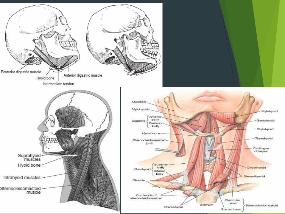

Precise and complex balance of the head and neck muscles must exist to maintain
proper head position and function. A, Muscle system. B, Each of the major muscles acts
like an elastic band. The tension provided must precisely contribute to the balance that
maintains the desired head position. If one elastic band breaks, the balance of the entire
system is disrupted and the head position altered.

Neurological structure

Neuromuscular Function
Function of the Sensory Receptors
Reflex Action
Reciprocal Innervation
Regulation of Muscle Activity
Influence from the Higher Centers

Reflex action
Myotic / Stretch Relex
Nociceptive reflex

MYOTIC REFLEX

Nociceptive reflex
Pain modulation in trigeminal nerve
The degree of suffering relates more closely to the patient’s perceived threat of
the injury and the amount of attention given to the injury
Pain modulation means that the impulses arising from a noxious stimulus, which
are primarily carried by the afferent neurons from the nociceptors, can be altered
before they reach the cortex for recognition.
This alteration or modulation of sensory input can occur while the primary neuron
synapses with the interneuron when it initially enters the CNS or while the input
ascends to the brainstem and cortex.
it is important to distinguish the differences among four terms:
nociception, pain, suffering, and pain behavior

Mechanism of pain modulation
Non painful cutaneous stimulation system
It has been postulated that if the larger fibers are stimulated at the same time as the smaller ones, the larger fibers will mask the input to the CNS of the smaller ones
The descending inhibitory system assists the brainstem in actively suppressing input to the cortex.
In order for an individual to sleep, the brainstem and descending inhibitory system must completely inhibit sensory input (e.g., sound, sight, touch) to the cortex. Without a well-functioning descending inhibitory system, sleep would be impossible.
Transcutaneous electrical nerve stimulation (TENS) is an example of the nonpainful cutaneous stimulation system masking a painful sensation.
Constant subthreshold impulses in larger nerves near the site of an injury or other lesion block the smaller nerves’ input, preventing painful stimuli from reaching the brain.

Intermittent painful stimulation system
the stimulation of areas of the body that have high concentrations of
nociceptors and low electrical impedance. Stimulation of these areas may
reduce pain felt at a distant site.
Two basic types of endorphins have been identified:
(1) the enkephalins and (2) the betaendorphins.
This is the basis for acupuncture:
A needle placed in a specific area of the body having high concentrations of
nociceptors and low electrical impedance is twisted approximately two times a second
to create intermittent low levels of pain.
The stimulation causes the release of certain enkephalins in the cerebrospinal fluid,
and this reduces the pain felt in tissues innervated by that area.
Runner’s High – by Beta-endorphin
Psychologic modulating system
conditions that seem to intensify the pain experience are anxiety, fear,
depression, and despair.
Certainly the amount of attention drawn to an injury, as well as the
consequence of the injury, can greatly influence suffering.

CENTRAL EXCITATORY EFFECT
First explanation suggests that if the afferent
input is constant and prolonged, it continuously
bombards the interneuron, resulting in an
accumulation of neurotransmitter substance at
the synapses. If this accumulation becomes
great, neurotransmitter substance can spill over
to an adjacent interneuron, causing it also to
become excited.
second explanation of the central excitatory
effect is that of convergence. single interneuron
may itself be one of many neurons that converge
to synapse with the next ascending interneuron.
As this convergence nears the brainstem and
cortex, it can become increasingly difficult for
the cortex to evaluate the precise location of the
input.

ETIOLOGY OF MPDS
TISSUE INJURY
Major trauma
Exposure to extreme temperature
PHYSICAL STRESSES
Extreme fatigue
Repetitive micro trauma (Clenching & Bruxism)
Other disease processes

Psychological factors
- Pipe smoking
- Sleeping on stomach with mandible supported by forearm.
- Teeth clenching or grinding
- Jaw thrusting, tip sucking, tongue thrusting.
- Nail, pen / pencil biting
- Constant chewing of tobacco or gum
Occlusal factor
Developmental occlusal disharmony
Acquired occlusal disharmony
Iatrogenic occlusal disharmony

THEORIES OF MPDS
Neurophysiological hypothesis
Repetitive strain theory
Central hypothesis
Central biasing mechanism

DIGAMMATIC RERESENTATION OF ETIOLOGY OF MPDSPSYCHOPHYSIOLOGIC THEORY OF MPDS
(Modified by LASKIN in 1969)

PATHOPHYSIOLOGY OF MUSCLE PAIN
Muscular shortening(Calcium excess shortening)
Prolonged sustained and muscular contraction
Disruption of delicate sarcoplasmic reticulum
Release of free calcium ions that are stored within SR
Act on sarcomeres containing
actin-myosin complex

Shortened muscles experience increase in metabolic
demands due to more actin and myosin
Depletion of ATP
(Muscular fatigue)
Actin myosin binding intensified
(ATP depletion shortening)
Mechanical interruption of blood flow through
this area of biochemical derangement
Vasoconstriction decrease of oxygen in the affected muscular fibres (shift to anaerobic metabolism)

Anaerobic metabolism causes propagation of decreased
pH & accumulation of Nocigenic and Spasmogenic
by-products called the “BIOGENIC AMINES” like serotonin,
histamines, kinins & prostaglandins
Activation of group III and group IV
muscle nociceptive fibres
PAIN
Pain and further exaggerated central response (reflex response phenomenon) creates increased accumulation of
biogenic amines & intensified vasoconstriction
Local twitch response & jump signs of myofascial trigger points

CLINICAL FEATURES
Trigger point are present
Presence of zone of reference
Generally present at the end of tiresome day
Limitation of motion of the jaw
Chronic, focal or regional muscle Pain as discomfort (unexplained nature)
Continuous, dull to sharp ache in region of TMJ, preauricular or post auricular
areas and at the angle of mandible
Joint noises – grating, clicking, snapping etc.
Tenderness to palpation of the muscles of mastication.

ASSOCIATED SYMPTOMPS
Neurologic GIT Musculoskeletal Otologic
Tingling
Numbness
Blurred vision
Twiches
Trembling
Lacrimation
Nausea
Vomiting
Diarrhea
Constipation
Indigestion
Dry mouth
Fatigue
Tension
Stiff joint pain
Tiredness
Weakness
Tinnitus
Ear pain
Dizziness
Vertigo
Diminished hearing

TRIGGER POINTS
Manifestations of abnormal muscles spindles
Nodes of degenerated tissues
Hyperirritable, localized point of tenderness in muscles
**Stimulation of trigger points produces local and referred pain
**Pathophysiology unknown although many theories proposed

MUSCLES INVOLVED REFERRED PAIN
1. Masseter
2. Temporalis
3. Medial pterygoid
4. Lateral pterygoid
5.Sternocleidomastoid
Preauricular, post auricular
region and mandibular body
Side of the head, masseter
origin, orbit maxillary teeth
Retromandibular region
Ear and TMJ
Ear, mastoid and anterior
cervical region

TEETH source
1. MAXILLARY INCISORS
2. MAXILLARY CANINES
3. MAXILLARY
PREMOLARS
4. MAXILLARY MOLARS &
MANDIBULAR MOLARS
ANTERIOR TEMPORAL MUSCLE
ANTERIOR TEMPORAL MUSCLE
INTERMEDIATE TEMPORAL
MUSCLE,SUPERFICIAL MASSETER
MUSCLE,
POSTERIOR TEMPORAL MUSCLE,
TRAPEZIUS MUSCLE AND
STERNO-CLEIDOMASTOID
MUSCLE
MUSCULAR SOURCES OF REFERRED PAIN TO THE TEETH

PAIN REFERENCE POINTS FOR MASSETER
MUSCLES (TRIGGER POINTS)
SUPERFICIAL LAYER MIDDLE LAYER
LOWER DEEP

PAIN REFERENCE POINTS FOR
TEMPORALIS (TRIGGER POINTS)
MIDDLE FIFRESANTERIOR FIFRES
MIDDLE FIFRES POSTERIOR FIFRES

PAIN REFERENCE POINTS FOR
MEDIAL PTERYGOID (TRIGGER POINTS)
BEFORE AND AFTER REMOVAL OF CONDYLE
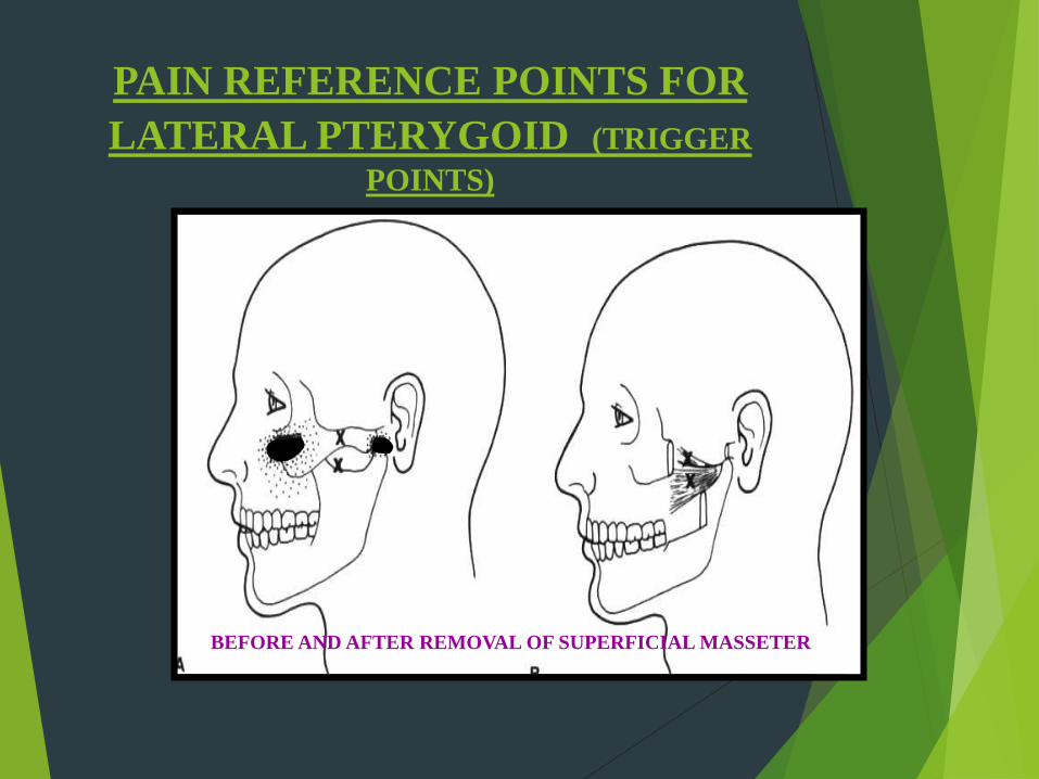
PAIN REFERENCE POINTS FOR
LATERAL PTERYGOID (TRIGGER
POINTS)
BEFORE AND AFTER REMOVAL OF SUPERFICIAL MASSETER

STERNAL DIVISION
CLAVICULAR DIVISION
PAIN REFERENCE POINTS FOR STERNOCLEIDO-MASTOID (TRIGGER POINTS)

PAIN REFERENCE POINTS FOR
TRAPEZIOUS (TRIGGER POINTS)
UPPER RIGHT TRAPEZIUS


KEYS IN MAKING A DIFFERENTIAL
DIAGNOSIS
History
Examination
Mandibular restriction
Mandibular interference
Acute malocclusion
Loading of the joint
Functional manipulation
Diagnostic anesthetic blockade
Diagnostic imaging & Investigations

GENERAL HISTORY: which includes medical, surgical, psychological,
occupational and social background
SPECIFIC HISTORY: related to present complaint i.e. onset and type of
pain, aggrevating and relieving, severity of symptoms, associated
symptoms and medicines taken for the problem.
HISTORY TAKING

CRANIAL NERVE EXAMINATION Olfactory nv
Optic nv
Occulomotor/ Trochlear/ Abducent nv
Trigeminal nerve
Facial nv
Acoustic nv
Glossopharyngeal nv
Accessory nv
Hypoglossal nerve

EYE EXAMINATION
Testing gross vision
Diplopia or blurriness of vision is noted
Reddening of the conjunctivae should be recorded
Any tearing or swelling of the eyelids
EAR EXAMINATION:

CERVICAL EXAMINATION
EXAMINATION FOR
CRANIOCERVICAL
DISORDERS.
asked to look to the extreme
right and the extreme left
look upward fully
Look downward fully
bend the neck to the right and
left

MUSCLE EXAMINTION Location of muscle pathology
Evaluation of muscle tone
Location of trigger point
Evaluation of temperature change
Location of swelling
Muscles are palpated bilaterally and simultaneously with firm but gentle pressure for 1-2min. Main pressure is exerted with the middle finger of each hand
During palpation subjective pain should be noted.
Patient is asked question regarding unilateral / bilateral pain, tenderness is mild / moderate or severe.
Reference zone of the pain should be noted

Temporalis Examination:

Masseter Examination:

Sternocleidomastoid Examination:

Posterior Neck Muscle examination

FUNCTIONAL MANIPULATION
Medial Pterygoid Muscle
Lateral pterygoid Muscle


Interincisal distance
Maximal comfortable mouth opening Maximal mouth opening
Checking “End Feel”

Alteration in Opening pathway

Dental / occlusal examination
Occlusal discrepancies, prematurities, or interference should be noted.
Anterior open bite, collapsed bite, cross bite, reduced vertical dimensions,
wear facets, mobility of teeth missing and teeth should be checked.
Type of occlusion, skeletal, dentofacial should be checked

Examination of Articular joint
JOINT SOUND
either clicks or crepitation
click is a sound of short duration. If it is relatively loud, it
is sometimes referred to as a “POP”
Crepitation is a multiple gravel-like sound described as
grating
JOINT RESTRICTION
The dynamic movements of the mandible are observed
for any irregularities or restrictions.
Diagnostic Blocking
INDICATIONS:
It is essential when differentiating primary from secondary pains
useful to identify the pathways that mediate peripheral pain and to localize pain sources
when the source of pain is difficult to identify, local anesthetic blocking of related tissues is the key to
making the proper diagnosis
educate the patient to the source of his or her pain problem
GENERAL RULE
purpose of an injection is to isolate the particular structure that is to be blocked
clinician should have a sound knowledge of the pharmacology of all solutions that will be used
clinician should avoid injecting into inflamed or diseased tissues
clinician should maintain strict asepsis at all times.
TYPES
Muscle block
Nerve block
Intra capsular

Technique of Trigger Point Injection

AURICULO-TEMPORAL NERVE BLOCKING

Radiological investigation
Helpful in diagnosis of
Intra articular pathologies
Osseous pathologies
Soft tissue pathologies
Conventional Radiograph
Panoramic radiograph
Transcranial projection
Transpharyngeal projection
Transmaxillary projection

Recent advances
CT
MRI
CBCT
Bone scaning
Other Investigations
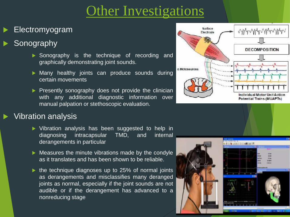
Electromyogram
Sonography
Sonography is the technique of recording and
graphically demonstrating joint sounds.
Many healthy joints can produce sounds during
certain movements
Presently sonography does not provide the clinician
with any additional diagnostic information over
manual palpation or stethoscopic evaluation.
Vibration analysis
Vibration analysis has been suggested to help in
diagnosing intracapsular TMD, and internal
derangements in particular
Measures the minute vibrations made by the condyle
as it translates and has been shown to be reliable.
the technique diagnoses up to 25% of normal joints
as derangements and misclassifies many deranged
joints as normal, especially if the joint sounds are not
audible or if the derangement has advanced to a
nonreducing stage

Thermography
Thermography is a technique that records and graphically
illustrates surface skin temperatures.
Various temperatures are recorded by different colors,
producing a map that depicts the surface being studied.
Recent studies shows Infrared imaging measurements can
provide a useful, non-invasive and nonionizing examination for
diagnosis of MTPs in masticatory muscles.
Mandibular tracking device
If a jaw-tracking device is used, the exact movement of the
mandible can be recorded
Unfortunately, many intracapsular and extracapsular disorders
create deviations and deflections in mandibular movement
pathways.
A particular deviation may not be specific for a particular
disorder, this information should only be used in conjunction
with history and examination findings.

MANAGEMENT OF MPDS
Patients counseling
Physiotherapy
Pharmacotherapy
Occlusal therapy

Patient concealing
Explaining patient about parafunctional habits such as clenching and bruxism.
Soft diet
Avoiding tooth to tooth contact.
Avoid stressful forces.
Resting of the jaw.
Relaxation therapy
Bio-feedback therapy – yoga, deep breathing, meditation, hypnosis

PHYSIOTHERAPY

Heat application
Superficial:
Hot packs, paraffin and radiants (Infra Red) Hot
moist application of towels for 15-20 min for 4
times.
Hydrocollator:
pad filled with clay and heated in water both for
70°-80°, wrapped in a protected towel and placed
over the affected area for 15-20 mins
Deep Heat application:
delivered by diathermy, ultrasound or
phonophorosis
DIATHERMY
ULTRASOUND
PHONOPHORESIS

DIATHERMY
Short Wave Diathermy
In chronic conditions, there will be increase in blood flow.
Increase in oxygenation on application for 10 mins
Mega Pulse
Rest period between pulse raise allows dissipation of heat by
blood flow.
Time of application – 10 mins
60 micro second pulse
100 pulse / sec.
Regime: 3 times / week for 4 weeks

Ultrasound:
Heat is placed on the skin which has to be coated with an acoustic coupling gel
and moved in parallel or circular over lapping sweeps 0.7 to 1 volts / cm2 for 10
mins.
Regime: 3 times / week for 4 weeks.
Uses:
Altered cell membrane permeability
Intracellular fluid absorption.
Decreased collagen viscosity.
Vasodilation
Relax muscles and analgesia.
Phonophorosis:
Application of ultrasound instead of acoustic coupling gel. It uses a pad filled with an anesthetic
or steroid cream is placed over the treatment kit
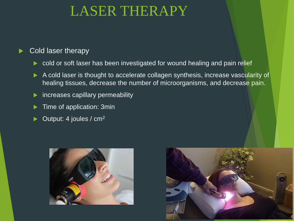
LASER THERAPY
Cold laser therapy
cold or soft laser has been investigated for wound healing and pain relief
A cold laser is thought to accelerate collagen synthesis, increase vascularity of
healing tissues, decrease the number of microorganisms, and decrease pain.
increases capillary permeability
Time of application: 3min
Output: 4 joules / cm2

Cryotherapy / Cold therapy :
Ice packs application to the painful area 4 times a day for 20
min.
Ice should not be placed over skin not more than 5 to 7 min
It lowers thermal gradient in skin, interrupting massive
concentration of Histamines, thus lowering pain threshold in the
skin.
Acupuncture:
It is based on a complex relationship between energy through
channels or natural elements (wood, earth and water) and
positive and negative elements.
Energy flow is done merely by placing a needle into a specific
site and adding either electric or heat to the needle.
It has minimal effect on reducing pain therefore not
recommended as primary therapy. Its used as an alternative
therapy.

Use of vasocoolent sprays:
Cold encourages the relaxation of muscles that are
in spasm and thus relieves the associated pain.
Most commonly used – ethyle chloride and
fluromethen
Fluromethane or ethylchloride spray is applied to
painful area for 5 min. Muscles are then gently
stretched after that.
Electrogalvanic stimulations:
Delivers a wide range of intensity to activate the
injured muscles.
It stimulate local circulation, achieves excitability and
conductivity without painful heating.
Pulse at 80 cycles / sec for 10 min followed by
excessive for 5 min.

TENS (Transcutaneous Electrical Nerve
Stimulation)
Produced by a continuous stimulation of cutaneousnerve fibers at a sub-painful level
When a TENS unit is placed over the tissues of apainful area, the electrical activity decreases painperception
TENS uses a low-voltage, low-amperage, biphasiccurrent of varied frequency and is designed primarilyfor sensory counter-stimulation in painful disorders.
It stimulate local circulation, achieves excitability andconductivity without painful heating.
Pulse at 80 cycles / sec for 10 min followed byexcessive for 5 min.

PENS (Percutaneous Electrical
Nerve Stimulation)
A new therapy for chronic pain sufferers that uses a low voltage electrical current delivered to the subcutaneous tissue or peripheral nerves to relieve chronic refractory neuropathic pain
It is a form of neurostimulation or neuromodultation that damping down overactive (sensitized) nerves that are causing pain
Does not destroy any nerves. It just makes them less sensitive to pain. A low voltage electrical current is delivered via a specially designed needle to a layer of tissue just below the surface of the skin close to the specific nerve, or to the nerve endings situated in an area that is painful
Some patients will have total pain relief, others experience prolonged pain relief for 3 months or more and others get relief for shorter periods of time

Manual therapy
Soft tissue mobilization
Joint mobilization
Muscle conditioning
Passive muscle stretching
Assisted muscle stretching
Resistance exercise
Postural training

PHARMACOTHERAPY
Anti inflammatory drugs:
NSAIDS: Reduces inflammation and provide pain relief both in the muscles and joints
for 14-21 days.
Aspirin 2 tab 0.3 to 0.6gm / 4th hourly
Piroxican 10-20 mg / 3-4 times /day
Ibiprofen 200-600mg / 3-4 times / day
Opoids: Pertazacine 50mg / 2-3 times /day.
Muscle relaxants:
It is used for short duration as they produce addiction.
Meprobamate 400mg TDS for 1 days.
Vallium 5-10mg 2-3 times /day.
It can be used as centrally acting eg Datrium, Succinyl colin, cusara, baclofin, and
peripherally acting.

ANTI ANXIETY MEDICATION:
Propylalcohol derivatives – Meprobamate 1200-1600 mg / day is divided doses.
Diphexyl methansis – Antilistamines are used in patients where benzyl diazapines are
contra indicated.
BENZODIAZEPIENES:
Alprazalam – 0.5mg 1-3 times / day
Diazepam – 2-5mg 1-4 times / day for 10 days
ANTI DEPRESSANT:
Amitriptyline 10-25 mg/day for 3 times
Fluoxitin 5mg / day
LOCAL ANAESTHETICS:
Procaine – 0.5%
Lidocain – 1%, 2%
Ethyl chloride spray or i.m.
Local anaesthetic at affected part give relief.

PCA (Patient Controlled
Analgesia) for MPDS
It is an effective method for administrating opiates to patient
for pain relief.
It gives patients a sense of control over pain

USE OF BOTOX
Botulinum toxin injections are currently the mainstay of treatment for most focal dystonias.
Neurotoxin botulinum toxin A, when injected into a muscle, causes a presynaptic blockade of the release of acetylcholine at the motor end plates.
End result is a muscle that can no longer contract (paralysis).
Normally takes 1 to 2 weeks for the effect to be clinically noticeable.
Normally, activity of the motor end plate is totally restored in 3 to 4 months
Approximately 25 U of botulinum toxin A is normally appropriate for each of these muscles.
The greatest number of motor end plates is found in the midbody of the muscle (halfway between the insertion and origin).

OCCLUSAL SPLINT
Purpose:
To create a balance joint tooth stabilization the mandible.
To reduce spasm, contracture and hyperactivity of musculature.
To restore vertical dimension
Types:
Stabilization splint
Relaxation splint
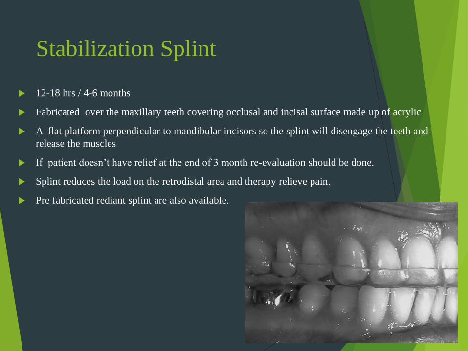
Stabilization Splint
12-18 hrs / 4-6 months
Fabricated over the maxillary teeth covering occlusal and incisal surface made up of acrylic
A flat platform perpendicular to mandibular incisors so the splint will disengage the teeth and
release the muscles
If patient doesn’t have relief at the end of 3 month re-evaluation should be done.
Splint reduces the load on the retrodistal area and therapy relieve pain.
Pre fabricated rediant splint are also available.

Relaxation splint
It is used for disengagement of teeth and for only short period (upto 4 wks)
They are fabricated over the maxillary incisor teeth
A platform is added to disengage mandibular anterior

Differential diagnosisType Cause History C/F Treatment
Muscle splinting 1. Altered sensory input
2. Constant deep pain
3. Increased stress
1. Recent alteration in
local structure
2. Source of deep pain
3. Recent increase in
emotional stress
1. Decrease ROM
2. But may achieve
normal ROM on
request
3. No pain at rest
4. Pain with function
5. Muscle weakness
1. Correction of local
causes
2. Removal of source of
deep pain
3. Psychological
regulation
4. Soft diet
5. Analgesic
Local muscle
soreness
1. h/o previous muscle splinting
2. Local tissue trauma
3. Emotional stress
1. Pain begun after
several hr/day of an
event
2. Pain started by-
injection, long
standing mouth
opening
3. Increased emotional
stress
1. Decrease ROM &
velocity but normal
range not achieve on
request
2. Minimum pain at rest
3. Pain increase with
function
4. Muscle fatigue
1. Elimination of
constant deep sensory
input
2. Patient motivation and
emotional stress
management
3. Supportive therapy to
control algesia
4. Stabilization
appliance
Myospasm 1. Continue deep pain
2. Local metabolic factors
within muscle tissues
3. Idiopathic myospasm
mechanism
1. Sudden onset of
restricted jaw
movement
1. Marked restriction of
jaw movement
2. Acute malocclusion
3. Pain at rest
4. Pain increase with
function
5. Affected muscle firm
and painful
6. Generalized muscle
tightness
1. Passive lightening/
stretching by manual
massage
2. 2% lidocaine without
vasopressor to stop
persistent spasm
3. Muscle rest
4. Reestablishment of
electrolyte balance

Type Cause History C/F Treatment
Myofascial pain 1. Continue deep pain
2. Increased emotional stress
3. Sleep disturbance
4. Local factors – habit, posture,
muscle strain, chilling
5. Systemic factors – nutritional
imbalance, fatigue, viral
infection
6. Idiopathic trigger point
1. c/o heterotropic pain
2. c/o headache or
muscle splinting
1. Slight decrease in
velocity and range of
motion of jaw
2. Presence of trigger
point
3. Presence of reference
zone
4. Heterotropic pain at
rest
5. Pain increase with
function
6. On provocation pain at
refer zone
1. Eleminate source of
deep pain
2. Soft diet
3. Life style modification
4. Analgesic, antianxyti,
muscle relaxant
5. Spray and stretch
6. Massage
7. Injection/ theraputic
blocking
Chronic myositis 1. Mediated by CNS not by
masticatory system
2. While CNS exposed to
prolonged pain – brain
pathway of pain deranged –
antidromic effect of afferent
nerve
1. Constant, primary,
myogenous pain
2. Associated with
prolonged history of
muscle complain
1. Significant decrease in
velocity and range of
movement
2. Significant pain at rest
3. Pain increase with rest
4. Generalized muscle
tightness
5. Significant pain on
muscle palpation
6. May induce muscle
atrophy
1. Restricted muscle use
2. Soft diet
3. Slower chewing and
smaller bite
4. Avoid exercise or
injection – may
increase pain – due to
neurogenic
inflammation
5. Disengage the teeth by
relaxation splint
6. Prescribe NSAIDs
Fibromyalgia 1. Still not cleared
2. Alteration in musculoskeletal
input by CNS
1. Chronic & generalized
musculoskeletal pain
in ¾ quadrant of body
since 3 month or more
2. Presence of sleep
disturbances
3. Clinical depression
1. Generalized
myogenous pain
2. Decreased ROM
3. Presence of numerous
myofascial trigger
point
4. Generalized muscle
fatigue & weakness
1. Definitive therapy to
treat underling causes
2. NSAIDs helpful to
some extent
3. If sleep problem –
antidepressant can be
given
