opean rev iewfor med icaland pharmacol sci 2016;20:4435 ... · 4438...
TRANSCRIPT

Abstract. – OBJECTIVE: Adult T cell lym-phoma is a highly aggressive T-cell malignancy.This study was designed to explore the expres-sion and functional significance of microRNA(miR)-373 in T cell lymphoma.
PATIENTS AND METHODS: We analyzed thelevels of CCND1 and miR-373 in T cell lymphomatissue and the relationship of miR-373 levelswith patients’ prognosis. We then overexpressedmiR-373 by miRNA mimics transfection and in-hibited miR-373 by miRNA antisense transfec-tion in T cell lymphoma cells. Cell survival andgrowth were analyzed by CCK-8 assay and MTTassay, respectively. Cell proliferation was ana-lyzed by flow cytometry. Bioinformatics analyseswere applied to predict miR-373 targets, whichwere then confirmed by luciferase reporter as-say.
RESULTS:We detected significantly higher lev-els of CCND1, and significantly lower levels ofmiR-373 in T cell lymphoma tissue, compared tothe adjacent non-tumor tissue. Moreover, the lowmiR-373 levels were associated with poor sur-vival of the patients. Overexpression of miR-373significantly inhibited cell growth, while deple-tion of miR-373 increased cell growth in T celllymphoma cells. Moreover, the effects of miR-373 on cell growth appeared to result from an al-teration in cell proliferation. Finally, miR-373 wasfound to bind to the 3'-UTR of CCND1 mRNA toinhibit its translation in T cell lymphoma cells.
CONCLUSIONS: Our study suggests that re-duced miR-373 levels in T cell lymphoma tissuemay promote T cell lymphoma growth, possiblythrough CCND1-mediated cell proliferation.
Key Words:T cell lymphoma; miR-373; cancer cell proliferation;
CCND1.
Introduction
Lymphoma is the most common blood cancer,which occurs when lymphocytes aberrantly out-
European Review for Medical and Pharmacological Sciences
Restoration of microRNA-373 suppressesgrowth of human T-cell lymphoma cellsby repressing CCND1
Y.-Y. TIAN, C.-M. JIA, Y. LI, Y. WANG, L. JIANG, A.-C. LIU
Department of Hematology, Harbin Medical University Cancer Hospital, Harbin, China
Yuyang Tian and Chuiming Jia contributed equally to this work
Corresponding Author: Aichun Liu, MD; e-mail: [email protected] 4435
grow, followed by metastasis into lymph nodes,spleen, bone marrow, blood, or other organs, andform tumor1-3. The two main forms of lymphomaare Hodgkin lymphoma and non-Hodgkin lym-phoma. Both B and T lymphocytes can developinto lymphomas. T cell lymphomas account forapproximately 15 percent of all non-Hodgkinlymphoma in the United States3-5. There are a va-riety of different types of T cell lymphoma, andthus the standard lymphoma therapies includechemotherapy, radiation, stem cell transplanta-tion and surgery6.Cyclin D1, also called CCND1, is a cell-cy-
cle activator. Regulatory component of the CC-ND1-CDK4 complex phosphorylates and in-hibits members of the retinoblastoma (RB)protein family including RB1 and regulates thecell-cycle during G1/S transition7-9. Compo-nent of the ternary complex, CCND1/CDK4/CDKN1B, is required for nuclear translocationand activity of the CCND1-CDK4 complex10-13.CCND1-CDK4 complexes are major integra-tors of various mitogenic and antimitogenicsignals14-18. CCND1/CDK4 complex is oftenover-activated in cancer cells and is a potent tu-mor enhancer19,20.MicroRNA (miRNA) is a group of non-coding
small RNAs of roughly 22 nucleotides, whichregulate many genes post-transcriptionally,through regulation of gene expression by repress-ing translation or directing sequence-specificdegradation of complementary mRNA by 3 -un-translated region (3 -UTR) binding21,22. MiRNAsplay a critical role in carcinogenesis of various tu-mors, as either tumor suppressor or enhancer23-30.Among all miRNAs, miR-373 has been rarelystudied. The first report on miR-373 showed aputative miR-373 target site in the promoter of E-cadherin. Transfection of miR-373 and its precur-sor hairpin RNA (pre-miR-373) readily induced
2016; 20: 4435-4444

Y.-Y. Tian, C.-M. Jia, Y. Li, Y. Wang, L. Jiang, A.-C. Liu
na). A null sequence was used as a control (null).The plasmids were transfected into cells at a con-centration of 50 nmol/l using Lipofectamine2000, according to the manufacturer’s instruc-tions (Invitrogen).
MiRNA Target Prediction and 3’-UTRLuciferase-Reporter AssayMiRNAs targets were predicted using the al-
gorithms TargetSan online software. The CCND13’-UTR reporter plasmid (pRL-CCND1) and theCCND1 3’-UTR reporter plasmid with a mutantat the miR-373 binding site (pRL-CCND1-mut)were purchased from Creative Biogene (Shirley,NY, USA). TT cells were co-transfected withpRL-CCND1/pRL-CCND1-mut and miR-373/as-miR-373/null by Lipofectamine 2000(5×10⁴ cells per well). Cells were collected 48hours after transfection for assay using the dual-luciferase reporter assay system gene assay kit(Promega, Madison, WI, USA), according to themanufacturer’s instructions.
Quantitative RT-PCR (RT-qPCR)Total RNA was extracted from resected speci-
mens or cultured cells with the miRNeasy minikit (Qiagen, Hilden, Germany). ComplementaryDNA (cDNA) was randomly primed from 2 g oftotal RNA using the Omniscript reverse tran-scription kit (Qiagen). Quantitative PCR (RT-qPCR) were performed in duplicates with Quan-tiTect SYBR Green PCR Kit (Qiagen). Allprimers were purchased from Qiagen. Data werecollected and analyzed, using 2-∆∆Ct method forquantification of the relative mRNA expressionlevels. Values of genes were first normalizedagainst α-tubulin and, then, compared to the ex-perimental controls.
Cell Counting Kit-8 (CCK-8) AssayThe CCK-8 detection kit (Sigma-Aldrich, St.
Louis, MO, USA) was used to measure cell vi-ability according to the manufacturer’s instruc-tions. Briefly, cells were seeded in a 96-wellmicroplate at a density of 5 × 10⁴/ml. After24h, cells were treated with resveratrol. Subse-quently, CCK-8 solution (20 ml/well) wasadded and the plate was incubated at 37oC for 2h. The viable cells were counted by absorbancemeasurements with a monochromator mi-croplate reader at a wavelength of 450 nm. Theoptical density value was reported as the per-centage of cell viability in relation to the con-trol group (set as 100%).
E-cadherin expression. Knockdown experimentsconfirmed that induction of E-cadherin by pre-miR-373 required the miRNA maturation proteinDicer31. Later on, miR-373 has been shown to beinvolved in the tumorigenesis of gastric cancer32,breast cancer33, lung cancer34, colon cancer35 andhepatocellular carcinoma36. However, the targetsof miR-373 in these cancers are very different,and thus the effects of miR-373 may be cancertype-specific. Moreover, a role of miR-373 in Tcell lymphoma has not been reported.Here, we analyzed the levels of CCND1 and
miR-373 in T cell lymphoma tissues, and studiedthe association of miR-373 with the prognosis ofthe patients. We then overexpressed miR-373 orinhibited miR-373 in 2 T cell lymphoma celllines and studied their effects on CCND1, cancercell growth, survival, and proliferation.
Patients and Methods
Patient SpecimensSurgical specimens from 30 T cell lymphoma
patients and matched tumor-adjacent normal tis-sues (NT) were obtained postoperatively in theHarbin Medical University Cancer Hospitalfrom 2011 to 2015. All patients gave signed, in-formed consent for the tissue to be used for sci-entific research. Ethical approval for the studywas obtained from the Harbin Medical Univer-sity Cancer Hospital. All diagnoses were basedon pathological and/or cytological evidence.The histological features of the specimens wereevaluated by senior pathologists according tothe World Health Organization classificationcriteria. All patients had been followed-up for30 months. Complete clinical data was electron-ically recorded.
Cell Line Culture and TransfectionH9 and HuT102 are two commonly human T
cell lymphoma lines used for research, and wereboth purchased from ATTC (American Type Cul-ture Collection, Manassas, VA, USA). Both celllines were cultured in Roswell Park Memorial In-stitute-1640 (RPMI-1640) medium (Invitrogen,Carlsbad, CA, USA) supplemented with 15% fe-tal bovine serum (FBS; Sigma-Aldrich, St Louis,MO, USA) in a humidified chamber with 5%CO2 at 37°C. MiRNAs mimics (miR-373) andmiRNAs antisense oligonucleotides (as-miR-373) were obtained from Origene (Beijing, Chi-
4436

followed by Fisher’ Exact Test for comparison oftwo groups. Patients’ survival was determined byKaplan-Meier analysis.
Results
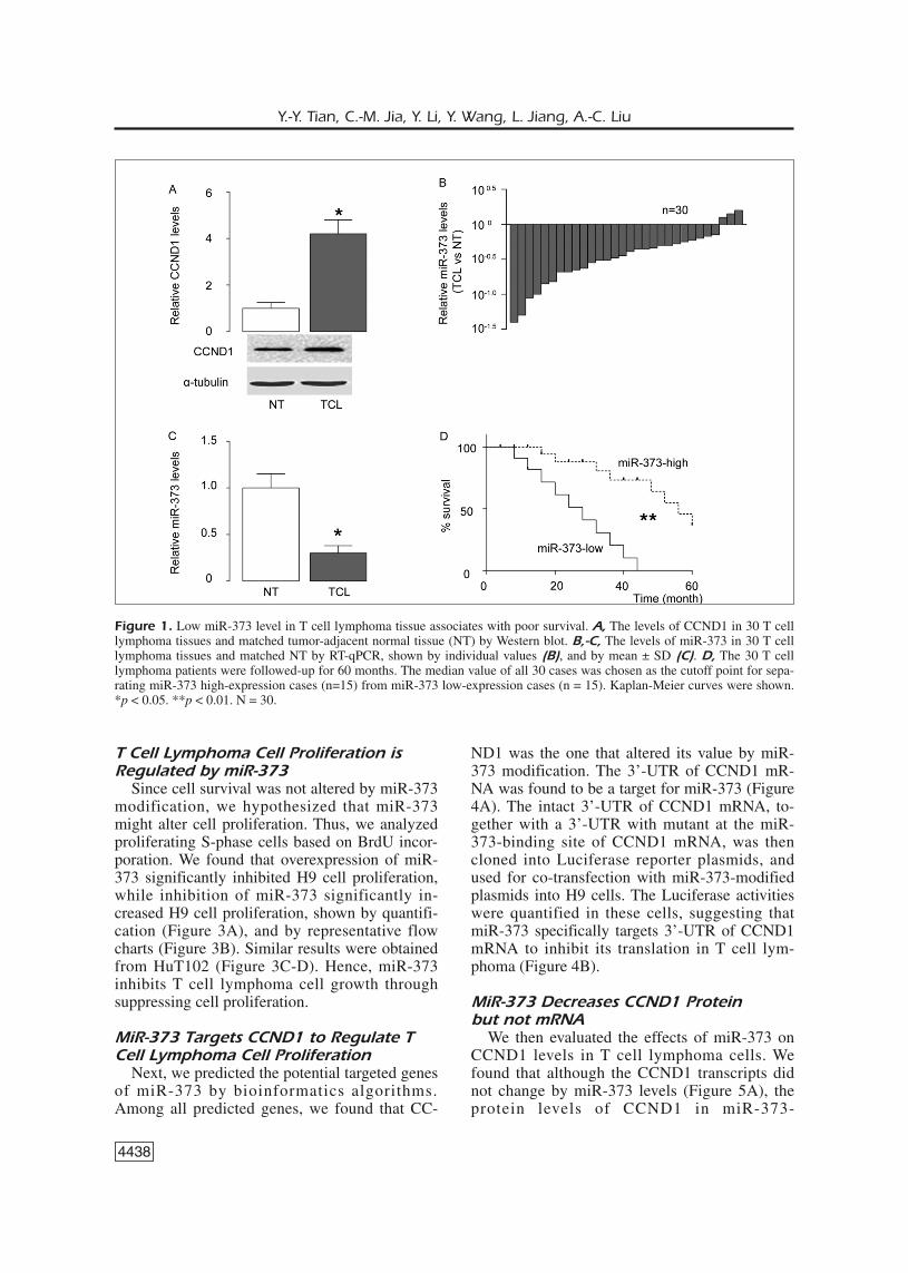
Low miR-373 Levels in T Cell LymphomaSpecimens Associate with Poor PrognosisThe levels of CCND1 and miR-373 in 30 pairs
of T cell lymphoma tissues and matched tumor-adjacent normal tissues (NT) were measured byWestern blot and RT-qPCR, respectively. Wefound that T cell lymphoma specimens expressedsignificantly higher levels of CCND1 (Figure1A), and significantly lower levels of miR-373,shown by individual values (Figure 1B), and bymean ± SD (Figure 1C). To examine the clinicalsignificance of low miR-373 levels in T cell lym-phoma, the 30 T cell lymphoma patients werefollowed-up for 30 months. The median value ofall 30 cases was chosen as the cutoff point forseparating miR-373 high-expression cases(n=15) from miR-373 low-expression cases(n=15). Kaplan-Meier curves indicated that Tcell lymphoma patients with low miR-373 levelshad a significantly worse prognosis than thosewith low miR-373 levels (Figure 1D).
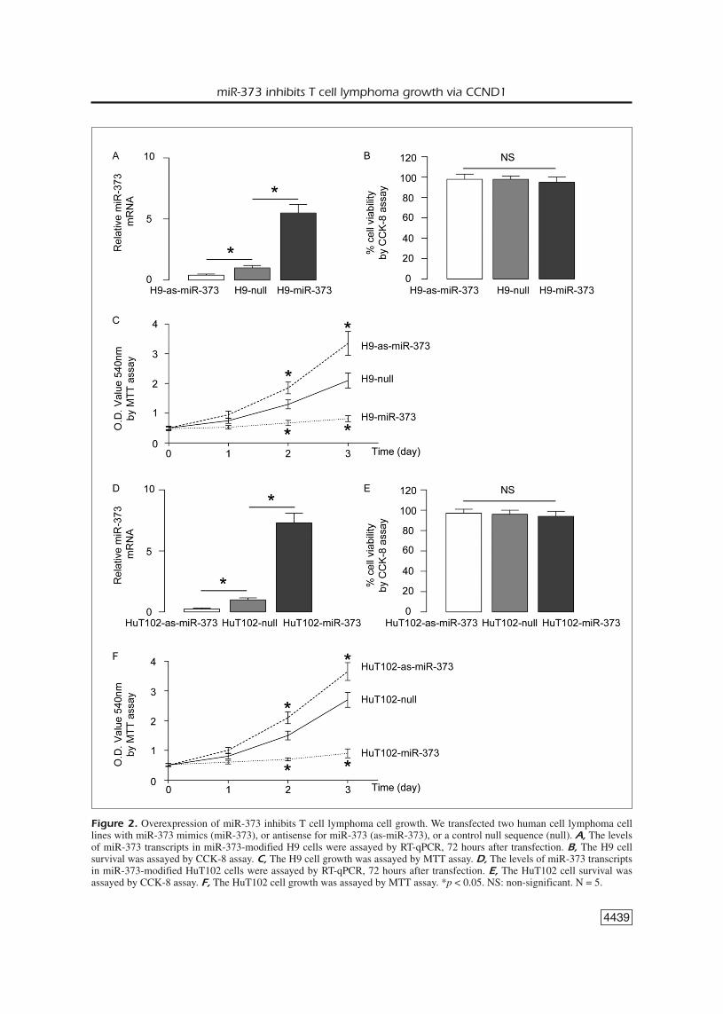
Overexpression of miR-373 Inhibits TCell Lymphoma Cell GrowthNext, the role of miR-373 in the growth of cul-
tured T cell lymphoma cells was investigated. Weused 2 human T cell lymphoma cell lines, H9 andHuT102, and transfected those cells with miR-373mimics (miR-373), or antisense for miR-373 (as-miR-373), or a control null sequence as a control(null). First, the levels of miR-373 in these modi-fied cells were assayed by RT-qPCR, 72 hours af-ter transfection. We found that the miR-373 levelsin H9 cells significantly increased by miR-373,while the miR-373 levels in H9 cells significantlydecreased by miR-373 suppression (Figure 2A).The cell survival and growth were then assayed byCCK-8 assay and by MTT assay, respectively. Wefound that the survival of H9 cells was not affect-ed by miR-373 modification (Figure 2B). Howev-er, overexpression of miR-373 significantly inhib-ited the cellular growth in H9 cells, while inhibi-tion of miR-373 significantly increased the cellu-lar growth in H9 cells, in an MTT assay (Figure2C). Similar results were obtained from HuT102cells (Figure 2D-F). Thus, miR-373 inhibits T celllymphoma cell growth in vitro.
4437
miR-373 inhibits T cell lymphoma growth via CCND1
MTT AssayFor assay of cell growth, 5×10³ cell per well
were seeded into 96 well-plate and subjected to aCell Proliferation Kit (MTT, Roche, Indianapo-lis, IN, USA), according to the instruction of themanufacturer. The MTT assay is a colorimetricassay for assessing viable cell number, taking ad-vantage that NADPH-dependent cellular oxidore-ductase enzymes in viable cells reduce the tetra-zolium dye 3-(4,5-dimethylthiazol-2-yl)-2,5-diphenyltetrazolium bromide (MTT) to its insol-uble formazan in purple readily being quantifiedby absorbance value (OD) at 570 nm in a mi-crotiter plate reader (Promega, Madison, WI,USA). Experiments were performed 5 times.
Proliferation AssayFor analysis of apoptosis, cultured cells were
dissociated and resuspended at a density of 106
cells/ml in PBS. After staining with FIT cell lym-phoma-conjugated BrdU antibody (FIT cell lym-phoma BrdU Flow Kit, Becton-Dickinson Bio-sciences, Franklin Lakes, NJ, USA), cells wereanalyzed using FACScan flow cytometer (Bec-ton-Dickinson Biosciences) equipped with CellQuest software (Becton-Dickinson Biosciences)for determination of FIT cell lymphoma+ S-phase proliferating cells.
Western BlotTotal Protein was extracted from the patients’
specimens or cultured cells by RIPA buffer (Sig-ma-Aldrich, St. Louis, MO, USA). An equalamount of proteins was loaded in the gel. Prima-ry antibodies for Western Blot are rabbit anti-CC-ND1 and anti-α-tubulin (all purchased from CellSignaling, St. Jose, LA, USA). The secondaryantibody is HRP-conjugated anti-rabbit (JacksonImmunoResearch Labs, West Grove, PA, USA).The protein levels were first normalized to α-tubulin and, then, normalized to control. Imagesshown in the figure were representatives from 3repeats. Densitometry of Western blots was quan-tified with NIH ImageJ software (Bethesda, MD,USA).
Statistical AnalysisAll statistical analyses were carried out using
the SPSS 18.0 statistical software package (SPSSInc., Chicago, IL, USA). All values in cell andanimal studies are depicted as mean ± standarddeviation and are considered significant if p <0.05. All data were statistically analyzed usingone-way ANOVA with a Bonferroni correction,
4438
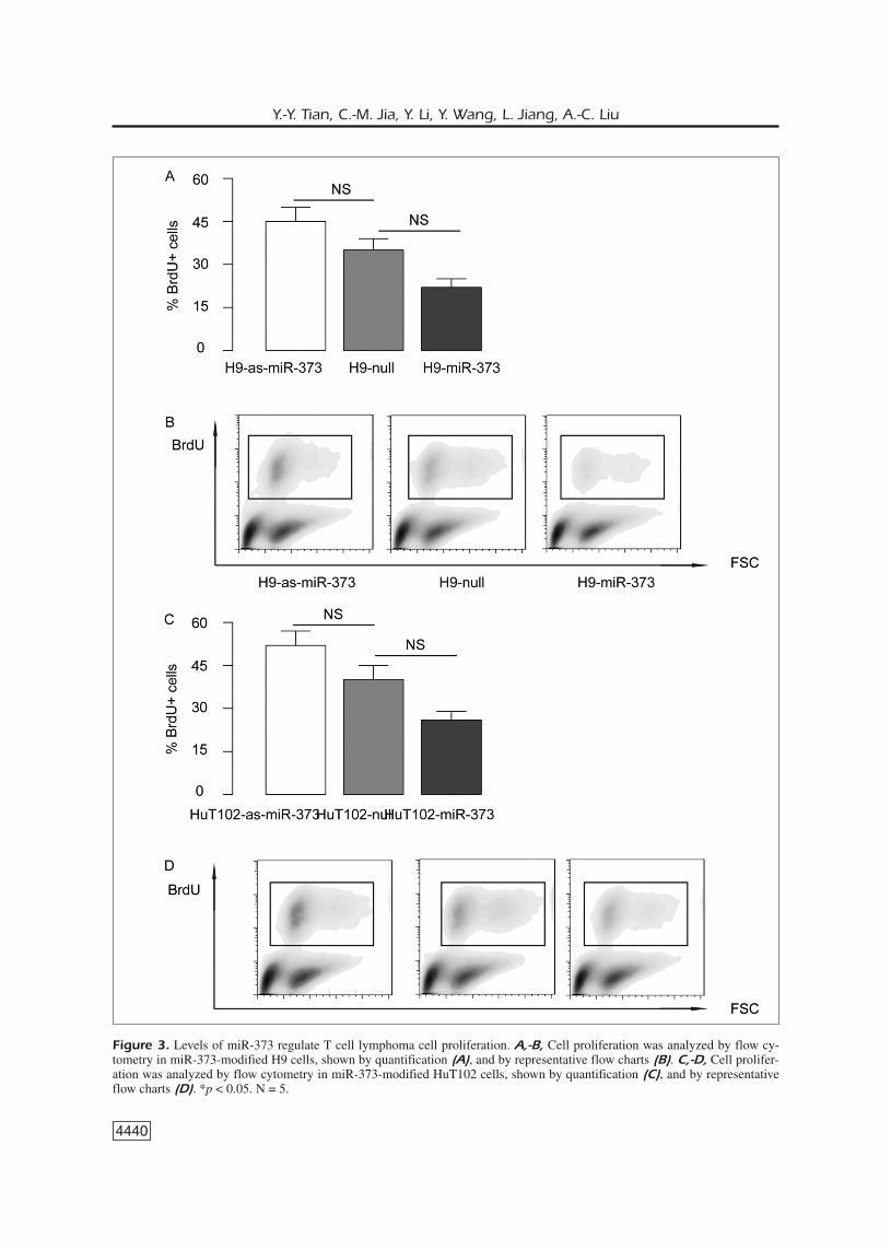
T Cell Lymphoma Cell Proliferation isRegulated by miR-373Since cell survival was not altered by miR-373
modification, we hypothesized that miR-373might alter cell proliferation. Thus, we analyzedproliferating S-phase cells based on BrdU incor-poration. We found that overexpression of miR-373 significantly inhibited H9 cell proliferation,while inhibition of miR-373 significantly in-creased H9 cell proliferation, shown by quantifi-cation (Figure 3A), and by representative flowcharts (Figure 3B). Similar results were obtainedfrom HuT102 (Figure 3C-D). Hence, miR-373inhibits T cell lymphoma cell growth throughsuppressing cell proliferation.
MiR-373 Targets CCND1 to Regulate TCell Lymphoma Cell ProliferationNext, we predicted the potential targeted genes
of miR-373 by bioinformatics algorithms.Among all predicted genes, we found that CC-
ND1 was the one that altered its value by miR-373 modification. The 3’-UTR of CCND1 mR-NA was found to be a target for miR-373 (Figure4A). The intact 3’-UTR of CCND1 mRNA, to-gether with a 3’-UTR with mutant at the miR-373-binding site of CCND1 mRNA, was thencloned into Luciferase reporter plasmids, andused for co-transfection with miR-373-modifiedplasmids into H9 cells. The Luciferase activitieswere quantified in these cells, suggesting thatmiR-373 specifically targets 3’-UTR of CCND1mRNA to inhibit its translation in T cell lym-phoma (Figure 4B).
MiR-373 Decreases CCND1 Proteinbut not mRNAWe then evaluated the effects of miR-373 on
CCND1 levels in T cell lymphoma cells. Wefound that although the CCND1 transcripts didnot change by miR-373 levels (Figure 5A), theprotein levels of CCND1 in miR-373-
Y.-Y. Tian, C.-M. Jia, Y. Li, Y. Wang, L. Jiang, A.-C. Liu
Figure 1. Low miR-373 level in T cell lymphoma tissue associates with poor survival. A, The levels of CCND1 in 30 T celllymphoma tissues and matched tumor-adjacent normal tissue (NT) by Western blot. B,-C, The levels of miR-373 in 30 T celllymphoma tissues and matched NT by RT-qPCR, shown by individual values (B), and by mean ± SD (C). D, The 30 T celllymphoma patients were followed-up for 60 months. The median value of all 30 cases was chosen as the cutoff point for sepa-rating miR-373 high-expression cases (n=15) from miR-373 low-expression cases (n = 15). Kaplan-Meier curves were shown.*p < 0.05. **p < 0.01. N = 30.
4439
miR-373 inhibits T cell lymphoma growth via CCND1
Figure 2. Overexpression of miR-373 inhibits T cell lymphoma cell growth. We transfected two human cell lymphoma celllines with miR-373 mimics (miR-373), or antisense for miR-373 (as-miR-373), or a control null sequence (null). A, The levelsof miR-373 transcripts in miR-373-modified H9 cells were assayed by RT-qPCR, 72 hours after transfection. B, The H9 cellsurvival was assayed by CCK-8 assay. C, The H9 cell growth was assayed by MTT assay. D, The levels of miR-373 transcriptsin miR-373-modified HuT102 cells were assayed by RT-qPCR, 72 hours after transfection. E, The HuT102 cell survival wasassayed by CCK-8 assay. F, The HuT102 cell growth was assayed by MTT assay. *p < 0.05. NS: non-significant. N = 5.
4440
Y.-Y. Tian, C.-M. Jia, Y. Li, Y. Wang, L. Jiang, A.-C. Liu
Figure 3. Levels of miR-373 regulate T cell lymphoma cell proliferation. A,-B, Cell proliferation was analyzed by flow cy-tometry in miR-373-modified H9 cells, shown by quantification (A), and by representative flow charts (B). C,-D, Cell prolifer-ation was analyzed by flow cytometry in miR-373-modified HuT102 cells, shown by quantification (C), and by representativeflow charts (D). *p < 0.05. N = 5.

4441
miR-373 inhibits T cell lymphoma growth via CCND1
Figure 4. MiR-373 targets CCND1 to regulate T cell lym-phoma cell proliferation. A, Bioinformatics algorithms analy-sis shows that the 3’-UTR of CCND1 mRNA is a target formiR-373. B, The intact 3'-UTR of CCND1 mRNA, togetherwith a 3'-UTR with mutant at miR-373-binding site of CC-ND1 mRNA, was then cloned into Luciferase reporter plas-mids, and used for co-transfection with miR-373-modifiedplasmids into H9 cells. The Luciferase activities were quanti-fied in these cells. *p < 0.05. NS: non-significant. N = 5.
Figure 5. MiR-373 decreases CCND1 protein but not mRNA. A,-B, We then evaluated the effects of miR-373 on CCND1levels in H9 cells, by mRNA (A), and by Western blot (B). C-D,We then evaluated the effects of miR-373 on CCND1 levels inHuT102 cells, by mRNA (C), and by Western blot (D). *p < 0.05. NS: non-significant. N = 5.

4442
overexpressing H9 cells was significantly de-creased, while the protein levels of CCND1 inmiR-373-depleted H9 cells was significantly in-creased (Figure 5B). Similar results were ob-tained from HuT102 cells (Figure 6C-D). Thesedata suggest that the translation of CCND1 in TTcells is suppressed by miR-373. Together, ourstudy demonstrates a role of miR-373 in controlof T cell lymphoma cell proliferation, throughCCND1 modulation (Figure 6).
Discussion
The participation of miRNAs in the T celllymphoma initiation and progression is not ade-quately investigated. Hence, elucidation of theaberrant expression of miRNAs in T cell lym-phoma carcinogenesis will help the physicianand tumor biologists to better characterize themolecular regulation of the tumorigenesis of Tcell lymphoma and may allow them to identifynovel targets to improve the levels of the currenttherapy.In this study, we showed that low level of
miR-373 in T cell lymphoma tissues were bothassociated with a low survival rate in T cell lym-phoma patients. Then, we showed that miR-373levels negatively regulated the cell growth in 2commonly used T cell lymphoma cell lines,through cell proliferation suppression, rather thanthrough interference with cell survival.Next, we examined how cell growth may be
regulated by miR-373 levels in T cell lymphomacells. Since the changes in cell number may re-sult from a summary of cell death and cell repli-cation but apoptosis seems unchanged in the cur-rent system, we thus hypothesize that the signaltransductions that regulate cell proliferation maybe involved. Among all candidates, we found thatCCND1 is a target for miR-373, and its value in-creased in T cell lymphoma and altered with theadaption of CCND1.Specifically, we found that the 3’-UTR of CC-
ND1 mRNA was targeted by miR-373, suggest-ing a strong effect on the translational regulationof CCND1 mRNA by miR-373. Moreover, wefurther proved that the binding of miR-373 to themRNA of CCND1 is functional in T cell lym-phoma cells, in which it suppressed the proteinlevel, but not mRNA level, consistent with thestructural analyses.Of note, when a miRNA molecule is attached
as a perfect match to a target mRNA, it causes
the mRNA degradation, resulting in a decrease inmRNA levels21-25. Here, it appears that there is apartial interaction between the miR-373 and the3’-UTR of the CCND1 mRNA. Thus, the miR-NA does not form a perfect match with the targetmRNA. Therefore, the translation process stopsat that point and hence the protein production isreduced, as described here.Apart from effects of CCND1 on cell growth,
it might affect also cell invasiveness. We did notexamine cell invasion in the current study. Be-sides regulation of CCND1 by miRNA, CCND1protein levels may be also affected by modula-tion of its degradation, e.g. through ubiquitina-tion. In future, it may be interesting to addressthese questions.
Conclusions
In T cell lymphoma, the levels of miR-373 ap-peared to be very low. Hence, miR-373 may be aspecific regulator of the tumor cell growth in Tcell lymphoma, and its loss may allow the tumorto grow. Further researches may address the mole-cular mechanisms underlying activation of miR-373 in T cell lymphoma, and these approachesmay provide additional evidence for using miR-373 as a novel target for treating T cell lymphoma.
–––––––––––––––––-––––Conflict of InterestThe Authors declare that there are no conflicts of interest.
References
1) BAGDONAITE I, WANDALL HH, LITVINOV IV, NASTASI C,BECKER JC, DABELSTEEN S, GEISLER C, BONEFELD CM,ZHANG Q, WASIK MA, ZHOU Y, SASSEVILLE D, ODUM N,WOETMANN A. Ectopic expression of a novel CD22splice-variant regulates survival and proliferationin malignant T cells from cutaneous T cell lym-phoma (CTCL) patients. Oncotarget 2015; 6:14374-14384.
Y.-Y. Tian, C.-M. Jia, Y. Li, Y. Wang, L. Jiang, A.-C. Liu
Figure 6. Schematic of the model. MiR-373 regulates Tcell lymphoma cell proliferation, through translational sup-pression of CCND1.

2) DERENZINI E, AGOSTINELLI C, IMBROGNO E, IACOBUCCI I,CASADEI B, BRIGHENTI E, RIGHI S, FULIGNI F, GHELLI
LUSERNA DI RORA A, FERRARI A, MARTINELLI G, PILERI S,ZINZANI PL. Constitutive activation of the DNAdamage response pathway as a novel therapeutictarget in diffuse large B-cell lymphoma. Oncotar-get 2015; 6: 6553-6569.
3) LAUENBORG B, CHRISTENSEN L, RALFKIAER U, KOPP KL,JONSON L, DABELSTEEN S, BONEFELD CM, GEISLER C,GJERDRUM LM, ZHANG Q, WASIK MA, RALFKIAER E,ODUM N, WOETMANN A. Malignant T cells expresslymphotoxin alpha and drive endothelial activationin cutaneous T cell lymphoma. Oncotarget 2015;6: 15235-15249.
4) MITOU G, FRENTZEL J, DESQUESNES A, LE GONIDEC S,ALSAATI T, BEAU I, LAMANT L, MEGGETTO F, ESPINOS E,CODOGNO P, BROUSSET P, GIURIATO S. Targeting au-tophagy enhances the anti-tumoral action ofcrizotinib in ALK-positive anaplastic large celllymphoma. Oncotarget 2015; 6: 30149-30164.
5) LEE S, PARK HY, KANG SY, KIM SJ, HWANG J, LEE S,KWAK SH, PARK KS, YOO HY, KIM WS, KIM JI, KO YH.Genetic alterations of JAK/STAT cascade and his-tone modification in extranodal NK/T-cell lym-phoma nasal type. Oncotarget 2015; 6: 17764-17776.
6) WU C, WU X, LIU X, YANG P, XU J, CHAI Y, GUO Q,WANG Z, ZHANG L. Prognostic significance ofmonocytes and monocytic myeloid-derived sup-pressor cells in diffuse large B-cell lymphomatreated with R-CHOP. Cell Physiol Biochem 2016;39: 521-530.
7) YE D, LUO H, LAI Z, ZOU L, ZHU L, MAO J, JACOB T,YE W, WANG L, CHEN L. ClC-3 chloride channel pro-teins regulate the cell cycle by up-regulating cy-clin D1-CDK4/6 through suppressing p21/p27 ex-pression in nasopharyngeal carcinoma cells. SciRep 2016; 6: 30276.
8) VEAS-PEREZ DE TUDELA M, MAESTRE C, DELGADO-ESTE-BAN M, BOLANOS JP, ALMEIDA A. Cdk5-mediated inhi-bition of APC/C-Cdh1 switches on the cyclin D1-Cdk4-pRb pathway causing aberrant S-phase en-try of postmitotic neurons. Sci Rep 2015; 5:18180.
9) ZAMPIERI A, CHAMPAGNE J, AUZEMERY B, FUENTES I,MAUREL B, BIENVENU F. Hyper sensitive protein de-tection by Tandem-HTRF reveals Cyclin D1 dy-namics in adult mouse. Sci Rep 2015; 5: 15739.
10) LIU YC, LEE CY, LIN CL, CHEN HY, LIU GY, HUNG HC.Multifaceted interactions and regulation betweenantizyme and its interacting proteins cyclin D1, or-nithine decarboxylase and antizyme inhibitor. On-cotarget 2015; 6: 23917-23929.
11) KENNEDY AL, VALLURUPALLI M, CHEN L, CROMPTON B,COWLEY G, VAZQUEZ F, WEIR BA, TSHERNIAK A, PARA-SURAMAN S, KIM S, ALEXE G, STEGMAIER K. Functional,chemical genomic, and super-enhancer screen-ing identify sensitivity to cyclin D1/CDK4 pathwayinhibition in Ewing sarcoma. Oncotarget 2015; 6:30178-30193.
12) HWANG SJ, LEE HW, KIM HR, SONG HJ, LEE DH, LEEH, SHIN CH, JOUNG JG, KIM DH, JOO KM, KIM HH.
Overexpression of microRNA-95-3p suppressesbrain metastasis of lung adenocarcinoma throughdownregulation of cyclin D1. Oncotarget 2015; 6:20434-20448.
13) CASIMIRO MC, DI SANTE G, CROSARIOL M, LORO E,DAMPIER W, ERTEL A, YU Z, SARIA EA, PAPANIKOLAOU A,LI Z, WANG C, ADDYA S, LISANTI MP, FORTINA P, CARDIFF
RD, TOZEREN A, KNUDSEN ES, ARNOLD A, PESTELL RG.Kinase-independent role of cyclin D1 in chromo-somal instability and mammary tumorigenesis.Oncotarget 2015; 6: 8525-8538.
14) ZHU A, LI Y, SONG W, XU Y, YANG F, ZHANG W, YIN Y,GUAN X. Antiproliferative effect of androgen recep-tor inhibition in mesenchymal stem-like triple-neg-ative breast cancer. Cell Physiol Biochem 2016;38: 1003-1014.
15) YU H, JIANG HL, XU D, JIN JZ, ZHAO ZM, MA YD,LIANG J. Transcription factor MafB promotes hepa-tocellular carcinoma cell proliferation through up-regulation of cyclin D1. Cell Physiol Biochem2016; 39: 700-708.
16) SCHENK LK, SCHULZE U, HENKE S, WEIDE T, PAVENSTADTH. TMEM16F regulates baseline phosphatidylser-ine exposure and cell viability in human embryon-ic kidney cells. Cell Physiol Biochem 2016; 38:2452-2463.
17) HU H, CHEN M, DAI G, DU G, WANG X, HE J, ZHAOY, HAN D, CAO Y, ZHENG Y, DING D. an inhibitoryrole of osthole in rat MSCs osteogenic differentia-tion and proliferation via Wnt/beta-catenin andErk1/2-MAPK pathways. Cell Physiol Biochem2016; 38: 2375-2388.
18) LUO G, WANG M, WU X, TAO D, XIAO X, WANG L,MIN F, ZENG F, JIANG G. Long non-coding RNAMEG3 inhibits cell proliferation and induces apop-tosis in prostate cancer. Cell Physiol Biochem2015; 37: 2209-2220.
19) SUN C, HUANG C, LI S, YANG C, XI Y, WANG L, ZHANGF, FU Y, LI D. Hsa-miR-326 targets CCND1 and in-hibits non-small cell lung cancer development.Oncotarget 2016; 7: 8341-8359.
20) HAN K, CHEN X, BIAN N, MA B, YANG T, CAI C, FAN Q,ZHOU Y, ZHAO TB. MicroRNA profiling identifiesMiR-195 suppresses osteosarcoma cell metasta-sis by targeting CCND1. Oncotarget 2015; 6:8875-8889.
21) DI LEVA G, CROCE CM. miRNA profiling of cancer.Curr Opin Genet Dev 2013; 23: 3-11.
22) PEREIRA DM, RODRIGUES PM, BORRALHO PM, RO-DRIGUES CM. Delivering the promise of miRNA can-cer therapeutics. Drug Discov Today 2013; 18:282-289.
23) MEI Q, LI F, QUAN H, LIU Y, XU H. Busulfan inhibitsgrowth of human osteosarcoma through miR-200family microRNAs in vitro and in vivo. Cancer Sci2014; 105: 755-762.
24) WANG F, XIAO W, SUN J, HAN D, ZHU Y. MiRNA-181c inhibits EGFR-signaling-dependent MMP9activation via suppressing Akt phosphorylationin glioblastoma. Tumour Biol 2014; 35: 8653-8658.
4443
miR-373 inhibits T cell lymphoma growth via CCND1

4444
25) LIU G, JIANG C, LI D, WANG R, WANG W. MiRNA-34ainhibits EGFR-signaling-dependent MMP7 activa-tion in gastric cancer. Tumour Biol 2014; 35: 9801-9806.
26) JIANG J, HUANG J, WANG XR, QUAN YH. MicroRNA-202 induces cell cycle arrest and apoptosis inlung cancer cells through targeting cyclin D1. EurRev Med Pharmacol Sci 2016; 20: 2278-2284.
27) ZHU SM, CHEN CM, JIANG ZY, YUAN B, JI M, WU FH,JIN J. MicroRNA-185 inhibits cell proliferation andepithelial-mesenchymal transition in hepatocellu-lar carcinoma by targeting Six2. Eur Rev MedPharmacol Sci 2016; 20: 1712-1719.
28) LI B, YANG XX, WANG D, JI HK. MicroRNA-138 in-hibits proliferation of cervical cancer cells by tar-geting c-Met. Eur Rev Med Pharmacol Sci 2016;20: 1109-1114.
29) JIN Z, GUAN L, SONG Y, XIANG GM, CHEN SX, GAO B.MicroRNA-138 regulates chemoresistance in hu-man non-small cell lung cancer via epithelial mes-enchymal transition. Eur Rev Med Pharmacol Sci2016; 20: 1080-1086.
30) ZHANG Y, LIU YJ, LIU T, ZHANG H, YANG SJ. PlasmamicroRNA-21 is a potential diagnostic biomarkerof acute myocardial infarction. Eur Rev Med Phar-macol Sci 2016; 20: 323-329.
31) PLACE RF, LI LC, POOKOT D, NOONAN EJ, DAHIYA R.MicroRNA-373 induces expression of genes with
complementary promoter sequences. Proc NatlAcad Sci U S A 2008; 105: 1608-1613.
32) ZHANG X, LI X, TAN Z, LIU X, YANG C, DING X, HU X,ZHOU J, XIANG S, ZHOU C, ZHANG J. MicroRNA-373is upregulated and targets TNFAIP1 in humangastric cancer, contributing to tumorigenesis. On-col Lett 2013; 6: 1427-1434.
33) EICHELSER C, STUCKRATH I, MULLER V, MILDE-LANGOSCH
K, WIKMAN H, PANTEL K, SCHWARZENBACH H. In-creased serum levels of circulating exosomal mi-croRNA-373 in receptor-negative breast cancerpatients. Oncotarget 2014; 5: 9650-9663.
34) SEOL HS, AKIYAMA Y, SHIMADA S, LEE HJ, KIM TI,CHUN SM, SINGH SR, JANG SJ. Epigenetic silencingof microRNA-373 to epithelial-mesenchymaltransition in non-small cell lung cancer throughIRAK2 and LAMP1 axes. Cancer Lett 2014; 353:232-241.
35) TANAKA T, ARAI M, WU S, KANDA T, MIYAUCHI H, IMAZE-KI F, MATSUBARA H, YOKOSUKA O. Epigenetic silenc-ing of microRNA-373 plays an important role inregulating cell proliferation in colon cancer. OncolRep 2011; 26: 1329-1335.
36) WU N, LIU X, XU X, FAN X, LIU M, LI X, ZHONG Q,TANG H. MicroRNA-373, a new regulator of proteinphosphatase 6, functions as an oncogene in he-patocellular carcinoma. FEBS J 2011; 278: 2044-2054.
Y.-Y. Tian, C.-M. Jia, Y. Li, Y. Wang, L. Jiang, A.-C. Liu