mmu-mir-125b overexpression suppresses no production in ... · research article open access...
TRANSCRIPT
Xu et al. BMC Cancer (2016) 16:252 DOI 10.1186/s12885-016-2288-z
RESEARCH ARTICLE Open Access
Mmu-miR-125b overexpression suppressesNO production in activated macrophagesby targeting eEF2K and CCNA2
Zhenbiao Xu, Lianmei Zhao, Xin Yang, Sisi Ma, Yehua Ge, Yanxin Liu, Shilian Liu, Juan Shi* and Dexian Zheng*Abstract
Background: MicroRNAs have been shown to be important regulators of the immune response and the developmentof the immune system. It was reported that microRNA-125b (miR-125b) was down-regulated in macrophageschallenged with endotoxin. However, little is known about the function and mechanism of action of miR-125b inmacrophage activation. Macrophages use L-arginine to synthesize nitric oxide (NO) through inducible NO synthase(iNOS), and the released NO contributes to the tumoricidal activity of macrophages.
Methods: Luciferase reporter assays were employed to validate regulation of a putative target of miR-125b. The effectof miR-125b on endogenous levels of this target were subsequently confirmed via Western blot. Quantitativereverse transcription-polymerase chain reaction (qRT-PCR) was performed to determine the expression level ofmiR-125b in macrophage. MTS assays were conducted to explore the impact of miR-125b overexpression onthe cell viability of 4T1 cells.
Results: Here, we demonstrate that mmu-miR-125b overexpression suppresses NO production in activatedmacrophages and that LPS-activated macrophages with overexpressed mmu-miR-125b promote 4T1 tumorcell proliferation in vitro and 4T1 tumor growth in vivo. CCNA2 and eEF2K are the direct and functionaltargets of mmu-miR-125b in macrophages; CCNA2 and eEF2K expression was knocked down, which mimickedthe mmu-miR-125b overexpression phenotype.
Conclusions: These data suggest that mmu-miR-125b decreases NO production in activated macrophages atleast partially by suppressing eEF2K and CCNA2 expression.
Keywords: Mmu-miR-125, Macrophages, Nitric oxide, eEF2K, CCNA2
BackgroundMacrophages are key components of the mammalian in-nate immune system, in which they function in cytokinerelease, pathogen killing and antigen presentation to theadaptive immune system. When cell surface sensing pro-teins, such as Toll-like receptors (TLRs), recognize andengage pathogens, macrophages are rapidly activated;these activated macrophages transform from a relativequiescent state to an effector state to perform defensefunctions [1–5]. Classically activated macrophages, orM1 macrophages, activate the Th1 immune response
* Correspondence: [email protected]; [email protected] Key Laboratory of Medical Molecular Biology, Institute of Basic MedicalSciences, Chinese Academy of Medical Sciences & Peking Union MedicalCollege, Beijing 100005, China
© 2016 Xu et al. Open Access This article is dInternational License (http://creativecommonsreproduction in any medium, provided you gthe Creative Commons license, and indicate if(http://creativecommons.org/publicdomain/ze
and secrete high amounts of pro-inflammatory media-tors, such as cytotoxic TNFα and nitric oxide (NO), tokill invading pathogens or tumor cells. In fact, the highexpression of inducible NO synthase (iNOS), whichproduces NO, is the hallmark of these macrophages.NO, a free radical gaseous molecule, is a mediator ofvital physiological functions, including host defense.Many cell types can produce NO using L-arginine viaiNOS. Macrophages are one of the best-characterizedsources of NO. Throughout the last decade, NO hasbeen identified to play an important role as a first line ofdefense against various pathogens. Macrophage uses L-arginine to synthesize NO via iNOS, and the released NOcontributes to the tumoricidal activity of macrophages. Inearly stages of tumor development, macrophages employ
istributed under the terms of the Creative Commons Attribution 4.0.org/licenses/by/4.0/), which permits unrestricted use, distribution, andive appropriate credit to the original author(s) and the source, provide a link tochanges were made. The Creative Commons Public Domain Dedication waiverro/1.0/) applies to the data made available in this article, unless otherwise stated.
Xu et al. BMC Cancer (2016) 16:252 Page 2 of 10
their killing mechanisms, particularly the generation ofhigh NO concentrations, to induce tumor cell apoptosisand destroy emerging transformed cells [6–8].It has been shown that microRNAs (miRs) are
important mediators of macrophage activation. It wasreported that miR-155, miR-146, miR-147, miR-9,miR-107 and miR-21 are induced by the TLR signal-ing pathway [9–13]. These miRs can inhibit theexpression of signaling proteins in the inflammatorysignaling cascade and therefore modulate immunitythrough feedback mechanisms [10, 12]. MiR-125b, ahomolog of C. elegans miR-lin-4, is deregulated inmost cancers and can regulate cancer cell proliferation viaits target genes [14–19]. It has also been demonstratedthat miR-125b is down-regulated in macrophages inresponse to TLR4 signaling [20–24] and enriched inhematopoietic stem cells, which then enhanceshematopoietic engraftment [25, 26]. The mechanisms bywhich macrophages respond to miR-125b and the func-tion of miR-125b in regulating macrophages remainunclear.In the present study, we demonstrate that mmu-
miR-125b (MIMAT0000136) is down-regulated in macro-phages activated by LPS. Mmu-miR-125b over-expressioninhibits NO production and thus promotes cancer cellgrowth both in vitro and in vivo. We further determinedthat eEF2K and CCNA2 are the important target genes ofmmu-miR-125b in macrophages. Knockdown of eEF2Kand CCNA2 expression mimics the phenotype of mmu-miR-125b overexpression in macrophages. These datasuggest that mmu-miR-125b decreases NO production inactivated macrophages to promote cancer cell growth, atleast partially by suppressing eEF2K and CCNA2expression.
MethodsIsolation of peritoneal macrophage and cell cultivationMice were injected intraperitoneally (i. p.) with 2 mL of3 % thioglycollate (Difco, Detroit, MI, USA). Three dayslater, mice were sacrificed by CO2 inhalation followed bycervical dislocation. Peritoneal exudate cells wereenriched for the peritoneal macrophages using themethod as described by Kumagai et al [27]. Briefly, theperitoneal cells were harvested by lavage and washed forthree times with the complete culture medium. Approxi-mately, 1 × 10⁶ cells per well were then cultured for twohours in six-well plates allowing the macrophages toadherent. The cells were washed three times with warmHank’s balanced salt solution to remove nonadhesivecells. The adherent macrophages were stimulated withvarious concentrations of stimuli and cultured at 37 °Cwith 5 % CO2 in DMEM or PRMI-1640 supplementedwith 10 % FBS, 100 U/ml penicillin, and 100 U/mlstreptomycin.
Cell lines of human HEK293T, mouse macrophageRAW264.7 and breast cancer 4T1 originated from theAmerican Type Culture Collection (Rockville, MD).These cells were cultured at 37 °C with 5 % CO2 inDMEM or PRMI-1640 supplemented with 10 % FBS, 100U/ml penicillin, and 100 U/ml streptomycin. RAW264.7cells stably transduced with lentivirals pLL3.7-miR-125b(named as RAW264.7-miR-125b) or control empty vectorpLL3.7 (named as RAW264.7-pLL3.7) were sorted byFACS. Mmu-miR-125b over-expression was verified byreal-time quantitative PCR (qPCR) carried out in a step-one Real-time PCR machine (Applied Biosystems, USA).
Quantitative real-time PCRRNA was isolated with TRIzol (Invitrogen, USA) reagentaccording to the manufacturer’s instructions. qPCR wasconducted using a step-one Real-time PCR machine(Applied Biosystems, USA). SYBR Green PCR MasterMix (Takara, Shiga, Japan) was used to analyze mmu-miR-125b, CCNA2 and eEF2K expression. Primersequences are listed in Additional file 1: Table S1.
DNA constructsMouse pre-miR-125b-2 gene and the 3’ UTR fragment ofCCNA2 and eEF2k containing the putative mmu-miR-125b target sites and the mutations were amplified byusing the specific PCR primers (The forward and reverseprimers were shown in the Additional file 1: Table S1)and mouse peripheral blood lymphocyte genomicDNA as template. The DNA fragments were respectivelycloned into the pLL3.7 vector (Promega, Madison, WI,USA) downstream of the U6 promoter and the psi-CHECK2.2 vector (Promega, Madison, WI, USA) down-stream of the renilla luciferase gene. The DNA constructswere verified with DNA sequencing by BGI Life Tech Co.Ltd. (China).
NO detectionNO was determined using a nitrate/nitrite assay kit(Beyotime Institute of Biotechnology, China). Briefly,cells were stimulated with LPS for 12 h and the superna-tants were collected by centrifugation. Concentration ofNO was determined by mixing 50 μl of the supernatantswith 50 μl Griess reagent I and 50 μl Griess reagent IIand measured in a Multiscan ELISA Reader (AssaysHiTech) at 540 nm with appropriate standards (0–60 M)and normalized by total protein concentration.
Coculture assay4T1 cells were cocultured with either RAW macro-phage cells. Briefly, for coculture without cell-cellcontact, 1 × 10⁵ LPS-activated RAW264.7-miR-125b orRAW264.7-pLL3.7 cells were seeded in BoydenTranswell inserts (0.4 μm pores; Corning) permeable
Xu et al. BMC Cancer (2016) 16:252 Page 3 of 10
for soluble factors but not cells. Transwells containingmacrophages were then inserted into a 24-well plateand seeded with 3 × 10⁵ 4T1 tumor cells in each well.The cell viability of 4T1 cells was measured with MTS (3-(4, 5-dimethylthiazol-2-yl) -5-(3– carboxymethoxyphe-nyl)-2-(4-sulfophenyl)-2H-tetazolium)) assay according tothe manufacturer’s instruction (Promega, Madison, WI) atdifferent time points and calculated by the followingformula: Viability (OD) =OD of mix well- OD of controlwell.
Cell viability assaysCell viability and growth cure was measured using MTSassay according to the manufacturer’s instruction. Briefly,the cells were seeded on 96-well plates at a density of2 000 cells/well, incubation for indicated time, MTSsolution was added (20μL/well) into the cells, and in-cubated for 2 h at 37 °C, followed by measuring theabsorbance at 492 nm with a microplate reader.
Animal experimentsAnimal experiments were performed in accordance withthe institutional guidelines for animal care and wereapproved by the committee for the use and care of ani-mals of the Chinese Academy of Medical Sciences andPeking Union Medical College, Beijing, China. Briefly,4T1 (2 × 10⁶) cells and LPS-activated RAW264.7-miR-125b or RAW264.7-pLL3.7 (5 × 10⁵) cells were subcuta-neously co-injected into the right flanks of 4 to 6-week-old BALB/c female mice. Mice were closely monitoredfor nearly 1 month. The tumor sizes were measuredevery 3 days with a caliper. The tumor volume (V) wascalculated using the formula: V = 0.5 × length × width2.At the experimental end point, animals were euthanizedand tumors were removed and weighed.
Sequence alignmentThe mmu-miR-125b seed region and CCNA2, eEF2K3’ UTR sequences from mouse (Mus musculus) wereobtained and aligned using micoRNA database (http://www.microrna.org/microrna/getGeneForm.do) or Targets-can (http://www.targetscan.org/mmu_61/) [28, 29].
Luciferase reporter assay293T cells were co-transfected with pLL3.7-125b orpLL3.7 and psiCHECK2.2 vector containing 3’ UTRs ofCCNA2, eEF2K or their mutations or miR-125b positivecontrol. The luciferase activity was quantified after 48 htransfection using a Dual Luciferase Assay kit (Promega,Madison, WI). Firefly luciferase activity was normal-ized to Renilla, and the ratio of Firefly/Renilla valuewas reported.
Western blotRAW264.7-miR-125b, RAW264.7-pLL3.7 or RAW264.7cells were lysed and total 40-60 ng proteins in loadingbuffer were denatured for 10 min at 95°C, and then theproteins were subjected to 10 % SDS-PAGE. Theproteins in the gel were electronically transferred to anImmobilon-P membrane (Millipore, Eschborn, Germany).After blocking with 5 % no-fat milk, the membrane wasincubated with a rabbit polyclonal anti-CCNA2 or anti-eEF2K or anti-GAPDH Ab (1:1000; Cell Signaling Tech-nology, Beverly, MA) overnight in TBS. The interestingproteins were visualized using a peroxidase-conjugatedanti-rabbit IgG Ab (1:10000, Cell Signaling Technology,Beverly, MA) for 1 h and detected by using ECL system(Amersham Pharmacia Biotech Europe, Freiburg, Germany)followed by exposure to an X-ray film.
RNA interferenceSiRNA used in the experiment was listed in Additionalfile 2: Table S2. siRNA duplexes were transfected intocells using Lipofectamine 2000 (Invitrogen) at a finalconcentration of 40 nM.
Statistical analysisAll experiments were at least repeated three times. Theresults are presented as mean ± SD. The data were sub-jected to the Student’s t-test. P < 0.05 was consideredsignificant.
ResultsMmu-miR-125b expression is down-regulated in activatedmacrophagesMiR-125b is an important microRNA in cancer and theimmune response. It has been reported that miR-125b isdown-regulated in macrophages in response to TLR4signaling [22]. However, little is known about the func-tion and mechanism of action of miR-125b in macro-phage activation. To determine the expression level ofmmu-miR-125b in macrophages, mouse RAW264.7 andperitoneal macrophages were stimulated with LPS atvarious concentrations for different time points, andtotal RNA was then extracted with TRIzol. Mmu-miR-125b expression was determined by reverse transcriptionusing a stem-loop primer (Additional file 1: Table S1)followed by SYBR Green quantitative PCR (qPCR). Asshown in Fig. 1a, mmu-miR-125b expression decreasedover time in RAW264.7 cells activated with 1 μg/mlLPS. Mmu-miR-125b expression was also down-regulated by different concentrations of LPS (Fig. 1b).Similar results were obtained in peritoneal macrophages(PMs) activated with LPS (Fig. 1c-d), indicating thatmmu-miR-125b expression is down-regulated in macro-phages activated by LPS.
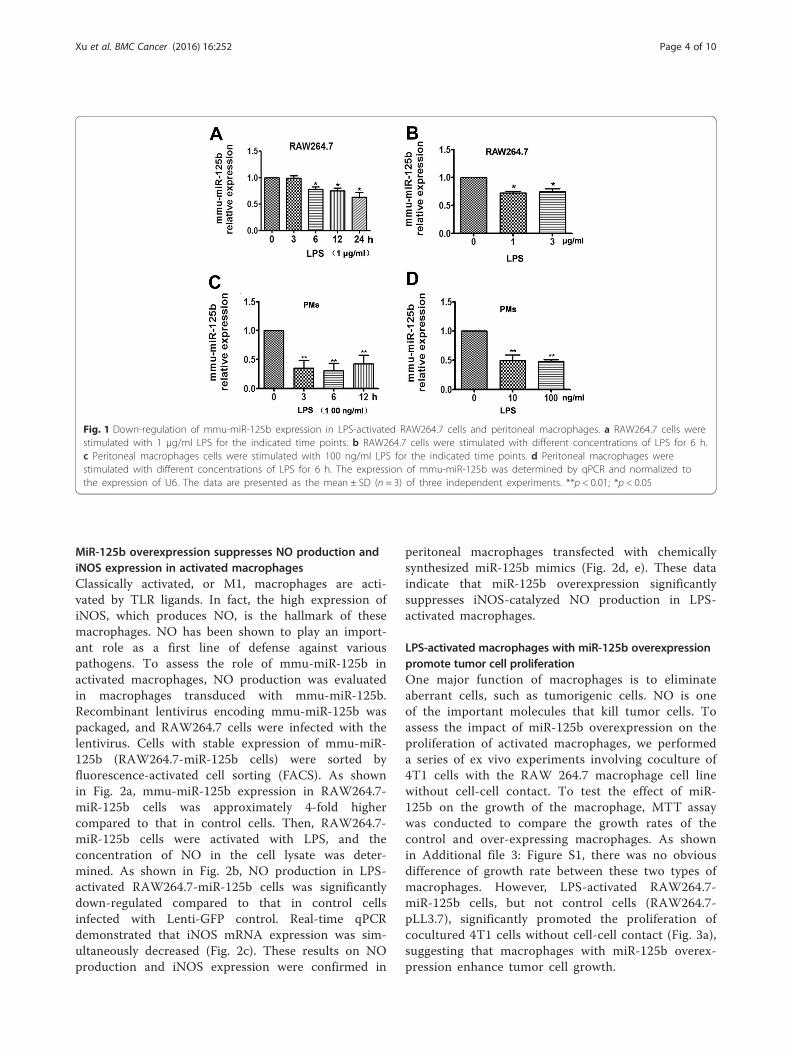
Fig. 1 Down-regulation of mmu-miR-125b expression in LPS-activated RAW264.7 cells and peritoneal macrophages. a RAW264.7 cells werestimulated with 1 μg/ml LPS for the indicated time points. b RAW264.7 cells were stimulated with different concentrations of LPS for 6 h.c Peritoneal macrophages cells were stimulated with 100 ng/ml LPS for the indicated time points. d Peritoneal macrophages werestimulated with different concentrations of LPS for 6 h. The expression of mmu-miR-125b was determined by qPCR and normalized tothe expression of U6. The data are presented as the mean ± SD (n = 3) of three independent experiments. **p < 0.01; *p < 0.05
Xu et al. BMC Cancer (2016) 16:252 Page 4 of 10
MiR-125b overexpression suppresses NO production andiNOS expression in activated macrophagesClassically activated, or M1, macrophages are acti-vated by TLR ligands. In fact, the high expression ofiNOS, which produces NO, is the hallmark of thesemacrophages. NO has been shown to play an import-ant role as a first line of defense against variouspathogens. To assess the role of mmu-miR-125b inactivated macrophages, NO production was evaluatedin macrophages transduced with mmu-miR-125b.Recombinant lentivirus encoding mmu-miR-125b waspackaged, and RAW264.7 cells were infected with thelentivirus. Cells with stable expression of mmu-miR-125b (RAW264.7-miR-125b cells) were sorted byfluorescence-activated cell sorting (FACS). As shownin Fig. 2a, mmu-miR-125b expression in RAW264.7-miR-125b cells was approximately 4-fold highercompared to that in control cells. Then, RAW264.7-miR-125b cells were activated with LPS, and theconcentration of NO in the cell lysate was deter-mined. As shown in Fig. 2b, NO production in LPS-activated RAW264.7-miR-125b cells was significantlydown-regulated compared to that in control cellsinfected with Lenti-GFP control. Real-time qPCRdemonstrated that iNOS mRNA expression was sim-ultaneously decreased (Fig. 2c). These results on NOproduction and iNOS expression were confirmed in
peritoneal macrophages transfected with chemicallysynthesized miR-125b mimics (Fig. 2d, e). These dataindicate that miR-125b overexpression significantlysuppresses iNOS-catalyzed NO production in LPS-activated macrophages.
LPS-activated macrophages with miR-125b overexpressionpromote tumor cell proliferationOne major function of macrophages is to eliminateaberrant cells, such as tumorigenic cells. NO is oneof the important molecules that kill tumor cells. Toassess the impact of miR-125b overexpression on theproliferation of activated macrophages, we performeda series of ex vivo experiments involving coculture of4T1 cells with the RAW 264.7 macrophage cell linewithout cell-cell contact. To test the effect of miR-125b on the growth of the macrophage, MTT assaywas conducted to compare the growth rates of thecontrol and over-expressing macrophages. As shownin Additional file 3: Figure S1, there was no obviousdifference of growth rate between these two types ofmacrophages. However, LPS-activated RAW264.7-miR-125b cells, but not control cells (RAW264.7-pLL3.7), significantly promoted the proliferation ofcocultured 4T1 cells without cell-cell contact (Fig. 3a),suggesting that macrophages with miR-125b overex-pression enhance tumor cell growth.
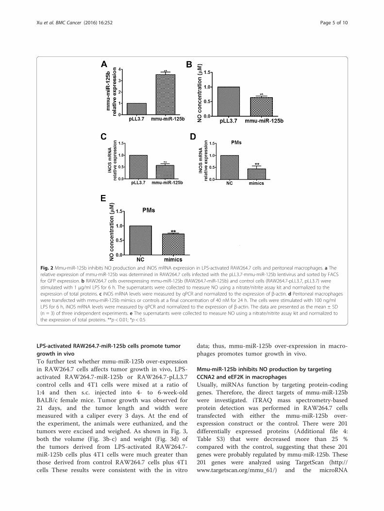
Fig. 2 Mmu-miR-125b inhibits NO production and iNOS mRNA expression in LPS-activated RAW264.7 cells and peritoneal macrophages. a Therelative expression of mmu-miR-125b was determined in RAW264.7 cells infected with the pLL3.7-mmu-miR-125b lentivirus and sorted by FACSfor GFP expression. b RAW264.7 cells overexpressing mmu-miR-125b (RAW264.7-miR-125b) and control cells (RAW264.7-pLL3.7, pLL3.7) werestimulated with 1 μg/ml LPS for 6 h. The supernatants were collected to measure NO using a nitrate/nitrite assay kit and normalized to theexpression of total proteins. c iNOS mRNA levels were measured by qPCR and normalized to the expression of β-actin. d Peritoneal macrophageswere transfected with mmu-miR-125b mimics or controls at a final concentration of 40 nM for 24 h. The cells were stimulated with 100 ng/mlLPS for 6 h, iNOS mRNA levels were measured by qPCR and normalized to the expression of β-actin. The data are presented as the mean ± SD(n = 3) of three independent experiments. e The supernatants were collected to measure NO using a nitrate/nitrite assay kit and normalized tothe expression of total proteins. **p < 0.01; *p < 0.5
Xu et al. BMC Cancer (2016) 16:252 Page 5 of 10
LPS-activated RAW264.7-miR-125b cells promote tumorgrowth in vivoTo further test whether mmu-miR-125b over-expressionin RAW264.7 cells affects tumor growth in vivo, LPS-activated RAW264.7-miR-125b or RAW264.7-pLL3.7control cells and 4T1 cells were mixed at a ratio of1:4 and then s.c. injected into 4- to 6-week-oldBALB/c female mice. Tumor growth was observed for21 days, and the tumor length and width weremeasured with a caliper every 3 days. At the end ofthe experiment, the animals were euthanized, and thetumors were excised and weighed. As shown in Fig. 3,both the volume (Fig. 3b-c) and weight (Fig. 3d) ofthe tumors derived from LPS-activated RAW264.7-miR-125b cells plus 4T1 cells were much greater thanthose derived from control RAW264.7 cells plus 4T1cells These results were consistent with the in vitro
data; thus, mmu-miR-125b over-expression in macro-phages promotes tumor growth in vivo.
Mmu-miR-125b inhibits NO production by targetingCCNA2 and eEF2K in macrophagesUsually, miRNAs function by targeting protein-codinggenes. Therefore, the direct targets of mmu-miR-125bwere investigated. iTRAQ mass spectrometry-basedprotein detection was performed in RAW264.7 cellstransfected with either the mmu-miR-125b over-expression construct or the control. There were 201differentially expressed proteins (Additional file 4:Table S3) that were decreased more than 25 %compared with the control, suggesting that these 201genes were probably regulated by mmu-miR-125b. These201 genes were analyzed using TargetScan (http://www.targetscan.org/mmu_61/) and the microRNA
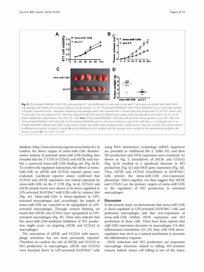
Fig. 3 LPS-activated RAW264.7-miR-125b cells promote 4T1 cell proliferation in vitro and in vivo. a 4T1 cells were cocultured with either RAWmacrophage cells. Briefly, for coculture without cell-cell contact, 1 × 10⁵ LPS-activated RAW264.7-miR-125b or RAW264.7-pLL3.7 cells were seededin Boyden Transwell inserts. Transwells containing macrophages were then inserted into a 24-well plate and seeded with 3 × 10⁵ 4T1 tumor cellsin each well. The cell viability of 4T1 cells was measured with MTS assay at different time points. Each bar represents the mean ± SD (n = 3) ofthree independent experiments. **p < 0.01; *p < 0.05. b-d. LPS-activated RAW264.7-miR-125b cells promote tumor growth in vivo. 4T1 cells andLPS-activated RAW264.7-miR-125b cells or LPS-activated RAW264.7-pLL3.7 cells were mixed at a ratio of 4:1 and then s.c. co-injected into 4- to6-week-old BALB/c female mice. After 5 days, tumor length and width were measured with a caliper every 3 days for 3 weeks. The tumor volumeat different time points is shown in panels b and c. Individual tumor weights and the average tumor weight at the experimental endpoint areshown in panels d. **p < 0.01; *p < 0.05
Xu et al. BMC Cancer (2016) 16:252 Page 6 of 10
database (http://www.microrna.org/microrna/home.do) toconfirm the direct targets of mmu-miR-125b. Bioinfor-matics analysis of potential mmu-miR-125b binding sitesrevealed that the 3’ UTR of CCNA2 and eEF2K each har-bor a conserved mmu-miR-125b binding site (Fig. 4a-b).To confirm the regulatory interaction, the effects of mmu-miR-125b on eEF2K and CCNA2 reporter genes wereevaluated. Luciferase reporter assays confirmed thatCCNA2 and eEF2K expression was indeed repressed bymmu-miR-125b via the 3’ UTR (Fig. 4c-d). CCNA2 andeEF2K protein levels were shown to be down-regulated inLPS-activated RAW264.7-miR-125b cells by western blot(Fig. 4e). Mmu-miR-125b is down-regulated in LPS-activated macrophages, and, accordingly, the targets ofmmu-miR-125b are expected to be upregulated in LPS-activated macrophages. Through western blotting, wefound that eEF2K and CCNA2 were upregulated in LPS-activated macrophages (Fig. 4f). These data indicate thatthe mmu-miR-125b-mediated inhibition of NO produc-tion might occur via targeting eEF2K and CCNA2 inmacrophages.The association of eEF2K and CCNA2 with macro-
phage activation has not been previously reported.Therefore, to confirm the role of eEF2K and CCNA2 inNO production in macrophages, eEF2K and CCNA2were knocked down in LPS-activated RAW264.7 cells
using RNA interference technology (siRNA sequencesare provided in Additional file 2: Table S2), and thenNO production and iNOS expression were examined. Asshown in Fig. 5, knockdown of eEF2K and CCNA2(Fig. 5a-b) resulted in a significant decrease in NOproduction (Fig. 5c) and iNOS gene expression (Fig. 5d).Thus, eEF2K and CCNA2 knockdown in RAW264.7cells mimics the mmu-miR-125b over-expressionphenotype. Taken together, our data suggest that eEF2Kand CCNA2 are the primary targets of mmu-miR-125bin the regulation of NO production in activatedmacrophages.
DiscussionIn the present study, we demonstrate that mmu-miR-125bis down-regulated in LPS-activated RAW264.7 cells andperitoneal macrophages and that over-expression ofmmu-miR-125b inhibits iNOS expression and NOproduction in these cells. There have been reports thatmiR-125b expression decreases in macrophages 3 h afterinflammatory stimulation [21, 23]; thus, miR-125b down-regulation may serve as a natural mechanism to promotethe inflammatory response.iNOS induction and NO production are important
macrophage functions related to killing NO-sensitivetumors; indeed, tumor cell killing is one of the major
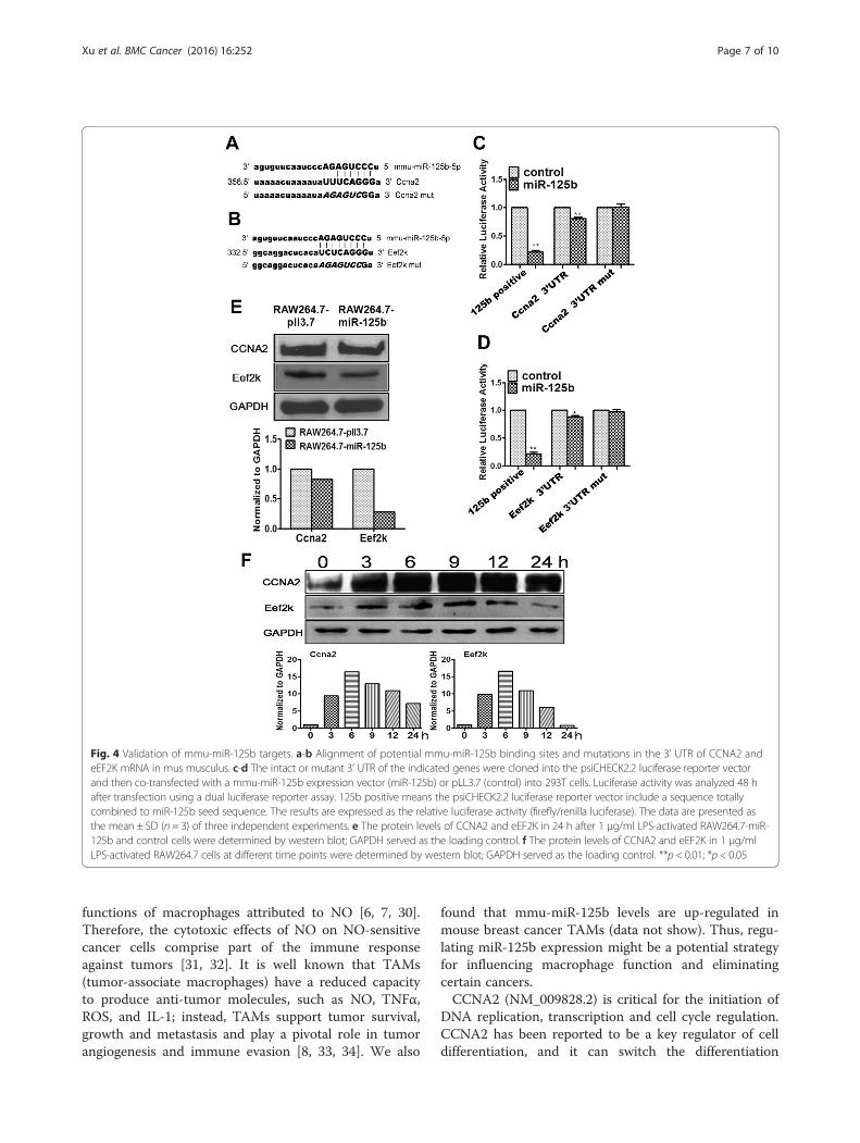
Fig. 4 Validation of mmu-miR-125b targets. a-b Alignment of potential mmu-miR-125b binding sites and mutations in the 3’ UTR of CCNA2 andeEF2K mRNA in mus musculus. c-d The intact or mutant 3’ UTR of the indicated genes were cloned into the psiCHECK2.2 luciferase reporter vectorand then co-transfected with a mmu-miR-125b expression vector (miR-125b) or pLL3.7 (control) into 293T cells. Luciferase activity was analyzed 48 hafter transfection using a dual luciferase reporter assay. 125b positive means the psiCHECK2.2 luciferase reporter vector include a sequence totallycombined to miR-125b seed sequence. The results are expressed as the relative luciferase activity (firefly/renilla luciferase). The data are presented asthe mean ± SD (n = 3) of three independent experiments. e The protein levels of CCNA2 and eEF2K in 24 h after 1 μg/ml LPS-activated RAW264.7-miR-125b and control cells were determined by western blot; GAPDH served as the loading control. f The protein levels of CCNA2 and eEF2K in 1 μg/mlLPS-activated RAW264.7 cells at different time points were determined by western blot; GAPDH served as the loading control. **p < 0.01; *p < 0.05
Xu et al. BMC Cancer (2016) 16:252 Page 7 of 10
functions of macrophages attributed to NO [6, 7, 30].Therefore, the cytotoxic effects of NO on NO-sensitivecancer cells comprise part of the immune responseagainst tumors [31, 32]. It is well known that TAMs(tumor-associate macrophages) have a reduced capacityto produce anti-tumor molecules, such as NO, TNFα,ROS, and IL-1; instead, TAMs support tumor survival,growth and metastasis and play a pivotal role in tumorangiogenesis and immune evasion [8, 33, 34]. We also
found that mmu-miR-125b levels are up-regulated inmouse breast cancer TAMs (data not show). Thus, regu-lating miR-125b expression might be a potential strategyfor influencing macrophage function and eliminatingcertain cancers.CCNA2 (NM_009828.2) is critical for the initiation of
DNA replication, transcription and cell cycle regulation.CCNA2 has been reported to be a key regulator of celldifferentiation, and it can switch the differentiation
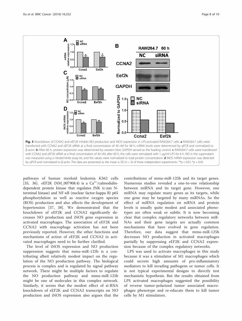
Fig. 5 Knockdown of CCNA2 and eEF2K inhibits NO production and iNOS expression in LPS-activated RAW264.7 cells. a RAW264.7 cells weretransfected with CCNA2 and eEF2K siRNA at a final concentration of 40 nM for 48 h; mRNA levels were determined by qPCR and normalized toβ-actin. b After 60 h, protein expression was determined by western blot; GAPDH served as the loading control. c RAW264.7 cells were transfectedwith CCNA2 and eEF2K siRNA at a final concentration of 40 nM; after 60 h, the cells were stimulated with 1 μg/ml LPS for 6 h. NO in the supernatantwas measured using a nitrate/nitrite assay kit, and the values were normalized to total protein concentration. d iNOS mRNA expression was detectedby qPCR and normalized to β-actin. The data are presented as the mean ± SD (n = 3) of three independent experiments. **p < 0.01; *p < 0.05
Xu et al. BMC Cancer (2016) 16:252 Page 8 of 10
pathways of human myeloid leukemia K562 cells[35, 36]. eEF2K (NM_007908.4) is a Ca2+/calmodulin-dependent protein kinase that regulates JNK (c-jun N-terminal kinase) and NF-κB (nuclear factor-kappa B) p65phosphorylation as well as reactive oxygen species(ROS) production and also affects the development ofhypertension [37, 38]. We demonstrated that theknockdown of eEF2K and CCNA2 significantly de-creases NO production and iNOS gene expression inactivated macrophages. The association of eEF2K andCCNA2 with macrophage activation has not beenpreviously reported. However, the other functions andmechanisms of action of eEF2K and CCNA2 in acti-vated macrophages need to be further clarified.The level of iNOS expression and NO production
suppression suggests that mmu-miR-125b is a con-tributing albeit relatively modest impact on the regu-lation of the NO production pathway. The biologicalprocess is complex and is regulated by signal pathwaynetwork. There might be multiple factors to regulatethe NO production pathway and mmu-miR-125bmight be one of molecules in this complex network.Similarly, it seems that the modest effect of si-RNAknockdown of eEF2K and CCNA2 transcripts on NOproduction and iNOS expression also argues that the
contributions of mmu-miR-125b and its target genes.Numerous studies revealed a one-to-one relationshipbetween miRNA and its target gene. However, onemiRNA may regulate many genes as its targets, whileone gene may be targeted by many miRNAs. So theeffect of miRNA regulation on mRNA and proteinlevels is usually quite modest and associated pheno-types are often weak or subtle. It is now becomingclear that complex regulatory networks between miR-NAs and their gene targets are actually commonmechanisms that have evolved in gene regulation.Therefore, our data suggest that mmu-miR-125bdecreases NO production in activated macrophagespartially by suppressing eEF2K and CCNA2 expres-sion because of the complex regulatory networks.LPS was used to activate macrophages in this study
because it was a stimulator of M1 macrophages whichcould secrete high amounts of pro-inflammatorymediators to kill invading pathogens or tumor cells. Itis not typical experimental designs to directly testmechanistic hypothesis. But the results obtained fromLPS activated macrophages suggested the possibilityof reverse tumor-polarized tumor associated macro-phages phenotype and re-educate them to kill tumorcells by M1 stimulators.
Xu et al. BMC Cancer (2016) 16:252 Page 9 of 10
ConclusionsWe have shown in this study that increased mmu-miR-125b expression in macrophages promotes 4T1 cellgrowth in vitro and in vivo. Therefore, knockdown ofmiR-125b expression in macrophages in the tumormicroenvironment may be a useful strategy for the treat-ment of certain cancers. These findings may extend ourunderstanding of the function of miR-125b in regulatingmacrophage activation and the immune response.
Additional files
Additional file 1: Table S1. Primers used in this study. (DOC 32 kb)
Additional file 2: Table S2. Ccna2 and Eef2k siRNA sequences. (DOC 30 kb)
Additional file 3: Figure S1. Proliferation of RAW264.7 cells stablyoverexpressing mmu-miR-125b was assessed by MTS assay. (TIFF 434 kb)
Additional file 4: Table S3. 201 differentially expressed proteins thatdecreased more than 25 % compared to the controls were detected byiTRAQ mass spectrometry. (XLSX 25 kb)
AbbreviationsmiRNAs (miR): microRNAs; mRNA: messenger RNA; 3’-UTR: 3’ untranslatedregion; NC: negative control; RNAi: RNA interference; siRNA: small interferingRNA; DMEM: Dulbecco’s modified Eagle’s medium; FBS: fetal bovine serum;TBS: Tris-buffered saline; PBS: phosphate-buffered saline; CCNA2: cyclin A2;eEF2K: eukaryotic elongation factor 2 kinase; iNOS: inducible nitric oxidesynthase; LPS: lipopolysaccharide; qPCR: quantitative real-time PCR; NO: nitricoxide; PMs: peritoneal macrophages; TLRs: Toll-like receptors.
Competing interestsThe authors declare that they have no competing interests.
Authors’ contributionsXZB and ZLM: study concept and design, acquisition of data, analysis andinterpretation of data, statistical analysis, and drafting of the manuscript. YX,MSS: statistical analysis. GYH: data and material support. LYX and LSL: studyconcept and design. SJ and ZDX: study concept and design, analysis andinterpretation of data, statistical analysis, drafting of the manuscript, studysupervision. All authors read and approved the final manuscript.
AcknowledgementsThis work was partially supported by the State Key Basic Research Programof China (Grant No. 2013CB530805) and the Natural Science Foundation ofChina (Grant No. 30972684 and 30972699).
Received: 26 May 2015 Accepted: 22 March 2016
References1. Krutzik SR, Tan B, Li H, et al. TLR activation triggers the rapid differentiation of
monocytes into macrophages and dendritic cells. Nat Med. 2005;11(6):653–60.2. Mantovani A, Sica A. Macrophages, innate immunity and cancer: balance,
tolerance, and diversity. Curr Opin Immunol. 2010;22(2):231–7.3. Geissmann F, Manz MG, Jung S, Sieweke MH, Merad M, Ley K.
Development of monocytes, macrophages, and dendritic cells.Science. 2010;327(5966):656–61.
4. Takeuchi O, Akira S. Pattern recognition receptors and inflammation. Cell.2010;140(6):805–20.
5. Schroder K, Sweet MJ, Hue DA. Signal integration between IFNgamma and TLRsignalling pathways in macrophages. Immunobiology. 2006;211(6-8):511–24.
6. Hibbs Jr JB, Taintor RR, Vavrin Z. Macrophage cytotoxicity: role forL-arginine deiminase and imino nitrogen oxidation to nitrite. Science.1987;235(4787):473–6.
7. Fukumura D, Kashiwagi S, Jain RK. The role of nitric oxide in tumourprogression. Nat Rev Cancer. 2006;6(7):521–34.
8. Pollard JW. Tumour-educated macrophages promote tumour progressionand metastasis. Nat Rev Cancer. 2004;4(1):71–8.
9. Chen Y, Liu W, Sun T, et al. 1,25-Dihydroxyvitamin D promotes negativefeedback regulation of TLR signaling via targeting microRNA-155-SOCS1 inmacrophages. J Immunol. 2013;190(7):3687–95.
10. O’Connell RM, Rao DS, Chaudhuri AA, Baltimore D. Physiological andpathological roles for microRNAs in the immune system. Nat Rev Immunol.2010;10(2):111–22.
11. Hennessy EJ, Sheedy FJ, Santamaria D, Barbacid M, O’Neill LA. Toll-likereceptor-4 (TLR4) down-regulates microRNA-107, increasing macrophageadhesion via cyclin-dependent kinase 6. J Biol Chem. 2011;286(29):25531–9.
12. O’Neill LA, Sheedy FJ, McCoy CE. MicroRNAs: the fine-tuners of Toll-likereceptor signalling. Nat Rev Immunol. 2011;11(3):163–75.
13. O’Connell RM, Kahn D, Gibson WS, et al. MicroRNA-155 promotesautoimmune inflammation by enhancing inflammatory T cell development.Immunity. 2010;33(4):607–19.
14. Shi L, Zhang J, Pan T, et al. MiR-125b is critical for the suppression ofhuman U251 glioma stem cell proliferation. Brain Res. 2010;1312:120–6.
15. Scott GK, Goga A, Bhaumik D, Berger CE, Sullivan CS, Benz CC. Coordinatesuppression of ERBB2 and ERBB3 by enforced expression of micro-RNAmiR-125a or miR-125b. J Biol Chem. 2007;282(2):1479–86.
16. Mizuno Y, Yagi K, Tokuzawa Y, et al. miR-125b inhibits osteoblasticdifferentiation by down-regulation of cell proliferation. Biochem Biophys ResCommun. 2008;368(2):267–72.
17. Le MT, Teh C, Shyh-Chang N, et al. MicroRNA-125b is a novel negativeregulator of p53. Genes Dev. 2009;23(7):862–76.
18. Huang L, Luo J, Cai Q, et al. MicroRNA-125b suppresses the development ofbladder cancer by targeting E2F3. Int J Cancer. 2011;128(8):1758–69.
19. Shi XB, Xue L, Yang J, et al. An androgen-regulated miRNA suppresses Bak1expression and induces androgen-independent growth of prostate cancercells. Proc Natl Acad Sci U S A. 2007;104(50):19983–8.
20. Lee YS, Kim HK, Chung S, Kim KS, Dutta A. Depletion of human micro-RNAmiR-125b reveals that it is critical for the proliferation of differentiated cellsbut not for the down-regulation of putative targets during differentiation.J Biol Chem. 2005;280(17):16635–41.
21. Androulidaki A, Iliopoulos D, Arranz A, et al. The kinase Akt1 controlsmacrophage response to lipopolysaccharide by regulating microRNAs.Immunity. 2009;31(2):220–31.
22. Tili E, Michaille JJ, Cimino A, et al. Modulation of miR-155 and miR-125blevels following lipopolysaccharide/TNF-alpha stimulation and their possibleroles in regulating the response to endotoxin shock. J Immunol.2007;179(8):5082–9.
23. Murphy AJ, Guyre PM, Pioli PA. Estradiol suppresses NF-kappa B activationthrough coordinated regulation of let-7a and miR-125b in primary humanmacrophages. J Immunol. 2010;184(9):5029–37.
24. Sonoki T, Iwanaga E, Mitsuya H, Asou N. Insertion of microRNA-125b-1, ahuman homologue of lin-4, into a rearranged immunoglobulin heavy chaingene locus in a patient with precursor B-cell acute lymphoblastic leukemia.Leukemia. 2005;19(11):2009–10.
25. O’Connell RM, Chaudhuri AA, Rao DS, Gibson WS, Balazs AB, Baltimore D.MicroRNAs enriched in hematopoietic stem cells differentially regulatelong-term hematopoietic output. Proc Natl Acad Sci U S A.2010;107(32):14235–40.
26. Ooi AG, Sahoo D, Adorno M, Wang Y, Weissman IL, Park CY. MicroRNA-125bexpands hematopoietic stem cells and enriches for the lymphoid-balancedand lymphoid-biased subsets. Proc Natl Acad Sci U S A. 2010;107(50):21505–10.
27. Kumagai K, Itoh K, Hinuma S, Tada M. Pretreatment of plastic Petri disheswith fetal calf serum. A simple method for macrophage isolation.J Immunol Methods. 1979;29(1):17–25.
28. Lewis BP, Burge CB, Bartel DP. Conserved seed pairing, often flanked byadenosines, indicates that thousands of human genes are microRNAtargets. Cell. 2005;120(1):15–20.
29. Friedman RC, Farh KK, Burge CB, Bartel DP. Most mammalian mRNAs areconserved targets of microRNAs. Genome Res. 2009;19(1):92–105.
30. Lala PK, Chakraborty C. Role of nitric oxide in carcinogenesis and tumourprogression. Lancet Oncol. 2001;2(3):149–56.
31. Jadeski LC, Chakraborty C, Lala PK. Role of nitric oxide in tumourprogression with special reference to a murine breast cancer model.Can J Physiol Pharmacol. 2002;80(2):125–35.
32. Thomsen LL, Miles DW. Role of nitric oxide in tumour progression: lessonsfrom human tumours. Cancer Metastasis Rev. 1998;17(1):107–18.
Xu et al. BMC Cancer (2016) 16:252 Page 10 of 10
33. Lewis CE, Pollard JW. Distinct role of macrophages in different tumormicroenvironments. Cancer Res. 2006;66(2):605–12.
34. Mantovani A, Allavena P, Sica A. Tumour-associated macrophages as aprototypic type II polarised phagocyte population: role in tumourprogression. Eur J Cancer. 2004;40(11):1660–7.
35. Bendris N, Arsic N, Lemmers B, Blanchard JM. Cyclin A2, Rho GTPases andEMT. Small GTPases. 2012;3(4):225–8.
36. Wang X, Song Y, Ren J, Qu X. Knocking-down cyclin A(2) by siRNAsuppresses apoptosis and switches differentiation pathways in K562 cellsupon administration with doxorubicin. PLoS One. 2009;4(8):e6665.
37. Cheng Y, Ren X, Zhang Y, et al. Integrated regulation of autophagy andapoptosis by EEF2K controls cellular fate and modulates the efficacy ofcurcumin and velcade against tumor cells. Autophagy. 2013;9(2):208–19.
38. Rose AJ, Alsted TJ, Jensen TE, et al. A Ca(2+)-calmodulin-eEF2K-eEF2signalling cascade, but not AMPK, contributes to the suppression of skeletalmuscle protein synthesis during contractions. J Physiol. 2009;587(Pt 7):1547–63.
• We accept pre-submission inquiries
• Our selector tool helps you to find the most relevant journal
• We provide round the clock customer support
• Convenient online submission
• Thorough peer review
• Inclusion in PubMed and all major indexing services
• Maximum visibility for your research
Submit your manuscript atwww.biomedcentral.com/submit
Submit your next manuscript to BioMed Central and we will help you at every step: