life in the trauma room: an into to thoracic injury - krcs · 1 life in the trauma room: an into to...
TRANSCRIPT

11
Life in the Trauma Room: An Into to Thoracic Injury
James Haan MD
Via Christi- St Francis
KU-Wichita
Sections
• Introduction to ThoracoAbdominal Injury
• Anatomy and Physiology of the Thorax & Abdomen
• Pathophysiology of Torso Trauma / Mechanism of Injury
• Assessment of the Torso Trauma Patient
• Management of the Torso Injury Patient
Thoracic Trauma
• Second leading cause of trauma deaths
• 85% treated with general resuscitation measures
• 15% require thoracotomy

22
• Vital Structures– Heart, Great Vessels, Esophagus, Tracheobronchial
Tree, & Lungs• 25% of MVC deaths are due to thoracic trauma
– 12,000 annually in US• Abdominal & Head injuries are common with chest
trauma.• Prevention Focus
– Legislation– Improved motor vehicle restraint systems
• Passive Restraint Systems• Airbags
Introduction to Thoracic Injury
• Trachea, Bronchi & Lungs
– Pleura• Visceral Pleura
– Cover lungs
• Parietal Pleura
– Lines inside of thoracic cavity
• Pleural Space
– POTENTIAL SPACE
» Air in Space = PNEUMOTHORAX
» Blood in Space = HEMOTHORAX
– Serous (pleural) fluid within
» Lubricates & permits ease of expansion
Anatomy and Physiology of the Thorax
• Trachea, Bronchi & Lungs
– Trachea• Hollow & cartilage supported structure
– Bronchi• Right & left extend for 3 centimeters• Enters lungs at Pulmonary Hilum
– Also where pulmonary arteries & veins enter• Further subdivide and terminate as alveoli
– Basic unit of structure & function in the lungs– Single cell membrane– External versus Internal Respiration
– Lungs• Right = 3 lobes• Left = 2 lobes
Anatomy and Physiology of the Thorax
33
• Diaphragm– Muscular, dome-like structure– Separates abdomen from the thoracic cavity– Affixed to the lower border of the rib cage– Central and superior margin extends to the level
of the 4th rib anteriorly and 6th rib posteriorly– Major muscle of respiration
• Draws downward during inspiration• Moves upward during exhalation
Anatomy and Physiology of the Thorax
• Thoracic Skeleton
– Topographical Thoracic Reference Lines• Midclavicular line
• Anterior axillary line
• Mid-axillary line
• Posterior axillary line
– Intercostal space• Artery, Vein and Nerve on inferior margin of each rib
– Thoracic Inlet• Superior opening of the thorax
• Curvature of 1st rib with associated structures
– Thoracic Outlet• Inferior opening of the thorax
• 12th rib and associated structures & Xiphisternal joint
Anatomy and Physiology of the Thorax
• Associated Musculature– Shoulder girdle
– Muscles of respiration• Diaphragm
– Primary muscle of respiration
– Inhalation: Contracts downward
– Exhalation: Relaxes upward
• Intercostal muscles– Contract to elevate the ribs and increase thoracic diameter
– Increase depth of respiration
• Sternocleidomastoid– Raise upper rib and sternum
Anatomy and Physiology of the Thorax
44
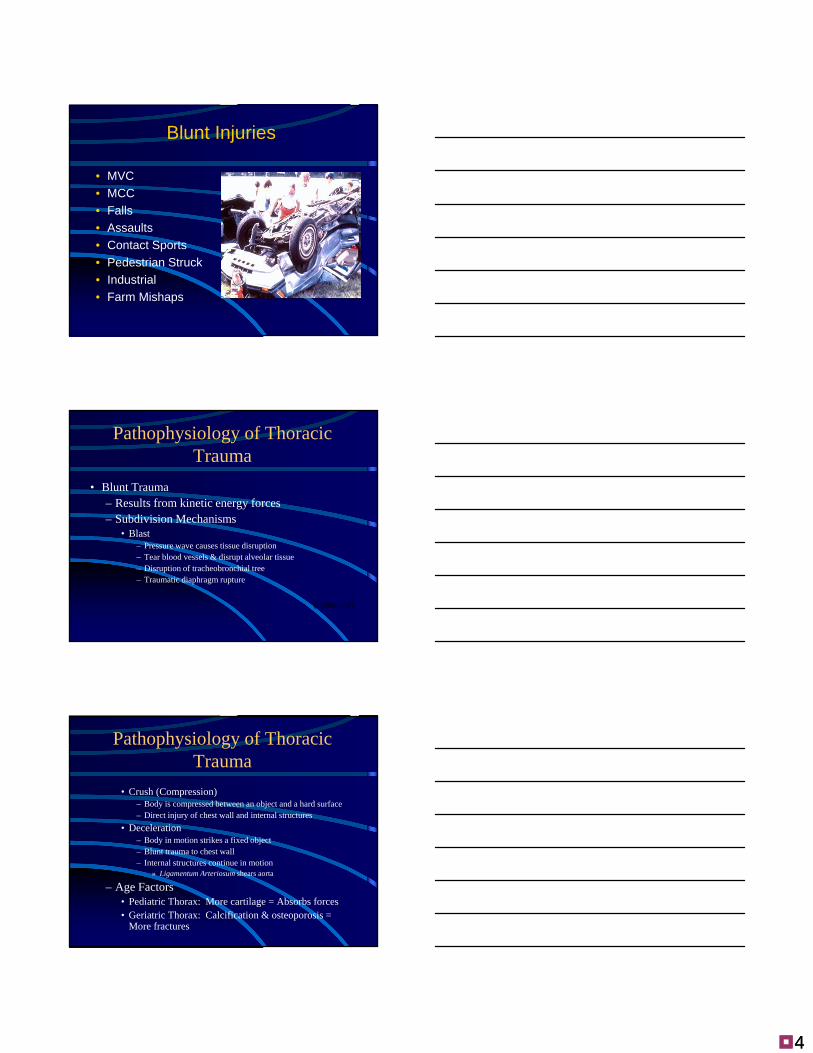
Blunt Injuries
• MVC
• MCC
• Falls
• Assaults
• Contact Sports
• Pedestrian Struck
• Industrial
• Farm Mishaps
• Blunt Trauma– Results from kinetic energy forces– Subdivision Mechanisms
• Blast– Pressure wave causes tissue disruption– Tear blood vessels & disrupt alveolar tissue– Disruption of tracheobronchial tree– Traumatic diaphragm rupture
Pathophysiology of Thoracic Trauma
(continued)
• Crush (Compression)– Body is compressed between an object and a hard surface– Direct injury of chest wall and internal structures
• Deceleration– Body in motion strikes a fixed object– Blunt trauma to chest wall– Internal structures continue in motion
» Ligamentum Arteriosum shears aorta
– Age Factors• Pediatric Thorax: More cartilage = Absorbs forces• Geriatric Thorax: Calcification & osteoporosis =
More fractures
Pathophysiology of Thoracic Trauma

55
• Traumatic Aortic Rupture– Aorta most commonly injured in severe blunt trauma
• 85-95% mortality
– Typically patients 50% will survive the initial injury insult
• 30% mortality in 6 hrs
• 50% mortality in 24 hrs
• 70% mortality in 1 week
– Injury may be confined to areas of aorta attachment
– Signs & Symptoms
• Rapid and deterioration of vitals
• Pulse deficit between right and left upper or lower extremities
• May be hemodynamically stable
Pathophysiology of Thoracic Trauma Cardiovascular Injuries
• Penetrating Trauma– Low Energy
• Arrows, knives, handguns• Injury caused by direct contact and cavitation
– High Energy• Military, hunting rifles & high powered hand guns• Extensive injury due to higher kinetic energy
– Shotgun• Injury severity based upon the distance between the victim and
shotgun & caliber of shot• Type I: >7 meters from the weapon
– Soft tissue injury• Type II: 3-7 meters from weapon
– Penetration into deep fascia and some internal organs• Type III: <3 meters from weapon
– Massive tissue destruction
Pathophysiology of Thoracic Trauma
Penetrating Injuries
• GSW
• Stab
• Impalements

66
• Closed pneumothorax
• Open pneumothorax (including sucking chest wound)
• Tension pneumothorax
• Pneumomediastinum
• Hemothorax
• Hemopneumothorax
• Laceration of vascular structures
Injuries Associated with Penetrating Thoraco Abdominal Trauma
Tracheobronchial tree lacerationsEsophageal lacerationsPenetrating cardiac injuriesPericardial tamponadeSpinal cord injuriesDiaphragm traumaIntra-abdominal penetration with associated organ injury
Tracheobronchial tree lacerationsEsophageal lacerationsPenetrating cardiac injuriesPericardial tamponadeSpinal cord injuriesDiaphragm traumaIntra-abdominal penetration with associated organ injury
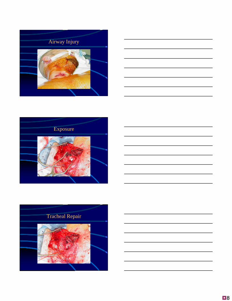
Airway Injuries
• Protect Aiway
• Primary repair if able– Buttress
– Segmental Resection
• Vent Strategy– Early extubation
– Minimize Peak Pressures
Airway: Resuscitative Procedures
• DAI/RSI– Maintain C-
spineimmobilization
– ETT size– Cricoid
pressure(Sellick Maneuver)
• Occlude esophagus to prevent aspiration
Esophagus
Cervical Vertebrae

77
Airway: Resuscitative Procedures - Optional
• Combitube– Not for use in children
• Cricothyroidotomy– Not recommended for
child < 12 yr. Old
Airway: Resuscitative Procedures
• DAI/RSI– Pre-oxygenate with
100% Oxygen– DAI Medications
• Succinylcholine • IV sedation• Etomidate
– Visualize vocal cords
Airway: Resuscitative Procedures
• Reassessment of airway– End tidal CO2 if
tracheal intubation
– Auscultation-Chest/Abdomen
– Chest wall rise
– Pulse oximeter
– Vital signs
88
Airway Injury
Exposure
Tracheal Repair

99
GSW
Bronchoscopy
Retrieval

1010
GSW Repair
Exposure
Repair
1111
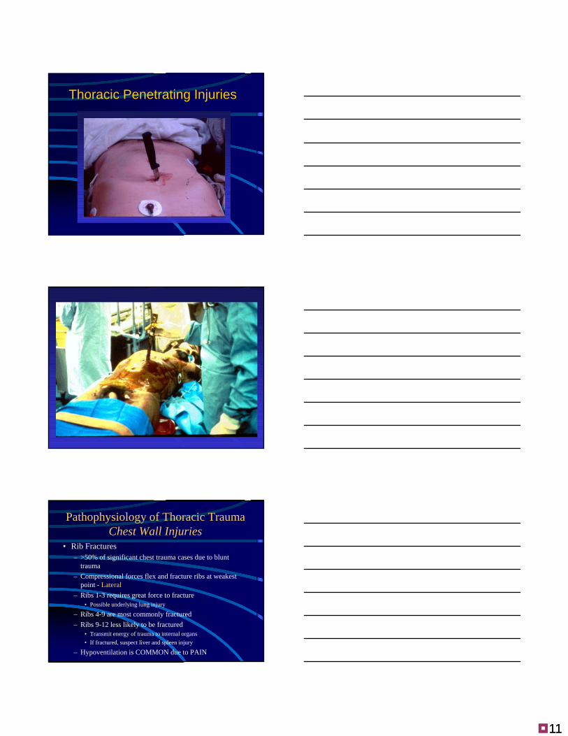
Thoracic Penetrating Injuries
• Rib Fractures– >50% of significant chest trauma cases due to blunt
trauma
– Compressional forces flex and fracture ribs at weakest point - Lateral
– Ribs 1-3 requires great force to fracture• Possible underlying lung injury
– Ribs 4-9 are most commonly fractured
– Ribs 9-12 less likely to be fractured• Transmit energy of trauma to internal organs
• If fractured, suspect liver and spleen injury
– Hypoventilation is COMMON due to PAIN
Pathophysiology of Thoracic Trauma Chest Wall Injuries

1212
• Flail Chest– Segment of the chest that becomes free to move with
the pressure changes of respiration
– Three or more adjacent rib fracture in two or more places
– Serious chest wall injury with underlying pulmonary injury- Especially Contusions
• Reduces volume of respiration/pneumonia
• Adds to increased mortality
– Paradoxical flail segment movement
– Positive pressure ventilation can restore tidal volume
– Pain control critical
Pathophysiology of Thoracic Trauma Chest Wall Injuries

1313
• Open Pneumothorax– Free passage of air between atmosphere and pleural
space
– Air replaces lung tissue
– Mediastinum shifts to uninjured side
– Air will be drawn through wound if wound is 2/3 diameter of the trachea or larger
– Signs & Symptoms• Penetrating chest trauma
• Sucking chest wound
• Frothy blood at wound site
• Severe Dyspnea
• Hypovolemia
Pathophysiology of Thoracic Trauma Pulmonary Injuries
• Tension Pneumothorax- Life Threatening– Buildup of air under pressure in the thorax.
– Excessive pressure reduces effectiveness of respiration
– Air is unable to escape from inside the pleural space
– Progression of Simple or Open Pneumothorax
– Decreased venus return
Pathophysiology of Thoracic Trauma Pulmonary Injuries

1414

1515
• Dyspnea– Tachypnea at first
• Progressive ventilation/perfusion mismatch
– Atelectasis on uninjured side• Hypoxemia• Hyperinflation of injured side of
chest• Hyperresonance of injured side of
chest
Pathophysiology of Thoracic Trauma Pulmonary Injuries
Tension Pneumothorax Signs & Symptoms
Diminished then absent breath sounds on injured sideCyanosisDiaphoresisAMSJVDHypotensionHypovolemiaTracheal Shifting
LATE SIGN
Diminished then absent breath sounds on injured sideCyanosisDiaphoresisAMSJVDHypotensionHypovolemiaTracheal Shifting
LATE SIGN
• Hemothorax– Accumulation of blood in the pleural space
– Serious hemorrhage may accumulate 1,500 mL of blood - Indication for Thoracotomy
• Mortality rate of 75%
• Each side of thorax may hold up to 3,000 mL
– Blood loss in thorax causes a decrease in tidal volume• Ventilation/Perfusion Mismatch & Shock
– Typically accompanies pneumothorax• Hemopneumothorax
Pathophysiology of Thoracic Trauma Pulmonary Injuries
• Blunt or penetrating chest trauma• Shock
– Dyspnea– Tachycardia– Tachypnea– Diaphoresis– Hypotension
• Dull to percussion over injured side
Pathophysiology of Thoracic Trauma Pulmonary InjuriesHemothorax Signs & Symptoms

1616
• Picture
1717
• Pulmonary Contusion– Soft tissue contusion of the lung– 30-75% of patients with significant blunt chest trauma– Frequently associated with rib fracture– Typical MOI
• Deceleration– Chest impact on steering wheel
• Bullet Cavitation– High velocity ammunition
– Microhemorrhage may account for 1- 1 ½ L of blood loss in alveolar tissue
• Progressive deterioration of ventilatory status
– Hemoptysis - Not Typical
Pathophysiology of Thoracic Trauma Pulmonary Injuries
• Contusion– Most Common result of blunt injury
– Signs & Symptoms (often none)• Erythema
• Ecchymosis
• DYSPNEA
• PAIN on breathing
• Limited breath sounds
• HYPOVENTILATION– BIGGEST CONCERN = “HURTS TO BREATHE”
Pathophysiology of Thoracic Trauma Chest Wall Injuries

1818
Pulmonary Contusion
• 70 % of patients demonstrate changes 1-hour post injury
• Other patients have a 4-6 hour time lag
• Initial x-ray findings have NO correlation with severity of contusion even CCT limited due to progression

1919
• Pericardial Tamponade– Restriction to cardiac filling caused by blood or other
fluid within the pericardium
– Occurs in <2% of all serious chest trauma• However, very high mortality
– Results from tear in the coronary artery or penetration of myocardium
• Blood seeps into pericardium and is unable to escape
• 200-300 ml of blood can restrict effectiveness of cardiac contractions
– Removing as little as 20 ml can provide relief
Pathophysiology of Thoracic Trauma Cardiovascular Injuries
• Dyspnea
• Possible cyanosis
• Beck’s Triad– JVD
– Distant heart tones
– Hypotension or narrowing pulse pressure
• Weak, thready pulse
• Shock
Pathophysiology of Thoracic Trauma Cardiovascular Injuries
Pericardial Tamponade Signs & Symptoms
Kussmaul’s signDecrease or absence of JVD during inspiration
Pulsus ParadoxusDrop in SBP >10 during inspirationDue to increase in CO2 during inspiration
Electrical AlteransP, QRS, & T amplitude changes in every other cardiac cycle
PEA
Kussmaul’s signDecrease or absence of JVD during inspiration
Pulsus ParadoxusDrop in SBP >10 during inspirationDue to increase in CO2 during inspiration
Electrical AlteransP, QRS, & T amplitude changes in every other cardiac cycle
PEA

2020
• Other Vascular Injuries– Rupture or laceration
• Superior Vena Cava• Inferior Vena Cava• General Thoracic Vasculature
– Blood Localizing in Mediastinum– Compression of:
• Great vessels• Myocardium• Esophagus
– General Signs & Symptoms• Penetrating Trauma• Hypovolemia & Shock• Hemothorax or hemomediastinum
Pathophysiology of Thoracic Trauma Cardiovascular Injuries
Aortic Injury

2121
• Traumatic Asphyxia– Results from severe compressive forces applied to the
thorax– Causes backwards flow of blood from right side of
heart into superior vena cava and the upper extremities– Signs & Symptoms
• Head & Neck become engorged with blood– Skin becomes deep red, purple, or blue– NOT RESPIRATORY RELATED
• JVD• Hypotension, Hypoxemia, Shock• Face and tongue swollen• Bulging eyes with conjunctival hemorrhage
Pathophysiology of Thoracic Trauma Other Thoracic Injuries

2222
• Scene Size-up
• Initial Assessment
• Rapid Trauma Assessment– Observe
• JVD, SQ Emphysema, Expansion of chest
– Question
– Palpate
– Auscultate
– Percuss
– Blunt Trauma Assessment
– Penetrating Trauma Assessment
• Ongoing Assessment
Assessment of the Thoraco Abdominal Trauma Patient
PT. ASSESSMENT
• Difficult to assess pain (ABD vs.Ribs) • Pain may be masked by drugs, head injury, ETOH• Observation
– Distention – Contusions
• Cullens sign – ecchymosis around umbilicus = spleenic injury
• Grey Turners sign – Flank ecchymosis• Kehrs sign – referred pain to shoulders from ABD
Injury, worse when lying flat = diaphragm and phrenic nerve)
PT. ASSESSMENT (CON’D)
• Observation (con’d)– Penetration
– Evisceration
– Impaled object
– Obvious bleeding
– Scaphoid abdomen – Sign of herniated diaphragm
– Encapsulating Injury – bleeding into itself without rupturing. (Ex. Spleen or Liver)

2323
• Ensure ABCDE’s– High flow O2 via NRB– Intubate if indicated– Consider RSI– No role noninvasive ventilation– CXR/ FAST exam
• Tension PTX is a CLINICAL diagnosis and can be delayed• Shock Management
– Fluid Bolus: 2 liters or 20 mL/kg– Constant Reevluation
Management of the Chest Injury PatientGeneral Management
• Rib Fractures– Consider analgesics for pain and to improve
chest excursion• Epidural
• Rib blocks
• Continuous infusion
– Indications for rib fixation
Management of the Chest Injury Patient
• Flail Chest
– Place patient on side of injury• ONLY if spinal injury is NOT suspected
– Expose injury site– Pain control
– High flow O2
• Consider PPV or ET if decreasing respiratory status
• Myth “ internal stenting/stabilization”
– DO NOT USE SANDBAGS/DRESSINGS TO STABILIZE FX
Management of the Chest Injury Patient

2424
• Open Pneumothorax– High flow O2
– Cover site with sterile occlusive dressing taped on three sides
– Progressive airway management if indicated
Management of the Chest Injury Patient
• Tension Pneumothorax– Confirmation
• Auscultaton & Percussion
– Pleural Decompression• 2nd intercostal space in
mid-clavicular line– TOP OF RIB
• Consider multiple decompression sites if patient remains symptomatic
• Create one-way valve• CT placement
Management of the Chest Injury Patient
(continued)
2525
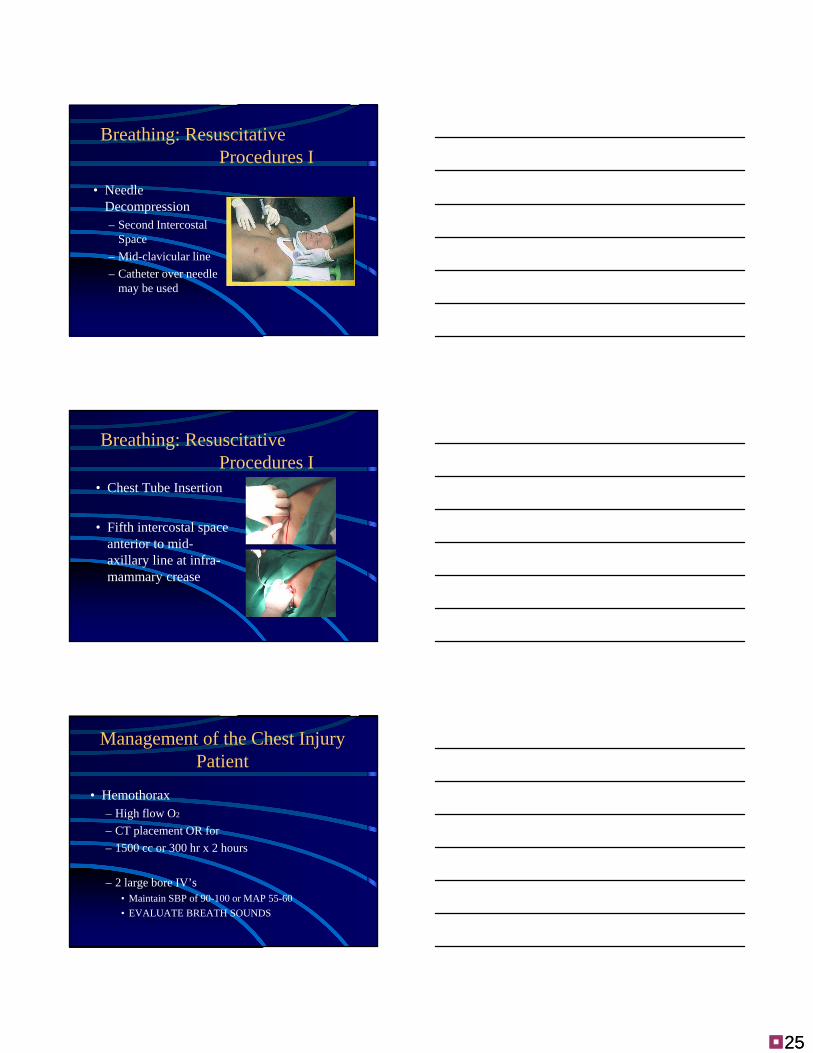
Breathing: Resuscitative Procedures I
• Needle Decompression– Second Intercostal
Space
– Mid-clavicular line
– Catheter over needle may be used
Breathing: Resuscitative Procedures I
• Chest Tube Insertion
• Fifth intercostal space anterior to mid-axillary line at infra-mammary crease
• Hemothorax– High flow O2
– CT placement OR for
– 1500 cc or 300 hr x 2 hours
– 2 large bore IV’s• Maintain SBP of 90-100 or MAP 55-60
• EVALUATE BREATH SOUNDS
Management of the Chest Injury Patient

2626
Management of the Chest Injury Patient
• Pulmonary contusion
• Injuried lung poorly compliant
• MAP = oxygenation– PC inverse ratio
– APRV
– Bilevel
– High frequency ventilation
• Proning?
• ECLA
Prone Ventilation
• Usually used late in the course of ARDS
• Decreased dependent atelectasis – Weight of the heart is removed
– Lower weight of lung pressing down on the dependent prone lung
– Prone positioning may shift the diaphragm down, decreasing the compressive effect of abdominal contents
Prone Ventilation
• Benefits Continued– Change from supine to prone with the same
level of PEEP may keep the now dependent portions open while allowing the non-dependent portions to re-expand (prevents derecruitment)
– Change in position does not completely change blood flow to the lung (good lung on the bottom may continue to receive increased flow)

2727
Prone Ventilation
• Hemodynamic instability (1.1% per prone cycle)
• Extubation (0.4%)• Decreased O2 sat
(0.3%)• Apical atelectasis
(0.3%)• Kinked ETT (0.1%)
• Obstructed CT (0.1%)
• Dislodged central lines (0.2%)
• Supraventricular tachycardia (0.1%)
• Possible aspiration (tube feeding rate must be decreased
• Traumatic Asphyxia– Support airway
• Provide O2
• PPV with BVM to assure adequate ventilation
– 2 large bore IV’s– Evaluate and treat for concomitant injuries– If entrapment > 20 min with chest compression
• Consider 1mEq/kg of Sodium Bicarbonate
Management of the Chest Injury Patient
Conclusions
• Early Diagnosis and Interventions
• Judicious use of Fluids
• Appropriate pain control
• MAP based Ventilator Strategy
• Early mobilization and physiotherapy