further observations on the histology of striped muscle
TRANSCRIPT
OBSERVATIONS ON THE HISTOLOGY OP STRIPJiD MUSOLE. 65
Further Observations on the Histology ofStriped Muscle.
By
C. F. Marshall, M.B., M.Sc,Late Platt Physiological Scholar in the Owens College.
With Plate XI.
IN a recent paper published in this Journal1 I gave theresults of some researches on the histology of muscular fibre.The present paper is a record of my further investigations onthis subject. These are somewhat incomplete owing to wantof time to finish them satisfactorily, but several results havebeen obtained which I trust are worthy of publication.
In order to render the questions dealt with more intelligiblethe summary of results arrived at in the former paper is repro-duced.
Summary of Former Paper .
1. In all muscles which have to perform rapid or frequentmovements a certain portion of the muscle is differentiatedto perform the function of contraction, and this portion takeson the form of a very regular and highly modified iutra-cellular network.
2. This network, by its regular arrangement, gives rise tocertain optical effects which cause the peculiar appearances ofstriped muscle.
1 " Observations on the Structure and Distribution of Striped and Un-striped Muscle in the Animal Kingdom, and a Theory of Muscular Contrac-tion/' 'Quart. Journ. Micr. Sci.,' 1887.
VOL. XXXI, PART I. NEW SER. E
66 0. F. MARSHALL.
3. The contraction of the striped muscle-fibre is probablycaused by the active contraction of the longitudinal fibrilsof the intra-cellular network : the transverse networks appearto be passively elastic, and by their elastic rebound cause themuscle to rapidly resume its relaxed condition when the longi-tudinal fibrils have ceased to contract; they are possibly alsopaths for the nervous impulses.
4. In some cases where muscle has been hitherto describedas striped, but gives no appearance of the network on treat-ment with the gold or other methods, the apparent striation isdue to optical effects caused by a corrugated outline to thefibre.
5. In muscles which do not perform rapid movements, butwhere contraction is comparatively slow and peristaltic innature, this peculiar network is not developed. In most if notall of the invertebrate unstriped muscle there does not appearto be an intra-cellular network present in any form j but in thevertebrate unstriped muscle a network is present in the form oflongitudinal fibrils only; this possibly represents a form of net-work intermediate between the typical irregular intra-cellularnetwork of other cells and the highly modified network ofstriped muscle.
6. The cardiac muscle-cells contain a network similar tothat of ordinary striped muscle.
Discuss ion of t he Views of R e c e n t O b s e r v e r s .Before commencing the subject-matter of this paper I proposeto discuss the results arrived at by several recent observersconcerning the structure of muscle-fibre, and also some of thecriticisms which have lately appeared concerning the existenceof an intra-cellular network in striped muscle.
The most important paper to discuss is that of Rollett.1
He considers the muscle-fibre to consist of longitudinal fibrillaegrouped together into " muscle-columns," the cross-sections ofwhich correspond to Cohnheim's areas. Filling up the spacesbetween the muscle-columns is the interfibrillar material or
1 * Arch. f. raikr. Anat.,' 1888, pp. 233—265.

OBSERVATIONS ON THE HISTOLOGY OF STRIPED MUSCLE. 67
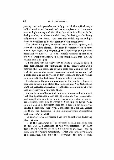
" sarcoplasma." Each muscle-column consists of alternatingthick and thin segments; in the centre of each thin segment isa dark granule. The thick segments of the muscle-columnscorrespond to " Bowman's sarcous elements," and the darkgranules in the centre of the thin segments correspond to" Krause's membrane" or the "transverse network" (fig. 13and diagram B).
Diagram A (high focus). Diagram B (low focus).
The sarcoplasma is the part which is stained in gold prepara-tions. In short, Rollett regards the appearance of goldpreparations as due to the staining of the sarcoplasma, andconsiders this to be a honeycomb of interfibrillar material, andnot a true intra-cellular network.
The latter portion of Rollett's paper is devoted to anelaborate criticism of the network view of the structure heldby Melland, Van Gehuchten, Carnoy, and myself. He statesthat what we describe as the fibrils of the network are onlytransverse and longitudinal sections of the walls of the honey-comb or sarcoplasma (" Was beide Autoreu an diesen als Padenbeschreiben sind nur Q,uer- oder Langsschnitte der Wande desWabenwerkes, welches das Sarcoplasma um die Muskelsaulchenbildet," p. 253).
He describes the appearances seen in fresh muscle-fibre asfollows :—At low focus (diagram B) the muscle-columns appeardark and in a line with the granules, the sarcoplasma appearinglight. At high focus (diagram A) the sarcoplasma appears dark,the muscle-columus light, and two rows of granules appear in aline with the sarcoplasma and alternating with the muscle-columns. He states that the dark lines drawn by Melland
68 0. P. MARSHALL.
joining the dark granules are only parts of the optical longi-tudinal sections of the walls of the sarcoplasma and are onlyseen at high focus; and that they do not lie in a line with thedark granules, but alternabe with them, the dark granules beingonly seen at low focus. The granules which appear at highfocus he considers to be thickenings of the sarcoplasma.
The above diagrams, modified from Rollett's figures, willmake these points clearer. Diagram B represents the appear-ance at low focus, and diagram A the appearance at high focusaccording to Rollett. In B the muscle-columns appear darkand the sarcoplasma light; in A the sarcoplasma dark and themuscle columns light.
In the same way he states that the rows of granules seen ingold preparations are thickenings of the sarcoplasma lyingbetween the thin segments of the muscle-columns, and that thetrue row of granules which correspond to and are parts of themuscle-columns are only seen at low focus, and then do not liein a line with the dark lines, but alternate with them.
He describes the same appearance at low and high focus inhardened muscle, and states that Melland and Van Gehuchtenplace the granules alternating with the muscle-columns, whereasthey are really in a line with them.
In short, he concludes that a network does not exist, andthat the appearances described by Melland, Van Gehuchten,and myself are due to errors in the interpretation of micro-scopic appearances and confusion of high and low focus (" Ichkomme also zum Resultate dass ein Netzwerk im Sinne vonMelland, Marshall, und Van Gehuchten und ein Enchylemeim Sinne des Letzteren in der quergestreiften Muskelfasernicht existirt," p. 262).
In answer to this criticism I venture to .make the followingobservations.
1. If the appearance of the network in fresh muscle is dueto the optical appearance of the " sarcoplasma" at highfocus, there must always be a double row of granules—one oneach side of Krause's membrane. (I use this term for the sakeof convenience, and take it to represent the row of dark
OBSERVATIONS ON THIS HISTOLOGY OF STRIPED MUSCLE. 69
granules of Rollett's muscle-columns, and the row of granulesof the transverse network of Melland, fee).
2. If the granules seen at high focus in gold preparationsare thickenings of the sarcoplasma stained by gold, theremust again be a double row, one on each side of the true rowof granules which are only seen on the low focus, and further,the former rows of granules must alternate with the latter.(Vide diagram A.) Although I have examined several hun-dred preparations of muscle-fibre, I have never seen tworows of granules in any gold or fresh preparation, nor have Iseen any change in the position of the granules at high andlow focus. The granules have always appeared to me to bein a line with, and connected with the longitudinal lines of, thenetwork (or " sarcoplasma") at both high and low focus.
3. Rollett states that the " sarcoplasma " is a honeycomb,and that the appearance of the network is due to the opticalsections of the walls of that honeycomb. If this is true, I donot understand why there is never any appearance of honey-comb structure in finely teased preparations, whereas isolatedpieces of network are easily obtained. Moreover, one prepara-tion obtained by Melland is, I think, almost conclusive infavour of a network (fig. 15).
4. According to Rollett, the " muscle-columns" are theessential parts of the fibre, and the " sarcoplasma " is simplyinterfibrillar material; we should therefore expect the latterto be least abundant in the most perfectly developed muscles.Now, in insects which possess the most powerful and mostrapidly contracting muscles of all animals, the part stained bygold is more strongly marked than in other animals. Thisseems to point to the fact that it is the most essential part ofthe fibre and not interfibrillar material, and is therefore infavour of the network view.
5. Again, in developing muscle-fibre I have shown that thenetwork is present but only demonstrated with difficulty jwhereas if it were inert interfibrillar substance, one would expectit to be relatively more abundant in the embryonic fibre.
6. If, as I have attempted to show, the nerve-ending is

70 0. F. MARSHALL.
connected with the part of the fibre stained by gold, thisagain points to the latter being the essential portion of thefibre; while if Rollett's view is correct, we should expect thenerve-ending to be connected with the muscle-columns.
7. The apparent connection in some cases of the networkwith the iutra-nuclear network of the muscle-corpuscles is infavour of the network view.
8. Lastly, the network view places the muscle-cell on a basisof comparison with other cells having intra-cellular networks,whereas all other views of the structure are at variance withsuch a comparison.
Dr; Klein1 adopts Rollett's view in the new edition of histext-book, and states that " t he reticulation described byMelland, Marshall, and others, is due to the coagulation ofthe sarcoplasma brought about by certain hardening reagents."
Dr. Michael Foster apparently holds the same view of thestructure in the new edition of his book,2 for he states that themuscle-substance is composed of longitudinal fibrillse, embeddedin interfibrillar substance which stains with gold, and henceappears as a network. He says, " The interfibrillar substanceis relatively to the fibrillse more abundant in the musclesof some animals than in those of others, being, for instance,very conspicuous in the muscles of insects, in which animalswe should naturally expect the less differentiated material tobe more plentiful than in the muscles of the more highlydeveloped mammal."
Now, I think I am right in saying that the muscular systemof insects is the most highly developed in the animal kingdom;certainly the muscles are far more powerful in comparison withthe size of the animal than in mammals, and among insectsare found the most rapid movements. I t therefore appears tome that the fact of the meshwork being more conspicuous inthe muscles of insects is strongly in favour of its being theactive part of the pike, and not of the nature of interfibrillarsubstance.
1 ' Elements of Histology,' p. 76.3 'Text-book of Physiology,' vol. i, p. 91.

OBSERVATIONS ON THE HISTOLOGY OF STRIPED MUSCLE. 71
I shall next give a r e s u m e o f the results of some receutobservers who are in favour of the existence of a network.
Van Gehuchten1 has described a network in striped musclesimilar in most respects to that described by Melland, myself,and others, but differing in some details.
Carnoy2 also adopts the network view, and remarks that" La cellule musculaire est une cellule ordinaire dont le reti-culum s'est regularise, et l'enchyleme charge de myosine."
Haswell3 has recently published an important paper on thissubject. His observations were made on the gizzard ofvarious species of Syllis, where he claims to have found veryprimitive forms of striped muscle. He divides striped muscleinto simple and compound types, the simple type showingonly transverse striation, not due to network; the com-pound both transverse and longitudinal. These two typescorrespond to what I have termed true and false striation ;the characteristics of the compound or true striped musclebeing as follows :
1. Each fibre consists of a bundle of fibrils.2. Each fibre is composed of two alternating series of
anisotropous and isotropous segments, the former of whichare more easily stained than the latter.
3. Eunning across the fibre in the middle of each iso-tropous segment is the transverse network, or Krause's mem-brane.
4. Between the fibrils run the strands of the longitudinalnetwork.
5. Each fibre is formed from a single cell, the nucleus ofwhich divides and forms " a multinucleated protoplasmicbody, by modification of whose protoplasm the muscle sub-stance and networks are formed."
He states that in these animals the elements of the fibreare on a larger scale than in Vertebrates and Arthropods, and
1 " Etude sur la structure intime de la cellule musculaire Stri6e." Extraitde la Revue 'La Cellule,' t. ii, 2 fascicul, Louvain, Gand et Liege, 1886.
2 ' La Biologie cellulaire,' 1884.3 ' Quart. Journ. Micr. Sci.,' 1889.
72 0. F. MARSHALL.
are hence favorable for the study of the structure of mus-cular fibre, which "seems to lead to that view of the structureof compound striated fibres advocated by Retzius, Bremer,Melland, C. F. Marshall, and others; the only poinb ofimportance in which there seems reason for dissenting fromthat view being with reference to the relation of the trans-verse networks to the fibrils." The chief differences betweenthe network Haswell describes and that described by theabove observers are these : (1) He states that the transversenetworks are not only in the interspaces between the" fibrils," but partly also penetrate them. (2) The longitu-ninal networks, though mostly longitudinal, have obliquestrands and anastomoses. (3) The transverse networks some-times appear as rows of spindle-shaped granular bodies, but incrushed specimens he says these are seen to consist of a closereticulum of delicate threads, the spindle bodies being con-densed parts of it. (4) The distance between the transversenetworks is much greater.
Haswell regards the " fibrils " as the contractile part, andnot the network.
I shall refer to Haswell's observations again in the subse-quent portions of this paper.
A. B. Macallum1 has published some interesting observa-tions dealing with striped muscle. He confirms the existenceof the network of Retzius, Melland, and others, and considersit the contractile part of the fibre. He states that the muscle-nuclei are marked by furrows, sometimes transverse and some-times longitudinal, and that these are probably caused by pres-sure of the trabeculse of the network. He also states that insome nuclei there is an intra-nuclear network similar to thatof the fibre itself.
I t thus appears that the view of the existence of a trueintra-cellular network in striped muscle-fibre has receivedmuch support, and has been confirmed by several recentobservers; and that the view held by Rollett and others that it
1 "On the Nuclei of the Striated Muscle-fibre in Menobranchus," 'Quart.Journ. Micr. Soi.,' 1887.

OBSERVATIONS ON THE HISTOLOGY OP STRIPED MUSOLE. 73
is interfibrillar material has not sufficient evidence for it to beaccepted. I shall therefore, in accordance with the results ofRetzius, Bremer, Melland, Carnoy, Haswell, Macallum, andmyself, assume that the former view represents the truestructure of striped muscle.
The present paper deals with the following points :1. The connection of the transverse networks with the
muscle-corpuscles.2. The development of the network.3. The connection of the nerve-ending with the network.
Connect ion of N e t w o r k wi th Muscle-corpuscles .
It was shown by Retzius1 that the transverse portions of themuscle network were directly counected with the muscle-corpuscles. He states that the protoplasm of the muscle-corpuscle is produced into several processes from which finerprocesses arise forming the transverse networks. Retzius1
results were obtained by a modification of gold staining.The fresh muscle-fibre was first placed in a 1 per cent, solutionof formic acid for a few seconds, then in gold chloride \—\per cent, for twenty-five minutes, then in formic acid L percent., and exposed to light for 10—20 hours.
By a special method of staining I have been able to confirmRetzius' results, and have made specimens showing the un-doubted connection of the transverse networks with themuscle-corpuscles.
Method of Prepara t ion .—The method of staining Iadopted is a modification of that employed by Mays fordemonstrating nerve-endings in muscle. He uses the follow-ing solution :
Arsenic acid f per cent 20 parts.Gold cbloride 1 per cent . . . 4 „Osmic acid 2 per cent 1 „
This solution although it preserves the nerve-endings disinte-grates the muscle-fibre. This I found was due to the arsenic
' Zur Kenntniss der Quer̂ -estreiften Muskelfaser.' Biologisclie Unter-suchungen, 1881.

74 0. F. MARSHALL.
acid. I therefore tried various strengths of acetic and formicacid in place of the arsenic, and found the best combinationwas the following:
Acetic acid 1 per cent. . . . . 20 parts.Gold chloride 1 per cent. . . . 4 „Osmio acid 1 per cent. . . . . 1 „
The muscle-fibre was placed in this solution for fifteenminutes, after previous immersion in acetic acid 1 per cent,for a few seconds ; then in acetic acid 1 per cent, in a warmchamber for one or two hours.
1. Dytiscus.—Fresh muscle-fibre of Dytiscus stained bythe above method shows the muscle-corpuscles in the form ofone or more chains of nuclei in the substance of the fibre, thenuclei being surrounded by a small amount of undifferentiatedprotoplasm. The transverse networks are seen directly con-tinuous with the nuclei. This is well shown in fig. 1. Fig. 2shows a portion of fibre with two rows of muscle nuclei; thetransverse networks are seen to be connected with both sets ofnuclei in some places. Fig. 3 shows several isolated nucleiwith the transverse processes attached to them.
Transverse views of the network and nuclei are more diffi-cult to obtain. Fig. 4 shows a transverse view of an isolatednucleus, with part of the transverse network connected with it.
2. Dragon-fly.—The muscle-corpuscles of the muscle-fibre of the dragon-fly are situated peripherally, i. e. justunder the sarcolemma, contrary to the general rule in insects.
In one preparation of this muscle I could trace the trans-verse networks into the muscle-corpuscles; and, moreover, thenetworks appeared to be distinctly connected with the intra-nuclear networks of the muscle-corpuscles (fig. 5).
3. Crayfish.—In a preparation of crayfish muscle pre-pared by Retzius' method I could apparently trace theconnection of the muscle network with that of the muscle-corpuscle. In this case it was somewhat difficult to tellwhether the effect was not due to the network lying over themuscle-corpuscle; but by careful focussing I think the con-nection could be made out (fig. 6).
OBSERVATIONS OP THE HISTOLOGY OF STEIPED MUSOLE. 75
These observations confirm Retzius' results, viz. that thetransverse portions of the muscle network are directly con-nected with the muscle-corpuscles; and, furthermore, that thenetwork is directly continuous with the intra-nuclear networkof the corpuscles.
Development of t h e Network .
This I have studied in embryos of the trout and rat.Trout.—In some gold preparations of embryo trout, taken
from the ova, I found developing muscle-fibres in an earlystage. These consisted of a portion of undifferentiated proto-plasm containing the nucleus, and a portion already trans-versely striated. Under a comparatively high power thetransversely striped portion showed darkly stained masses ofan ellipsoidal shape arranged side by side, and causing theappearance of striation. Under a very high power (-^ immer-sion) the network could be seen between these darkly stainedmasses, and in the same form in which it appears in the adultfibre. The dark masses appear to be some substance alteredby the method of staining, and shrunken in the meshes of thenetwork (figs. 7, 8 and 9). No connection was seen betweenthe network and the nucleus.
In older fibres the network is more fully developed, but stillno connection appears to exist between the nuclei and thenetwork (fig. 10).
Rat.—In developing muscle-fibre from the embryo rat thefibres consist of an axial core of undifferentiated protoplasmcontaining the nuclei, and a peripheral part with developingnetwork. Here, again, no connection was observed betweenthe nuclei and the network.
It thus appears that—1. The network appears at a very early stage.2. It develops in its permanent form, and is not produced by
the transformation of an irregular network into the adult type.3. Each muscle-fibre is probably developed from a single
cell, and is not formed by a coalescence of cells, either end to
76 0. J\ MAESHALL.
end or laterally (as Calberla1 states), because the fibres oftrout muscle examined were evidently single cells, and had thenetwork well developed. I t is difficult to conceive that thesebecome fitted together, either end to end or laterally, so thatthe network of one cell should exactly fit on to that of thenext.
4. The network develops centripetally, and commences at thepart of the cell farthest away from the nucleus; moreover, itdoes not appear to become connected with the nuclei till thefibre is fully developed.
Haswell,3 from his observations on the muscle of the gizzardof Syllis, forms a view of the ontogeny of striped muscle whichdoes not agree with that described above.
In the same organ, in various species of Syllis, he finds inone case bundles of non-striped fibres j in another, compoundhollow striated fibres, consisting of bundles of fibrils similar tothe above, and bound together by a single transverse network.In a third there are three transverse networks, and so on up tothe fully developed type of striped muscle found in Vertebratesand Arthropods. He therefore regards each striped fibre asderived from a bundle of non-striped fibres. He thinks thetransverse network probably the equivalent of a transverseline of nuclei of the unstriped fibres, which occupies a similarposition.
If this is correct each striped fibre must be a multicellularstructure, and the network intercellular, and not intra-cellular.However, in another part of his paper he speaks of each fibrebeing formed from a single cell, the nucleus of which divides andforms " a multinucleated protoplasmic body, by modificationof whose protoplasm the muscle-substance and networks areformed."
1 ' Arch. f. mik. Anat.,' xi, 1875.s Loc. cit.
OBSERVATIONS ON THE HISTOLOGY 01 STRIPED MUSOLE. 77
Connect ion of N e r v e - e n d i n g with MuscleNe twork .
Nerve-endings may be divided into two main types, thecircumscribed and the diffused form. Between these typesthere are many intermediate forms, as shown by Kiihne in hisclassical work on ' Nerve-Endings.'l The nerve-endings in themuscle of the snake form a good example of the circumscribedvariety. Here we have a localised end-plate, compact and non-ramifying. This is also the common type in mammalian muscle.In Dytiscus, on the other hand, we find the diffuse form, whichbranches and ramifies nearly to the end of the fibre.
In each case the nerve-ending, when stained by any of thegold methods, is seen to consist of a slightly stained ramifyingportion, with darkly stained irregular masses between thebranches of the lighter stained portions. The lighter stained partappears to be continuous with the axis-cylinder of the nerve.
In attempting to demonstrate a connection between thenerve-ending and the muscle-network there is great difficulty,because the methods which best show the nerve-endings usuallydisintegrate the muscle-fibre and destroy the network. Thebest method of obtaining the nerve-endings is that employedby Mays,2 and also, with modifications, by Kiihne,3 viz. the mix-ture of arsenic acid, gold chloride, and osmic acid mentioned inthe first part of this paper. But this method usually destroysthe network, owing to the action of the arsenic acid. Anotherdifficulty arises from the fact that the connection, if it exists,must take place underneath the nerve-ending, and hence in thenormal position of the parts it could hardly be seen.
Dytiscus.—In one preparation of Dytiscus muscle preparedby May's method I succeeded in finding a portion of fibre withthe network still intact (fig. 11). A portion of the ramifyingnerve-ending is seen crossing the fibre transversely; both the
1 " Untersuchungen fiber motorische Nervenendigung," ' Zeit. f. Biol.,'Bd. xxiii.
2 ' Zeit. f. Biologie,' Bd. xx, 1884,3 Loc. cit.
78 0. F. MARSHALL.
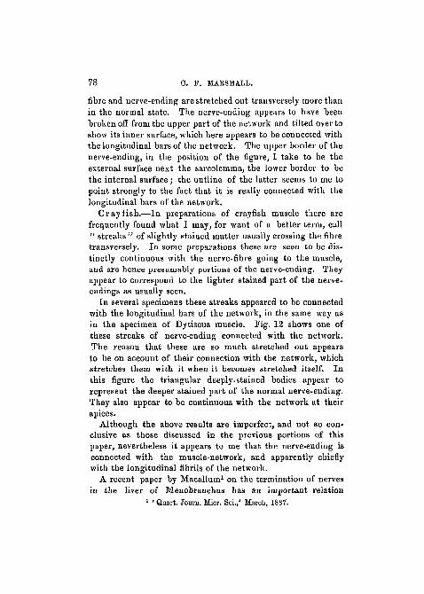
fibre and nerve-ending are stretched out transversely more thanin the normal state. The nerve-ending appears to have beenbroken off from the upper part of the network and tilted over toshow its inner surface, which here appears to be connected withthe longitudinal bars of the network. The upper border of thenerve-ending, in the position of the figure, I take to be theexternal surface next the sarcolemma, the lower border to bethe internal surface; the outline of the latter seems to me topoint strongly to the fact that it is really connected with thelongitudinal bars of the network.
Crayfish.—In preparations of crayfish muscle there arefrequently found what I may, for want of a better term, call" streaks" of slightly stained matter usually crossing the fibretransversely. In some preparations these are seen to be dis-tinctly continuous with the nerve-fibre going to the muscle,and are hence presumably portions of the nerve-ending. Theyappear to correspond to the lighter stained part of the nerve-endings as usually seen.
In several specimens these streaks appeared to be connectedwith the longitudinal bars of the network, in the same way asin the specimen of Dytiscus muscle. Fig. 12 shows one ofthese streaks of nerve-ending connected with the network.The reason that these are so much stretched out appearsto be on account of their connection with the network, whichstretches them with it when it becomes stretched itself. Inthis figure the triangular deeply-stained bodies appear torepresent the deeper stained part of the normal nerve-ending.They also appear to be continuous with the network at theirapices.
Although the above results are imperfect, and not so con-clusive as those discussed in the previous portions of thispaper, nevertheless it appears to me that the nerve-ending isconnected with the muscle-network, and apparently chieflywith the longitudinal fibrils of the network.
A recent paper by Macallum1 on the termination of nervesin the liver of Menobranchus has an important relation
1 • Quart. Jouin. Micr. Sci./ March, 1887.

OBSERVATIONS ON THE HISTOLOGY OF STRIPED MUSCLE. 79
to the above question. He states that he has traced theterminal fibrils of the nerves into direct connection withthe intra-nuclear network of the hepatic cells, and the figureshe gives seem to place this beyond doubt. This by analogygives support to the view that the nerve-ending of muscle isconnected with the network; and possibly this is the normalmethod of termination of nerves in connection with cells.
Haswell's1 observations are also interesting in connectionwith this question. He describes a ganglion-cell at the end ofeach fibre, and occupying the axis of the fibre. This sends outnumerous branching processes, which penetrate between thefibrils. He states that the core of the fibre is occupied bygranular protoplasmic material, through which runs a networkof fine threads, " connected with the fine branches of the nerve-processes of the ganglion-cell, and with the network of themuscle substance." Special branches of nerve-processes alsoramify on the surface of the fibre, and probably enter into con-nection with the transverse networks.
Hence both Macallum's and Haswell's observations lendsupport to the view that the network is connected with thenerve-ending.
Conclusion.
In reviewing the foregoing observations we are at once metby an apparent contradiction, viz. that in the young muscle-fibre the network does not appear to be connected with thenucleus, whereas the connection is definitely established(Retzius, Marshall) in the adult fibre. This at first sight is anabsolute contradiction : for, on the one hand, it is difficult toconceive that processes originating as outgrowths from thenucleus could exactly hit off and fuse with the already formednetwork ; on the other hand, it is almost equally inconceivablethat the fibres should grow into the nucleus. This apparentcontradiction can, I think, be to a great extent explained.Firstly, we must bear in mind that histological differentiationproceeds during its development from without inwards, i. e.
1 Loc. cit.
80 . 0. f. MARSHALL.
centripetally; and that the special characters of the adult cellappear first and are most marked at the periphery, i. e. farthestfrom the nucleus. Examples of this rule are seen in the de-velopment of epidermic- cells, dentine, cartilage, and bone. Inbone and dentine the processes are not supposed to grow fromthe cells, but to be formed by the lengthening of connectingstrands by deposit of new matter.
Secondly, the nerve-ending being on the surface of the fibre,and the network also appearing first on the surface, the con-nection between the two can be established from the first;whereas if the network grew out from the nuclei it could onlybe connected with the nerve-ending at a much later period.Hence the fibre would be useless till it was far advanced indevelopment, because for the network to be of any use it mustfrom the first be connected with the nerve-ending. For a newstructure of any kind to be developed it must always have beenof use from the first, either for its ultimate purpose or forsome other. If the network grew out from the nuclei itcould not be of use till it got to the nerve-ending, i. e. to thesurface.
In considering this point we may imagine that the celldivides into " formed and unformed matter" (Beale), theformed matter being characteristic of the particular cell. Inthe case of the muscle-fibre the protoplasm divides into net-work aad muscle-plasma all along, beginning at the peripheryand gradually extending to the nucleus. We are here met byan apparent difficulty, for in the muscle-fibre the " formedmatter" which is characteristic of the cell is the muscle-plasma, and not the network (which is presumed to be thecontractile part). But the special feature of striped muscle isnot the fact that it contracts, but the mode in which this isbrought about, i. e. the rapidity; and this will be a matter ofnutrition which will depend on the muscle-plasma. There-fore the special feature of striped muscle is probably the modeof nutrition of its specially contractile part, i. e. quick repair.The active network bathed in such a fluid is placed in a goodposition for such rapidity.
OBSERVATIONS ON THE HISTOLOGY OF STRIPED MUSOLE. 81
Summary.
1. The transverse portions of the network of the stripedmuscle-fibre are directly connected with the muscle-cor-puscles.
2. The nerve-ending appears to be connected with themuscle-network, and chiefly with the longitudinal bars of thenetwork.
3. The development of the network takes place at a veryearly stage in the development of the fibre, and the networkdevelops from the first in its permanent form.
4. The network develops first at the surface, and grows cen-tripetally. It does not appear to be connected with themuscle-corpuscles till the fibre is fully developed.
5. Each muscle-fibre appears to be developed from a singlecell, and not by a coalescence of cells.
The investigations described in this paper were carried onin the Physiological Laboratory of Owens College during thewinter of 1887. My thanks are due to the Council of the Collegefor a special grant to enable me to carry on the research.
I must express my thanks to my brother Professor MilnesMarshall for many valuable suggestions in producing thispaper, and for examining several of the preparations. I mustalso thank Professor Stirling for much assistance to me in mywork.
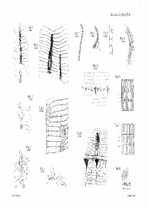
DESCRIPTION OP PLATE XI,
Illustrating Mr. C. F. Marshall's paper, " Further Observa-tions on the Histology of Striped Muscle."
[The main details were drawn under the camera in all the figures.]
FIG. 1.—Muscle-fibre of Djtiscus, showing transverse networks connectedwith muscle-corpuscles. The longitudinal bars of the network are omitted
VOL. XXXI, PART I. NEW SEK. F
82 0. F. MARSHALL.
for the sake of distinctness, ith obj. Acetic acid, osmic acid, and goldchloride.
FIG. 2.—Portion of another fibre, with transverse networks connected withtwo rows of muscle-corpuscles. -^th immersion obj. Same method.
FIG. 3.—Three isolated muscle-corpuscles of Dytiscus muscle, with trans-verse networks attached, j^th immersion obj. Same method.
FIG. i.—Muscle-corpuscle of Dytiscus, with portion of transverse networkconnected with it. Transverse view. Gold preparation, -^th immersion obj.
FIG. 5.—Muscle-fibre of dragon-fly, showing two muscle-corpuscles withtheir intra-nuclear network. Some of the transverse networks are seen tobe connected with the intra-nuclear network of the upper corpuscle, x&thimmersion obj. Acetic acid, osmic acid, and gold chloride.
FIG. 6.—Muscle-corpuscle of crayfish, with portion of the muscle networkapparently connected with its intra-nuclear network, j^th immersion obj.
FIGS. 7 and 8.—Developing muscle-fibres of trout, showing striation. n.Nucleus, ^gth immersion obj. Acetic acid 2 per cent., a few seconds ; goldchloride 1 per cent., fifteen minutes; formic acid 25 per cent., thirty minutesin warm chamber.
FIG. 9.—Portion of the striated part of one of the above fibres, showingthe network and the darkly stained bodies in its meshes. Jjth immersion obj.
FIG. 10.—More fully developed fibre of trout, n. Nucleus. T\jtli im-mersion obj.
FIG. 11.—Muscle-fibre of Dytiscus, with a portion of nerve-ending appa-rently connected with the longitudinal bars of the network, -j^th imm. obj.Mays' method.
FIG. 12.—Muscle of crayfish, showing a "streak" of nerve-ending appa-rently connected with the longitudinal bars of the network. t\jth imm. obj.Retzius' method.
FIGS. 13 and 14.—Diagrams comparing the view of Rollett and otherswith the network. In Fig. 13 the structure, according to Rollett, is markedin full lines, and the network marked in dotted lines. In Fig. 14 the thicksegments of Rollett's muscle-columns are shown by dotted lines in themeshes of the network, n. Network, s. Muscle-columns, g. Granules.
FIG. 15.—Portion of muscle-fibre of Dytiscus, showing network veryplainly. One of the transverse networks is split off, and some of the longi-tudinal bars are shown broken off. (Copied from Melland, loc. cit., fig. 6.)
[Note.—In figure 13 the lines connecting the thick parts of Rollett'smuscle-columns should be much thicker. They represent the thin segmentsof the columns.]
1 Fig.2 L
< 899 , „ •; 2SI
;ili
|"l
!'
i) ,'\ ("i
u 'J i>
! i