differential dx and management four questions to begin of ... · osteoarthritis labral pathology...
TRANSCRIPT

Differential Diagnosis and Management of Intra-articular Hip Pathology
CPTA Annual Conference 2011Long Beach, CA
J. Meyer, PT, DPT,OCS, FAFSPersonal Teaching Property 1
Differential Dx and Management of Intra-articular Hip Pathology
CPTA Annual Conference Long Beach, CA September 22, 2011
John Meyer, DPT, OCS, FAFSUniversity of Southern CaliforniaDepartment of Athletic Medicine
Four Questions to Begin
• Why are you here?• What do you hope to achieve?
• What type of dx/patients do you see?
• What has been successful for you in the past?
• Frustrations?
Hip Pain: “The Challenge”
• The non-arthritic hip poses a diagnostic dilemma– Extraarticular vs. intraarticular – Pain is difficult to localize– 60% of patients are initially misdiagnosed
Inaccurate Diagnosis
• Misdiagnosed for an average of 7 months to 3.1 years
• Patient’s evaluated by an average of 4.2 healthcare providers prior to correct diagnosis
• Goal of today is to change this !!!
Clohisky JC:Clin Orthop Relat Res 2009
Differential Diagnosis of Hip Pain
• Intra-articular– Osteoarthritis – Labral pathology– Chondral pathology– FAI– Capsular laxity– Injury to the ligamentum
teres– Loose bodies– Benign intra-articular
tumors
• Extra-articular– Hip tendonitis/avulsion
injuries– Femoral neck stress fx– Snapping hip– Trochanteric bursitis– Abductor tears– Athletic pubalgia– Osteitis pubis– Nerve compression
typologies
• Systemic Causes– Testicular Cancer– Inflammatory bowel
disease– Endometriosis– Ovarian Cyst– Pelvic inflammatory
disease– Appendicitis– Osteomyelitis– Septic arthritis– UTI
• Referred Pain– Sacroiliac– Lumbar Spine

Differential Diagnosis and Management of Intra-articular Hip Pathology
CPTA Annual Conference 2011Long Beach, CA
J. Meyer, PT, DPT,OCS, FAFSPersonal Teaching Property 2
Differential Diagnosis of Hip Pain
• Age of the patient• Prior trauma
– direct-impact activities• Duration and location of symptoms• Morning stiffness
– less than 1 hour• Childhood or family hx
– Developmental Disorders– Dad had a THR @ 50 years of age
Age of the PatientHip Pain
Adolescent 18-50 y/o 50-?? y/o Extraarticular
SCFELCPDDH
Labral TearFAI
Labral TearFAIAS
Athletic PubalgiaAVN
OASeptic Arthritis
NeoplasmPaget’s Disease
Snapping HipMuscle Strain
TendonitisBursitisSI joint
Referred pain
Systemic
RAPolyarticular
ArthritidesHormonal
CRPS
Slipped Capital Femoral Epiphysis
• Occurs during adolescent rapid growth period (10-15 years)
• More common in obese males @ age 13– Females @ age 12
• Most common disorder of the adolescent hip
Clinical Signs of SCFE
• Onset of limp• Hip pain referred to the knee• Limited Hip Abd and IR• Progressive adduction and ER with
shortening of the leg• Bilateral cases yield “waddling” gait
Legg-Calve-Perthes (LCP)
• AVN of the femoral capital epiphysis• Occurs age 3 to 12• 5:1 male predominance• Short history of
– Painful limp: Flex, Abd, ER– Reduced mobility– Muscle atrophy– Trendelenburg test positive
• Leads to adult DJD
Ankylosing Spondylitis
• Chronic progressive inflammatory arthritis• More frequently seen in men than women • Age of onset is usually between 15 and 35
years of age (average of 26)• 95% positive for HLAB-27• Primarily affects synovial joints of the
spine and sacroiliac joints

Differential Diagnosis and Management of Intra-articular Hip Pathology
CPTA Annual Conference 2011Long Beach, CA
J. Meyer, PT, DPT,OCS, FAFSPersonal Teaching Property 3
Clinical Presentation
• Gradual onset of pain and morning stiffness in the spine– improves with exercise
• Referred pain to the buttock or posterior thigh
• Generalized fatigue, nausea and iritis• Reduced chest expansion
– Less than 2.5 cm
Ankylosing Spondylitis
• Sacroiliac involvement is the hallmark of AS– Pseudowidening of the joint space– Loss of the articular cortical bone– Erosive and sclerotic changes– Irregular joint margins– Ankylosis
Normal A/P of Pelvis
Pseudowidening of the joint space
AP Pelvic Radiograph
Athletic Pubalgia/Sports Hernia 2 Major Schools of Thought
• Regarding the cause and pathogenesis of athletic pubalgia/sports hernia
1. Muscular injury and disruption from chronic stress to pubic joint
– Rectus abdominis– Adductor longus– External oblique aponeurosis– Conjoined tendon– Inguinal ligament

Differential Diagnosis and Management of Intra-articular Hip Pathology
CPTA Annual Conference 2011Long Beach, CA
J. Meyer, PT, DPT,OCS, FAFSPersonal Teaching Property 4
Anatomy of Athletic Pubalgia Anatomy of Athletic Pubalgia
Athletic Pubalgia/Sports Hernia
• Encompasses a wide variety of injuries to the anterior pelvis outside of the hip joint– Term sports hernia is inaccurate– Need to understand the “pubic bone” joint
• Complex rotational joint of both pubic bones and the entire anterior pelvic musculo-skeleton
• 2008 recognize at least 17 different nonhip, soft-tissue structures as causes of primary pain.
Meyers WC:Oper Tech Sports Med. 2005
Athletic Pubalgia
Athletic Pubalgia
• Primary mechanism involves hyper extension of the abdomen and/or hyper abduction of the thigh
• Resulting in multiple areas of fraying on the lateral and anterior aspects of the fascia of the muscles
2 Major Schools of Thought
• Regarding the cause and pathogenesis of athletic pubalgia/sports hernia
2. Described as an occult hernia process– Major abnormality is a defect in the posterior
wall of the inguinal canal– Involvement of the genital branch of the
gentio-femoral nerve– Often concurrent muscular dysfunction

Differential Diagnosis and Management of Intra-articular Hip Pathology
CPTA Annual Conference 2011Long Beach, CA
J. Meyer, PT, DPT,OCS, FAFSPersonal Teaching Property 5
History and Prevalence
• Athletic pubalgia affects both professional and recreational athletes
• Involved in activities such as running, kicking, cutting an explosive change of direction– Soccer, football and hockey
• More common in men than women– Pelvic shape and force distribution
Presentation
• Complaint of exercise induced lower abdomen and groin pain
• May radiate to the inner thigh and scrotum• Pain relieved by rest but returns upon
activity– Describes deep and intense pain in the
unilateral– Insidious onset and or distinct injury
Presentation
• Aggravating factors include– Sprinting– Twisting and cutting– Kicking– Sit-ups– Coughing– Sneezing
• Pain is usually present for a few days after the activity and associated with stiffness
Athletic Pubalgia
• Cluster of 5 signs and symptoms– Subjective complaint of deep groin/lower
abdominal pain• Localized tenderness pubic tubercle, inguinal
canal, rectus insertion and adductor origin– Pain that is exacerbated with sport-specific
activities and relieved with rest– Palpable tenderness over the pubic ramus– Pain with resisted hip adduction, flexion or
abdominal curl-upMeyers WC et al: AM J Sports Med 2000
Imaging Findings
Meyers WC:Oper Tech Sports Med. 2005
OSTEITIS PUBIS

Differential Diagnosis and Management of Intra-articular Hip Pathology
CPTA Annual Conference 2011Long Beach, CA
J. Meyer, PT, DPT,OCS, FAFSPersonal Teaching Property 6
Various Surgical Findings Conservative Treatment Tips
• Look for adductor dysfunction and address with aggressive soft tissue strategies.
• Balance forces on the pelvis• Teach the patient to control the hip and
trunk eccentrically• Train pelvic and thoracic disassociation• Avoid traditional concentric core work
Underlying Hip Dysfunction
• Always screen for underlying hip FAI in cases of athletic pubalgia
• Asymmetrical hip flexion and IR creates shearing and abnormal movement at the pubic joint– Cam lesion with Flex/IR– Symphysis motion occurred primarily in the transverse
plane opening the joint anteriorly• Can also lead to lubo-pelvic compensatory
movements and added pelvic stress
Kelly BT et al: AOSSM June 20211
FEMORAL STRESS FX
Femoral Neck Stress Fx
• Relatively uncommon etiology of hip pain• If not dx in a timely fashion serious
complications can occur• 7% of all stress fractures involve the
femoral neck– 50% of these occur in the femoral shaft
• Medial aspect of the proximal third being the most common
Matheson GO:Am J Sports Med 1987
Presentation
• Thigh and hip pain from a femoral stress fracture is generally vague
• Location of pain may not correlate with location of fracture
• Clinical findings are often variable

Differential Diagnosis and Management of Intra-articular Hip Pathology
CPTA Annual Conference 2011Long Beach, CA
J. Meyer, PT, DPT,OCS, FAFSPersonal Teaching Property 7
Risk Factors
• Previous history of stress fracture• Increases in training volume
– Recreational runners > 35 miles per week– Marathon runners > 45 miles per week– Competitive runners > 70 miles per week
• Corticosteroids• Restrictive eating patterns• History of amenorrhea
Clement DB:Int J Sports Med 1993
Clinical Findings
• Often insidious onset– Deep aching pain in the hip or groin region– Can radiate to a knee
• Worse with activity improves with rest – Night pain is not uncommon
• Mild pain at the extremes of hip range of motion• Lack of local bone tenderness with palpation• Positive hop test?• Positive fulcrum test?
Fulcrum Test
Hop TestApproximately 70% of patients with a positive hop test have a femoral stress fracture
Monteleone G:Orthop Clin North Am 1995
Johnson A et al: Am J Sports Med 1994
Imaging & Diagnosis
• Average delay in diagnosis 14 weeks• Sensitivity of radiographs for diagnosis as
low as 15%• 1-3 week window before bony changes
are visualized• MRI is the gold standard for diagnosis
– ↓ signal intensity of fracture line on T1 images– ↑ signal intensity on T2 images
Internal Snapping Hip External Snapping Hip Snapping Hip
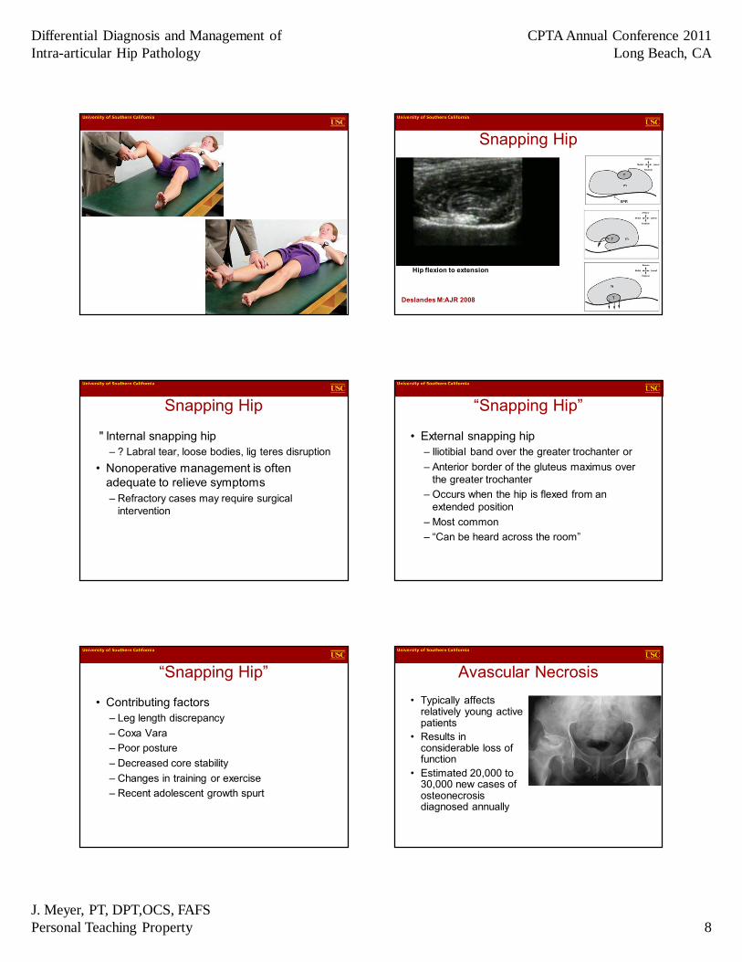
• Internal snapping hip– Iliopsoas over the iliopectineal eminence???
• Characterized by an audible snap or pop• Usually occurs during hip ext. from a flexed position• Often accompanied by pain and generally occurs during
physical activity
• Dynamic Sonography gives us new info– the snapping was provoked by the sudden flipping of
the iliopsoas tendon around the iliac muscle, allowing abrupt contact of the tendon against the pubic bone and producing an audible snap
Differential Diagnosis and Management of Intra-articular Hip Pathology
CPTA Annual Conference 2011Long Beach, CA
J. Meyer, PT, DPT,OCS, FAFSPersonal Teaching Property 8
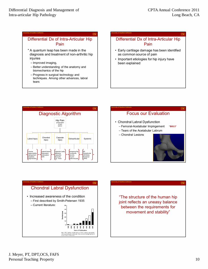
Snapping Hip
Deslandes M:AJR 2008
Hip flexion to extension
Snapping Hip
• Internal snapping hip– ? Labral tear, loose bodies, lig teres disruption
• Nonoperative management is often adequate to relieve symptoms– Refractory cases may require surgical
intervention
“Snapping Hip”
• External snapping hip– Iliotibial band over the greater trochanter or– Anterior border of the gluteus maximus over
the greater trochanter – Occurs when the hip is flexed from an
extended position– Most common– “Can be heard across the room”
“Snapping Hip”
• Contributing factors– Leg length discrepancy– Coxa Vara– Poor posture– Decreased core stability– Changes in training or exercise– Recent adolescent growth spurt
Avascular Necrosis• Typically affects
relatively young active patients
• Results in considerable loss of function
• Estimated 20,000 to 30,000 new cases of osteonecrosis diagnosed annually

Differential Diagnosis and Management of Intra-articular Hip Pathology
CPTA Annual Conference 2011Long Beach, CA
J. Meyer, PT, DPT,OCS, FAFSPersonal Teaching Property 9
AVN
• Etiology remains unknown– alcohol use– high-dose corticosteroid administration– coagulation abnormalities
• Pathophysiology of AVN points toward – alterations in intravascular blood flow– direct cellular toxicity– impaired mesenchymal cellular differentiation
Lateral Hip Pain
• Frequently assigned a diagnosis of trochanteric bursitis– Postmenopausal women
• Injection into the site of maximal tenderness can provide relief
• Recurrence of symptoms after injection often occur
Lateral Hip Pain
• Correlation demonstrated in the literature between– lumbar degenerative disease– gluteus medius tendinopathy or tears– trochanteric bursitis
• Major predictor of relapse of pain: – lumbar degenerative disease
• Must effectively screen the lumbar spineWalker et al:Jour Clin Orthop Relat Res2007
Osteoarthritis
• Hip pain associated with OA is the most common cause of the pain in older adults
• Prevalence studies have shown the rates of adult hip OA range from .4% to 27%
• The most common predisposing factor for hip OA is age
Dagenais S:Clin Orthop Relat Res 2009
Clinical Criteria for OA
Hip Pain(loss of joint space)
Moderate lateral
or anterior hip pain
Morningstiffness < 1hour
> 50 y/oPain with
weightbearing
Limited Hip Flexion & IR >15°
Pain with Hip IR Hip Abductor Weakness
Cibulka CT et al:JOSPT 2009
Questions?
Intra-articular vs. Extra-articular
Differential Diagnosis and Management of Intra-articular Hip Pathology
CPTA Annual Conference 2011Long Beach, CA
J. Meyer, PT, DPT,OCS, FAFSPersonal Teaching Property 10
Differential Dx of Intra-Articular Hip Pain
• A quantum leap has been made in the diagnosis and treatment of non-arthritic hip injuries– Improved imaging,– Better understanding of the anatomy and
biomechanics of the hip– Progress in surgical technology and
techniques. Among other advances, labral tears
Differential Dx of Intra-Articular Hip Pain
• Early cartilage damage has been identified as common source of pain
• Important etiologies for hip injury have been explained
Diagnostic AlgorithmHip Pain(preservation
of jointspace)
Labral Injury Extraarticular SystemicCapsularInjury
ChondralInjury
TraumaHypermobilityImpingementDysplasiaDegenerative
Lateral ImpactDislocationSubluxationAVN
Capsular LaxityAdhesive CapsulitisSynovitisLig. Teres
Snapping HipTendonitisBursitisSI jointReferred pain
RAPolyarticularArthritidesHormonalCRPS
Focus our Evaluation
• Chondral Labral Dysfunction– Femoral-Acetabular Impingement– Tears of the Acetabular Labrum– Chondral Lesions
“BIG 3”
Chondral Labral Dysfunction
• Increased awareness of the condition– First described by Smith-Petersen 1935– Current literature:
“The structure of the human hip joint reflects an uneasy balance between the requirements for
movement and stability”

Differential Diagnosis and Management of Intra-articular Hip Pathology
CPTA Annual Conference 2011Long Beach, CA
J. Meyer, PT, DPT,OCS, FAFSPersonal Teaching Property 11
Anatomical Understanding
• Bony Morphology of Impingement
• Function of the Labrum– Labro-Acetabular complex– Movements that strain or stress labrum
• Capsular Ligaments– Overlying anatomy– Symptoms that can fool you!!
Femoralacetabular Impingement
• FAI is a morphologic condition that predisposes hip to intraarticular pathology that then becomes painful
Pincer Cam > 70% Combination
Types Of Impingement
CombinationCAM and Pincer
OvercoveragePincer
Insufficient OffsetCAM
Etiology of FAI
• CAM– Abnormal physeal development– Slipped capital femoral epiphysis (SCFE)– Legg-Calve-Perthes disease– Malunited femoral neck fractures
• Pincer– General (coxa profunda/ acetabuli protrusio)– Local (acetabular retroversion)
Incidence of FAI• Copenhagen Osteoarthritis Study revealed
– Cam deformities in 17% of men and 4% of women in a cohort of 3,202
• Reichenbach et al and Hack et al. found an incidence of – Cam deformities in 25% of men and 5% of women
• The combination of cam and pincer deformities is the most frequent FAI finding– pure cam deformities seem to be more common in
men– pure pincer deformities are seen in women9
Function of the Labrum• Complex
fibrocartilaginous rim• Spans entire acetabulum• Becomes contiguous
with trans. acteab lig• Protective seal
– Enabling increased hydrostatic fluid pressure to facilitate synovial lubrication and resistance to joint distraction

Differential Diagnosis and Management of Intra-articular Hip Pathology
CPTA Annual Conference 2011Long Beach, CA
J. Meyer, PT, DPT,OCS, FAFSPersonal Teaching Property 12
Physiologic Function
• Currently not completely defined• Appears to serve multiple functions
– Most important role is dissipating large forces across the hip during weight-bearing activity
– Deepens the acetabulum to enhance stability• Increases the volume of the acetabulum by 33%
– Limits extreme range of motion
Tan V: Am J Orthop 2001
Labro-Acetabular Complex
• Junctional interface– 7 structures
• Labral injury leads to– Decreased articular
cartilage• Fluid content• Nutrition• Lubrication
– May lead to articular cartilage degeneration
Ferguson SJ: Clin Biomech 2000
Strain Across Labrum During Hip Movements
• Flexion and adduction places the greatest strain on anterior labrum
• ER strain is > IR strain• Lateral strain is greatest at 90° of hip
flexion with abduction and external rotation
Safran et al: AJSM 2011
Strain Across Labrum During Special Test
• The impingement position increases the anterior lateral strain the most
• Posterior labrum strain is significantly increased with hip external rotation in neutral or full extension
Safran et al: AJSM 2011
Stress of Repetitive AB/ER Loading
Dy et al:JBJS 2008
• Strain to the labral chondral interface occurs in flexion or extension
• Greatest stress is in the anterior labrum– 2-3 O'clock position
• Femoral head displaces 1.3-1.6 mm anteriorly
Waterpolo

Differential Diagnosis and Management of Intra-articular Hip Pathology
CPTA Annual Conference 2011Long Beach, CA
J. Meyer, PT, DPT,OCS, FAFSPersonal Teaching Property 13
Stress of Repetitive Loading in AB/ER
• 22 y/o Fb player• Bilateral CAM• Alpha angle 84/89
degrees
Capsular Ligaments• Iliofemoral ligament
– taut in extension & ER– resists anterior translation
• Pubofemoral– provides resistance to
hyperextension & hyperabduction
• Ischiofemoral ligament– becomes loose with flexion
and tight with extension
• Ligamentum teres
Extracapsular Relationships
• Iliopsoas acts as a dynamic stabilizer
• Rectus tendon close proximity to hip capsule
• Role of the iliocapsularis muscle
a = anterior-inferior iliac spineb = origin rectus femorisc = origin of iliocapsularisd = capsule overlying femoral necke = iliacus muscle f = iliocapsularis muscle
Iliocapsularis
• Little known stabilizer of the anterior hip
• AIIS to the lesser trochanter– Pincer impingement
• atrophied & fatty infiltrated iliocapsularis
– Acetabular dysplasia• large iliocapsularis
muscle without fatty infiltration
Babst et al. Clin Orth Rel Res 2010
Consequences of FAI
• Cam Impingement– Femoral head asphericity– Early acetabular chondral delamination– Progression to full thickness chondral lesions and
labral detachment• Pincer impingement
– Labrum affected primarily, cystic changes and degeneration
– Narrow peripheral acetabular cartilage affected
“New” hypothesis of OA in the non-dysplastic hip
Labral Tears
• Mechanism of injury1. Trauma: 5.3% to 15% of cases
Twisting/falling2. FAI 3. Dysplasia4. Joint instability5. Joint degeneration
87% of patients Wenger: Clin Orth Rel Res 2007

Differential Diagnosis and Management of Intra-articular Hip Pathology
CPTA Annual Conference 2011Long Beach, CA
J. Meyer, PT, DPT,OCS, FAFSPersonal Teaching Property 14
Mechanisms of Labral Injury
• Hip IR at 90° of hip flexion– Femoral neck impinging on acetabular margin
• Ganz et al: J Bone Joint Surg Br 2005
• Repetitive strain during ER and abduction in slight flexion or extension– Dy et al: JBJS 2008– Crawford: Clin Orth Rel Res 2010– Safran et al: AJSM 2011
Consequences of Labral Tears
1. Separation of the labrum from the articular margin
• “Watershed lesion”2. Destabilization of the hip
• Disruption of the negative pressure seal• instability
Ferguson SJ: J Biomech 2000
Consequences of Labral Tears
1. Loss of function2. Degenerative joint disease
• Chondral damage
McCarthy GC:Clinical Orthop Rel Res 2001
436 consecutive hip arthroscopies261 labral tears73% of the patients with a labral tear had chondral damage
How do we identify patient’s at risk and achieve an accurate
diagnosis?
Comprehensive Evaluation
Chondral Labral Dysfunction• USC Athletic Medicine Experience
– Prior to our initial work: 18 hip chondral labral injuries
– 40 surgeries (4 athletes in 2005/2006 had revision arthroscopy)
• 5 from 2000/2003• 16 in 2005/2006• 2 in 2007• 4 in 2008• 2 in 2009• 10 in 2010• 1 in 2011
Football 12, Women's Basketball 4, Baseball 6, Water Polo 12, Soccer 2,Track 4All athletes have returned to sport
Clinical Presentation
• Insidious onset of anterior hip & groin pain– Lateral hip, thigh,
buttock and lumbar• Moderate to severe at
presentation• About 35% report a
specific injury
• Describe pain as aching and sharp
• Often a mechanical component of catching & or locking
• C sign
Byrd T JW: NAJSPT 2007

Differential Diagnosis and Management of Intra-articular Hip Pathology
CPTA Annual Conference 2011Long Beach, CA
J. Meyer, PT, DPT,OCS, FAFSPersonal Teaching Property 15
Pain location and frequency or 51 patients with symptomatic FAI
Clohisy et al: Clin Orthop Relat Res 2009
Hip Joint Pain
• Receives innervations from branches of L2 to S1 of the lumbosacral plexus,– predominantly L3.
• Hip symptoms may be referred to the L3 dermatome– anterior and medial thigh– knee
Aggravating Factors
• Symptoms worse with activities• Twisting, turning or changing directions is
difficult• Seated position may be uncomfortable,
especially with > hip flexion• Rising from seated position often painful
(catching)• Difficulty ascending and descending stairs• Symptoms with entering /exiting an automobile
Functional Limitations
Clohisy et al: Clin Orthop Relat Res 2009
Components of Eval
PROMSpecial
Tests
Lunge Tests
Eccentric Core Tests
HipExcursion
Testing
Leg Length
&Hurdle S/O
SquatSLB
S/L Squat
Posture &
Gait
HipEval
Posture
• Slouching & listing to the uninvolved side– Decreases hip flexion– Abduction and ER
• relaxes the capsule
• Stand with the hip and knee joint slightly flexed
Byrd T JW: NAJSPT 2007

Differential Diagnosis and Management of Intra-articular Hip Pathology
CPTA Annual Conference 2011Long Beach, CA
J. Meyer, PT, DPT,OCS, FAFSPersonal Teaching Property 16
Gait Examination
• Assess hip mobility with gait alterations• Focus on hip extension, adduction, IR, hip
flexion and ankle dorsiflexion• Look for change in hip pain symptoms and
at the quality of the movement
Long Stride
• Assess hip extension• The restricted side hip
will rotate back towards you as the patient walks away
• Causing excessive lumbopelvic rotation or extension
Crossover Stride
• Assess hip adduction• Looking for the
smooth movement without excessive lumbar sidebend
Toe In Walking
• Assess hip IR• Looking for
symmetrical motion– Reproduction of
symptoms• Also looks at tri-plane
iliopsoas mobility– Ext/IR/Abd
Crouched Walking
• Utilize to assess ankle dorsiflexion and hip flexion
• Look for asymmetries in motion and reproduction of pain
FAI and Gait
• During level gait CAM FAI patient’s had significantly – decreased hip abduction motion– less total frontal hip ROM– decreased pelvic mobility– lower total sagittal hip ROM
• Predominately decreased extension
M.J. Kennedy et al: (2009)
Differential Diagnosis and Management of Intra-articular Hip Pathology
CPTA Annual Conference 2011Long Beach, CA
J. Meyer, PT, DPT,OCS, FAFSPersonal Teaching Property 17
1st MTP Function• Assess NWB ROM
– 60 degrees• Assess WB ROM
– Place patient in a stride stance with the lead leg elevated
– Perform a SL heel raise
• Assess 1st MTP ROM• Weight distribution• Rearfoot inversion
WB 1st MTP ROM
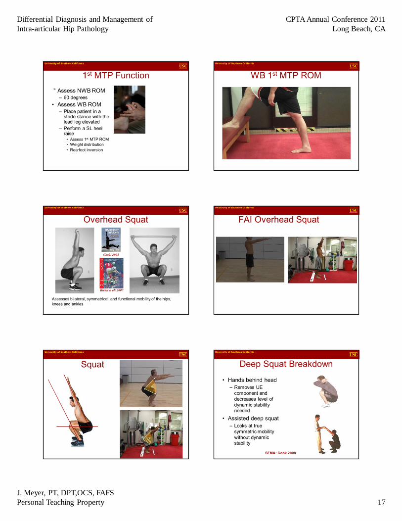
Overhead Squat
Kiesel et al: 2007
Cook: 2003
Assesses bilateral, symmetrical, and functional mobility of the hips, knees and ankles
FAI Overhead Squat
Squat Deep Squat Breakdown
• Hands behind head– Removes UE
component and decreases level of dynamic stability needed
• Assisted deep squat– Looks at true
symmetric mobility without dynamic stability
SFMA: Cook 2008

Differential Diagnosis and Management of Intra-articular Hip Pathology
CPTA Annual Conference 2011Long Beach, CA
J. Meyer, PT, DPT,OCS, FAFSPersonal Teaching Property 18
Deep Squat Breakdown
• Half kneeling ankle dorsiflexion– Flexibility vs. mobility
issue– Achilles and soleus
complex vs. anterior talocrual joint
– Excellent interrater and intrarater reliability
• .82• .89
Krause et al: J of Sport Rehab 2011
The Effect of Cam FAI on Pelvic Motion during Maximum Squat
• The FAI group had decreased sagittal pelvic ROM
• Also had decreased maximal squat depth
• Reduced sagittal mobility may lead to impingement
Lamontagne et al:Clin Orthop Relat Res 2009
The Biomechanics Associated with Squatting and FAI
SquatCONTROL FAI
Squat Results Squat Results

Differential Diagnosis and Management of Intra-articular Hip Pathology
CPTA Annual Conference 2011Long Beach, CA
J. Meyer, PT, DPT,OCS, FAFSPersonal Teaching Property 19
Squat S/L Squat
• Stability• Control• Strength• Range of motion
Hip Hiking & Single Leg Balance
• Screen for leg length differences
• Assess balance and glut medius strength
• Screen for stress fractures of the pubic ramus Noakes TD et al:
Am J Sports Med.1985
Hurdle Step Over• Assess ability to flex
hip w/o L/S compensation
• Assess dynamic balance and glut medius strength
Kiesel et al: 2007
Hurdle StepCONTROL FAI
Hurdle Results

Differential Diagnosis and Management of Intra-articular Hip Pathology
CPTA Annual Conference 2011Long Beach, CA
J. Meyer, PT, DPT,OCS, FAFSPersonal Teaching Property 20
Hurdle Results Hurdle Results
At 90 Degrees Femur Flexion Hip Flexion
• What muscles can assist hip flexion?– TFL, rectus femoris &
sartorius• Capable of hip flexion
up to 90°– Psoas and iliacus
• Flex hip above 90°– Weakness often in FAI
& athletic pubalgia
Active Hip Flexion Test
• Patient stand with one foot on a 18”or 24” plyo box
• Hands behind the head or at side• Attempt to lift the foot off the box and hold
for 5 sec• Inability to lift and hold is indicative of a
weak psoas and or iliacus.
Active Hip Flexion Test

Differential Diagnosis and Management of Intra-articular Hip Pathology
CPTA Annual Conference 2011Long Beach, CA
J. Meyer, PT, DPT,OCS, FAFSPersonal Teaching Property 21
Hip Excursion Testing
• Assess amount of motion that can be controlled within a specific plane while in unilateral stance
• “Standing hip ROM assessment”• Document quality and quantity of motion
Hip Flexion Excursion
• Single leg balance hip flexion
• Evaluate quality and quantity
• Contribution from the lumbar spine
• Reproduction of pain
19 y/o CAM impingement
Hip Extension Excursion
• Single leg balance hip extension
• Assess quality and quantity
• Contribution from the lumbar spine
• Reproduction of hip symptoms
Hip IR Excursion• Single leg balance hip
internal rotation• Be sure the foot is
kept flat• Utilize toe touch to
assess for range of motion restriction versus strength/balance deficit
Hip Adduction Excursion
• Single leg balance hip adduction/abduction
• Be sure the foot is kept flat
• Watch for lumbar spine sidebend
Eccentric-Concentric Control of the Core
• These tests look at the ability of the core muscle groups to control the body’s center of mass– Sagittal ECC SLB Posterior Reach– Frontal ECC SLB Medial Reach– Transverse ECC SLB PL Reach
Geraci et al: 2005 Phys Med Rehabil Clin N AmKibler, BW: Sports Medicine 2006

Differential Diagnosis and Management of Intra-articular Hip Pathology
CPTA Annual Conference 2011Long Beach, CA
J. Meyer, PT, DPT,OCS, FAFSPersonal Teaching Property 22
SLB Posterior Reach• Assesses the ability of
the psoas, rectus femoris, and rectus abdominus to control eccentrically extension of the spine and anterior translation of the pelvis
• A successful base test requires 10 controlled repetition on each side
SLB Posterior Reach
• Common compensatory patterns include– Restricted anterior translation of the pelvis
with overextension by the lumbar or thoracic spine
– Excessive flexion of the knee– ‘‘Clawing’’ of the toes, or hyperactivity of the
toe extensors to stabilize distally
SLB Medial Reach• Challenges pelvic stability
in the frontal plane • Excellent test of gluteus
medius function
• Loss of balance or a Trendelenburg sign mark the onset of fatigue in this muscle and lateral stabilizers of the pelvis
SLB PL Reach• The pelvis and spine
should rotate synchronously
• Look for motion in the spine that markedly exceeds pelvic motion in relationship to the femur of the stance leg
• Indicates tightness in the short hip external rotators and posterior hip capsule
Lunge Tests
• Assess dynamic loading capacity of the lower extremities
• Balance and control• Lumbar spine
movement compensations
• Dynamic flexibility
Anterior
Lateral
Posterior Lateral
Geraci et al: 2005 Phys Med Rehabil Clin N Am
PROM & Special Tests
• Hip IR/ER Seated• Hip Flexion MMT• Log Roll• Hip Flexion• Hip IR/ER• Impingement Test• FABER distance• Resisted Active SLR
• Resisted Adduction• Ober Test • Sit up• Thomas Test• Posterior Impingement• Prone IR• Craig’s Test• MMT
Martin HD et al: Arthroscopy 2010
Differential Diagnosis and Management of Intra-articular Hip Pathology
CPTA Annual Conference 2011Long Beach, CA
J. Meyer, PT, DPT,OCS, FAFSPersonal Teaching Property 23
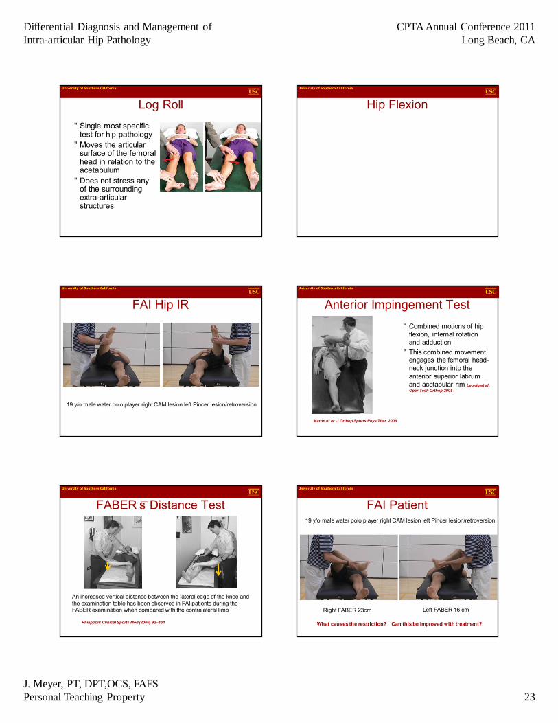
Log Roll• Single most specific
test for hip pathology• Moves the articular
surface of the femoral head in relation to the acetabulum
• Does not stress any of the surrounding extra-articular structures
Hip Flexion
FAI Hip IR
19 y/o male water polo player right CAM lesion left Pincer lesion/retroversion
Anterior Impingement Test• Combined motions of hip
flexion, internal rotation and adduction
• This combined movement engages the femoral head-neck junction into the anterior superior labrum and acetabular rim Leunig et al: Oper Tech Orthop.2005
Martin et al: J Orthop Sports Phys Ther. 2006
FABER’s Distance Test
An increased vertical distance between the lateral edge of the knee and the examination table has been observed in FAI patients during the FABER examination when compared with the contralateral limb
Philippon: Clinical Sports Med (2000) 92–101
FAI Patient
Right FABER 23cm Left FABER 16 cm
19 y/o male water polo player right CAM lesion left Pincer lesion/retroversion
What causes the restriction? Can this be improved with treatment?

Differential Diagnosis and Management of Intra-articular Hip Pathology
CPTA Annual Conference 2011Long Beach, CA
J. Meyer, PT, DPT,OCS, FAFSPersonal Teaching Property 24
FABER Test Reliability
• Philippon: unpublished data– Found as FABER distance increased – alpha
angle increased (r=0.2;p=0.002)– FABER difference >5cm è Alpha Angle = 72– FABER difference <5cm è Alpha Angle = 57
• *p=0.001
• Growing evidence this test can be a affective part of a screening exam for FAI
Active Straight Leg Raise
• Against resistance• Compressive forces
of multiple times body weight
• Often painful when mild degree of underlying disease
FAI Hip IR Understanding Hip IR• Craig’s test• 8-15° of anteversion in the adult• Anteverted = > IR vs. ER• Retroverted = > ER vs. IR
Patterns of Hip Muscle Weakness with FAI
• Patients with FAI (n=18) show significant muscle weakness compared to asymptomatic controls– Hip flexion– Hip adduction,– Hip external rotation – Hip abduction– Hip flexor muscle weakness
• was accompanied by reduced muscle activation of TFL
Casartelli et al: Osteoarthritis and Cartilage: 2011
How do we utilize this info?
Evaluation
PainLimited ROM &
Functional Strength
Abnormal MovementPatterns
Can we prevent this cycle from occurring?

Differential Diagnosis and Management of Intra-articular Hip Pathology
CPTA Annual Conference 2011Long Beach, CA
J. Meyer, PT, DPT,OCS, FAFSPersonal Teaching Property 25
Pre-participation Physical Evaluation (PPE)
• Prior to the 2006 season screened 81 Division 1 football players– 9 physical exam tests – Video analysis of overhead squat– Imaging studies – Physician consult
9 Physical Exam Tests• Range of Motion
– Hip Flexion (Supine)
– Hip IR (Prone/Seated)
– Hip ER (Prone/Seated)
– Thomas Test• Psoas, Quad, TFL
– Van Dillen et al: JOSPT 2000
– Straight Leg Raise (SLR)
• Provocation Tests– Anterior Impingement– FABER’s– Log Roll– Lumbar Spine Flexion
Abnormal Exam
• Athletes with hip pain and an asymmetrical FABER test greater than 4 cm
• Underwent MRI examination• Alpha angle measurements were obtained • Suspected pathologies of the chondral
labral junction were recorded
Alpha Angle
Used to quantify the degree of abnormality and cam impingement on the anterior aspect of the femoral head neck junction. An angle of 55°or greater is considered abnormal
Notzli et al: J Bone Joint Surg Br 2002
Abnormal Alpha Angle Results
• Thirty-three athletes (41%) had abnormal hip exams– Asymmetrical FABER– Hip Pain
• There was no difference in hip flexion between hips with normal exams(101°) and hips with abnormal exams(98°)

Differential Diagnosis and Management of Intra-articular Hip Pathology
CPTA Annual Conference 2011Long Beach, CA
J. Meyer, PT, DPT,OCS, FAFSPersonal Teaching Property 26
Results
• Hips with abnormal exams had significantly less external rotation(39°vs.49°; p<0.01)
• And significantly less internal rotation (28°vs. 33°;p<0.05) than hips with normal exams
MRI Examination• 27 hips were evaluated
by MRI • Average alpha angle
measurements were 67°(range 44° to 83°)– 26 hips with alpha angle
greater than 50°• 11 hips had suspected
pathologies of the chondral labral junction– Higher alpha angle(73°
vs.62°;p<0.01).
Screening Outcomes• Abnormal hips have reduced range of
motion• High alpha angles correlated with
suspicion of chondral labral pathologies• PPE identified athletes at risk of chondral
labral dysfunction– A modified screening exam has continued to
identify athletes at risk from 2007-2011The clinical exam had a positive predictive value for α>64° of 0.750 [0.588 to 0.817] and a negative predictive value for α≤64° of 0.833
Clinical Examination with MRI Validation to Assess High Hip Alpha Angle: A Prospective Study Among Asymptomatic Elite Youth and Pre-Collegiate Ice Hockey Players
Stull JD et al: 2011 AOSSM Meeting
Alpha Angle and Chondral Labral Dysfunction
• Higher alpha angles in FAI patient’s correlate with a greater magnitude of– Decreased range of motion – Chondral damage– Labral injury
Johnston et al: 2008
Multifactorial Nature of Intra-articular Hip Dysfunction
• CAM and Pincer bony abnormalities– Chondral-labral injury
• Decreased hip ROM• Abnormal gait• Reduced pelvic
mobility• Hip and trunk muscle
weakness

Differential Diagnosis and Management of Intra-articular Hip Pathology
CPTA Annual Conference 2011Long Beach, CA
J. Meyer, PT, DPT,OCS, FAFSPersonal Teaching Property 27
Diff Diagnosis, Evaluation and Screening
Question’s Additions???
Imaging & Surgical Procedures
Rehabilitation After HipFemoroacetabular ImpingementArthroscopy
More than 30,000 hip arthroscopies were performed in 2008. This number is expected
to grow at a rate of 15% over the next 5 years, resulting in more then 70,000 hip
arthroscopies performed each year by 2013
US markets for arthroscopy devices 2009. Report by Millennium Research Group
Goals of Surgery• Hip arthroscopic
procedures are used– Correct the bony geometry– Provide an intact labral
complex and ligamentous structure
– Improved hip congruency• Femoral osteoplasty• Acetabular rim trimming• Labral repair• Microfracture• Capsular closure
Rehabilitation Principles
• Individualized and evaluation-based (not time-based) program designed to address– Surgical Findings– Impairments – Functional Limitations– Lifestyle, work and sport requirements
Goals of Hip Rehab
• Protect the integrity of the healing tissue• Prevent negative effects of immobilization• Diminish pain and inflammation• Identify the primary causes and
compensations of the hip dysfunction

Differential Diagnosis and Management of Intra-articular Hip Pathology
CPTA Annual Conference 2011Long Beach, CA
J. Meyer, PT, DPT,OCS, FAFSPersonal Teaching Property 28
Goals of Hip Rehab• Normalize gait• Restore ROM
– Hip– Thoracic Spine– Screen
• Lumbar spine• Foot and Ankle
• Promote dynamic LE mobility and stability• Address motor control deficiencies• Restore strength and functional capacity
Rehab Progression• Respect differences in a
– Patient’s age– Magnitude of chondral
injury• Grade I-IV• More severe leads to
poorer prognosis – Nutrition and systemic
health– Concomitant injuries or
impairments– Goals and sport-specific
demands
Components of Rehabilitation and/or Prevention Program
Modify TrainingStressSafety
Reinforce Sport
MvmntSLLE
Strength
Eccentric Core
Training
Improve Hip/TrunkStability
Restore Hip
Mobility
CompetePainFree
Restore Hip Mobility
• Individualized specific time lines for weight bearing and ROM restrictions – Determined by the surgical procedure– Compliance with these restrictions by patients
and therapists is critical– Error on the side of safety– “Don’t push hip ROM through pain”
Precautions for Hip Mobility
Wahoff M et al: Clin Sports Med 2011
Range of Motion Strategies
• Utilize– Passive and active range of motion– Bottom-Up: femur on pelvis movements– Top-Down: pelvis on femur movements– Restore range of motion in all three planes of
motion while protecting the surgical repair– Avoid impingement symptoms
Differential Diagnosis and Management of Intra-articular Hip Pathology
CPTA Annual Conference 2011Long Beach, CA
J. Meyer, PT, DPT,OCS, FAFSPersonal Teaching Property 29
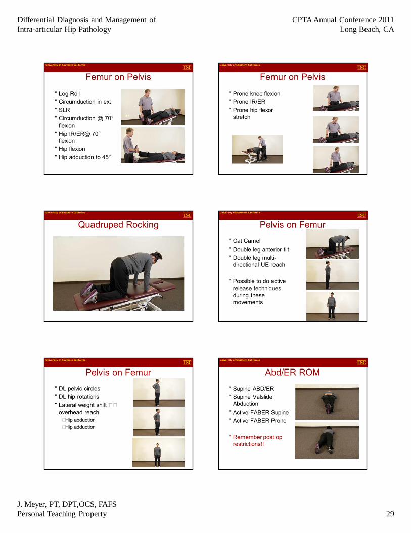
Femur on Pelvis
• Log Roll• Circumduction in ext• SLR• Circumduction @ 70°
flexion• Hip IR/ER@ 70°
flexion• Hip flexion• Hip adduction to 45°
Femur on Pelvis
• Prone knee flexion• Prone IR/ER• Prone hip flexor
stretch
Quadruped Rocking Pelvis on Femur
• Cat Camel• Double leg anterior tilt• Double leg multi-
directional UE reach
• Possible to do active release techniques during these movements
Pelvis on Femur
• DL pelvic circles• DL hip rotations• Lateral weight shift č
overhead reach– Hip abduction– Hip adduction
Abd/ER ROM
• Supine ABD/ER• Supine Valslide
Abduction• Active FABER Supine• Active FABER Prone
• Remember post op restrictions!!

Differential Diagnosis and Management of Intra-articular Hip Pathology
CPTA Annual Conference 2011Long Beach, CA
J. Meyer, PT, DPT,OCS, FAFSPersonal Teaching Property 30
Soft Tissue, Joint & Fascial Mobility
• Deep tissue and self soft tissue massage• Tri-plane hip musculature stretching
– With focus on pelvic mobility moving proximal on distal segment
– Top-down approach• Hip joint mobilization
– Athlete guided mobilization with movement
Soft Tissue Work• Often focus on
iliopsoas, adductor longus, hip ext/er, gracilis and pectineus
Stress of Repetitive Loading in AB/ER
• 22 y/o Fb player• Bilateral CAM• Alpha angle 84/89
degrees
Stress of Repetitive AB/ER Loading
Dy et al:JBJS 2008
• Strain to the labral chondral interface occurs in flexion or extension
• Greatest stress is in the anterior labrum– 2-3 O'clock position
• Femoral head displaces 1.3-1.6 mm anteriorly
Waterpolo Tri-plane Stretching
Adductors
Hamstrings

Differential Diagnosis and Management of Intra-articular Hip Pathology
CPTA Annual Conference 2011Long Beach, CA
J. Meyer, PT, DPT,OCS, FAFSPersonal Teaching Property 31
Tri-plane Stretching
Hip Flexors
Quads
Psoas Rectus Adductors
• After you have restored hip flexion ROM
• Outside of timeline for precautions for hip extension ROM
Hip Rotation
• Swiss Ball Active IR• Standing Hip ER
Table Stretch
Hip Joint Mobilizations
Cliborne et alNovember 2004
Caudal Glide Posterior Glide
Lateral Distraction Anterior Glide
Standing AP Glidewith or without thoracic rotation
Posterior Complex Mobilizationwith thoracic rotation
Differential Diagnosis and Management of Intra-articular Hip Pathology
CPTA Annual Conference 2011Long Beach, CA
J. Meyer, PT, DPT,OCS, FAFSPersonal Teaching Property 32
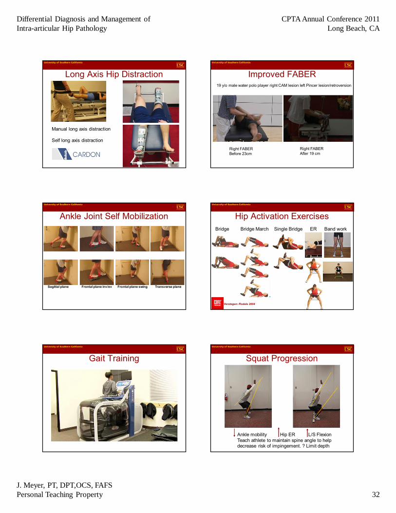
Long Axis Hip Distraction
Manual long axis distraction
Self long axis distraction
Improved FABER
Right FABER Before 23cm
Right FABER After 19 cm
19 y/o male water polo player right CAM lesion left Pincer lesion/retroversion
Ankle Joint Self Mobilization
Frontal plane inv/evSagittal plane Frontal plane swing Transverse plane
Hip Activation ExercisesBridge Bridge March Single Bridge ER Band work
Verstegen: Rodale 2004
Gait Training Squat Progression
Ankle mobility Hip ER L/S FlexionTeach athlete to maintain spine angle to help decrease risk of impingement. ? Limit depth

Differential Diagnosis and Management of Intra-articular Hip Pathology
CPTA Annual Conference 2011Long Beach, CA
J. Meyer, PT, DPT,OCS, FAFSPersonal Teaching Property 33
Lots of possibilities
What to you expect to see based on their age and injury?
Squat Variety
• Squat from a variety of foot positions to attempt to change the loading on the hip and simulate various everyday movements
Step Ups• Step Up
– Anterior• hip demand
– Lateral• ankle and knee
demand
Flanagan et al: 2004
Single Leg Mobility & Strength
• SL RDL• SL MD Reaching RDL• SL IR Excursion• SL ER Excursion
Standing Hip Abduction
• Early activation minimizes hip compressive forces
Hip Flexion Progressions• Supine Abdominal
Marching• Supine Abdominal 1 Leg
Slide• Sitting Isometric Hip
Flexion• Supine Band Hip Flexion• Standing Box Hip Flexion• Standing Dynamic Hip
Extension to Flexion

Differential Diagnosis and Management of Intra-articular Hip Pathology
CPTA Annual Conference 2011Long Beach, CA
J. Meyer, PT, DPT,OCS, FAFSPersonal Teaching Property 34
Single Leg Mobility & Strength
• SL overhead UE R/L lateral reach– Eccentric hip abductor
control
Balance Reach Training
Posterior UE reach - hip extension Medial reach - hip adduction
Posterior lateral - hip ER Posterior medial - hip ER
Plisky et al: 2006
Gray: 1995
Anterior lateral - hip IR
Single Leg Training Progression
Split Squat Bench Split Squat 1 Leg Squat
Verstegen: Rodale 2004
Single Leg DL
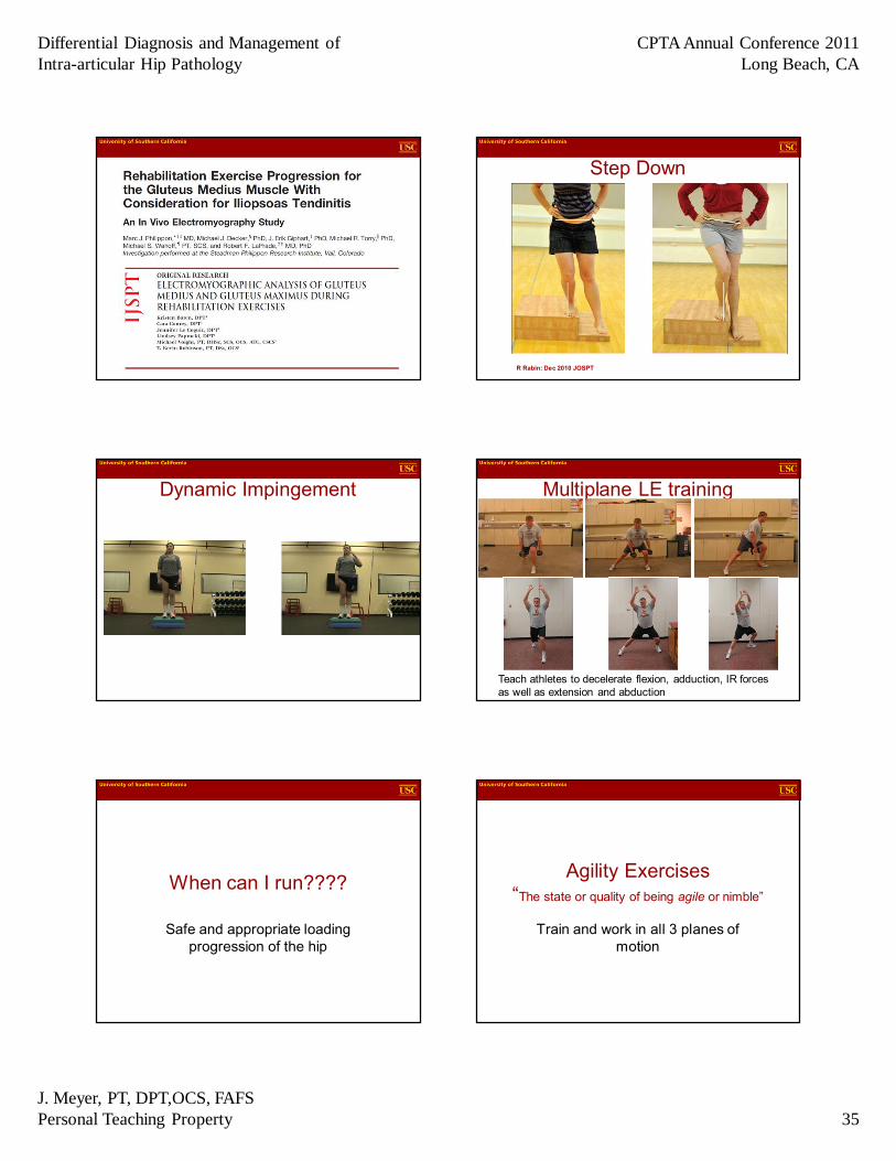
Glut Max Activation DuringCommon Therapeutic Exercises
Distefano et al 2009 JOSPT
Glut Medius Activation DuringCommon Therapeutic Exercises
Distefano et al 2009 JOSPT
Hip-Muscle Activation During the Lunge, Single-Leg Squat, and Step-Up-and-Over
Boudreau et al: JSR 2009
Differential Diagnosis and Management of Intra-articular Hip Pathology
CPTA Annual Conference 2011Long Beach, CA
J. Meyer, PT, DPT,OCS, FAFSPersonal Teaching Property 35
Step Down
R Rabin: Dec 2010 JOSPT
Dynamic Impingement Multiplane LE training
Teach athletes to decelerate flexion, adduction, IR forces as well as extension and abduction
When can I run????
Safe and appropriate loading progression of the hip
Agility Exercises“The state or quality of being agile or nimble”
Train and work in all 3 planes of motion

Differential Diagnosis and Management of Intra-articular Hip Pathology
CPTA Annual Conference 2011Long Beach, CA
J. Meyer, PT, DPT,OCS, FAFSPersonal Teaching Property 36
Parameters for Agility Training
• Based on qualitative and quantitative data– Attributes of movement
• Velocity, accuracy, control, rhythm, ROM– Numerical results
• Time, reps per bout of agility• Self select a work/rest interval based on
the patient’s functional level– 3:1, 2: 1, 4:1 10sec/30sec, 8 sec/16 sec– Adjust depending on the patient’s response
Base Front to Back Base Side to Side
STARTING POSITION:Stand in an athletic position, with your feet outside your hipsPROCEDURE:Keep both feet apart jump 4 inches to the right and then to the left as if you were jumping back and fourth over a line. Repeat as rapidly as possibleTEACHING KEY:As soon as you hit the ground spring back to the other side concentrate on quickness not height
Verstegen: Rodale 2004
Base Side to Side Base Rotations
STARTING POSITION:Stand with your shoulder square, your feet at a 45 degree anglePROCEDURE:Rotate your hips to the right and left at a 45 degree angle. Shoulders stay stationary; use the hips onlyTEACHING KEY:Rotate from the core not the shouldersImagine an X on the floor, rotate your hips so your feet move to the ends of the X
Verstegen: Rodale 2004
Differential Diagnosis and Management of Intra-articular Hip Pathology
CPTA Annual Conference 2011Long Beach, CA
J. Meyer, PT, DPT,OCS, FAFSPersonal Teaching Property 37
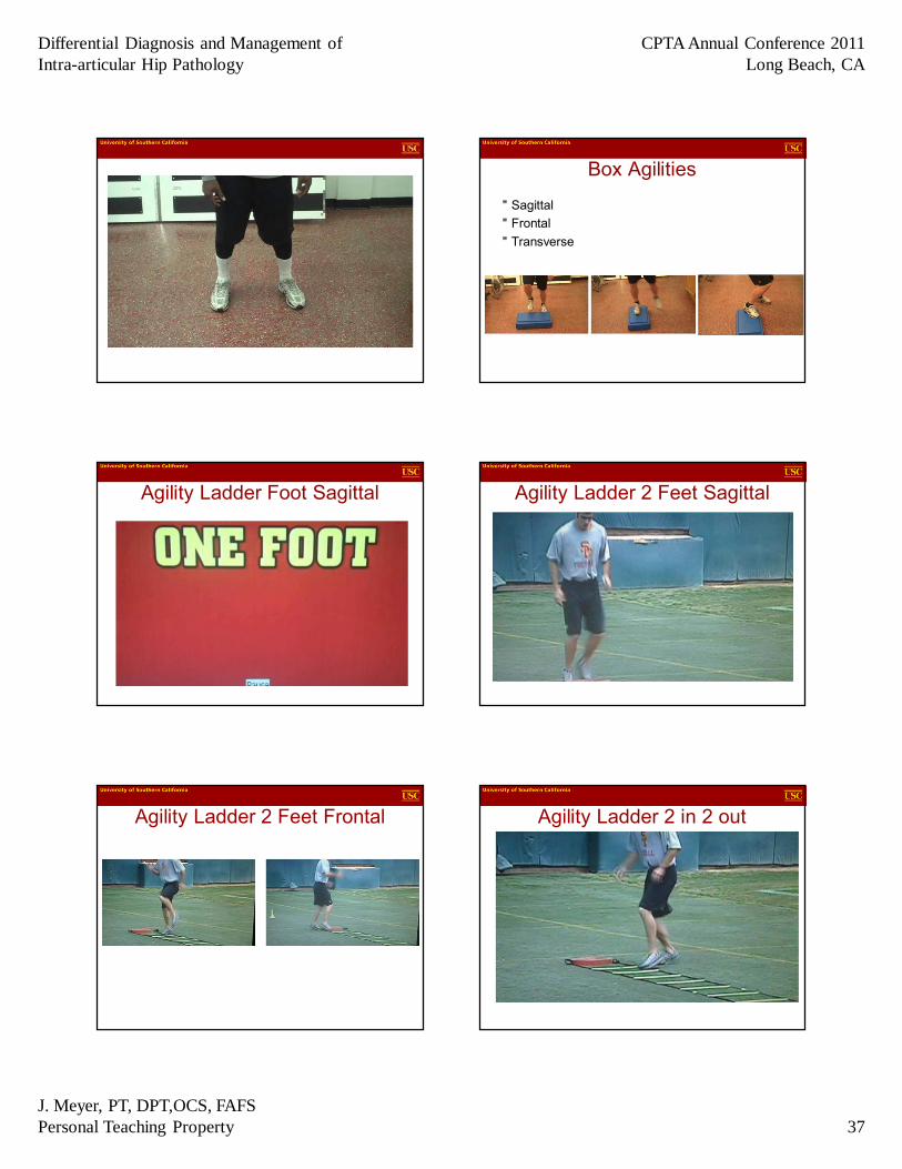
Box Agilities
• Sagittal• Frontal• Transverse
Agility Ladder Foot Sagittal Agility Ladder 2 Feet Sagittal
Agility Ladder 2 Feet Frontal Agility Ladder 2 in 2 out

Differential Diagnosis and Management of Intra-articular Hip Pathology
CPTA Annual Conference 2011Long Beach, CA
J. Meyer, PT, DPT,OCS, FAFSPersonal Teaching Property 38
Agility Ladder Scissor Agility Ladder Icky Shuffle
Common Agility Tests Common Agility Tests
Progressive Plyometric Program Keys
• Exercises are taught in a progressive manner
• Progress is based on competence, not a predetermined timeline
• Teach how to summate forces using the arms and the hips– Teach to “Land Softly” “Quiet”– Must learn to absorb force with the muscles
and not with the joints
Phase 1: Single Response with Stabilization
• Learn to jump and land– Tendon training– Summate forces using arms and hips– Land softly
• Purpose is to develop Eccentric Strength• Generally lasts 3-4 weeks• Most important and often overlooked
phase
Differential Diagnosis and Management of Intra-articular Hip Pathology
CPTA Annual Conference 2011Long Beach, CA
J. Meyer, PT, DPT,OCS, FAFSPersonal Teaching Property 39
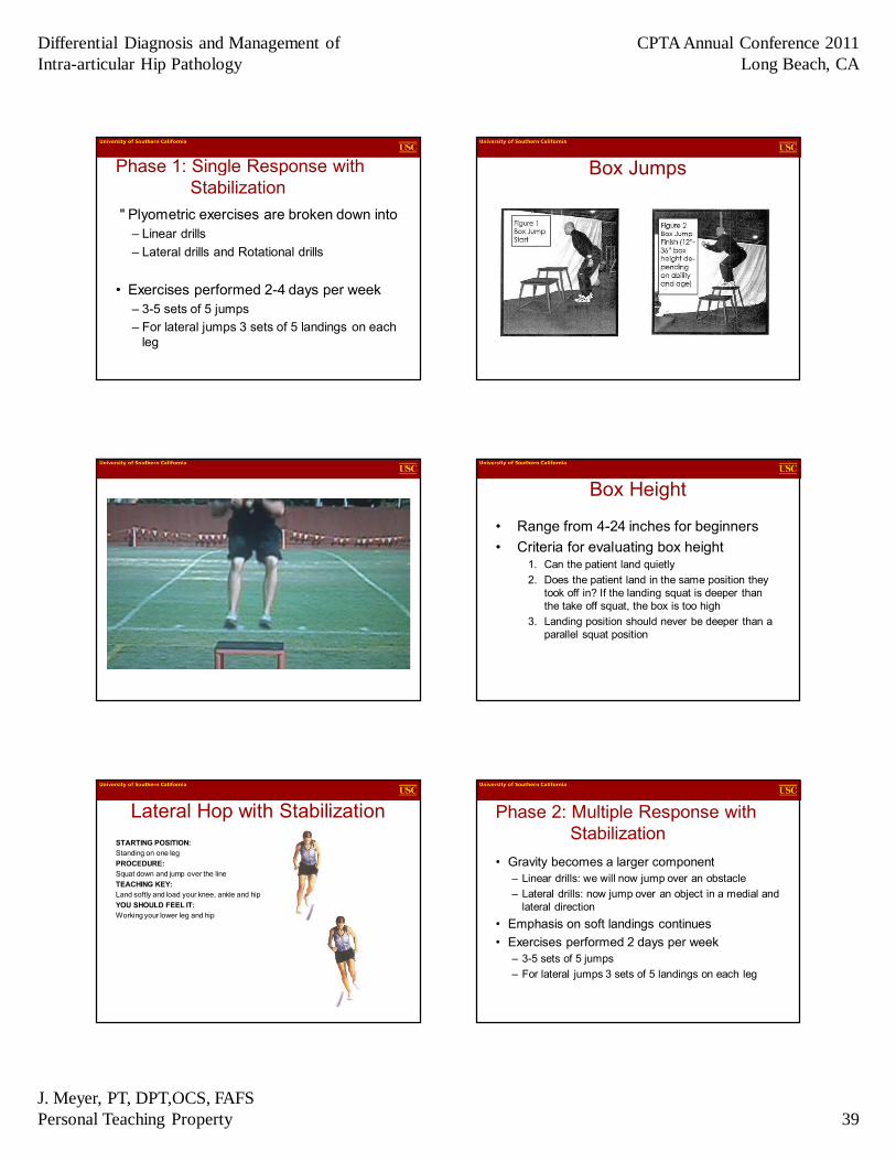
Phase 1: Single Response with Stabilization
• Plyometric exercises are broken down into– Linear drills– Lateral drills and Rotational drills
• Exercises performed 2-4 days per week– 3-5 sets of 5 jumps– For lateral jumps 3 sets of 5 landings on each
leg
Box Jumps
Box Height
• Range from 4-24 inches for beginners• Criteria for evaluating box height
1. Can the patient land quietly2. Does the patient land in the same position they
took off in? If the landing squat is deeper than the take off squat, the box is too high
3. Landing position should never be deeper than a parallel squat position
Lateral Hop with StabilizationSTARTING POSITION:Standing on one legPROCEDURE:Squat down and jump over the lineTEACHING KEY:Land softly and load your knee, ankle and hipYOU SHOULD FEEL IT:Working your lower leg and hip
Phase 2: Multiple Response with Stabilization
• Gravity becomes a larger component– Linear drills: we will now jump over an obstacle– Lateral drills: now jump over an object in a medial and
lateral direction• Emphasis on soft landings continues• Exercises performed 2 days per week
– 3-5 sets of 5 jumps– For lateral jumps 3 sets of 5 landings on each leg

Differential Diagnosis and Management of Intra-articular Hip Pathology
CPTA Annual Conference 2011Long Beach, CA
J. Meyer, PT, DPT,OCS, FAFSPersonal Teaching Property 40
Hurdle Hop and Stick
STARTING POSITION:Stand behind a hurdle on two feetPROCEDURE:Squat and load your knee, ankle and hip and jump over the hurdle and stick the landing. Hold this position for 2 seconds and repeat over the remaining hurdlesTEACHING KEY:Land soft, stick the landing , use your arms
Single Leg Lateral Hop and Stick Single Leg Medial Hop and Stick
STARTING POSITION:Stand next to the hurdle on one foot PROCEDURE:Squat and load your knee, ankle and hip and jump over the hurdle and stick the landing. Hold this position for 2 seconds and repeat.TEACHING KEY:Land soft, stick the landing , use your arms
Phase 3: Multiple Jumps
• Emphasis switches from development of eccentric strength to eccentric to concentric switching
• Less emphasis on stabilization• Introduction of elastic component• Most injury occurs from ignoring phase
1&2
Hurdle Hops
STARTING POSITION:Stand behind a hurdle on two feetPROCEDURE:Squat and load your knee, ankle and hip and jump over the hurdle and stick the landing. Rapidly repeat this movement over the remaining hurdlesTEACHING KEY:Land soft, stick the landing , use your arms

Differential Diagnosis and Management of Intra-articular Hip Pathology
CPTA Annual Conference 2011Long Beach, CA
J. Meyer, PT, DPT,OCS, FAFSPersonal Teaching Property 41
Single Leg Medial and Lateral Hurdle Hops
STARTING POSITION:Stand next to a hurdle on one footPROCEDURE:Squat and load your knee, ankle and hip and jump over the hurdle and stick the landing. Repeat over the remaining hurdlesTEACHING KEY:Land soft, stick the landing , use your arms
Other Testing Options
• Triple Hop• Multidirectional SL Hop• Drop Jump
– Assess landing strategy– Hip, knee and ankle
• Lateral Shuffle – Assess change of direction strategy
• Cutting 45° or 90°– Assess LE position & control during cutting
Drop Jump
Pre Training Post Training
Lateral Shuffle
Pre Training Post Training
Bad Cutting Mechanics
Differential Diagnosis and Management of Intra-articular Hip Pathology
CPTA Annual Conference 2011Long Beach, CA
J. Meyer, PT, DPT,OCS, FAFSPersonal Teaching Property 42
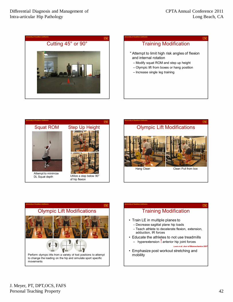
Cutting 45° or 90° Training Modification
• Attempt to limit high risk angles of flexion and internal rotation– Modify squat ROM and step up height– Olympic lift from boxes or hang position– Increase single leg training
Squat ROM Step Up Height
Attempt to minimize DL Squat depth Utilize a step below 90°
of hip flexion
Olympic Lift Modifications
Hang Clean Clean Pull from box
Olympic Lift Modifications
Perform olympic lifts from a variety of foot positions to attempt to change the loading on the hip and simulate sport specific movements
Training Modification
• Train LE in multiple planes to – Decrease sagittal plane hip loads– Teach athlete to decelerate flexion, extension,
adduction, IR forces• Educate the athletes to not use treadmills
– hyperextension anterior hip joint forces
• Emphasize post workout stretching and mobility
Lewis et al: Jour of Biomechanics 2007

Differential Diagnosis and Management of Intra-articular Hip Pathology
CPTA Annual Conference 2011Long Beach, CA
J. Meyer, PT, DPT,OCS, FAFSPersonal Teaching Property 43
Management of Intra-articular Hip Injuries
• Screening - Identify hip at risk• Early diagnosis• Modify training program and educate patient and family
• Importance of comprehensive evaluation and treatment
• Total body movement assessment• Imaging (Extra-articular Injections: PRP)• Modify strength training
• Surgical intervention if necessary• Safe appropriate rehabilitation progressions
Components of Rehabilitation and Prevention Program
Modify TrainingStressSafety
Reinforce Sport
MvmntSLLE
Strength
Eccentric Core
Training
Improve Hip
Stability
Restore Hip
Mobility
CompetePainFree
Biomechanical Changes Long Term Outcomes
Thanks to
• USC Athletic Medicine and Physical Therapy– Dr. Snibbe– Dr. Tibone– Dr. Philippon– Dr. Byrd– Dr. Sampson
• USC Athletes • CPTA