management of patellar chondral defects with autologous
TRANSCRIPT
Life 2021, 11, 141. https://doi.org/10.3390/life11020141 www.mdpi.com/journal/life
Communication
Management of Patellar Chondral Defects with Autologous Matrix Induced Chondrogenesis (AMIC) Com-pared to Microfractures: A Four Years Follow-Up Clinical Trial Filippo Migliorini 1, Jörg Eschweiler 1, Nicola Maffulli 2,3,4,*, Arne Driessen 1, Björn Rath 1,5, Markus Tingart 1 and Hanno Schenker 1
1 Department of Orthopedics and Trauma Surgery, University Clinic Aachen, RWTH Aachen University Clinic, 52064 Aachen, Germany; [email protected] (F.M.); [email protected] (J.E.); [email protected] (A.D.); [email protected] (B.R.); [email protected] (M.T.); [email protected] (H.S.)
2 Department of Medicine, Surgery and Dentistry, University of Salerno, Via S. Allende, 84081 Baronissi (SA), Italy
3 School of Pharmacy and Bioengineering, Keele University School of Medicine, Thornburrow Drive, Stoke on Trent ST4 7QB, UK
4 Barts and the London School of Medicine and Dentistry, Queen Mary University of London, Centre for Sports and Exercise Medicine, Mile End Hospital, 275 Bancroft Road, London E1 4DG, UK
5 Department of Orthopedics, Klinikum Wels-Grieskirchen, A-4600 Wels, Austria * Correspondence: [email protected]
Abstract: Introduction: Evidence on the management of chondral defects of the patella arises from studies in which the patellofemoral joint was treated together with the femorotibial joint and pri-mary and revision settings. Furthermore, the superiority of Autologous Matrix Induced Chondro-genesis (AMIC) over microfractures (MFx) for patellar chondral defects is uncertain. Therefore, the present study compared primary isolated AMIC versus MFx for focal unipolar chondral defects of the patellar facet joints at midterm follow-up. Methods: Patients undergoing AMIC or isolated MFx surgery for borderline-sized focal unipolar chondral defects of the patellar facet joints were fol-lowed at our institution. All surgeries were performed in the same fashion by experienced surgeons. A parapatellar arthrotomy was adopted in all surgeries. The outcomes of interest were: Visual An-alogic Scale (VAS), Tegner Activity Scale, International Knee Documentation Committee (IKDC), and the Lysholm scores. The Magnetic Resonance Observation of Cartilage Repair Tissue (MOCART) was assessed by a blinded radiologist, who had not been involved in the clinical man-agement of the patients. Results: 38 patients were enrolled in the present study: 27 underwent AMIC, and 11 MFx. The mean follow-up was 45.1 months. The mean age of the patients at baseline was 34.5 years. The mean size of the defect was 2.6 cm2. The MFx cohort experienced a shorter length of the hospitalization (p = 0.008). There was no difference in terms of follow-up and previous symp-toms duration, mean age, sex, side, defect size, and BMI. At last follow-up, the AMIC cohort re-ported greater IKDC (p = 0.01), Lysholm (p = 0.009), and Tegner (p = 0.02), along with a low rate of failure (p = 0.02). VAS was lower in the AMIC group (p = 0.002). No difference was found in the MOCART score (p = 0.09), rates of revision (p = 0.06), and arthroplasty (p = 0.2). Conclusion: The AMIC procedure achieves greater IKDC and Lysholm score, and a significant reduction of the VAS score in the management of patellar chondral defects. The Tegner scale demonstrated greater activ-ity after AMIC procedure. Finally, the AMIC group evidenced a lower rate of failure. Similarity was found on MOCART score, rates of revision, and arthroplasty between the two procedures.
Keywords: knee; patella; autologous matrix induced chondrogenesis; AMIC; microfractures; chon-dral defects; management
Citation: Migliorini, F.; Eschweiler,
J.; Maffulli, N.; Driessen, A.; Rath, B.;
Tingart, M.; Schenker, H.
Management of Patellar Chondral
Defects with Autologous Matrix
Induced Chondrogenesis (AMIC)
Compared to Microfractures: A Four
Years Follow-up Clinical Trial. 2021,
11, 141. https://doi.org/10.3390/
life11020141
Academic Editor: Michael Tanzer
Received: 1 February 2021
Accepted: 10 February 2021
Published: 13 February 2021
Publisher’s Note: MDPI stays neu-
tral with regard to jurisdictional
claims in published maps and insti-
tutional affiliations.
Copyright: © 2021 by the authors.
Licensee MDPI, Basel, Switzerland.
This article is an open access article
distributed under the terms and con-
ditions of the Creative Commons At-
tribution (CC BY) license (http://cre-
ativecommons.org/licenses/by/4.0/).
Life 2021, 11, 141 2 of 11
1. Introduction Symptomatic focal chondral defects of the patella are common [1,2]. If left untreated,
they can lead to early osteoarthritis [3]. Several surgical approaches have been proposed to manage chondral defects [4–9]. Microfractures (MFx) is cost-effective and has been largely used to manage smaller chondral defects [4,5]. Autologous chondrocyte implan-tation (ACI) techniques evolved from the periosteal patch (pACI), to chondral patch (cACI), to third generation procedures named matrix-induced ACI (mACI) [2,10–12]. Ir-respective of the generation, ACI requires harvesting, external chondrocyte expansion, and a second stage surgery for re-implantation [13–15]. The superiority of ACI compared to MFx is still controversial [16–18]. Autologous Matrix-Induced Chondrogenesis (AMIC) avoids cartilage harvest and external expansion and is performed in a single surgical ses-sion [19,20]. Differently to ACI, which requires expanded autologous chondrocytes, AMIC exploits the potential of bone marrow-derived mesenchymal stem cells [21,22]. Iso-lated AMIC for chondral defects of the patella joint surface in a primary setting has been poorly investigated. Evidence on the management of chondral defects of the patella mainly arises from studies in which the patellofemoral joint was treated together with the femorotibial joint, and primary and revision settings were mixed [23–26]. Especially for borderline sized defects, the superiority of AMIC with respect to MFx for patellar defects is uncertain. The present study compared primary isolated AMIC versus MFx for focal unipolar chondral defects of the patellar facet joints at midterm follow-up.
2. Material and Methods 2.1. Patients Recruitment
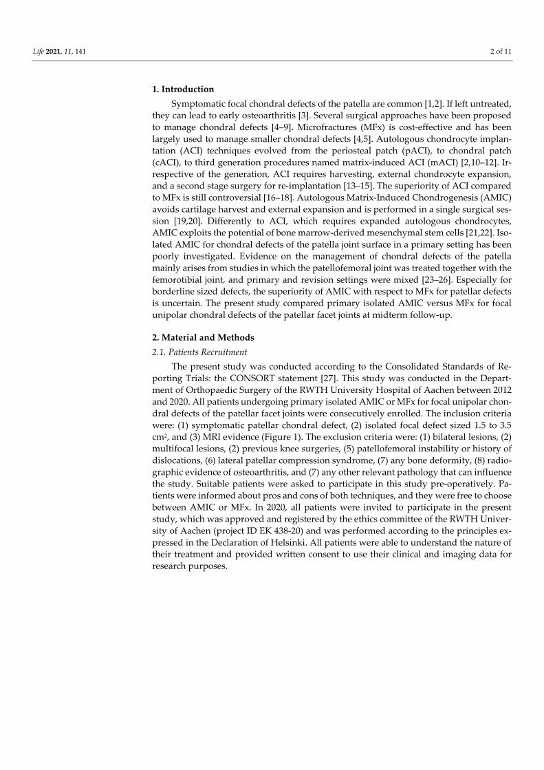
The present study was conducted according to the Consolidated Standards of Re-porting Trials: the CONSORT statement [27]. This study was conducted in the Depart-ment of Orthopaedic Surgery of the RWTH University Hospital of Aachen between 2012 and 2020. All patients undergoing primary isolated AMIC or MFx for focal unipolar chon-dral defects of the patellar facet joints were consecutively enrolled. The inclusion criteria were: (1) symptomatic patellar chondral defect, (2) isolated focal defect sized 1.5 to 3.5 cm2, and (3) MRI evidence (Figure 1). The exclusion criteria were: (1) bilateral lesions, (2) multifocal lesions, (2) previous knee surgeries, (5) patellofemoral instability or history of dislocations, (6) lateral patellar compression syndrome, (7) any bone deformity, (8) radio-graphic evidence of osteoarthritis, and (7) any other relevant pathology that can influence the study. Suitable patients were asked to participate in this study pre-operatively. Pa-tients were informed about pros and cons of both techniques, and they were free to choose between AMIC or MFx. In 2020, all patients were invited to participate in the present study, which was approved and registered by the ethics committee of the RWTH Univer-sity of Aachen (project ID EK 438-20) and was performed according to the principles ex-pressed in the Declaration of Helsinki. All patients were able to understand the nature of their treatment and provided written consent to use their clinical and imaging data for research purposes.
Life 2021, 11, 141 3 of 11
Figure 1. MRI of a focal chondral defect of the patella.
2.2. Surgical Technique All the surgeries were performed in the same fashion by two experienced surgeons
according to a previous report [28]. Briefly, preliminary arthroscopy was performed through traditional anteromedial and anterolateral portals. Debridement and curettage of the non-viable tissues surrounding the lesion were performed. MFx to a depth of 4 mm were performed using a 65° and 90° pick. In those patients who underwent AMIC, a mini medial parapatellar approach was performed. The perforation of the subchondral bone was performed with a 40° pick or a 1.2/1.4 mm Kischer wire under constant irrigation. An aluminum template was trimmed according to the defect. A type I/III porcine resorbable collagen membrane was used in all procedures (Chondro-Gide, Geistlich Pharma AG, Wolhusen, Switzerland). The membrane was trimmed according to the aluminum tem-plate to slightly undersize the defect to avoid displacement. The membrane was hydrated in a saline solution and placed into the lesion and attached with fibrin glue. The stability of the membrane was checked by repeatedly flexing and extending the knee. The rehabil-itation process was performed according to our previous study [29].
2.3. Outcomes of Interest On admission, the following data were recorded: age, gender, side, area of defect,
BMI (Kg/m2), symptoms duration prior to surgery, and length of hospital stay. The pri-mary outcomes of interest were to compare clinical findings using patient reported out-comes measures (PROMs) and complications of both groups. The secondary outcomes of interest were to compare MRI findings according to the Magnetic Resonance Observation of Cartilage Repair Tissue (MOCART) score [30]. At last follow-up, patients performed independently the following scores: Visual Analog Scale (VAS) [31], Tegner Activity Scale [32], International Knee Documentation Committee (IKDC) [33], and the Lysholm [34] scores. Data concerning the rate of complications (failure, revision, arthroplasty, delami-nation, hypertrophy) and additional procedures were also collected. Failure was defined as persistent pain that affected negatively the quality of life and limited the participation to recreational activities. The MOCART score was performed by a blinded radiologist, who had not been involved in the clinical management of the patients.
Life 2021, 11, 141 4 of 11
2.4. Statistical Analysis All statistical analyses were performed using the software IBM SPSS version 25. Con-
tinuous data were analysed using the mean difference (MD), while dichotomic data with odd ratio (OR) effect measures for parametric data. The T-test and ߯2 tests were per-formed, respectively, with values of p < 0.05 considered statistically significant. The con-fidence interval (CI) was set at 95% in all comparisons.
3. Results 3.1. Recruitment Process
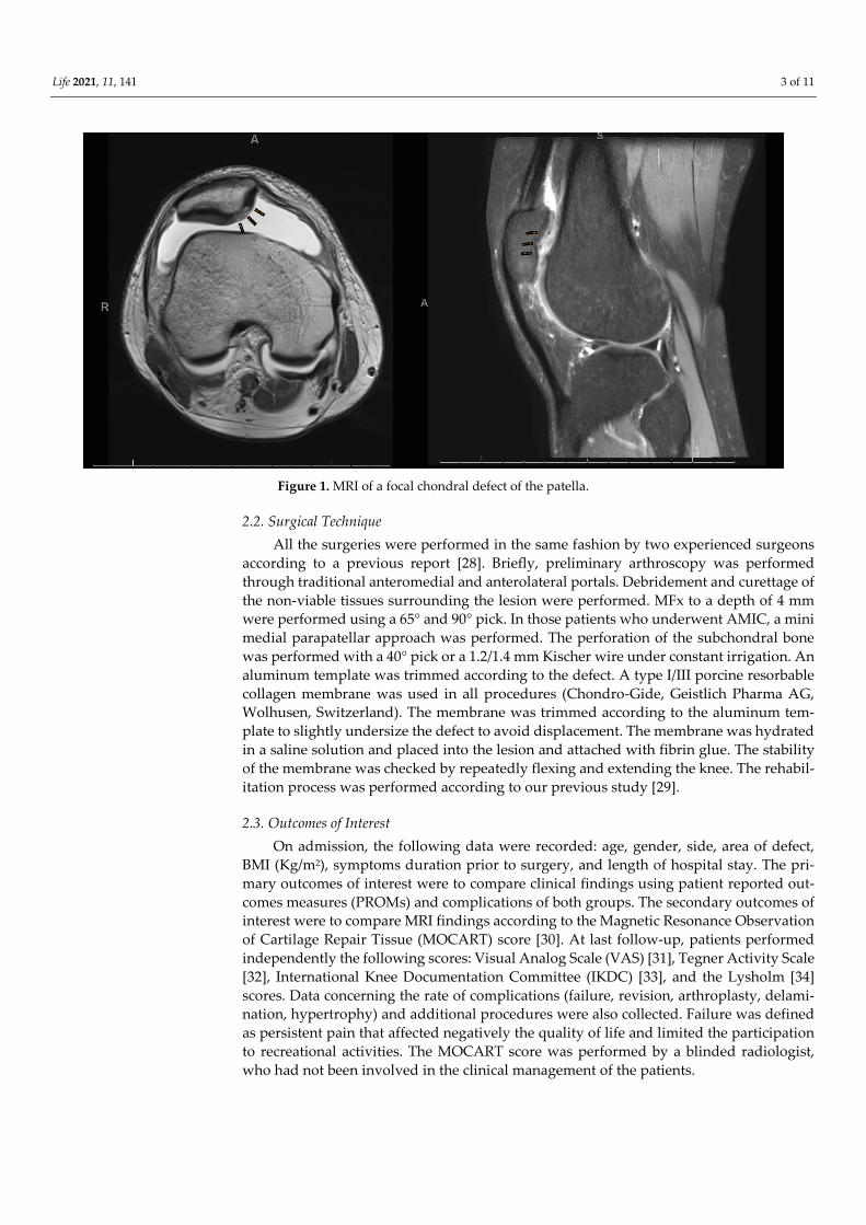
A total of 76 patients were initially screened. Of them, 25 were not eligible: kissing lesions (N = 1), bilateral lesions (N = 1), multiple lesions (N = 5), previous knee surgeries (N = 14), metabolic bone disease (N = 1), and patellofemoral instability or history of dislo-cations (N = 3). Fifty-one patients were available and operated: 32 underwent AMIC and 19 microfractures. At last follow-up, five patients who had undergone AMIC and eight microfractures were not available. Eventually, 38 patients were enrolled in the present study: 27 underwent AMIC, and 11 MFx. The diagram of the recruitment process is shown in Figure 2.
Figure 2. Diagram of the recruitment process.
Life 2021, 11, 141 5 of 11
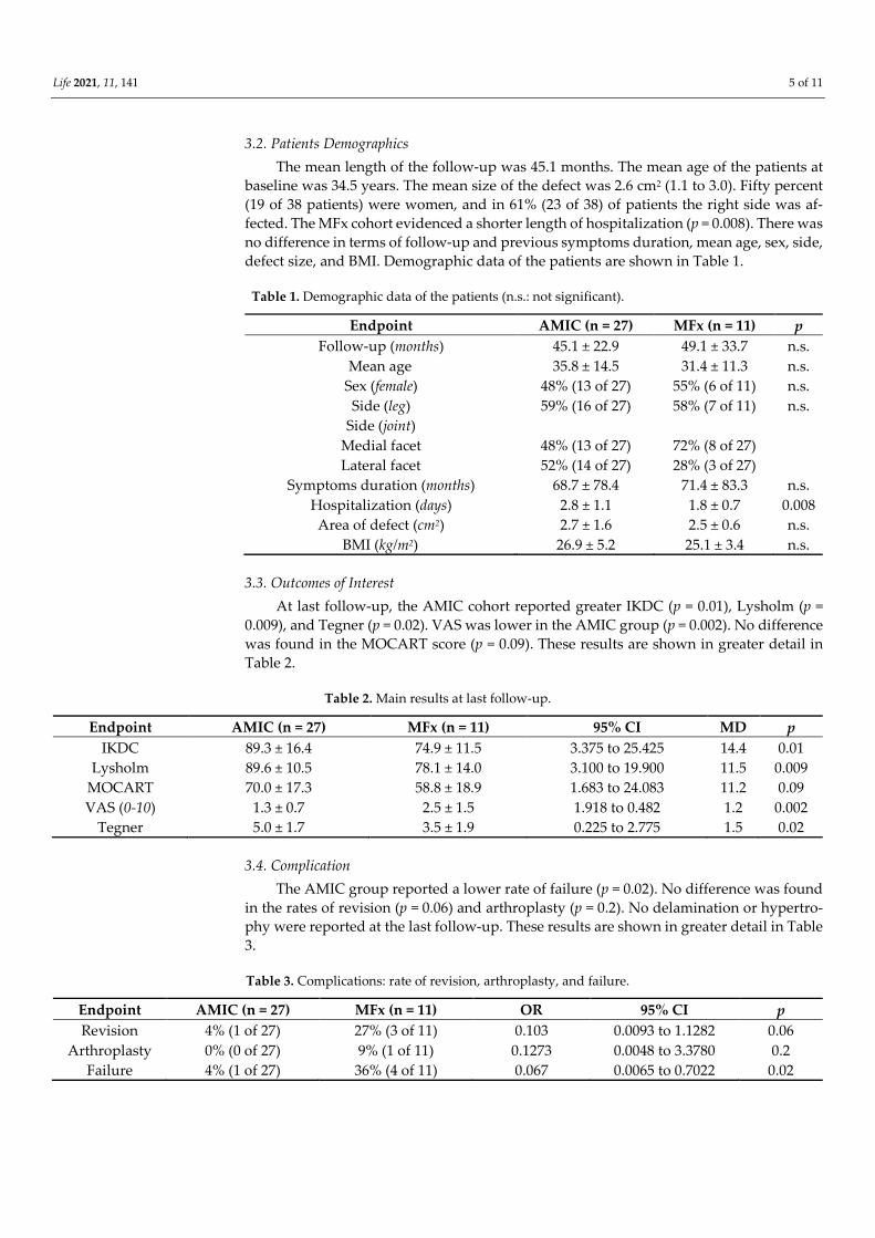
3.2. Patients Demographics The mean length of the follow-up was 45.1 months. The mean age of the patients at
baseline was 34.5 years. The mean size of the defect was 2.6 cm2 (1.1 to 3.0). Fifty percent (19 of 38 patients) were women, and in 61% (23 of 38) of patients the right side was af-fected. The MFx cohort evidenced a shorter length of hospitalization (p = 0.008). There was no difference in terms of follow-up and previous symptoms duration, mean age, sex, side, defect size, and BMI. Demographic data of the patients are shown in Table 1.
Table 1. Demographic data of the patients (n.s.: not significant).
Endpoint AMIC (n = 27) MFx (n = 11) p Follow-up (months) 45.1 ± 22.9 49.1 ± 33.7 n.s.
Mean age 35.8 ± 14.5 31.4 ± 11.3 n.s. Sex (female) 48% (13 of 27) 55% (6 of 11) n.s.
Side (leg) 59% (16 of 27) 58% (7 of 11) n.s. Side (joint)
Medial facet 48% (13 of 27) 72% (8 of 27) Lateral facet 52% (14 of 27) 28% (3 of 27)
Symptoms duration (months) 68.7 ± 78.4 71.4 ± 83.3 n.s. Hospitalization (days) 2.8 ± 1.1 1.8 ± 0.7 0.008 Area of defect (cm2) 2.7 ± 1.6 2.5 ± 0.6 n.s.
BMI (kg/m2) 26.9 ± 5.2 25.1 ± 3.4 n.s.
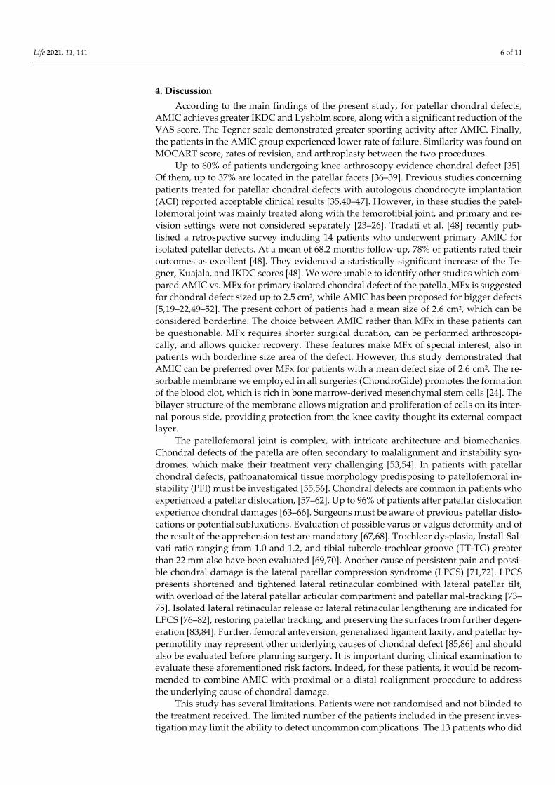
3.3. Outcomes of Interest At last follow-up, the AMIC cohort reported greater IKDC (p = 0.01), Lysholm (p =
0.009), and Tegner (p = 0.02). VAS was lower in the AMIC group (p = 0.002). No difference was found in the MOCART score (p = 0.09). These results are shown in greater detail in Table 2.
Table 2. Main results at last follow-up.
Endpoint AMIC (n = 27) MFx (n = 11) 95% CI MD p IKDC 89.3 ± 16.4 74.9 ± 11.5 3.375 to 25.425 14.4 0.01
Lysholm 89.6 ± 10.5 78.1 ± 14.0 3.100 to 19.900 11.5 0.009 MOCART 70.0 ± 17.3 58.8 ± 18.9 1.683 to 24.083 11.2 0.09 VAS (0-10) 1.3 ± 0.7 2.5 ± 1.5 1.918 to 0.482 1.2 0.002
Tegner 5.0 ± 1.7 3.5 ± 1.9 0.225 to 2.775 1.5 0.02
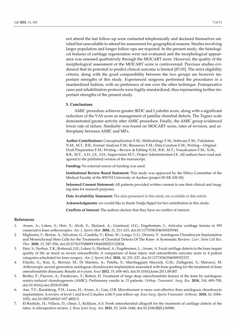
3.4. Complication The AMIC group reported a lower rate of failure (p = 0.02). No difference was found
in the rates of revision (p = 0.06) and arthroplasty (p = 0.2). No delamination or hypertro-phy were reported at the last follow-up. These results are shown in greater detail in Table 3.
Table 3. Complications: rate of revision, arthroplasty, and failure.
Endpoint AMIC (n = 27) MFx (n = 11) OR 95% CI p Revision 4% (1 of 27) 27% (3 of 11) 0.103 0.0093 to 1.1282 0.06
Arthroplasty 0% (0 of 27) 9% (1 of 11) 0.1273 0.0048 to 3.3780 0.2 Failure 4% (1 of 27) 36% (4 of 11) 0.067 0.0065 to 0.7022 0.02
Life 2021, 11, 141 6 of 11
4. Discussion According to the main findings of the present study, for patellar chondral defects,
AMIC achieves greater IKDC and Lysholm score, along with a significant reduction of the VAS score. The Tegner scale demonstrated greater sporting activity after AMIC. Finally, the patients in the AMIC group experienced lower rate of failure. Similarity was found on MOCART score, rates of revision, and arthroplasty between the two procedures.
Up to 60% of patients undergoing knee arthroscopy evidence chondral defect [35]. Of them, up to 37% are located in the patellar facets [36–39]. Previous studies concerning patients treated for patellar chondral defects with autologous chondrocyte implantation (ACI) reported acceptable clinical results [35,40–47]. However, in these studies the patel-lofemoral joint was mainly treated along with the femorotibial joint, and primary and re-vision settings were not considered separately [23–26]. Tradati et al. [48] recently pub-lished a retrospective survey including 14 patients who underwent primary AMIC for isolated patellar defects. At a mean of 68.2 months follow-up, 78% of patients rated their outcomes as excellent [48]. They evidenced a statistically significant increase of the Te-gner, Kuajala, and IKDC scores [48]. We were unable to identify other studies which com-pared AMIC vs. MFx for primary isolated chondral defect of the patella. MFx is suggested for chondral defect sized up to 2.5 cm2, while AMIC has been proposed for bigger defects [5,19–22,49–52]. The present cohort of patients had a mean size of 2.6 cm2, which can be considered borderline. The choice between AMIC rather than MFx in these patients can be questionable. MFx requires shorter surgical duration, can be performed arthroscopi-cally, and allows quicker recovery. These features make MFx of special interest, also in patients with borderline size area of the defect. However, this study demonstrated that AMIC can be preferred over MFx for patients with a mean defect size of 2.6 cm2. The re-sorbable membrane we employed in all surgeries (ChondroGide) promotes the formation of the blood clot, which is rich in bone marrow-derived mesenchymal stem cells [24]. The bilayer structure of the membrane allows migration and proliferation of cells on its inter-nal porous side, providing protection from the knee cavity thought its external compact layer.
The patellofemoral joint is complex, with intricate architecture and biomechanics. Chondral defects of the patella are often secondary to malalignment and instability syn-dromes, which make their treatment very challenging [53,54]. In patients with patellar chondral defects, pathoanatomical tissue morphology predisposing to patellofemoral in-stability (PFI) must be investigated [55,56]. Chondral defects are common in patients who experienced a patellar dislocation, [57–62]. Up to 96% of patients after patellar dislocation experience chondral damages [63–66]. Surgeons must be aware of previous patellar dislo-cations or potential subluxations. Evaluation of possible varus or valgus deformity and of the result of the apprehension test are mandatory [67,68]. Trochlear dysplasia, Install-Sal-vati ratio ranging from 1.0 and 1.2, and tibial tubercle-trochlear groove (TT-TG) greater than 22 mm also have been evaluated [69,70]. Another cause of persistent pain and possi-ble chondral damage is the lateral patellar compression syndrome (LPCS) [71,72]. LPCS presents shortened and tightened lateral retinacular combined with lateral patellar tilt, with overload of the lateral patellar articular compartment and patellar mal-tracking [73–75]. Isolated lateral retinacular release or lateral retinacular lengthening are indicated for LPCS [76–82], restoring patellar tracking, and preserving the surfaces from further degen-eration [83,84]. Further, femoral anteversion, generalized ligament laxity, and patellar hy-permotility may represent other underlying causes of chondral defect [85,86] and should also be evaluated before planning surgery. It is important during clinical examination to evaluate these aforementioned risk factors. Indeed, for these patients, it would be recom-mended to combine AMIC with proximal or a distal realignment procedure to address the underlying cause of chondral damage.
This study has several limitations. Patients were not randomised and not blinded to the treatment received. The limited number of the patients included in the present inves-tigation may limit the ability to detect uncommon complications. The 13 patients who did
Life 2021, 11, 141 7 of 11
not attend the last follow-up were contacted telephonically and declared themselves sat-isfied but unavailable to attend for assessment for geographical reasons. Studies involving larger population and longer follow-ups are required. In the present study, the histologi-cal features of cartilage regeneration were not evaluated and the morphological appear-ance was assessed qualitatively through the MOCART score. However, the quality of the morphological assessment of the MOCART score is controversial. Previous studies evi-denced that its potential to predict clinical outcome is limited [87,88]. The strict eligibility criteria, along with the good comparability between the two groups are however im-portant strengths of this study. Experienced surgeons performed the procedures in a standardized fashion, with no preference of one over the other technique. Postoperative cares and rehabilitation protocols were highly standardized, thus representing further im-portant strengths of the present study.
5. Conclusions AMIC procedure achieves greater IKDC and Lysholm score, along with a significant
reduction of the VAS score as management of patellar chondral defects. The Tegner scale demonstrated greater activity after AMIC procedure. Finally, the AMIC group evidenced lower rate of failure. Similarity was found on MOCART score, rates of revision, and ar-throplasty between AMIC and MFx.
Author Contributions: Conceptualization F.M.; Methodology F.M.; Software F.M.; Validation N.M., M.T., B.R.; Formal Analysis F.M.; Resources F.M.; Data Curation F.M.; Writing—Original Draft Preparation F.M.; Writing—Review & Editing N.M., B.R., M.T.; Visualization F.M., N.M., B.R., M.T., A.D., J.E., S.H.; Supervision M.T.; Project Administration J.E. All authors have read and agreed to the published version of the manuscript.
Funding: No external source of funding was used.
Institutional Review Board Statement: This study was approved by the Ethics Committee of the Medical Faculty of the RWTH University of Aachen (project ID EK 438-20).
Informed Consent Statement: All patients provided written consent to use their clinical and imag-ing data for research purposes.
Data Availability Statement: The data presented in this study are available in this article
Acknowledgments: we would like to thank Nadja Sippel for her contribution in this study.
Conflicts of Interest: The authors declare that they have no conflict of interest.
References 1. Aroen, A.; Loken, S.; Heir, S.; Alvik, E.; Ekeland, A.; Granlund, O.G.; Engebretsen, L. Articular cartilage lesions in 993
consecutive knee arthroscopies. Am. J. Sports Med. 2004, 32, 211–215, doi:10.1177/0363546503259345. 2. Migliorini, F.; Berton, A.; Salvatore, G.; Candela, V.; Khan, W.; Longo, U.G.; Denaro, V. Autologous Chondrocyte Implantation
and Mesenchymal Stem Cells for the Treatments of Chondral Defects Of The Knee- A Systematic Review. Curr. Stem Cell Res. Ther. 2020, 15, 547–556, doi:10.2174/1574888X15666200221122834.
3. Heir, S.; Nerhus, T.K.; Rotterud, J.H.; Loken, S.; Ekeland, A.; Engebretsen, L.; Aroen, A. Focal cartilage defects in the knee impair quality of life as much as severe osteoarthritis: A comparison of knee injury and osteoarthritis outcome score in 4 patient categories scheduled for knee surgery. Am. J. Sports Med. 2010, 38, 231–237, doi:10.1177/0363546509352157.
4. Filardo, G.; Kon, E.; Berruto, M.; Di Martino, A.; Patella, S.; Marcheggiani Muccioli, G.M.; Zaffagnini, S.; Marcacci, M. Arthroscopic second generation autologous chondrocytes implantation associated with bone grafting for the treatment of knee osteochondritis dissecans: Results at 6 years. Knee 2012, 19, 658–663, doi:10.1016/j.knee.2011.08.007.
5. Bertho, P.; Pauvert, A.; Pouderoux, T.; Robert, H. Treatment of large deep osteochondritis lesions of the knee by autologous matrix-induced chondrogenesis (AMIC): Preliminary results in 13 patients. Orthop. Traumatol. Surg. Res. 2018, 104, 695–700, doi:10.1016/j.otsr.2018.05.008.
6. Aae, T.F.; Randsborg, P.H.; Luras, H.; Aroen, A.; Lian, O.B. Microfracture is more cost-effective than autologous chondrocyte implantation: A review of level 1 and level 2 studies with 5 year follow-up. Knee Surg. Sports Traumatol. Arthrosc. 2018, 26, 1044–1052, doi:10.1007/s00167-017-4802-5.
7. El-Rashidy, H.; Villacis, D.; Omar, I.; Kelikian, A.S. Fresh osteochondral allograft for the treatment of cartilage defects of the talus: A retrospective review. J. Bone Joint Surg. Am. 2011, 93, 1634–1640, doi:10.2106/JBJS.J.00900.
Life 2021, 11, 141 8 of 11
8. Behrens, P.; Bitter, T.; Kurz, B.; Russlies, M. Matrix-associated autologous chondrocyte transplantation/implantation (MACT/MACI)--5-year follow-up. Knee 2006, 13, 194–202, doi:10.1016/j.knee.2006.02.012.
9. Basad, E.; Wissing, F.R.; Fehrenbach, P.; Rickert, M.; Steinmeyer, J.; Ishaque, B. Matrix-induced autologous chondrocyte implantation (MACI) in the knee: Clinical outcomes and challenges. Knee Surg. Sports Traumatol. Arthrosc. 2015, 23, 3729–3735, doi:10.1007/s00167-014-3295-8.
10. Basad, E.; Ishaque, B.; Bachmann, G.; Sturz, H.; Steinmeyer, J. Matrix-induced autologous chondrocyte implantation versus microfracture in the treatment of cartilage defects of the knee: A 2-year randomised study. Knee Surg. Sports Traumatol. Arthrosc. 2010, 18, 519–527, doi:10.1007/s00167-009-1028-1.
11. Bartlett, W.; Skinner, J.A.; Gooding, C.R.; Carrington, R.W.; Flanagan, A.M.; Briggs, T.W.; Bentley, G. Autologous chondrocyte implantation versus matrix-induced autologous chondrocyte implantation for osteochondral defects of the knee: A prospective, randomised study. J. Bone Joint Surg. Br. 2005, 87, 640–645, doi:10.1302/0301-620X.87B5.15905.
12. Shimozono, Y.; Hurley, E.T.; Nguyen, J.T.; Deyer, T.W.; Kennedy, J.G. Allograft Compared with Autograft in Osteochondral Transplantation for the Treatment of Osteochondral Lesions of the Talus. J. Bone Joint Surg. Am. 2018, 100, 1838–1844, doi:10.2106/JBJS.17.01508.
13. Siebold, R.; Suezer, F.; Schmitt, B.; Trattnig, S.; Essig, M. Good clinical and MRI outcome after arthroscopic autologous chondrocyte implantation for cartilage repair in the knee. Knee Surg. Sports Traumatol. Arthrosc. 2018, 26, 831–839, doi:10.1007/s00167-017-4491-0.
14. Niemeyer, P.; Laute, V.; Zinser, W.; Becher, C.; Kolombe, T.; Fay, J.; Pietsch, S.; Kuzma, T.; Widuchowski, W.; Fickert, S. A Prospective, Randomized, Open-Label, Multicenter, Phase III Noninferiority Trial to Compare the Clinical Efficacy of Matrix-Associated Autologous Chondrocyte Implantation With Spheroid Technology Versus Arthroscopic Microfracture for Cartilage Defects of the Knee. Orthop. J. Sports Med. 2019, 7, 2325967119854442, doi:10.1177/2325967119854442.
15. Saris, D.B.; Vanlauwe, J.; Victor, J.; Almqvist, K.F.; Verdonk, R.; Bellemans, J.; Luyten, F.P. Treatment of symptomatic cartilage defects of the knee: Characterized chondrocyte implantation results in better clinical outcome at 36 months in a randomized trial compared to microfracture. Am. J. Sports Med. 2009, 37, 10S–19S, doi:10.1177/0363546509350694.
16. Van Assche, D.; Staes, F.; Van Caspel, D.; Vanlauwe, J.; Bellemans, J.; Saris, D.B.; Luyten, F.P. Autologous chondrocyte implantation versus microfracture for knee cartilage injury: A prospective randomized trial, with 2-year follow-up. Knee Surg. Sports Traumatol. Arthrosc. 2010, 18, 486–495, doi:10.1007/s00167-009-0955-1.
17. Vasiliadis, H.S.; Wasiak, J.; Salanti, G. Autologous chondrocyte implantation for the treatment of cartilage lesions of the knee: A systematic review of randomized studies. Knee Surg. Sports Traumatol. Arthrosc. 2010, 18, 1645–1655, doi:10.1007/s00167-010-1050-3.
18. Gotze, C.; Nieder, C.; Felder, H.; Migliorini, F. AMIC for Focal Osteochondral Defect of the Talar Shoulder. Life 2020, 10, 328, doi:10.3390/life10120328.
19. De Girolamo, L.; Schonhuber, H.; Vigano, M.; Bait, C.; Quaglia, A.; Thiebat, G.; Volpi, P. Autologous Matrix-Induced Chondrogenesis (AMIC) and AMIC Enhanced by Autologous Concentrated Bone Marrow Aspirate (BMAC) Allow for Stable Clinical and Functional Improvements at up to 9 Years Follow-Up: Results from a Randomized Controlled Study. J. Clin. Med. 2019, 8, 392, doi:10.3390/jcm8030392.
20. Schiavone Panni, A.; Del Regno, C.; Mazzitelli, G.; D′Apolito, R.; Corona, K.; Vasso, M. Good clinical results with autologous matrix-induced chondrogenesis (Amic) technique in large knee chondral defects. Knee Surg. Sports Traumatol. Arthrosc. 2018, 26, 1130–1136, doi:10.1007/s00167-017-4503-0.
21. Schagemann, J.; Behrens, P.; Paech, A.; Riepenhof, H.; Kienast, B.; Mittelstadt, H.; Gille, J. Mid-term outcome of arthroscopic AMIC for the treatment of articular cartilage defects in the knee joint is equivalent to mini-open procedures. Arch. Orthop. Trauma Surg. 2018, 138, 819–825, doi:10.1007/s00402-018-2887-z.
22. Volz, M.; Schaumburger, J.; Frick, H.; Grifka, J.; Anders, S. A randomized controlled trial demonstrating sustained benefit of Autologous Matrix-Induced Chondrogenesis over microfracture at five years. Int. Orthop. 2017, 41, 797–804, doi:10.1007/s00264-016-3391-0.
23. Anders, S.; Volz, M.; Frick, H.; Gellissen, J. A Randomized, Controlled Trial Comparing Autologous Matrix-Induced Chondrogenesis (AMIC(R)) to Microfracture: Analysis of 1- and 2-Year Follow-Up Data of 2 Centers. Open Orthop. J. 2013, 7, 133–143, doi:10.2174/1874325001307010133.
24. Chung, J.Y.; Lee, D.H.; Kim, T.H.; Kwack, K.S.; Yoon, K.H.; Min, B.H. Cartilage extra-cellular matrix biomembrane for the enhancement of microfractured defects. Knee Surg. Sports Traumatol. Arthrosc. 2014, 22, 1249–1259, doi:10.1007/s00167-013-2716-4.
25. Sadlik, B.; Puszkarz, M.; Kosmalska, L.; Wiewiorski, M. All-Arthroscopic Autologous Matrix-Induced Chondrogenesis-Aided Repair of a Patellar Cartilage Defect Using Dry Arthroscopy and a Retraction System. J. Knee Surg. 2017, 30, 925–929, doi:10.1055/s-0037-1599246.
26. Moher, D.; Hopewell, S.; Schulz, K.F.; Montori, V.; Gotzsche, P.C.; Devereaux, P.J.; Elbourne, D.; Egger, M.; Altman, D.G. CONSORT 2010 explanation and elaboration: Updated guidelines for reporting parallel group randomised trials. BMJ 2010, 340, c869, doi:10.1136/bmj.c869.
27. Benthien, J.P.; Behrens, P. The treatment of chondral and osteochondral defects of the knee with autologous matrix-induced chondrogenesis (AMIC): Method description and recent developments. Knee Surg. Sports Traumatol. Arthrosc. 2011, 19, 1316–1319, doi:10.1007/s00167-010-1356-1.
Life 2021, 11, 141 9 of 11
28. Kusano, T.; Jakob, R.P.; Gautier, E.; Magnussen, R.A.; Hoogewoud, H.; Jacobi, M. Treatment of isolated chondral and osteochondral defects in the knee by autologous matrix-induced chondrogenesis (AMIC). Knee Surg. Sports Traumatol. Arthrosc. 2012, 20, 2109–2115, doi:10.1007/s00167-011-1840-2.
29. Marlovits, S.; Singer, P.; Zeller, P.; Mandl, I.; Haller, J.; Trattnig, S. Magnetic resonance observation of cartilage repair tissue (MOCART) for the evaluation of autologous chondrocyte transplantation: Determination of interobserver variability and correlation to clinical outcome after 2 years. Eur. J. Radiol. 2006, 57, 16–23, doi:10.1016/j.ejrad.2005.08.007.
30. Hawker, G.A.; Mian, S.; Kendzerska, T.; French, M. Measures of adult pain: Visual Analog Scale for Pain (VAS Pain), Numeric Rating Scale for Pain (NRS Pain), McGill Pain Questionnaire (MPQ), Short-Form McGill Pain Questionnaire (SF-MPQ), Chronic Pain Grade Scale (CPGS), Short Form-36 Bodily Pain Scale (SF-36 BPS), and Measure of Intermittent and Constant Osteoarthritis Pain (ICOAP). Arthritis Care Res. 2011, 63, S240–S252, doi:10.1002/acr.20543.
31. Mostafaee, N.; Negahban, H.; Shaterzadeh Yazdi, M.J.; Goharpey, S.; Mehravar, M.; Pirayeh, N. Responsiveness of a Persian version of Knee Injury and Osteoarthritis Outcome Score and Tegner activity scale in athletes with anterior cruciate ligament reconstruction following physiotherapy treatment. Physiother. Theory Pract. 2020, 36, 1019–1026, doi:10.1080/09593985.2018.1548672.
32. Collins, N.J.; Misra, D.; Felson, D.T.; Crossley, K.M.; Roos, E.M. Measures of knee function: International Knee Documentation Committee (IKDC) Subjective Knee Evaluation Form, Knee Injury and Osteoarthritis Outcome Score (KOOS), Knee Injury and Osteoarthritis Outcome Score Physical Function Short Form (KOOS-PS), Knee Outcome Survey Activities of Daily Living Scale (KOS-ADL), Lysholm Knee Scoring Scale, Oxford Knee Score (OKS), Western Ontario and McMaster Universities Osteoarthritis Index (WOMAC), Activity Rating Scale (ARS), and Tegner Activity Score (TAS). Arthritis Care Res. 2011, 63, S208–S228, doi:10.1002/acr.20632.
33. Briggs, K.K.; Lysholm, J.; Tegner, Y.; Rodkey, W.G.; Kocher, M.S.; Steadman, J.R. The reliability, validity, and responsiveness of the Lysholm score and Tegner activity scale for anterior cruciate ligament injuries of the knee: 25 years later. Am. J. Sports Med. 2009, 37, 890–897, doi:10.1177/0363546508330143.
34. Becher, C.; Ettinger, M.; Ezechieli, M.; Kaps, C.; Ewig, M.; Smith, T. Repair of retropatellar cartilage defects in the knee with microfracture and a cell-free polymer-based implant. Arch Orthop. Trauma Surg. 2015, 135, 1003–1010, doi:10.1007/s00402-015-2235-5.
35. Curl, W.W.; Krome, J.; Gordon, E.S.; Rushing, J.; Smith, B.P.; Poehling, G.G. Cartilage injuries: A review of 31,516 knee arthroscopies. Arthroscopy 1997, 13, 456–460, doi:10.1016/s0749-8063(97)90124-9.
36. Hjelle, K.; Solheim, E.; Strand, T.; Muri, R.; Brittberg, M. Articular cartilage defects in 1000 knee arthroscopies. Arthroscopy 2002, 18, 730–734, doi:10.1053/jars.2002.32839.
37. Widuchowski, W.; Lukasik, P.; Kwiatkowski, G.; Faltus, R.; Szyluk, K.; Widuchowski, J.; Koczy, B. Isolated full thickness chondral injuries. Prevalance and outcome of treatment. A retrospective study of 5233 knee arthroscopies. Acta Chir. Orthop. Traumatol. Cech. 2008, 75, 382–386.
38. Widuchowski, W.; Widuchowski, J.; Trzaska, T. Articular cartilage defects: Study of 25,124 knee arthroscopies. Knee 2007, 14, 177–182, doi:10.1016/j.knee.2007.02.001.
39. Buda, R.; Baldassarri, M.; Perazzo, L.; Ghinelli, D.; Pagliazzi, G. A useful combination for the treatment of patellofemoral chondral lesions: Realignment procedure plus mesenchymal stem cell-retrospective analysis and clinical results at 48 months of follow-up. Eur. J. Orthop. Surg. Traumatol. 2019, 29, 461–470, doi:10.1007/s00590-018-2310-z.
40. Ebert, J.R.; Fallon, M.; Smith, A.; Janes, G.C.; Wood, D.J. Prospective clinical and radiologic evaluation of patellofemoral matrix-induced autologous chondrocyte implantation. Am. J. Sports Med. 2015, 43, 1362–1372, doi:10.1177/0363546515574063.
41. Gomoll, A.H.; Madry, H.; Knutsen, G.; van Dijk, N.; Seil, R.; Brittberg, M.; Kon, E. The subchondral bone in articular cartilage repair: Current problems in the surgical management. Knee Surg. Sports Traumatol. Arthrosc. 2010, 18, 434–447, doi:10.1007/s00167-010-1072-x.
42. Macmull, S.; Jaiswal, P.K.; Bentley, G.; Skinner, J.A.; Carrington, R.W.; Briggs, T.W. The role of autologous chondrocyte implantation in the treatment of symptomatic chondromalacia patellae. Int. Orthop. 2012, 36, 1371–1377, doi:10.1007/s00264-011-1465-6.
43. Meyerkort, D.; Ebert, J.R.; Ackland, T.R.; Robertson, W.B.; Fallon, M.; Zheng, M.H.; Wood, D.J. Matrix-induced autologous chondrocyte implantation (MACI) for chondral defects in the patellofemoral joint. Knee Surg. Sports Traumatol. Arthrosc. 2014, 22, 2522–2530, doi:10.1007/s00167-014-3046-x.
44. Perdisa, F.; Filardo, G.; Sessa, A.; Busacca, M.; Zaffagnini, S.; Marcacci, M.; Kon, E. One-Step Treatment for Patellar Cartilage Defects With a Cell-Free Osteochondral Scaffold: A Prospective Clinical and MRI Evaluation. Am. J. Sports Med. 2017, 45, 1581–1588, doi:10.1177/0363546517694159.
45. Teo, B.J.; Buhary, K.; Tai, B.C.; Hui, J.H. Cell-based therapy improves function in adolescents and young adults with patellar osteochondritis dissecans. Clin. Orthop. Relat. Res. 2013, 471, 1152–1158, doi:10.1007/s11999-012-2338-z.
46. Mehl, J.; Huck, J.; Bode, G.; Hohloch, L.; Schmitt, A.; Sudkamp, N.P.; Niemeyer, P. Clinical mid- to long-term outcome after autologous chondrocyte implantation for patellar cartilage lesions and its correlation with the geometry of the femoral trochlea. Knee 2019, 26, 364–373, doi:10.1016/j.knee.2019.01.019.
47. Tradati, D.; De Luca, P.; Maione, A.; Uboldi, F.M.; Volpi, P.; de Girolamo, L.; Berruto, M. AMIC-Autologous Matrix-Induced Chondrogenesis Technique in Patellar Cartilage Defects Treatment: A Retrospective Study with a Mid-Term Follow-Up. J. Clin. Med. 2020, 9, 1184, doi:10.3390/jcm9041184.
Life 2021, 11, 141 10 of 11
48. Gudas, R.; Kalesinskas, R.J.; Kimtys, V.; Stankevicius, E.; Toliusis, V.; Bernotavicius, G.; Smailys, A. A prospective randomized clinical study of mosaic osteochondral autologous transplantation versus microfracture for the treatment of osteochondral defects in the knee joint in young athletes. Arthroscopy 2005, 21, 1066–1075, doi:10.1016/j.arthro.2005.06.018.
49. Richter, D.L.; Schenck, R.C., Jr.; Wascher, D.C.; Treme, G. Knee Articular Cartilage Repair and Restoration Techniques: A Review of the Literature. Sports Health 2016, 8, 153–160, doi:10.1177/1941738115611350.
50. Smith, G.D.; Knutsen, G.; Richardson, J.B. A clinical review of cartilage repair techniques. J. Bone Joint Surg. Br. 2005, 87, 445–449, doi:10.1302/0301-620X.87B4.15971.
51. Steinwachs, M.R.; Guggi, T.; Kreuz, P.C. Marrow stimulation techniques. Injury 2008, 39, S26–S31, doi:10.1016/j.injury.2008.01.042.
52. Minas, T.; Bryant, T. The role of autologous chondrocyte implantation in the patellofemoral joint. Clin. Orthop. Relat. Res. 2005, 436, 30–39, doi:10.1097/01.blo.0000171916.40245.5d.
53. Rhee, S.J.; Pavlou, G.; Oakley, J.; Barlow, D.; Haddad, F. Modern management of patellar instability. Int. Orthop. 2012, 36, 2447–2456, doi:10.1007/s00264-012-1669-4.
54. Migliorini, F.; Maffulli, N.; Eschweiler, J.; Quack, V.; Tingart, M.; Driessen, A. Lateral retinacular release combined with MPFL reconstruction for patellofemoral instability: A systematic review. Arch. Orthop. Trauma Surg. 2020, 1–10, doi:10.1007/s00402-020-03689-9.
55. Migliorini, F.; Baroncini, A.; Eschweiler, J.; Tingart, M.; Maffulli, N. Interference screws vs. suture anchors for isolated medial patellofemoral ligament femoral fixation: A systematic review. J. Sport Health Sci. 2020, doi:10.1016/j.jshs.2020.11.011.
56. Nomura, E.; Inoue, M. Cartilage lesions of the patella in recurrent patellar dislocation. Am. J. Sports Med. 2004, 32, 498–502, doi:10.1177/0095399703258677.
57. Vollnberg, B.; Koehlitz, T.; Jung, T.; Scheffler, S.; Hoburg, A.; Khandker, D.; Hamm, B.; Wiener, E.; Diederichs, G. Prevalence of cartilage lesions and early osteoarthritis in patients with patellar dislocation. Eur. Radiol. 2012, 22, 2347–2356, doi:10.1007/s00330-012-2493-3.
58. Migliorini, F.; Driessen, A.; Quack, V.; Gatz, M.; Tingart, M.; Eschweiler, J. Surgical versus conservative treatment for first patellofemoral dislocations: A meta-analysis of clinical trials. Eur. J. Orthop Surg. Traumatol. 2020, 30, 771–780, doi:10.1007/s00590-020-02638-x.
59. Migliorini, F.; Driessen, A.; Quack, V.; Schenker, H.; Tingart, M.; Eschweiler, J. Patellar fixation graft via suture anchors versus tunnel techniques during isolated MPFL reconstruction for recurrent patellofemoral instability: A systematic review of the literature. Arch. Orthop. Trauma Surg. 2020, 140, 1201–1210, doi:10.1007/s00402-020-03420-8.
60. Diederichs, G.; Issever, A.S.; Scheffler, S. MR imaging of patellar instability: Injury patterns and assessment of risk factors. Radiographics 2010, 30, 961–981, doi:10.1148/rg.304095755.
61. Elias, D.A.; White, L.M.; Fithian, D.C. Acute lateral patellar dislocation at MR imaging: Injury patterns of medial patellar soft-tissue restraints and osteochondral injuries of the inferomedial patella. Radiology 2002, 225, 736–743, doi:10.1148/radiol.2253011578.
62. Guerrero, P.; Li, X.; Patel, K.; Brown, M.; Busconi, B. Medial patellofemoral ligament injury patterns and associated pathology in lateral patella dislocation: An MRI study. Sports Med. Arthrosc. Rehabil. Ther. Technol. 2009, 1, 17, doi:10.1186/1758-2555-1-17.
63. Nomura, E.; Inoue, M.; Kurimura, M. Chondral and osteochondral injuries associated with acute patellar dislocation. Arthroscopy 2003, 19, 717–721.
64. Sanders, T.G.; Paruchuri, N.B.; Zlatkin, M.B. MRI of osteochondral defects of the lateral femoral condyle: Incidence and pattern of injury after transient lateral dislocation of the patella. AJR Am. J. Roentgenol. 2006, 187, 1332–1337, doi:10.2214/AJR.05.1471.
65. Potter, H.G.; Linklater, J.M.; Allen, A.A.; Hannafin, J.A.; Haas, S.B. Magnetic resonance imaging of articular cartilage in the knee. An evaluation with use of fast-spin-echo imaging. J. Bone Joint. Surg. Am. 1998, 80, 1276–1284, doi:10.2106/00004623-199809000-00005.
66. Migliorini, F.; Trivellas, A.; Colarossi, G.; Eschweiler, J.; Tingart, M.; Rath, B. Single- versus double-bundle patellar graft insertion for isolated MPFL reconstruction in patients with patellofemoral instability: A systematic review of the literature. Arch. Orthop. Trauma Surg. 2020, 140, 769–776, doi:10.1007/s00402-020-03376-9.
67. Migliorini, F.; Trivellas, A.; Driessen, A.; Quack, V.; Tingart, M.; Eschweiler, J. Graft choice for isolated MPFL reconstruction: Gracilis versus semitendinosus. Eur. J. Orthop. Surg. Traumatol 2020, 30, 763–770, doi:10.1007/s00590-020-02636-z.
68. Migliorini, F.; Rath, B.; Tingart, M.; Meisen, N.; Eschweiler, J. Surgical management for recurrent patellar dislocations in skeletally immature patients. Eur. J. Orthop. Surg. Traumatol. 2019, 29, 1815–1822, doi:10.1007/s00590-019-02483-7.
69. Migliorini, F.; Rath, B.; Tingart, M.; Niewiera, M.; Eschweiler, J. Distal alignment procedures for patellofemoral instability: Comprehensive review of the literature. Eur. J. Orthop. Surg. Traumatol. 2019, 29, 1579–1588, doi:10.1007/s00590-019-02451-1.
70. Saper, M.G.; Shneider, D.A. Diagnosis and treatment of lateral patellar compression syndrome. Arthrosc. Tech. 2014, 3, e633–e638, doi:10.1016/j.eats.2014.07.004.
71. Chen, J.B.; Chen, D.; Xiao, Y.P.; Chang, J.Z.; Li, T. Efficacy and experience of arthroscopic lateral patella retinaculum releasing through/outside synovial membrane for the treatment of lateral patellar compression syndrome. BMC Musculoskelet. Disord. 2020, 21, 108, doi:10.1186/s12891-020-3130-y.
72. Ostermeier, S.; Holst, M.; Hurschler, C.; Windhagen, H.; Stukenborg-Colsman, C. Dynamic measurement of patellofemoral kinematics and contact pressure after lateral retinacular release: An in vitro study. Knee Surg. Sports Traumatol. Arthrosc. 2007, 15, 547–554, doi:10.1007/s00167-006-0261-0.
Life 2021, 11, 141 11 of 11
73. Bentley, G.; Dowd, G. Current concepts of etiology and treatment of chondromalacia patellae. Clin. Orthop. Relat. Res. 1984, 189, 209–228.
74. Dzioba, R.B. Diagnostic arthroscopy and longitudinal open lateral release. A four year follow-up study to determine predictors of surgical outcome. Am. J. Sports Med. 1990, 18, 343–348, doi:10.1177/036354659001800402.
75. Fulkerson, J.P.; Shea, K.P. Disorders of patellofemoral alignment. J. Bone Joint Surg. Am. 1990, 72, 1424–1429. 76. Ficat, P. The syndrome of lateral hyperpressure of the patella. Acta. Orthop. Belg. 1978, 44, 65–76. 77. Larson, R.L.; Cabaud, H.E.; Slocum, D.B.; James, S.L.; Keenan, T.; Hutchinson, T. The patellar compression syndrome: Surgical
treatment by lateral retinacular release. Clin. Orthop. Relat. Res. 1978, 134, 158–167. 78. Gecha, S.R.; Torg, J.S. Clinical prognosticators for the efficacy of retinacular release surgery to treat patellofemoral pain. Clin.
Orthop. Relat. Res. 1990, 253, 203–208. 79. Krompinger, W.J.; Fulkerson, J.P. Lateral retinacular release for intractable lateral retinacular pain. Clin. Orthop. Relat. Res. 1983,
179, 191–193. 80. Pagenstert, G.; Wolf, N.; Bachmann, M.; Gravius, S.; Barg, A.; Hintermann, B.; Wirtz, D.C.; Valderrabano, V.; Leumann, A.G.
Open lateral patellar retinacular lengthening versus open retinacular release in lateral patellar hypercompression syndrome: A prospective double-blinded comparative study on complications and outcome. Arthroscopy 2012, 28, 788–797, doi:10.1016/j.arthro.2011.11.004.
81. O′Neill, D.B. Open lateral retinacular lengthening compared with arthroscopic release. A prospective, randomized outcome study. J. Bone Joint Surg. Am. 1997, 79, 1759–1769.
82. Fithian, D.C.; Paxton, E.W.; Post, W.R.; Panni, A.S.; International Patellofemoral Study, G. Lateral retinacular release: A survey of the International Patellofemoral Study Group. Arthroscopy 2004, 20, 463–468, doi:10.1016/j.arthro.2004.03.002.
83. Mori, Y.; Fujimoto, A.; Okumo, H.; Kuroki, Y. Lateral retinaculum release in adolescent patellofemoral disorders: Its relationship to peripheral nerve injury in the lateral retinaculum. Bull. Hosp. Jt. Dis. Orthop. Inst. 1991, 51, 218–229.
84. Parikh, S.; Noyes, F.R. Patellofemoral disorders: Role of computed tomography and magnetic resonance imaging in defining abnormal rotational lower limb alignment. Sports Health 2011, 3, 158–169, doi:10.1177/1941738111399372.
85. Nomura, E.; Inoue, M.; Kobayashi, S. Generalized joint laxity and contralateral patellar hypermobility in unilateral recurrent patellar dislocators. Arthroscopy 2006, 22, 861–865, doi:10.1016/j.arthro.2006.04.090.
86. ASTM F2706-18, S.T.M.f.O.-C.a.O.-C.-T.S.I.C.i.a.V.M.; ASTM International: West Conshohocken, PA, USA, 2018. Available online: www.astm.org (accessed on 27 October 2020).
87. Blackman, A.J.; Smith, M.V.; Flanigan, D.C.; Matava, M.J.; Wright, R.W.; Brophy, R.H. Correlation between magnetic resonance imaging and clinical outcomes after cartilage repair surgery in the knee: A systematic review and meta-analysis. Am. J. Sports Med. 2013, 41, 1426–1434, doi:10.1177/0363546513485931.
88. Shive, M.S.; Stanish, W.D.; McCormack, R.; Forriol, F.; Mohtadi, N.; Pelet, S.; Desnoyers, J.; Methot, S.; Vehik, K.; Restrepo, A. BST-CarGel(R) Treatment Maintains Cartilage Repair Superiority over Microfracture at 5 Years in a Multicenter Randomized Controlled Trial. Cartilage 2015, 6, 62–72, doi:10.1177/1947603514562064.