management of patellofemoral chondral injuries of patellofemoral chondral injuries 479. ... starting...
TRANSCRIPT
Management ofPatellofemoral Chondral
InjuriesAdam B. Yanke, MD*, Thomas Wuerz, MD,Bryan M. Saltzman, MD, Davietta Butty, BS, Brian J. Cole, MD, MBA
KEYWORDS
� Articular cartilage � Patellofemoral � Autologous chondrocyte implantation� Osteochondral allograft � Microfracture � Patellofemoral chondral defects� Tibial tubercle osteotomy � Articular cartilage techniques
KEY POINTS
� Proper clinical indications is the keystone to successful outcomes in patellofemoral carti-lage lesion treatment.
� Overlooking an unloading or realignment osteotomy may lead to clinical failure.
� There is limited data to recommend microfracture of the patellofemoral joint.
� Improved reliability in surgical treatment is seen with: low BMI, pain for less than a year,objective effusions, and no prior surgery.
INTRODUCTION: NATURE OF THE PROBLEM
Patients can develop patellofemoral pain for several reasons, including acutetrauma and overuse injuries. The underlying cause may be rooted in a chondraldefect. In the professional athlete, the prevalence of patellofemoral defects was37%, with 64% of these being patellar.1 Similar findings have been described in pa-tients undergoing routine knee arthroscopy, with patellar lesions present in 36% ofknees.2
Despite the relatively high prevalence of incidental lesions, no data exist to supportprophylactic treatment. Although chondral lesions may progress in size,3 cliniciansshould focus on short-term improvement in patient symptoms, including objectivefindings, such as swelling.
Department of Orthopedic Surgery, Rush University Medical Center, 1611 W Harrison Street,Chicago, IL 60612, USA* Corresponding author.E-mail address: [email protected]
Clin Sports Med 33 (2014) 477–500http://dx.doi.org/10.1016/j.csm.2014.03.004 sportsmed.theclinics.com0278-5919/14/$ – see front matter � 2014 Elsevier Inc. All rights reserved.
Yanke et al478
Although patellofemoral defects are commonly associated with valgus malalign-ment or patellar instability, this review focuses on the treatment of the defect itself.Associated osteotomies and their role are also included; however, the general treat-ment of patellar dislocations is not covered.
HISTORY
Successful treatment hinges on accurate diagnosis, which can be obtained from athorough history and physical examination. Factors that can modify patient outcomeare workers’ compensation status, and previous surgery. Body mass index (BMI,calculated as weight in kilograms divided by the square of height in meters) maynot have the same role in progression of patellofemoral defects as it does in tibiofe-moral defects.4 Typically, patients complain of anterior knee pain that is deep to thepatella, and patients gesture with 1 finger to the patella or describe a band inferiorto the patella adjacent to the infrapatellar fat pad. Trochlear lesions can also causeposterior knee pain. Symptoms are exacerbated by going down stairs, which requiresthe most knee flexion of activities of daily living. Stairs also place the largest load onthe patellofemoral joint, causing symptom flares. Running, jumping, kneeling, andsquatting also exacerbate pain. Patients also describe the movie theater sign, in whichanterior knee pain is increased after prolonged sitting. Symptoms are typically notworsened with walking on level ground.Although these are classic symptoms, a history of knee swelling and symptoms
caused by a traumatic event is more focal and indicates a true lesion. The durationof pain should be evaluated, because patients with more acute onset and shorterduration of symptoms are more likely to have predictable pain relief. Catching,popping, or clicking that is not associated with true mechanical symptoms or pain iso-lated to these events are nonspecific and unlikely to be addressed successfully withsurgery.If the patient has a history of patellar instability, the clinician should be diligent to
determine if pain and discomfort are present when the knee is stable or only whendislocation/subluxation events occur. If it is the former, there is a possibility that achondral defect is the culprit. However, our preference is not to treat lesions thatare found incidentally in patients with symptoms related only to instability events.This history is not always clear; therefore, using a patellar stabilization brace can aidpatients in determining if instability is the inciting factor. Similarly, a positive yet tran-sient response to an intra-articular injection can correlate with improved response toforetell the response that a patient might have to a cartilage procedure.Nonoperative management should include injections and bracing, as discussed
earlier. However, the mainstay of treatment is physical therapy, which includes quad-riceps strengthening, peripatellar mobilization, core strengthening, abductor strength-ening, and physiotaping. Antiinflammatories in conjunction with an injection can alsodecrease the effect of the inflammatory cascade. This treatment should be continuedfor 6 weeks to 6 months, depending on the patient’s response. Continued pain in thesetting of normal range of motion and symmetric thigh circumference are concerningfor failure of nonoperative management.
PHYSICAL EXAMINATION
� General
� Gait (antalgic, Trendelenburg, in-toeing)� Lower extremity alignment
Box 1
Gener
� Cha
� Pain
� Failu
� Join
� Corr
� Posi
� Out
Management of Patellofemoral Chondral Injuries 479
- Q angle (anterior superior iliac spine–central patella–tibial tuberosity): male:14 � 3�, female: 17 � 3�5
� Measure at 0� and 30� of flexion when patella is engaged in the trochlea- Femoral anteversion
� Inspection� Patellar tracking with flexion and extension� If J sign is present, determine at what degree of flexion it occurs
- Hold medializing force on the patella during knee range of motion to deter-mine if improved symptoms of instability� Improvement of symptoms is most likely related to medial patellofemoralligament laxity opposed to lateral contracture6
� Vastus medialis atrophy and thigh circumference difference� Palpation
� Joint effusion� Patellar tilt, translation, and apprehension� Patellar grind test and crepitus� Decreased range of motion� Iliotibial band contracture� Provocative maneuvers (deep squat, laterally directed patellar force with kneerange of motion)
INDICATIONS/CONTRAINDICATIONS
Indications and contraindications are listed in Boxes 1–4.
SURGICAL TECHNIQUE/PROCEDURE
� Preoperative planning
� Radiographs- Standing anteroposterior, Rosenberg posteroanterior (45� of flexion), lateraland sunrise or Merchant view (45�–60� of flexion)
- 30� flexion views of the patellofemoral joint are best to assess patellar mal-tracking and condylar dysplasia
- Lateral view to asses for patella alta (Blumensaat line, Blackburn-Peel, orInsall-Salvati methods)
- Standing mechanical axis in the setting of instability� Advanced imaging
- Magnetic resonance imaging (MRI)
al indications for cartilage restoration
racteristic anterior knee pain
not always associated with instability
re of aggressive nonsurgical management
t effusions
esponding lesion on radiographs with possible bone marrow edema
tive response to injection even if temporary
erbridge grade III to IV lesion
Box 2
Relative contraindications for cartilage restoration
� Increased BMI
� Worker’s compensation
� Significant bone marrow edema at the time of surgery
� Radiographic evidence of joint space narrowing (Kellgren Lawrence grade III–IV)
Box 3
Indica
� Sym
� Late
� Dire
� Pate
� Pate
� Faile
Yanke et al480
� Axial T1 and T2 useful for detecting effusions and to evaluate the patello-femoral chondral surface
� Axial T2 evaluation for subchondral edema� Axial view for trochlear dysplasia or lateral patellar facet prominence� The TT-TG distance can be measured on axial views, with normal being
w15 mm and 50% of patients with symptomatic patellofemoral diseasehave TT-TG greater than 20mm, whereas this is true in only 5% of asymp-tomatic knees (Fig. 1)7
� Sagittal views aid in determining proximal/distal aspect of the lesion andevaluation of the suprapatellar pouch for loose bodies
� Determine presence of acute chondral or osteochondral fragment associ-ated with patellar dislocation
� Remainder is important to rule out any concomitant disease- Computed tomography
� Similar to MRI, the TT-TG distance can be measured on axial views
� Preparation and patient positioning� Supine on operating table� Bump placed under the operative hip is optional based on alignment� Tourniquet (we prefer to use throughout the procedure)
� Surgical approach� Arthroscopy should be performed using an inferolateral and inferomedial para-patellar portal with an optional outflow portal
- Because these lesions are rarely treated primarily unless debridement or mi-crofracture is performed, arthroscopy should be performed first for appro-priate staging and possible cartilage biopsy
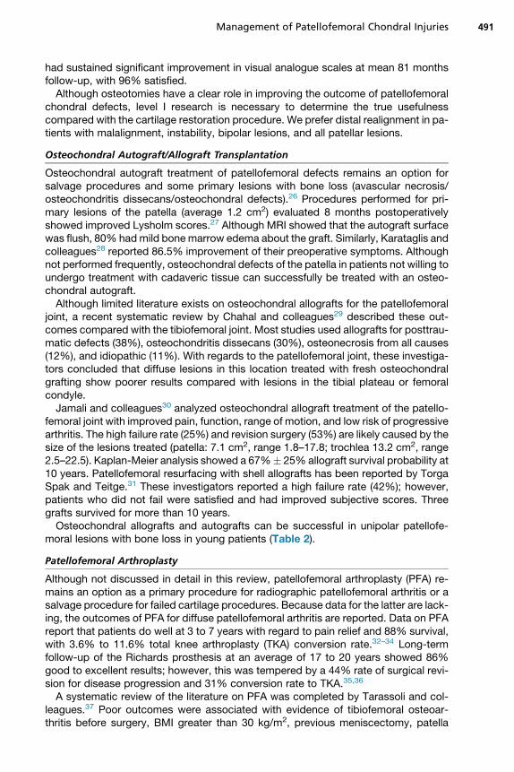
� Three incisions can be chosen based on the likelihood of an isolated cartilageprocedure, isolated osteotomy, or combined treatment (Fig. 2)- Incision is based centrally parallel to the tibial crest from the proximal pole of
the patella to the 5 to 7 cm distal to the tibial tuberosity
tions for tibial tubercle (TT) osteotomy
ptomatic patellar or bipolar lesion
ral or central patellar defect
ct anteriorization for isolated central/medial defect
llar instability with increased TT–trochlear groove (TG) distance
lla alta
d primary cartilage procedure with proper indication/technique

Box 4
Guidelines for specific cartilage procedures
Debridement
� Large flap component
� Not indicated for incidental lesions
� Lesions staged to undergo autologous chondrocyte implantation (ACI)
Microfracture
� Low demand
� Unipolar (trochlea>>patella)
� Lesion size less than 2 to 3 cm2
� Augmentation of other cartilage procedures
Osteochondral allograft/autograft
� High demand
� Lesion size smaller than 3 cm2 or lesions larger than 3 cm2 with bone loss
� Revision of failed cartilage procedure
Cell-based cartilage therapies (ACI [Carticel, Genzyme, Cambridge, MA], DeNovo NT [Zimmer,Warsaw, IN])
� Lesion size greater than 3 cm2 without bone loss
� Bipolar lesions are a relative contraindication
� Minimal bone marrow edema at time of treatment
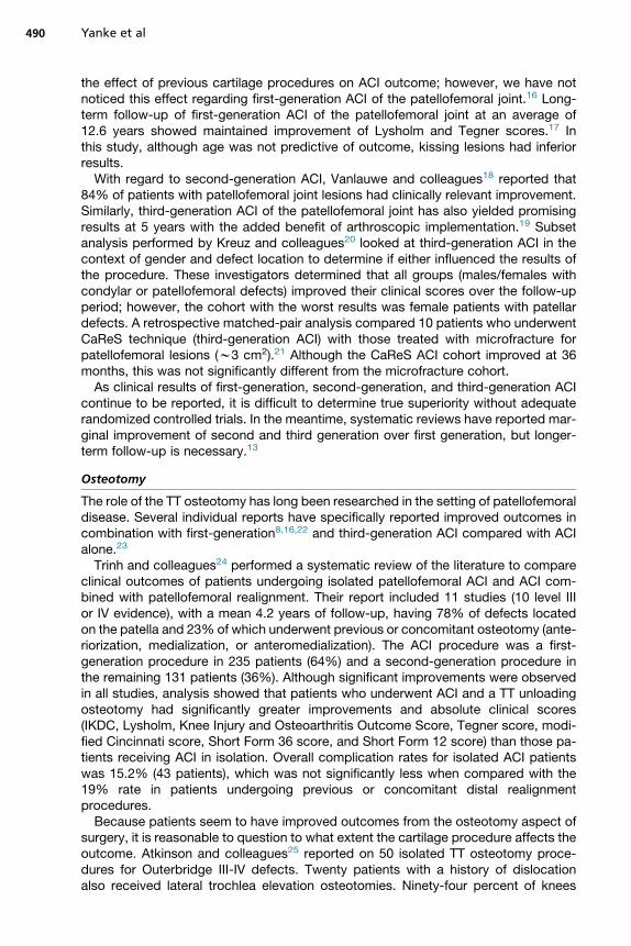
Fig. 1postebetw
Management of Patellofemoral Chondral Injuries 481
� We prefer a lateral versus medial arthrotomy- Increased ability to access the patellofemoral joint without entering the
quadriceps (vastus lateralis relatively more proximal than the vastusmedialis)
- The arthrotomy is closed only proximal to the superior pole of the patella toact as a lateral release
. The TT-TG distance as measured on MRI. Starting with a line perpendicular to therior condylar axis, a second line is drawn parallel to this through the TG. The distanceeen these 2 lines (yellow line) represents the TT-TG distance.
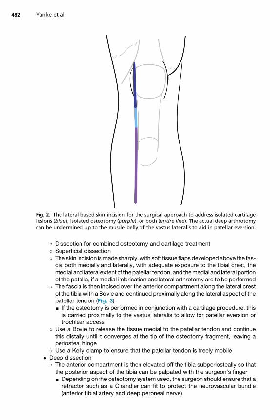
Fig. 2. The lateral-based skin incision for the surgical approach to address isolated cartilagelesions (blue), isolated osteotomy (purple), or both (entire line). The actual deep arthrotomycan be undermined up to the muscle belly of the vastus lateralis to aid in patellar eversion.
Yanke et al482
� Dissection for combined osteotomy and cartilage treatment� Superficial dissection� The skin incision ismade sharply, with soft tissue flaps developed above the fas-cia both medially and laterally, with adequate exposure to the tibial crest, themedial and lateral extent of thepatellar tendon, and themedial and lateral portionof the patella, if a medial imbrication and lateral arthrotomy are to be performed
� The fascia is then incised over the anterior compartment along the lateral crestof the tibia with a Bovie and continued proximally along the lateral aspect of thepatellar tendon (Fig. 3)- If the osteotomy is performed in conjunction with a cartilage procedure, this
is carried proximally to the vastus lateralis to allow for patellar eversion ortrochlear access
� Use a Bovie to release the tissue medial to the patellar tendon and continuethis distally until it converges at the tip of the osteotomy fragment, leaving aperiosteal hinge
� Use a Kelly clamp to ensure that the patellar tendon is freely mobile� Deep dissection
� The anterior compartment is then elevated off the tibia subperiosteally so thatthe posterior aspect of the tibia can be palpated with the surgeon’s finger
- Depending on the osteotomy system used, the surgeon should ensure that aretractor such as a Chandler can fit to protect the neurovascular bundle(anterior tibial artery and deep peroneal nerve)
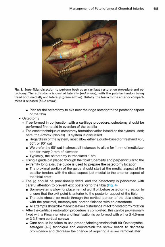
Fig. 3. Superficial dissection to perform both open cartilage restoration procedure and os-teotomy. The arthrotomy is created laterally (red arrow), with the patellar tendon beingfreed both medially and laterally (green arrows). Distally, the fascia to the anterior compart-ment is released (blue arrow).
Management of Patellofemoral Chondral Injuries 483
- Plan for the osteotomy to exit near the ridge anterior to the posterior aspectof the tibia
� Osteotomy� If performed in conjunction with a cartilage procedure, osteotomy should beperformed first to aid in eversion of the patella
� The exact technique of osteotomy formation varies based on the system used;here, the Arthrex (Naples) T3 system is discussed
- Regardless of the system, most allow either a guide-based or freehand 45�,60�, or 90� cut- We prefer the 60� cut in almost all instances to allow for 1 mm of medializa-
tion for every 2 mm of elevation- Typically, the osteotomy is translated 1 cm
� Using a guide pin placed through the tibial tuberosity and perpendicular to theextremity long axis, the guide is used to prepare the osteotomy location- The proximal portion of the guide should start at the medial aspect of the
patellar tendon, with the distal aspect just medial to the anterior aspect ofthe tibial crest
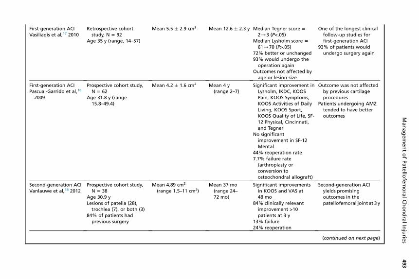
� The jig should be provisionally fixed, and the osteotomy is performed withcareful attention to prevent exit posterior to the tibia (Fig. 4)- Some systems allow for placement of a drill bit before osteotomy creation to
ensure that the exit point is anterior to the posterior aspect of the tibia- The cuts should be made through the cortical portion of the tibia distally,
with the proximal, metaphyseal portion finished with an osteotome- All attemptsshouldbemadeto leaveadistal hinge intact forosteotomy rotation
� After the cartilage restoration procedure is completed, this can be provisionallyfixed with a Kirschner wire and final fixation is performed with either 2 4.5-mmor 3 3.5-mm cortical screws- Care should be taken to use proper Arbeitsgemeinschaft fur Osteosynthe-
sefragen (AO) technique and countersink the screw heads to decreaseprominence and decrease the chance of requiring a screw removal later
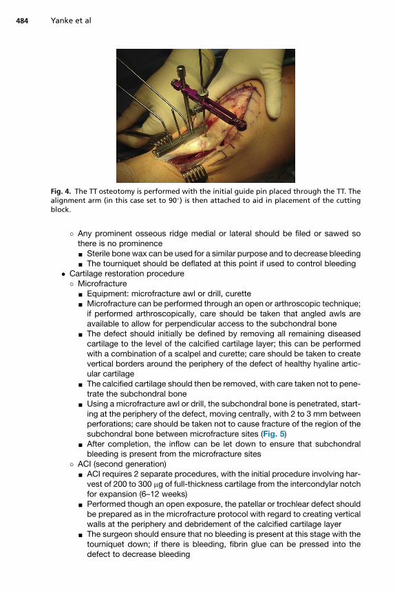
Fig. 4. The TT osteotomy is performed with the initial guide pin placed through the TT. Thealignment arm (in this case set to 90�) is then attached to aid in placement of the cuttingblock.
Yanke et al484
� Any prominent osseous ridge medial or lateral should be filed or sawed sothere is no prominence- Sterile bone wax can be used for a similar purpose and to decrease bleeding- The tourniquet should be deflated at this point if used to control bleeding
� Cartilage restoration procedure� Microfracture
- Equipment: microfracture awl or drill, curette- Microfracture can be performed through an open or arthroscopic technique;
if performed arthroscopically, care should be taken that angled awls areavailable to allow for perpendicular access to the subchondral bone
- The defect should initially be defined by removing all remaining diseasedcartilage to the level of the calcified cartilage layer; this can be performedwith a combination of a scalpel and curette; care should be taken to createvertical borders around the periphery of the defect of healthy hyaline artic-ular cartilage
- The calcified cartilage should then be removed, with care taken not to pene-trate the subchondral bone
- Using a microfracture awl or drill, the subchondral bone is penetrated, start-ing at the periphery of the defect, moving centrally, with 2 to 3 mm betweenperforations; care should be taken not to cause fracture of the region of thesubchondral bone between microfracture sites (Fig. 5)
- After completion, the inflow can be let down to ensure that subchondralbleeding is present from the microfracture sites
� ACI (second generation)- ACI requires 2 separate procedures, with the initial procedure involving har-
vest of 200 to 300 mg of full-thickness cartilage from the intercondylar notchfor expansion (6–12 weeks)
- Performed though an open exposure, the patellar or trochlear defect shouldbe prepared as in the microfracture protocol with regard to creating verticalwalls at the periphery and debridement of the calcified cartilage layer
- The surgeon should ensure that no bleeding is present at this stage with thetourniquet down; if there is bleeding, fibrin glue can be pressed into thedefect to decrease bleeding
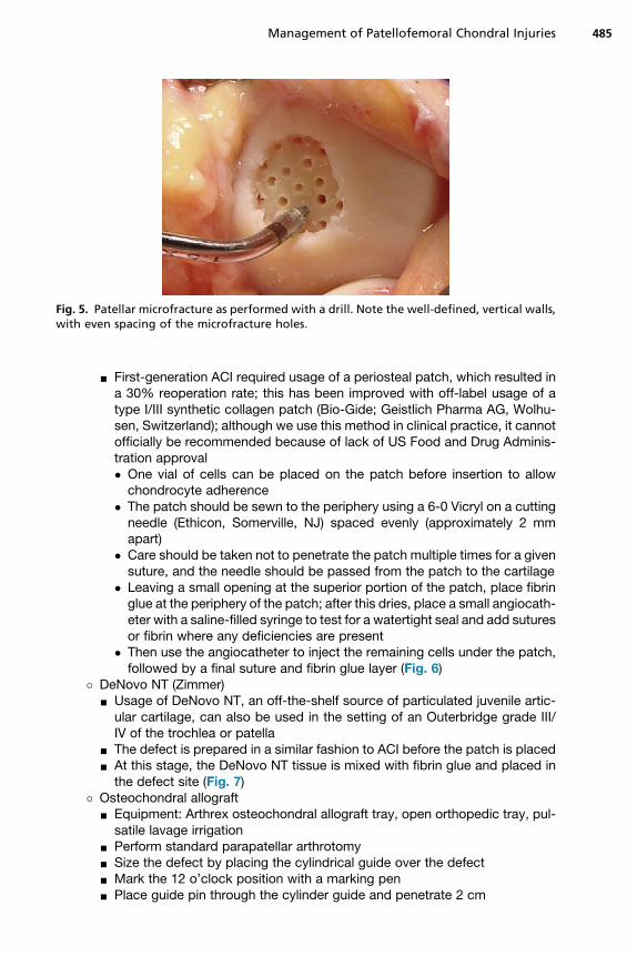
Fig. 5. Patellar microfracture as performed with a drill. Note the well-defined, vertical walls,with even spacing of the microfracture holes.
Management of Patellofemoral Chondral Injuries 485
- First-generation ACI required usage of a periosteal patch, which resulted ina 30% reoperation rate; this has been improved with off-label usage of atype I/III synthetic collagen patch (Bio-Gide; Geistlich Pharma AG, Wolhu-sen, Switzerland); although we use this method in clinical practice, it cannotofficially be recommended because of lack of US Food and Drug Adminis-tration approval� One vial of cells can be placed on the patch before insertion to allowchondrocyte adherence
� The patch should be sewn to the periphery using a 6-0 Vicryl on a cuttingneedle (Ethicon, Somerville, NJ) spaced evenly (approximately 2 mmapart)
� Care should be taken not to penetrate the patch multiple times for a givensuture, and the needle should be passed from the patch to the cartilage
� Leaving a small opening at the superior portion of the patch, place fibringlue at the periphery of the patch; after this dries, place a small angiocath-eter with a saline-filled syringe to test for a watertight seal and add suturesor fibrin where any deficiencies are present
� Then use the angiocatheter to inject the remaining cells under the patch,followed by a final suture and fibrin glue layer (Fig. 6)
� DeNovo NT (Zimmer)- Usage of DeNovo NT, an off-the-shelf source of particulated juvenile artic-
ular cartilage, can also be used in the setting of an Outerbridge grade III/IV of the trochlea or patella
- The defect is prepared in a similar fashion to ACI before the patch is placed- At this stage, the DeNovo NT tissue is mixed with fibrin glue and placed in
the defect site (Fig. 7)� Osteochondral allograft
- Equipment: Arthrex osteochondral allograft tray, open orthopedic tray, pul-satile lavage irrigation
- Perform standard parapatellar arthrotomy- Size the defect by placing the cylindrical guide over the defect- Mark the 12 o’clock position with a marking pen- Place guide pin through the cylinder guide and penetrate 2 cm
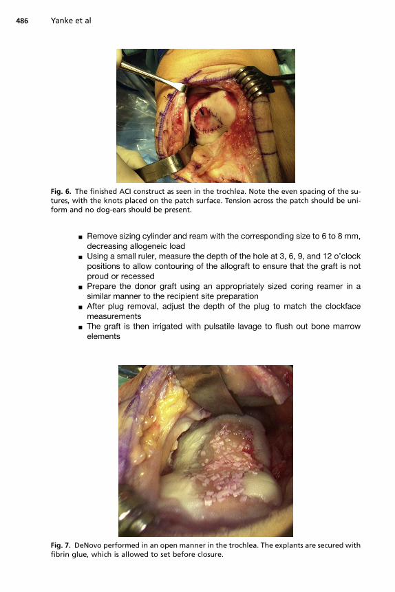
Fig. 7. Dfibrin g
Fig. 6. The finished ACI construct as seen in the trochlea. Note the even spacing of the su-tures, with the knots placed on the patch surface. Tension across the patch should be uni-form and no dog-ears should be present.
Yanke et al486
- Remove sizing cylinder and ream with the corresponding size to 6 to 8 mm,decreasing allogeneic load
- Using a small ruler, measure the depth of the hole at 3, 6, 9, and 12 o’clockpositions to allow contouring of the allograft to ensure that the graft is notproud or recessed
- Prepare the donor graft using an appropriately sized coring reamer in asimilar manner to the recipient site preparation
- After plug removal, adjust the depth of the plug to match the clockfacemeasurements
- The graft is then irrigated with pulsatile lavage to flush out bone marrowelements
eNovo performed in an open manner in the trochlea. The explants are secured withlue, which is allowed to set before closure.
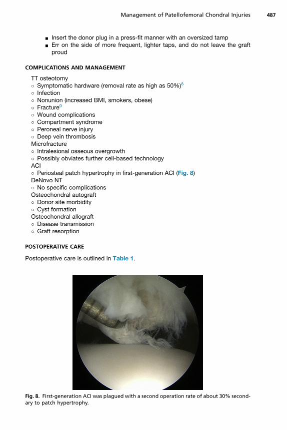
Fig. 8. Fary to p
Management of Patellofemoral Chondral Injuries 487
- Insert the donor plug in a press-fit manner with an oversized tamp- Err on the side of more frequent, lighter taps, and do not leave the graft
proud
COMPLICATIONS AND MANAGEMENT
TT osteotomy� Symptomatic hardware (removal rate as high as 50%)8
� Infection� Nonunion (increased BMI, smokers, obese)� Fracture9
� Wound complications� Compartment syndrome� Peroneal nerve injury� Deep vein thrombosisMicrofracture� Intralesional osseous overgrowth� Possibly obviates further cell-based technologyACI� Periosteal patch hypertrophy in first-generation ACI (Fig. 8)DeNovo NT� No specific complicationsOsteochondral autograft� Donor site morbidity� Cyst formationOsteochondral allograft� Disease transmission� Graft resorption
POSTOPERATIVE CARE
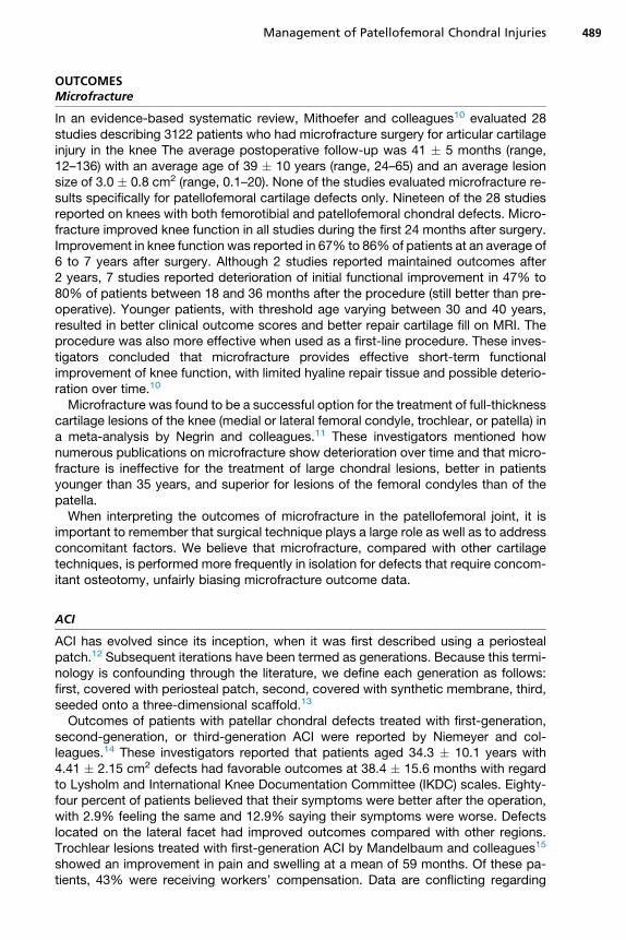
Postoperative care is outlined in Table 1.
irst-generation ACI was plagued with a second operation rate of about 30% second-atch hypertrophy.
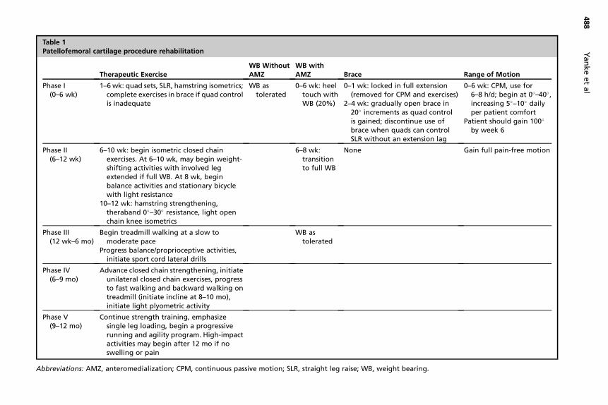
Table 1Patellofemoral cartilage procedure rehabilitation
Therapeutic ExerciseWB WithoutAMZ
WB withAMZ Brace Range of Motion
Phase I(0–6 wk)
1–6 wk: quad sets, SLR, hamstring isometrics;complete exercises in brace if quad controlis inadequate
WB astolerated
0–6 wk: heeltouch withWB (20%)
0–1 wk: locked in full extension(removed for CPM and exercises)
2–4 wk: gradually open brace in20� increments as quad controlis gained; discontinue use ofbrace when quads can controlSLR without an extension lag
0–6 wk: CPM, use for6–8 h/d; begin at 0�–40�,increasing 5�–10� dailyper patient comfort
Patient should gain 100�
by week 6
Phase II(6–12 wk)
6–10 wk: begin isometric closed chainexercises. At 6–10 wk, may begin weight-shifting activities with involved legextended if full WB. At 8 wk, beginbalance activities and stationary bicyclewith light resistance
10–12 wk: hamstring strengthening,theraband 0�–30� resistance, light openchain knee isometrics
6–8 wk:transitionto full WB
None Gain full pain-free motion
Phase III(12 wk–6 mo)
Begin treadmill walking at a slow tomoderate pace
Progress balance/proprioceptive activities,initiate sport cord lateral drills
WB astolerated
Phase IV(6–9 mo)
Advance closed chain strengthening, initiateunilateral closed chain exercises, progressto fast walking and backward walking ontreadmill (initiate incline at 8–10 mo),initiate light plyometric activity
Phase V(9–12 mo)
Continue strength training, emphasizesingle leg loading, begin a progressiverunning and agility program. High-impactactivities may begin after 12 mo if noswelling or pain
Abbreviations: AMZ, anteromedialization; CPM, continuous passive motion; SLR, straight leg raise; WB, weight bearing.
Yankeetal
488
Management of Patellofemoral Chondral Injuries 489
OUTCOMESMicrofracture
In an evidence-based systematic review, Mithoefer and colleagues10 evaluated 28studies describing 3122 patients who had microfracture surgery for articular cartilageinjury in the knee The average postoperative follow-up was 41 � 5 months (range,12–136) with an average age of 39 � 10 years (range, 24–65) and an average lesionsize of 3.0 � 0.8 cm2 (range, 0.1–20). None of the studies evaluated microfracture re-sults specifically for patellofemoral cartilage defects only. Nineteen of the 28 studiesreported on knees with both femorotibial and patellofemoral chondral defects. Micro-fracture improved knee function in all studies during the first 24 months after surgery.Improvement in knee function was reported in 67% to 86%of patients at an average of6 to 7 years after surgery. Although 2 studies reported maintained outcomes after2 years, 7 studies reported deterioration of initial functional improvement in 47% to80% of patients between 18 and 36 months after the procedure (still better than pre-operative). Younger patients, with threshold age varying between 30 and 40 years,resulted in better clinical outcome scores and better repair cartilage fill on MRI. Theprocedure was also more effective when used as a first-line procedure. These inves-tigators concluded that microfracture provides effective short-term functionalimprovement of knee function, with limited hyaline repair tissue and possible deterio-ration over time.10
Microfracture was found to be a successful option for the treatment of full-thicknesscartilage lesions of the knee (medial or lateral femoral condyle, trochlear, or patella) ina meta-analysis by Negrin and colleagues.11 These investigators mentioned hownumerous publications on microfracture show deterioration over time and that micro-fracture is ineffective for the treatment of large chondral lesions, better in patientsyounger than 35 years, and superior for lesions of the femoral condyles than of thepatella.When interpreting the outcomes of microfracture in the patellofemoral joint, it is
important to remember that surgical technique plays a large role as well as to addressconcomitant factors. We believe that microfracture, compared with other cartilagetechniques, is performed more frequently in isolation for defects that require concom-itant osteotomy, unfairly biasing microfracture outcome data.
ACI
ACI has evolved since its inception, when it was first described using a periostealpatch.12 Subsequent iterations have been termed as generations. Because this termi-nology is confounding through the literature, we define each generation as follows:first, covered with periosteal patch, second, covered with synthetic membrane, third,seeded onto a three-dimensional scaffold.13
Outcomes of patients with patellar chondral defects treated with first-generation,second-generation, or third-generation ACI were reported by Niemeyer and col-leagues.14 These investigators reported that patients aged 34.3 � 10.1 years with4.41 � 2.15 cm2 defects had favorable outcomes at 38.4 � 15.6 months with regardto Lysholm and International Knee Documentation Committee (IKDC) scales. Eighty-four percent of patients believed that their symptoms were better after the operation,with 2.9% feeling the same and 12.9% saying their symptoms were worse. Defectslocated on the lateral facet had improved outcomes compared with other regions.Trochlear lesions treated with first-generation ACI by Mandelbaum and colleagues15
showed an improvement in pain and swelling at a mean of 59 months. Of these pa-tients, 43% were receiving workers’ compensation. Data are conflicting regarding
Yanke et al490
the effect of previous cartilage procedures on ACI outcome; however, we have notnoticed this effect regarding first-generation ACI of the patellofemoral joint.16 Long-term follow-up of first-generation ACI of the patellofemoral joint at an average of12.6 years showed maintained improvement of Lysholm and Tegner scores.17 Inthis study, although age was not predictive of outcome, kissing lesions had inferiorresults.With regard to second-generation ACI, Vanlauwe and colleagues18 reported that
84% of patients with patellofemoral joint lesions had clinically relevant improvement.Similarly, third-generation ACI of the patellofemoral joint has also yielded promisingresults at 5 years with the added benefit of arthroscopic implementation.19 Subsetanalysis performed by Kreuz and colleagues20 looked at third-generation ACI in thecontext of gender and defect location to determine if either influenced the results ofthe procedure. These investigators determined that all groups (males/females withcondylar or patellofemoral defects) improved their clinical scores over the follow-upperiod; however, the cohort with the worst results was female patients with patellardefects. A retrospective matched-pair analysis compared 10 patients who underwentCaReS technique (third-generation ACI) with those treated with microfracture forpatellofemoral lesions (w3 cm2).21 Although the CaReS ACI cohort improved at 36months, this was not significantly different from the microfracture cohort.As clinical results of first-generation, second-generation, and third-generation ACI
continue to be reported, it is difficult to determine true superiority without adequaterandomized controlled trials. In the meantime, systematic reviews have reported mar-ginal improvement of second and third generation over first generation, but longer-term follow-up is necessary.13
Osteotomy
The role of the TT osteotomy has long been researched in the setting of patellofemoraldisease. Several individual reports have specifically reported improved outcomes incombination with first-generation8,16,22 and third-generation ACI compared with ACIalone.23
Trinh and colleagues24 performed a systematic review of the literature to compareclinical outcomes of patients undergoing isolated patellofemoral ACI and ACI com-bined with patellofemoral realignment. Their report included 11 studies (10 level IIIor IV evidence), with a mean 4.2 years of follow-up, having 78% of defects locatedon the patella and 23% of which underwent previous or concomitant osteotomy (ante-riorization, medialization, or anteromedialization). The ACI procedure was a first-generation procedure in 235 patients (64%) and a second-generation procedure inthe remaining 131 patients (36%). Although significant improvements were observedin all studies, analysis showed that patients who underwent ACI and a TT unloadingosteotomy had significantly greater improvements and absolute clinical scores(IKDC, Lysholm, Knee Injury and Osteoarthritis Outcome Score, Tegner score, modi-fied Cincinnati score, Short Form 36 score, and Short Form 12 score) than those pa-tients receiving ACI in isolation. Overall complication rates for isolated ACI patientswas 15.2% (43 patients), which was not significantly less when compared with the19% rate in patients undergoing previous or concomitant distal realignmentprocedures.Because patients seem to have improved outcomes from the osteotomy aspect of
surgery, it is reasonable to question to what extent the cartilage procedure affects theoutcome. Atkinson and colleagues25 reported on 50 isolated TT osteotomy proce-dures for Outerbridge III-IV defects. Twenty patients with a history of dislocationalso received lateral trochlea elevation osteotomies. Ninety-four percent of knees
Management of Patellofemoral Chondral Injuries 491
had sustained significant improvement in visual analogue scales at mean 81 monthsfollow-up, with 96% satisfied.Although osteotomies have a clear role in improving the outcome of patellofemoral
chondral defects, level I research is necessary to determine the true usefulnesscompared with the cartilage restoration procedure. We prefer distal realignment in pa-tients with malalignment, instability, bipolar lesions, and all patellar lesions.
Osteochondral Autograft/Allograft Transplantation
Osteochondral autograft treatment of patellofemoral defects remains an option forsalvage procedures and some primary lesions with bone loss (avascular necrosis/osteochondritis dissecans/osteochondral defects).26 Procedures performed for pri-mary lesions of the patella (average 1.2 cm2) evaluated 8 months postoperativelyshowed improved Lysholm scores.27 Although MRI showed that the autograft surfacewas flush, 80% hadmild bonemarrow edema about the graft. Similarly, Karataglis andcolleagues28 reported 86.5% improvement of their preoperative symptoms. Althoughnot performed frequently, osteochondral defects of the patella in patients not willing toundergo treatment with cadaveric tissue can successfully be treated with an osteo-chondral autograft.Although limited literature exists on osteochondral allografts for the patellofemoral
joint, a recent systematic review by Chahal and colleagues29 described these out-comes compared with the tibiofemoral joint. Most studies used allografts for posttrau-matic defects (38%), osteochondritis dissecans (30%), osteonecrosis from all causes(12%), and idiopathic (11%). With regards to the patellofemoral joint, these investiga-tors concluded that diffuse lesions in this location treated with fresh osteochondralgrafting show poorer results compared with lesions in the tibial plateau or femoralcondyle.Jamali and colleagues30 analyzed osteochondral allograft treatment of the patello-
femoral joint with improved pain, function, range of motion, and low risk of progressivearthritis. The high failure rate (25%) and revision surgery (53%) are likely caused by thesize of the lesions treated (patella: 7.1 cm2, range 1.8–17.8; trochlea 13.2 cm2, range2.5–22.5). Kaplan-Meier analysis showed a 67%� 25% allograft survival probability at10 years. Patellofemoral resurfacing with shell allografts has been reported by TorgaSpak and Teitge.31 These investigators reported a high failure rate (42%); however,patients who did not fail were satisfied and had improved subjective scores. Threegrafts survived for more than 10 years.Osteochondral allografts and autografts can be successful in unipolar patellofe-
moral lesions with bone loss in young patients (Table 2).
Patellofemoral Arthroplasty
Although not discussed in detail in this review, patellofemoral arthroplasty (PFA) re-mains an option as a primary procedure for radiographic patellofemoral arthritis or asalvage procedure for failed cartilage procedures. Because data for the latter are lack-ing, the outcomes of PFA for diffuse patellofemoral arthritis are reported. Data on PFAreport that patients do well at 3 to 7 years with regard to pain relief and 88% survival,with 3.6% to 11.6% total knee arthroplasty (TKA) conversion rate.32–34 Long-termfollow-up of the Richards prosthesis at an average of 17 to 20 years showed 86%good to excellent results; however, this was tempered by a 44% rate of surgical revi-sion for disease progression and 31% conversion rate to TKA.35,36
A systematic review of the literature on PFA was completed by Tarassoli and col-leagues.37 Poor outcomes were associated with evidence of tibiofemoral osteoar-thritis before surgery, BMI greater than 30 kg/m2, previous meniscectomy, patella
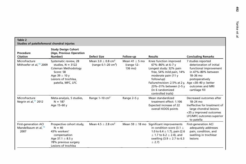
Table 2Studies of patellofemoral chondral injuries
ProcedureCitation
Study Design Cohort(Age, Previous OperationNumber) Defect Size Follow-up Results Concluding Remarks
MicrofractureMithoefer et al,10 2009
Systematic review, 28studies, N 5 3122
Coleman MethodologyScore: 58
Age 39 � 10 yLesions of trochlea,
patella, MFC, LFC
Mean 3.0 � 0.8 cm2
(range 0.1–20 cm2)Mean 41 � 5 mo
(range 12–136 mo)
Knee function improved67%–86% at 6–7 y
Longest study: 32% painfree, 54% mild pain, 14%moderate pain (11 yfollow/up)
Failure/revision: 2.5% at 2 y,23%–31% between 2–5 y(in 6 randomizedcontrolled trails)
7 studies reporteddeterioration of initialfunctional improvementin 47%–80% between18–36 mopostoperatively
Age <30–40 y: betteroutcomes and MRIcartilage fill
MicrofractureNegrin et al,11 2012
Meta-analysis, 5 studies,N 5 187
Age 15–60 y
Range 1–10 cm2 Range 2–5 y Mean standardizedtreatment effect: 1.106
Expected increase of 22overall KOOS points
Decreased outcomes after18–24 mo
Ineffective for treatment oflarge chondral lesions
<35 y improved outcomesLFC/MFC outcomes superior
to patella
First-generation ACIMandelbaum et al,15
2007
Prospective cohort study,N 5 40
43% workers’compensation
Age 37.1 � 8.5 y78% previous surgeryLesions of trochlea
Mean 4.5 � 2.8 cm2 Mean 59 � 18 mo Significant improvementsin condition score (3.1 �1.0 to 6.4 � 1.7), pain (2.6� 1.7 to 6.2 � 2.4), andswelling (3.9 � 2.7 to 6.3� 2.7)
First-generation ACIadequately addressespain, condition, andswelling in trochlearlesions
Yankeetal
492
First-generation ACIVasiliadis et al,17 2010
Retrospective cohortstudy, N 5 92
Age 35 y (range, 14–57)
Mean 5.5 � 2.9 cm2 Mean 12.6 � 2.3 y Median Tegner score 5
2/3 (P<.05)Median Lysholm score 5
61/70 (P>.05)72% better or unchanged93% would undergo theoperation again
Outcomes not affected byage or lesion size
One of the longest clinicalfollow-up studies forfirst-generation ACI
93% of patients wouldundergo surgery again
First-generation ACIPascual-Garrido et al,16
2009
Prospective cohort study,N 5 62
Age 31.8 y (range15.8–49.4)
Mean 4.2 � 1.6 cm2 Mean 4 y(range 2–7)
Significant improvement inLysholm, IKDC, KOOSPain, KOOS Symptoms,KOOS Activities of DailyLiving, KOOS Sport,KOOS Quality of Life, SF-12 Physical, Cincinnati,and Tegner
No significantimprovement in SF-12Mental
44% reoperation rate7.7% failure rate(arthroplasty orconversion toosteochondral allograft)
Outcome was not affectedby previous cartilageprocedures
Patients undergoing AMZtended to have betteroutcomes
Second-generation ACIVanlauwe et al,18 2012
Prospective cohort study,N 5 38
Age 30.9 yLesions of patella (28),
trochlea (7), or both (3)84% of patients had
previous surgery
Mean 4.89 cm2
(range 1.5–11 cm2)Mean 37 mo
(range 24–72 mo)
Significant improvementsin KOOS and VAS at48 mo
84% clinically relevantimprovement >10patients at 3 y
13% failure24% reoperation
Second-generation ACIyields promisingoutcomes in thepatellofemoral joint at 3 y
(continued on next page)
ManagementofPatello
femoralChondralInjurie
s493
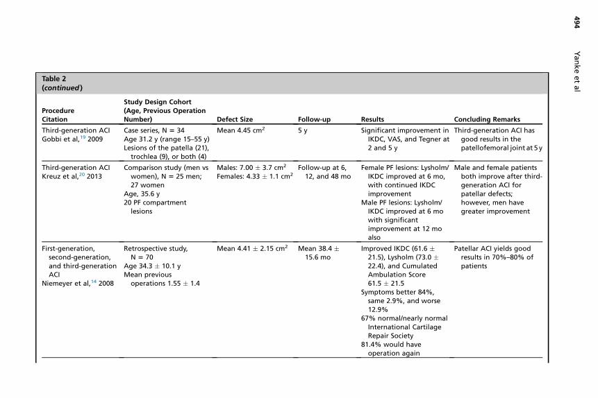
Table 2(continued )
ProcedureCitation
Study Design Cohort(Age, Previous OperationNumber) Defect Size Follow-up Results Concluding Remarks
Third-generation ACIGobbi et al,19 2009
Case series, N 5 34Age 31.2 y (range 15–55 y)Lesions of the patella (21),
trochlea (9), or both (4)
Mean 4.45 cm2 5 y Significant improvement inIKDC, VAS, and Tegner at2 and 5 y
Third-generation ACI hasgood results in thepatellofemoral joint at 5 y
Third-generation ACIKreuz et al,20 2013
Comparison study (men vswomen), N 5 25 men;27 women
Age, 35.6 y20 PF compartment
lesions
Males: 7.00 � 3.7 cm2
Females: 4.33 � 1.1 cm2
Follow-up at 6,12, and 48 mo
Female PF lesions: Lysholm/IKDC improved at 6 mo,with continued IKDCimprovement
Male PF lesions: Lysholm/IKDC improved at 6 mowith significantimprovement at 12 moalso
Male and female patientsboth improve after third-generation ACI forpatellar defects;however, men havegreater improvement
First-generation,second-generation,and third-generationACI
Niemeyer et al,14 2008
Retrospective study,N 5 70
Age 34.3 � 10.1 yMean previous
operations 1.55 � 1.4
Mean 4.41 � 2.15 cm2 Mean 38.4 �15.6 mo
Improved IKDC (61.6 �21.5), Lysholm (73.0 �22.4), and CumulatedAmbulation Score61.5 � 21.5
Symptoms better 84%,same 2.9%, and worse12.9%
67% normal/nearly normalInternational CartilageRepair Society
81.4% would haveoperation again
Patellar ACI yields goodresults in 70%–80% ofpatients
Yankeetal
494
First-generation ACI �AMZ (73.7%concomitant)
Farr,8 2007
Prospective study, N 5 39(38 knees)
Age 31.2 � 11.3 yPatella and trochlea
Trochlea: 4.3 � 1.9 cm2
(46% of cohort)Patella: 5.4 � 1.9 cm2
(36% of cohort)Bipolar: 8.8 � 3.5 cm2
(18% of cohort)
Mean 1.2 y Modified Cincinnati OverallCondition score: median3-point improvement
Lysholm score: median31-point improvement
VAS score resting: median2-point improvement
VAS score maximum:3-point improvement
25 patients had 32subsequent surgeries
3 patients failed ACI
Overall condition improvedregardless of concurrentAMZ or presence of >1lesion
First-generationACI � AMZ
Henderson & Lavigne,22
2006
Comparison study, N 5 22per group, lesions ofpatella
N/A Mean 2 y Osteotomy with greaterincrease in meanmodified Cincinnati KneeScore (4.5 vs 1.7 points),better function (1.7 vs2.5), better SF-36 physicalcomponent scores (70.9vs 55.4 points), higherIKDC scores (85.2 vs 60.6points)
Patellar ACI withosteotomy has betteroutcomes than ACI alone,possibly in patient withnormal PF biomechanics
Third-generation ACI 1distal realignment
Gigante et al,23 2009
Prospective cohort study,N 5 14 knees (12patients)
Age 31 y
Median 4 cm2
(range 3–9 cm2)Mean 3 y Improved Modified
Cincinnati and medianLysholm, Tegner, andKujala Score
13/14 patients satisfied50% excellent, 43% good,7% poor final outcomes
93% of patients with third-generation ACI andosteotomy have good/excellent results
(continued on next page)
ManagementofPatello
femoralChondralInjurie
s495
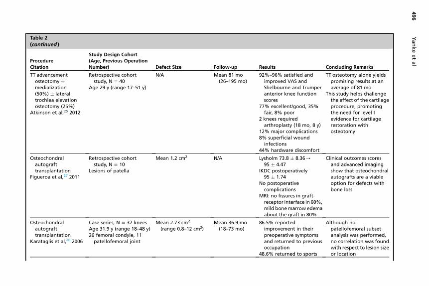
Table 2(continued )
ProcedureCitation
Study Design Cohort(Age, Previous OperationNumber) Defect Size Follow-up Results Concluding Remarks
TT advancementosteotomy �medialization(50%) � lateraltrochlea elevationosteotomy (25%)
Atkinson et al,25 2012
Retrospective cohortstudy, N 5 40
Age 29 y (range 17–51 y)
N/A Mean 81 mo(26–195 mo)
92%–96% satisfied andimproved VAS andShelbourne and Trumperanterior knee functionscores
77% excellent/good, 35%fair, 8% poor
2 knees requiredarthroplasty (18 mo, 8 y)
12% major complications8% superficial woundinfections
44% hardware discomfort
TT osteotomy alone yieldspromising results at anaverage of 81 mo
This study helps challengethe effect of the cartilageprocedure, promotingthe need for level Ievidence for cartilagerestoration withosteotomy
Osteochondralautografttransplantation
Figueroa et al,27 2011
Retrospective cohortstudy, N 5 10
Lesions of patella
Mean 1.2 cm2 N/A Lysholm 73.8 � 8.36/95 � 4.47
IKDC postoperatively95 � 1.74
No postoperativecomplications
MRI: no fissures in graft-receptor interface in 60%,mild bonemarrow edemaabout the graft in 80%
Clinical outcomes scoresand advanced imagingshow that osteochondralautografts are a viableoption for defects withbone loss
Osteochondralautografttransplantation
Karataglis et al,28 2006
Case series, N 5 37 kneesAge 31.9 y (range 18–48 y)26 femoral condyle, 11
patellofemoral joint
Mean 2.73 cm2
(range 0.8–12 cm2)Mean 36.9 mo
(18–73 mo)86.5% reportedimprovement in theirpreoperative symptomsand returned to previousoccupation
48.6% returned to sports
Although nopatellofemoral subsetanalysis was performed,no correlation was foundwith respect to lesion sizeor location
Yankeetal
496
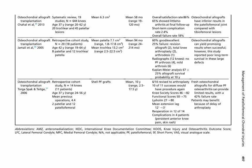
Osteochondral allografttransplantation
Chahal et al,29 2013
Systematic review, 19studies; N 5 644 knees
Age 37 y (range 20–62 y)20 trochlear and 45 patellar
lesions
Mean 6.3 cm2 Mean 58 mo(range 19–120 mo)
Overall satisfactionrate86%65% showed little/noarthritis at final follow-up
Short-term complicationrate 2.4%
Overall failure rate 18%
Osteochondral allograftshave inferior results inthe patellofemoral jointcompared withtibiofemoral lesions
Osteochondral allografttransplantation
Jamali et al,30 2005
Retrospective cohort study,N 5 20 (18 patients)
Age 42 y (range 19–64 y)8 patellar and 12 trochlea/
patella
Mean patella 7.1 cm2
(range, 1.8–17.8 cm2)Mean trochlea 13.2 cm2
(range 2.5–22.5 cm2)
Mean 94 mo(range 24–214 mo)
60% good/excellent25% failure: revisionallograft (2), total kneearthroplasty (2),arthrodesis (1)
Radiographs (12 knees): noPF arthrosis (4), mildarthrosis (6)
Kaplan-Meier analysis 67 �25% allograft survivalprobability at 10 y
Osteochondral allograftscan yield promisingresults when successful;however, this studyreported poor long-termsurvival in these largedefects
Osteochondral allografttransplantation
Torga Spak & Teitge,31
2006
Retrospective cohortstudy, N 5 14 knees(11 patients)
Age 37 y (range 24–56 y)Mean previous
operations, 4.42 patellar and 12
patellofemoral
Shell PF grafts Mean, 10 y(range, 2.5–17.5 y)
6/14 revised to arthroplasty10 of 11 successes wouldhave procedure again
Knee Society Scores 46/82Functional Scores 50/75Lysholm 27/80Mean extension lag12�/3�
Reoperation in 12 of 14Complications in 4 patients(persistent anterior kneepain, skin rash)
Fresh osteochondralallografts for diffuse PFosteoarthritis can providelimited results, with a42% failure rate
Patients may benefitbecause of delay ofarthroplasty
Abbreviations: AMZ, anteromedialization; IKDC, International Knee Documentation Committee; KOOS, Knee Injury and Osteoarthritis Outcome Score;LFC, Lateral Femoral Condyle; MFC, Medial Femoral Condyle; N/A, not applicable; PF, patellofemoral; SF, Short Form; VAS, visual analogue scale.
ManagementofPatello
femoralChondralInjurie
s497
Yanke et al498
alta or baja, and ligamentous instability. The most common reason that they foundcited for failure necessitating revision was progression of tibiofemoral osteoarthritis.However, it was concluded that PFA is a less invasive operation, with more rapid post-operative recovery and preservation of bone stock to allow for conversion to TKA at alater date.
SUMMARY
Treatment of patellofemoral chondral defects is fraught with difficulty because of thegenerally inferior outcomes and significant biomechanical complexity of the joint.Noyes and Barber-Westin38 performed a systematic review of large (>4 cm2) patello-femoral ACI (11 studies), PFA (5 studies), and osteochondral allografting (2 studies) inpatients younger than 50 years. Respectively, failures or poor outcomes were noted in8% to 60%after ACI, 22% after PFA, and 53% after osteochondral allograft treatment.As noted in the outcome reviews earlier, unacceptable complication and reoperationrates were reported from all 3 procedures, and it was concluded that each operationhad unpredictable results for this patient demographic. This study highlights theimportance of strict indications and working to address all concomitant diseases todecrease revision rate. Outcomes are most predictable in young patients with lowBMI and unipolar defects lower than 4 cm2.
REFERENCES
1. Flanigan DC, Harris JD, Trinh TQ, et al. Prevalence of chondral defects in athletes’knees: a systematic review. Med Sci Sports Exerc 2010;42:1795–801.
2. Widuchowski W, Widuchowski J, Trzaska T. Articular cartilage defects: study of25,124 knee arthroscopies. Knee 2007;14:177–82.
3. Wang Y, Ding C, Wluka AE, et al. Factors affecting progression of knee cartilagedefects in normal subjects over 2 years. Rheumatology (Oxford) 2006;45:79–84.
4. Ding C, Cicuttini F, Scott F, et al. Natural history of knee cartilage defects and fac-tors affecting change. Arch Intern Med 2006;166:651–8.
5. Mihalko WM, Boachie-Adjei Y, Spang JT, et al. Controversies and techniques inthe surgical management of patellofemoral arthritis. Instr Course Lect 2008;57:365–80.
6. Cole BJ, Gomoll AH, Minas T, et al. Treatment of chondral defects in the patello-femoral joint. J Knee Surg 2006;19:285–95.
7. Dejour H, Walch G, Nove-Josserand L, et al. Factors of patellar instability: ananatomic radiographic study. Knee Surg Sports Traumatol Arthrosc 1994;2:19–26.
8. Farr J. Autologous chondrocyte implantation improves patellofemoral cartilagetreatment outcomes. Clin Orthop Relat Res 2007;463:187–94.
9. Pidoriano AJ, Weinstein RN, Buuck DA, et al. Correlation of patellar articular le-sions with results from anteromedial tibial tubercle transfer. Am J Sports Med1997;25:533–7.
10. Mithoefer K, McAdams T, Williams RJ, et al. Clinical efficacy of the microfracturetechnique for articular cartilage repair in the knee: an evidence-based systematicanalysis. Am J Sports Med 2009;37:2053–63.
11. Negrin L, Kutscha-Lissberg F, Gartlehner G, et al. Clinical outcome after micro-fracture of the knee: a meta-analysis of before/after-data of controlled studies.Int Orthop 2012;36:43–50.
12. Brittberg M, Lindahl A, Nilsson A, et al. Treatment of deep cartilage defects in theknee with autologous chondrocyte transplantation. N Engl J Med 1994;331:889–95.
Management of Patellofemoral Chondral Injuries 499
13. Goyal D, Goyal A, Keyhani S, et al. Evidence-based status of second- and third-generation autologous chondrocyte implantation over first generation: a system-atic review of level I and II studies. Arthroscopy 2013;29:1872–8.
14. Niemeyer P, Steinwachs M, Erggelet C, et al. Autologous chondrocyteimplantation for the treatment of retropatellar cartilage defects: clinical re-sults referred to defect localisation. Arch Orthop Trauma Surg 2008;128:1223–31.
15. Mandelbaum BR, Browne J, Fu F, et al. Treatment outcomes of autologous chon-drocyte implantation for full-thickness articular cartilage defects of the trochlea.Am J Sports Med 2007;35:915–21.
16. Pascual-Garrido C, Slabaugh MA, L’Heureux DR, et al. Recommendations andtreatment outcomes for patellofemoral articular cartilage defects with autologouschondrocyte implantation: prospective evaluation at average 4-year follow-up.Am J Sports Med 2009;37(Suppl 1):33S–41S.
17. Vasiliadis HS, Wasiak J, Salanti G. Autologous chondrocyte implantation for thetreatment of cartilage lesions of the knee: a systematic review of randomizedstudies. Knee Surg Sports Traumatol Arthrosc 2010;18:1645–55.
18. Vanlauwe JJ, Claes T, Van Assche D, et al. Characterized chondrocyte implanta-tion in the patellofemoral joint: an up to 4-year follow-up of a prospective cohort of38 patients. Am J Sports Med 2012;40:1799–807.
19. Gobbi A, Kon E, Berruto M, et al. Patellofemoral full-thickness chondral defectstreated with second-generation autologous chondrocyte implantation: results at5 years’ follow-up. Am J Sports Med 2009;37:1083–92.
20. Kreuz PC, Niemeyer P, Muller S, et al. Influence of sex on the outcome of autol-ogous chondrocyte implantation in chondral defects of the knee. Am J SportsMed 2013;41:1541–8.
21. Petri M, Broese M, Simon A, et al. CaReS (MACT) versus microfracture in treatingsymptomatic patellofemoral cartilage defects: a retrospective matched-pair anal-ysis. J Orthop Sci 2013;18:38–44.
22. Henderson IJ, Lavigne P. Periosteal autologous chondrocyte implantation forpatellar chondral defect in patients with normal and abnormal patellar tracking.Knee 2006;13:274–9.
23. Gigante A, Enea D, Greco F, et al. Distal realignment and patellar autologouschondrocyte implantation: mid-term results in a selected population. Knee SurgSports Traumatol Arthrosc 2009;17:2–10.
24. Trinh TQ, Harris JD, Siston RA, et al. Improved outcomes with combined autolo-gous chondrocyte implantation and patellofemoral osteotomy versus isolatedautologous chondrocyte implantation. Arthroscopy 2013;29:566–74.
25. Atkinson HD, Bailey CA, Anand S, et al. Tibial tubercle advancement osteotomywith bone allograft for patellofemoral arthritis: a retrospective cohort study of 50knees. Arch Orthop Trauma Surg 2012;132:437–45.
26. Lu AP, Hame SL. Autologous osteochondral transplantation for simple cyst in thepatella. Arthroscopy 2005;21:1008.
27. Figueroa D, Melean P, Calvo R, et al. Osteochondral autografts in full thicknesspatella cartilage lesions. Knee 2011;18:220–3.
28. Karataglis D, Green MA, Learmonth DJ. Autologous osteochondral transplanta-tion for the treatment of chondral defects of the knee. Knee 2006;13:32–5.
29. Chahal J, Cole BJ, Gross AE, et al. Outcomes of osteochondral allograft trans-plantation in the knee. Arthroscopy 2013;29:575–88.
30. Jamali AA, Emmerson BC, Chung C, et al. Fresh osteochondral allografts: resultsin the patellofemoral joint. Clin Orthop Relat Res 2005;(437):176–85.
Yanke et al500
31. Torga Spak R, Teitge RA. Fresh osteochondral allografts for patellofemoralarthritis: long-term followup. Clin Orthop Relat Res 2006;444:193–200.
32. Ackroyd CE, Chir B. Development and early results of a new patellofemoral ar-throplasty. Clin Orthop Relat Res 2005;(436):7–13.
33. Leadbetter WB, Kolisek FR, Levitt RL, et al. Patellofemoral arthroplasty: a multi-centre study with minimum 2-year follow-up. Int Orthop 2009;33(6):1597–601.http://dx.doi.org/10.1007/s00264-008-0692-y.
34. Mont MA, Johnson AJ, Naziri Q, et al. Patellofemoral Arthroplasty 7-year MeanFollow-Up. J Arthroplasty 2011. http://dx.doi.org/10.1016/j.arth.2011.07.010.
35. Kooijman HJ, Driessen APPM, van Horn JR. Long-term results of patellofemoralarthroplasty. A report of 56 arthroplasties with 17 years of follow-up. J Bone JointSurg Br 2003;85(6):836–40.
36. van Jonbergen H-PW, Poolman RW, van Kampen A. Isolated patellofemoral osteo-arthritis. Acta Orthop 2010;81(2):199–205. http://dx.doi.org/10.3109/17453671003628756.
37. Tarassoli P, Punwar S, Khan W, et al. Patellofemoral arthroplasty: a systematic re-view of the literature. Open Orthop J 2012;6(1):340–7. http://dx.doi.org/10.2174/1874325001206010340.
38. Noyes FR, Barber-Westin SD. Advanced patellofemoral cartilage lesions in pa-tients younger than 50 years of age: is there an ideal operative option? Arthros-copy 2013;29:1423–36.