treating patellofemoral chondral lesions? personal...
TRANSCRIPT
Treating patellofemoral chondral lesions? Personal experience
Fernando Fonseca
http://rihuc.huc.min-saude.pt/handle/10400.4/1753
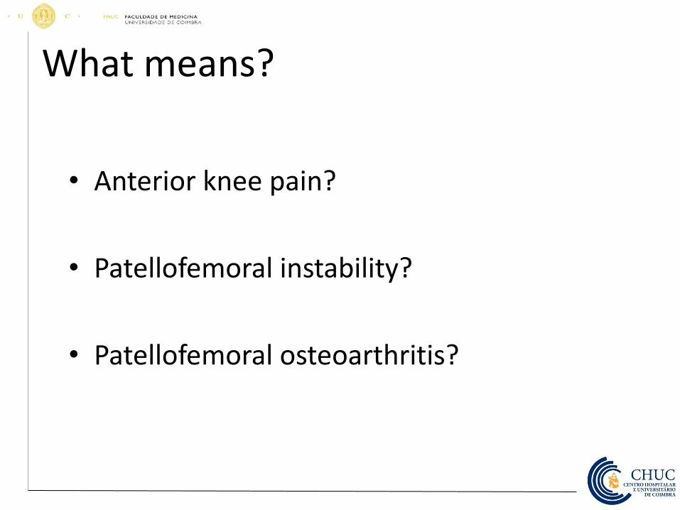
What means?
• Anterior knee pain?
• Patellofemoral instability?
• Patellofemoral osteoarthritis?
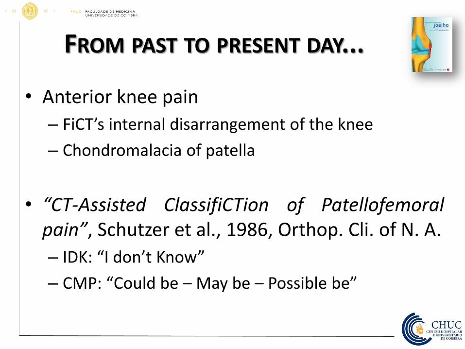
FROM PAST TO PRESENT DAY...
• Anterior knee pain
– FiCT’s internal disarrangement of the knee
– Chondromalacia of patella
• “CT-Assisted ClassifiCTion of Patellofemoral pain”, Schutzer et al., 1986, Orthop. Cli. of N. A.
– IDK: “I don’t Know”
– CMP: “Could be – May be – Possible be”
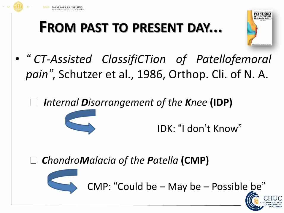
• “ CT-Assisted ClassifiCTion of Patellofemoral pain”, Schutzer et al., 1986, Orthop. Cli. of N. A.
Internal Disarrangement of the Knee (IDP)
IDK: “I don’t Know”
ChondroMalacia of the Patella (CMP)
CMP: “Could be – May be – Possible be”
FROM PAST TO PRESENT DAY...

Classification and aetiology
1) Painfull patellar syndrome
2) Potential patellar instability
3)Objective patellar instability
(CT; n=143 knees)
Trochlear dysplasia (85%)
Quadriceps Femoralis dysplasia (83%) – patella tilting> 20%
Patella alta – ICD > 1.2 (24%)
TT-TG > 20 mm (56%)

PATELLOFEMORAL BIOMECHANIC

PATELLOFEMORAL BIOMECHANIC
PatellaTracking
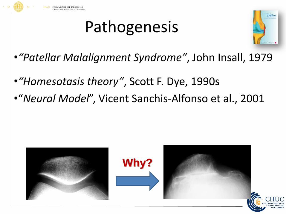
Pathogenesis
•“Patellar Malalignment Syndrome”, John Insall, 1979
•“Homesotasis theory”, Scott F. Dye, 1990s
•“Neural Model”, Vicent Sanchis-Alfonso et al., 2001
Why?
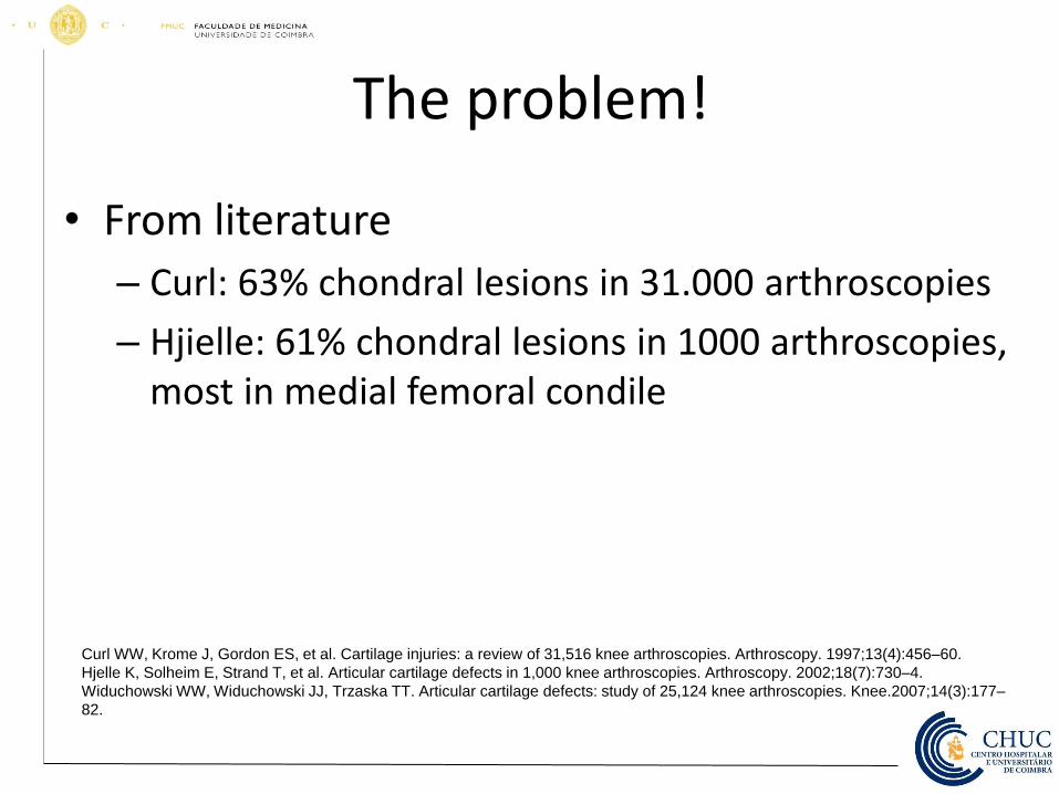
The problem!
• From literature
– Curl: 63% chondral lesions in 31.000 arthroscopies
– Hjielle: 61% chondral lesions in 1000 arthroscopies, most in medial femoral condile
Curl WW, Krome J, Gordon ES, et al. Cartilage injuries: a review of 31,516 knee arthroscopies. Arthroscopy. 1997;13(4):456–60.
Hjelle K, Solheim E, Strand T, et al. Articular cartilage defects in 1,000 knee arthroscopies. Arthroscopy. 2002;18(7):730–4.
Widuchowski WW, Widuchowski JJ, Trzaska TT. Articular cartilage defects: study of 25,124 knee arthroscopies. Knee.2007;14(3):177–
82.
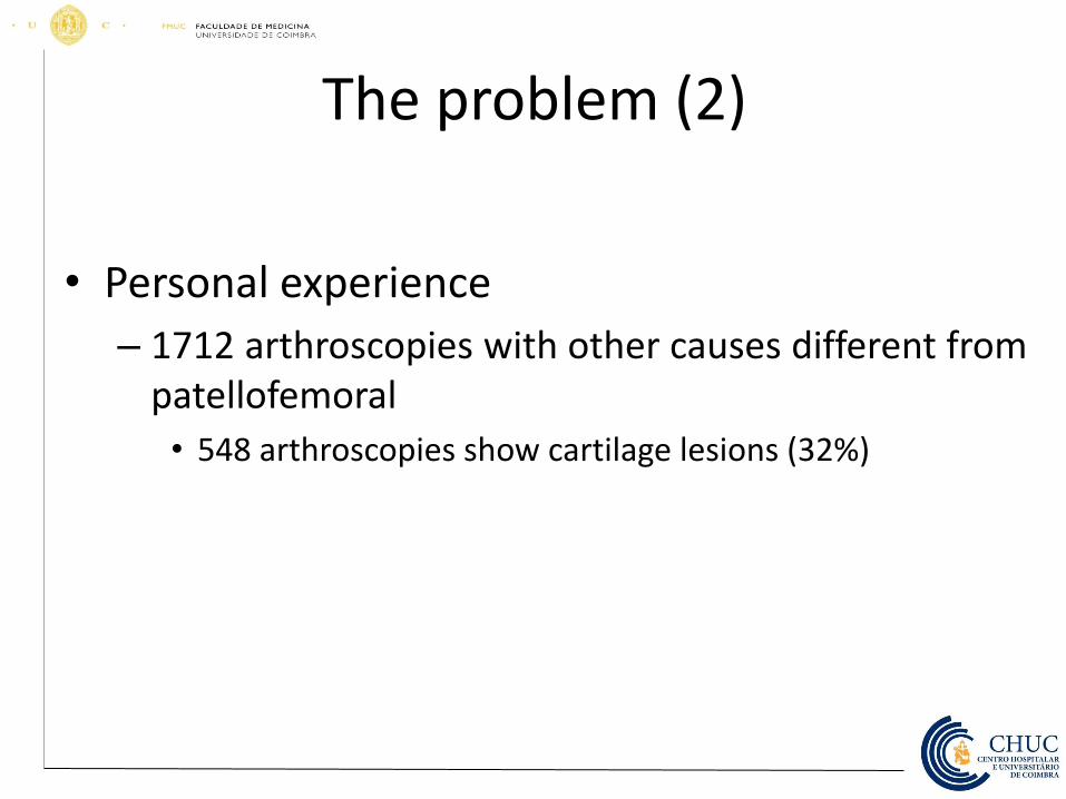
The problem (2)
• Personal experience
– 1712 arthroscopies with other causes different from patellofemoral
• 548 arthroscopies show cartilage lesions (32%)
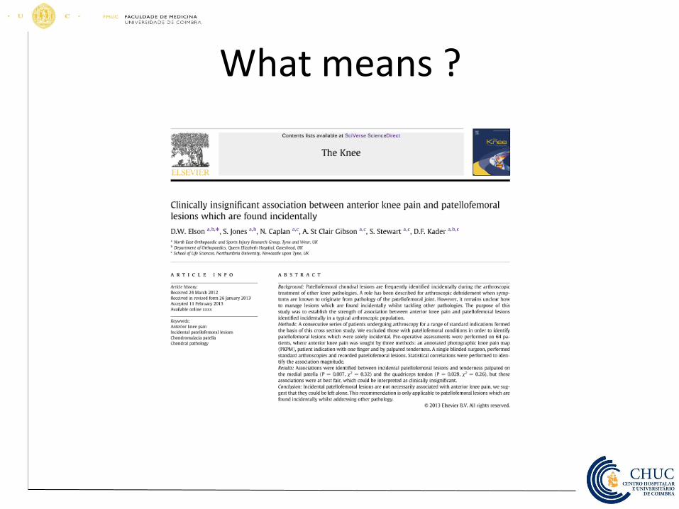
What means ?
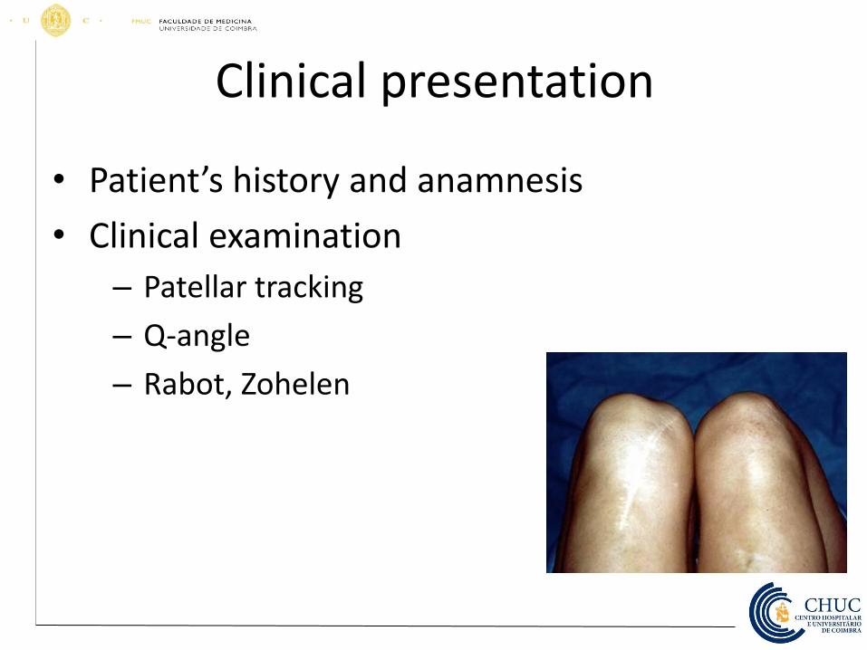
Clinical presentation
• Patient’s history and anamnesis
• Clinical examination
– Patellar tracking
– Q-angle
– Rabot, Zohelen
Patellar pain
• Anterior knee pain mainly
– Upstairs ou downstairs
– Prolonged sitting
– Nonspecific
• Frequent complaints
– “pseudo-blockade” (frequent)
– Knee effusion (rare)
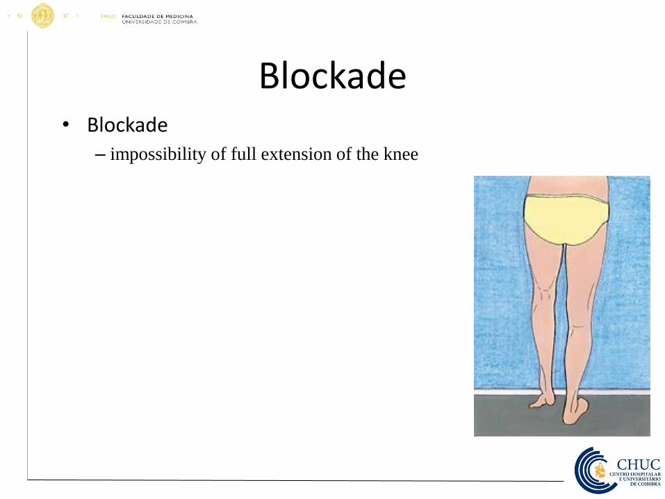
Blockade • Blockade
– impossibility of full extension of the knee
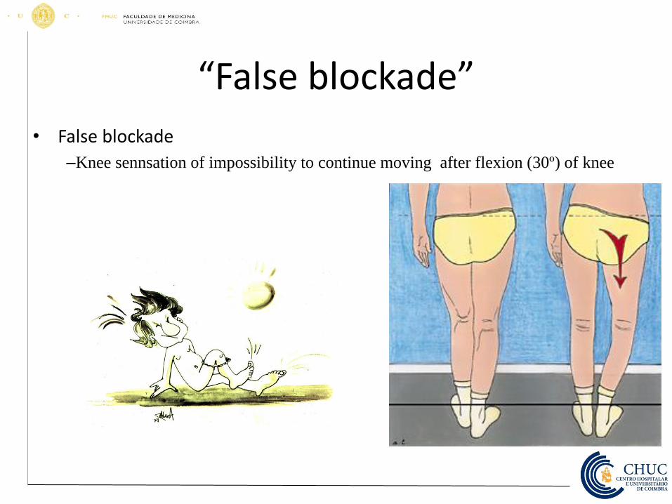
“False blockade”
• False blockade
–Knee sennsation of impossibility to continue moving after flexion (30º) of knee
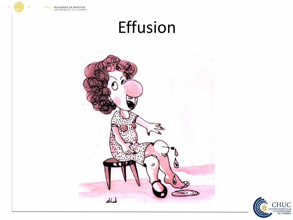
Effusion
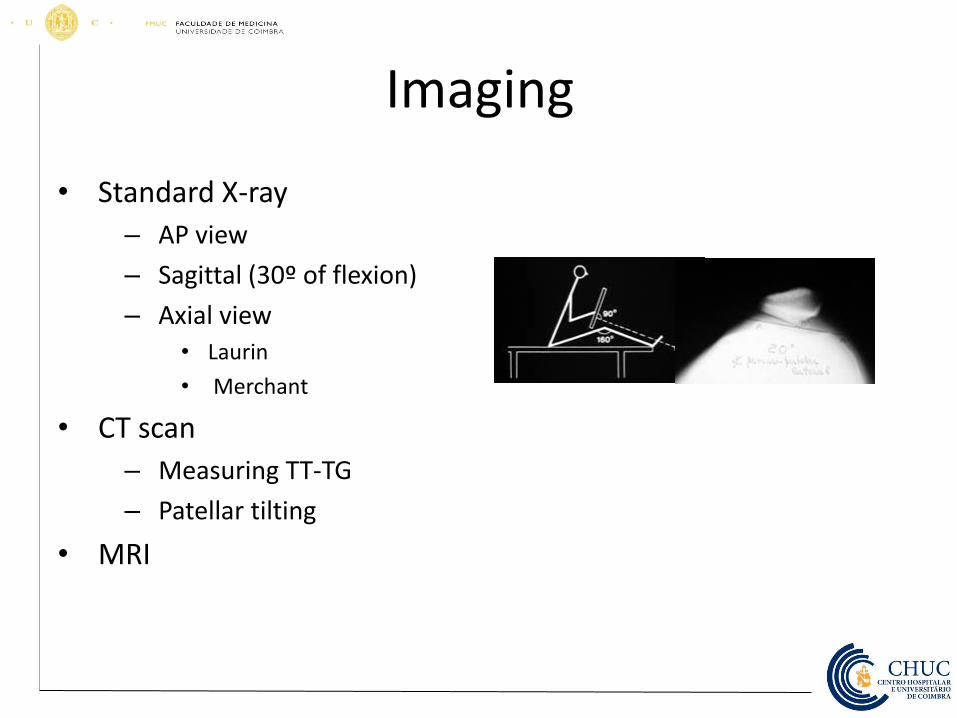
Imaging
• Standard X-ray
– AP view
– Sagittal (30º of flexion)
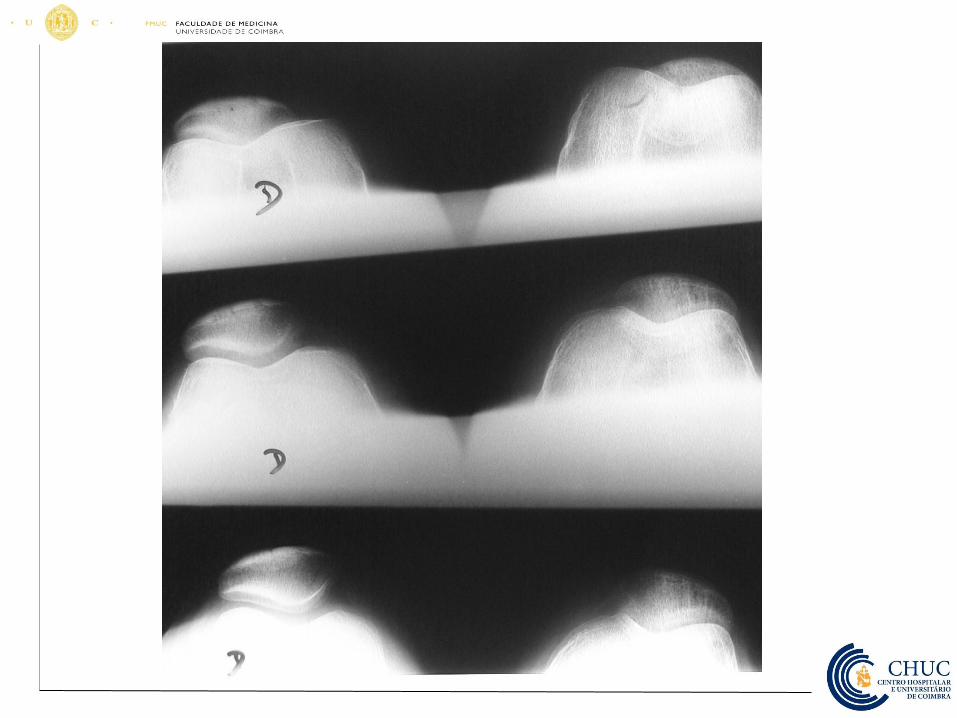
– Axial view
• Laurin
• Merchant
• CT scan
– Measuring TT-TG
– Patellar tilting
• MRI
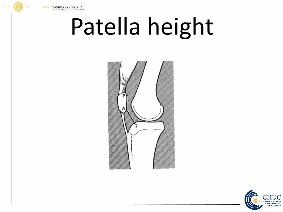
Patella height
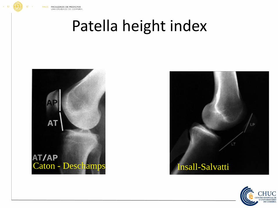
Patella height index
Caton - Deschamps Insall-Salvatti
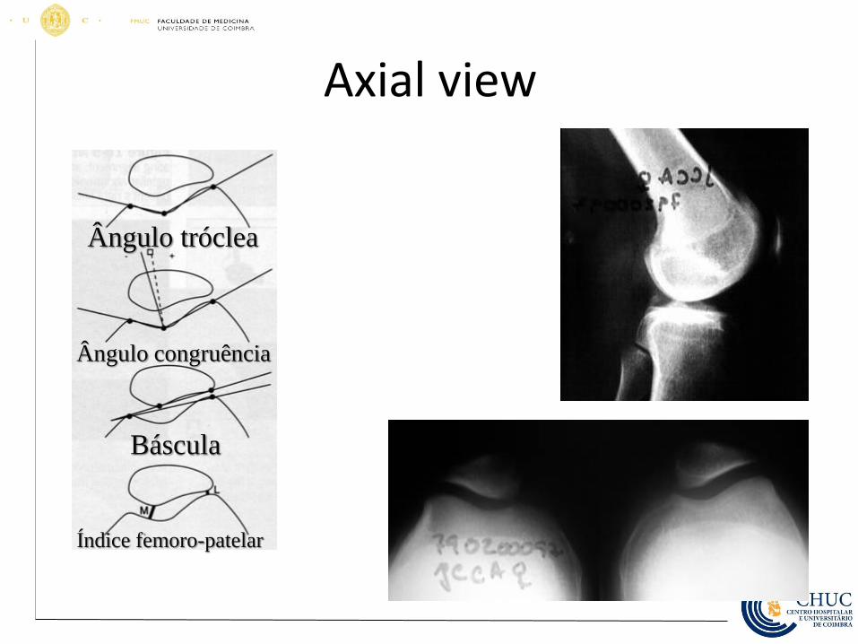
Axial view
Ângulo tróclea
Ângulo congruência
Báscula
Índice femoro-patelar
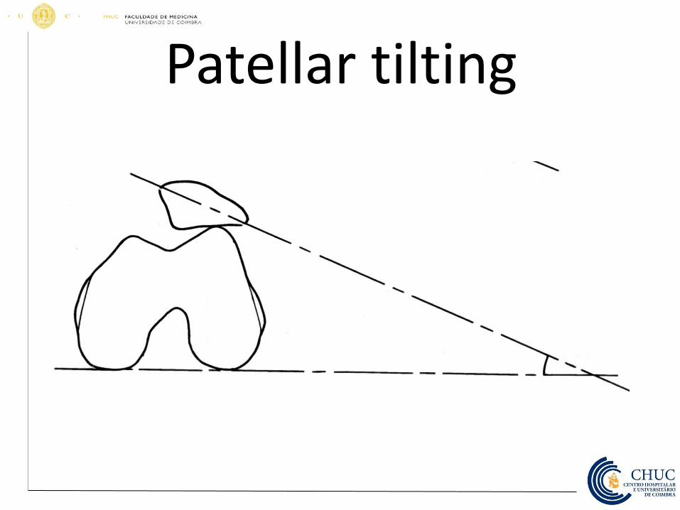
Patellar tilting
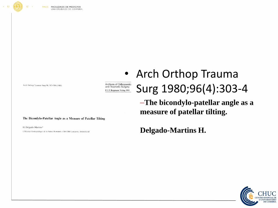
• Arch Orthop Trauma Surg 1980;96(4):303-4 –The bicondylo-patellar angle as a
measure of patellar tilting.
Delgado-Martins H.
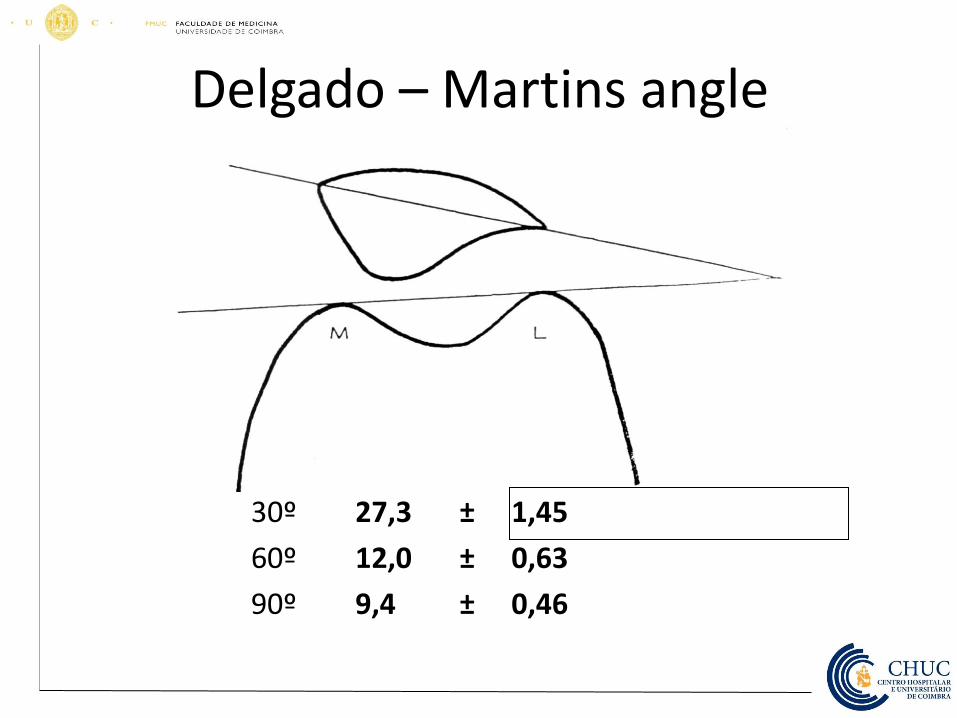
Delgado – Martins angle
30º 27,3 ± 1,45
60º 12,0 ± 0,63
90º 9,4 ± 0,46
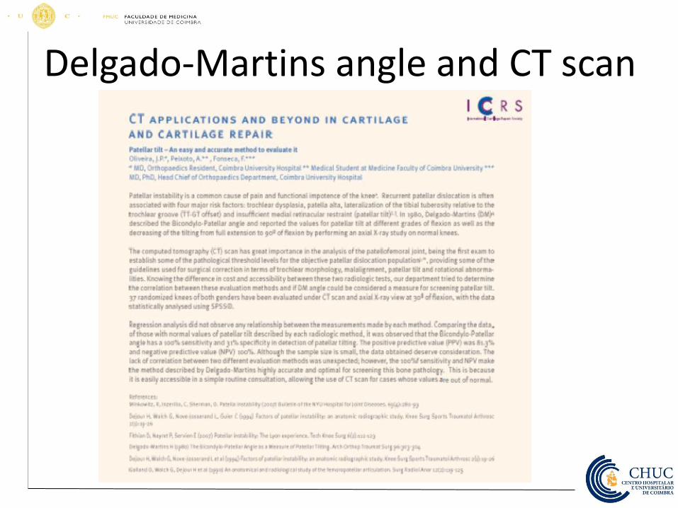
Delgado-Martins angle and CT scan
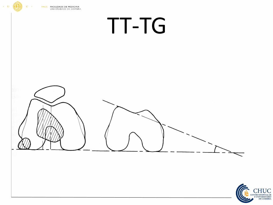
TT-TG
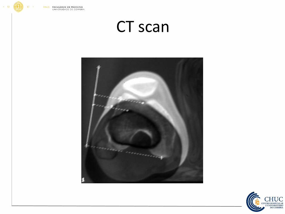
CT scan
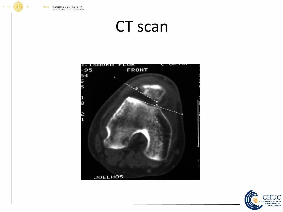
CT scan
Treatment • Conservative (6 months at least)
– Pain control and NSAID
– Viscosuplementation
– Physyoterapy
• Closed kinetics chain exercises
• Open kinetics chain exercises
• Reeducation – Muscular
– Postural
Kramer K. Management of patellar and trochlear
chondral injuries. Oper Tech Orthop. 2007;17(4):10–0
MD STC, MD JHB. Campbell's operative orthopaedics
e-dition: text with continually updated online reference,
11e. 11(null) ed. Mosby; 2007
.
Conservative traetment
• Rest
• Muscle exercises
• Hamstrings streching exercises
• Ice after exercises

Surgery • Last option!
– Arthroscopy
– Extension mechanism surgery • Lateral release of lateral retinaculum
– Ficat
s technique
– Larson-Slocum
• Modifying TTA
– Maquet III
– Fulkerson
– Cartilage • Microfractures
• Mosaicplasty
• ACI
Ficat´s release
• Described by Merchant and Mercer in 1974 and popularized by Ficat – Indication
• Patellofemoral pain with patellar tilt
Lateral retinaculum
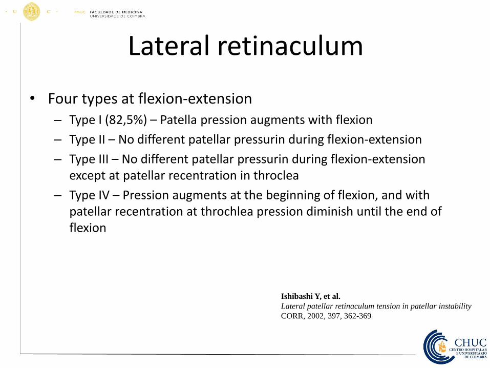
• Four types at flexion-extension – Type I (82,5%) – Patella pression augments with flexion
– Type II – No different patellar pressurin during flexion-extension
– Type III – No different patellar pressurin during flexion-extension except at patellar recentration in throclea
– Type IV – Pression augments at the beginning of flexion, and with patellar recentration at throchlea pression diminish until the end of flexion
Ishibashi Y, et al.
Lateral patellar retinaculum tension in patellar instability
CORR, 2002, 397, 362-369
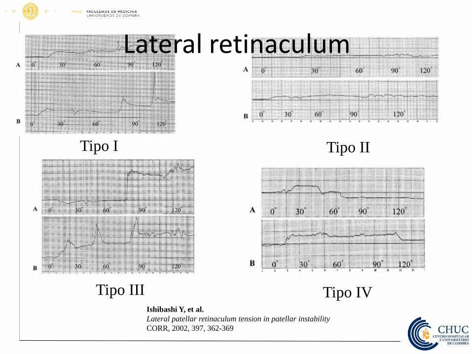
Tipo I Tipo II
Tipo III Tipo IV Ishibashi Y, et al.
Lateral patellar retinaculum tension in patellar instability
CORR, 2002, 397, 362-369
Lateral retinaculum
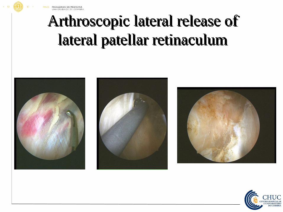
Arthroscopic lateral release of
lateral patellar retinaculum
Lateral release of lateral patellar retinaculum
• Be carefull ….
• There some long time complications !
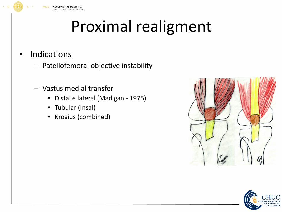
• Indications – Patellofemoral objective instability
– Vastus medial transfer
• Distal e lateral (Madigan - 1975)
• Tubular (Insal)
• Krogius (combined)
Proximal realigment
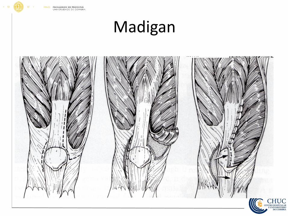
Madigan
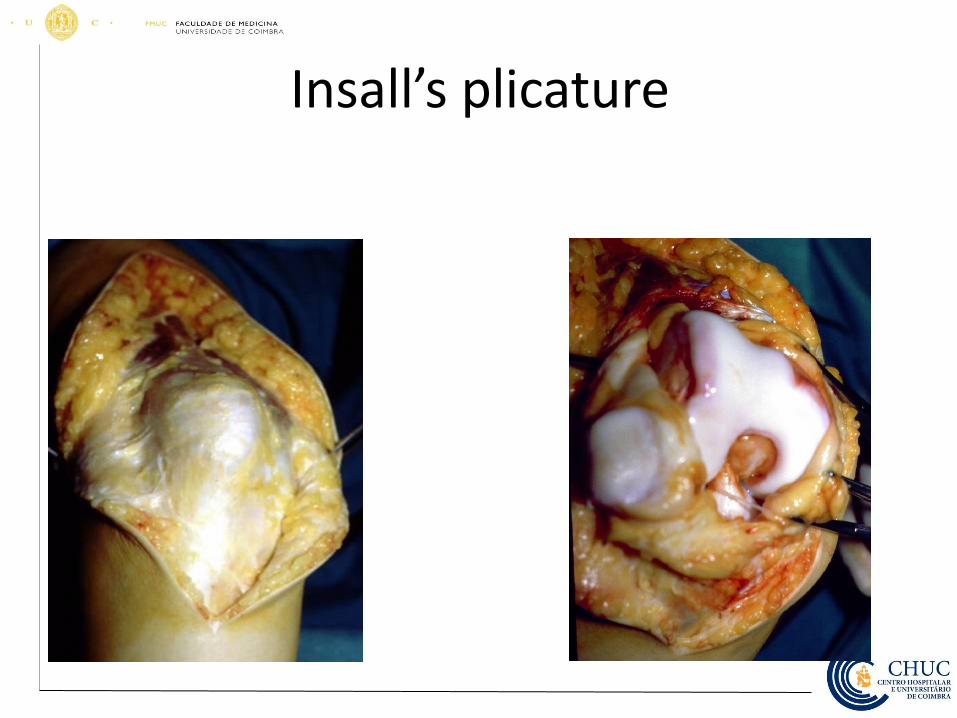
Insall’s plicature
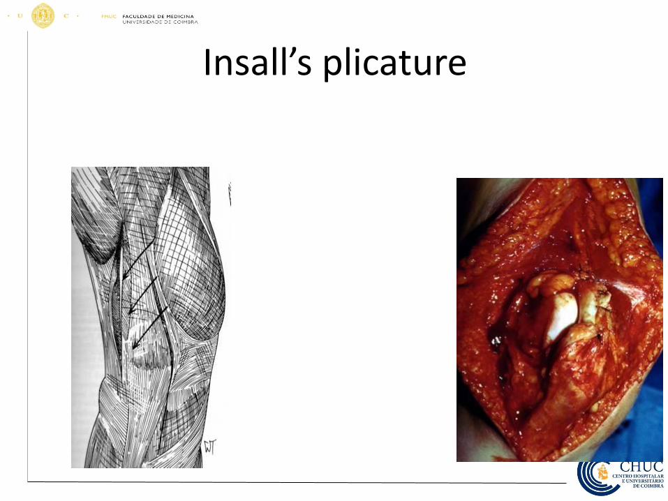
Insall’s plicature
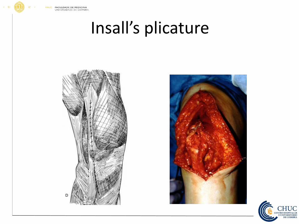
Insall’s plicature
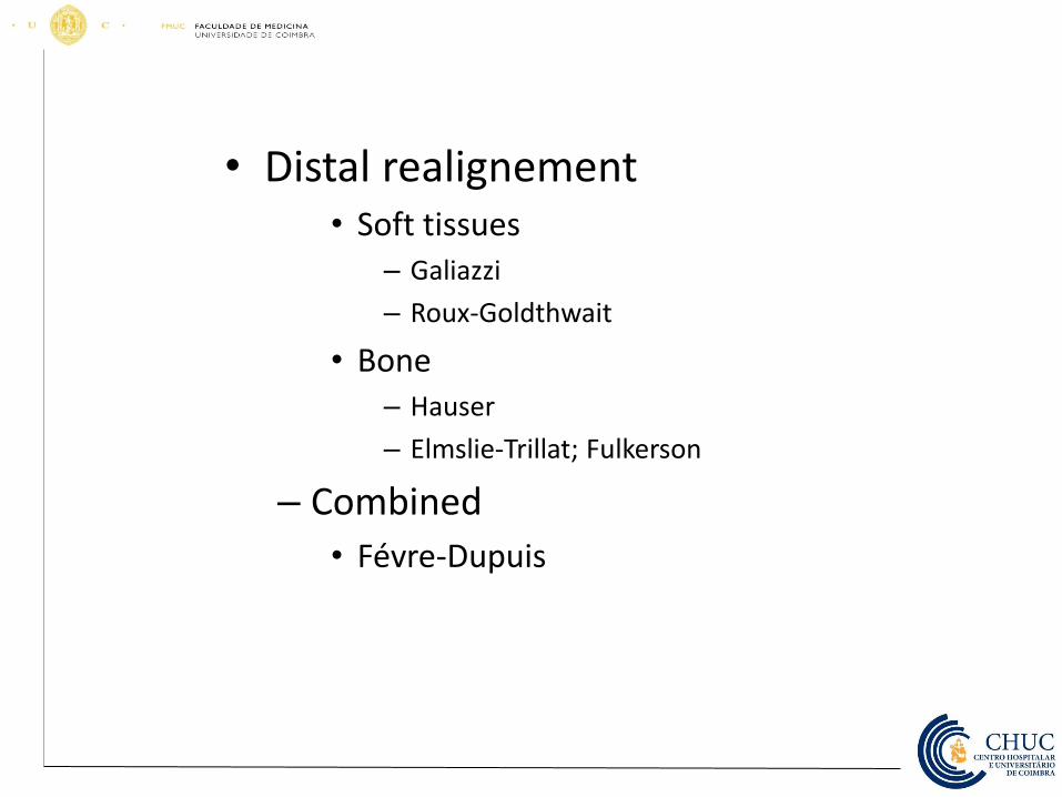
• Distal realignement • Soft tissues
– Galiazzi
– Roux-Goldthwait
• Bone – Hauser
– Elmslie-Trillat; Fulkerson
– Combined
• Févre-Dupuis
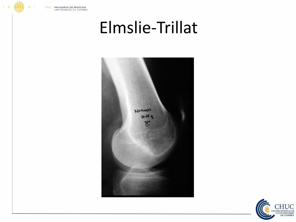
Elmslie-Trillat
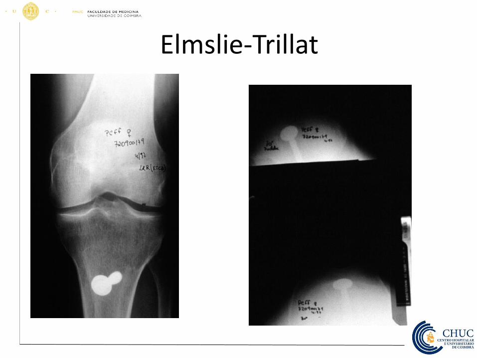
Elmslie-Trillat
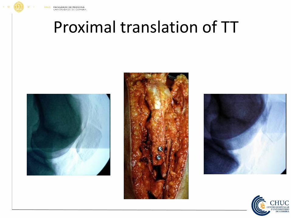
Proximal translation of TT
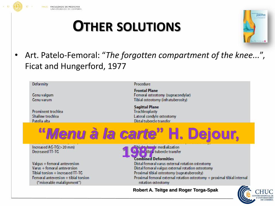
OTHER SOLUTIONS
• Art. Patelo-Femoral: “The forgotten compartment of the knee...”, Ficat and Hungerford, 1977
“Menu à la carte” H. Dejour,
1987
Robert A. Teitge and Roger Torga-Spak
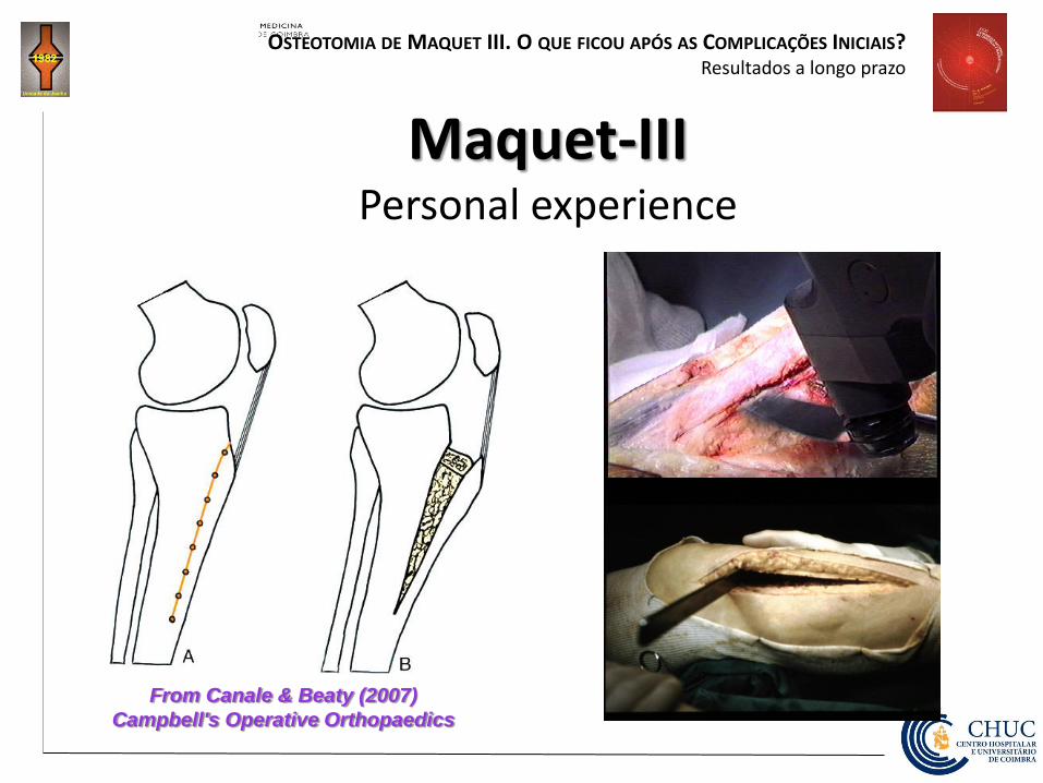
OSTEOTOMIA DE MAQUET III. O QUE FICOU APÓS AS COMPLICAÇÕES INICIAIS? Resultados a longo prazo
Maquet-III Personal experience
From Canale & Beaty (2007)
Campbell's Operative Orthopaedics
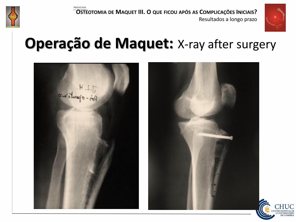
OSTEOTOMIA DE MAQUET III. O QUE FICOU APÓS AS COMPLICAÇÕES INICIAIS? Resultados a longo prazo
Operação de Maquet: X-ray after surgery
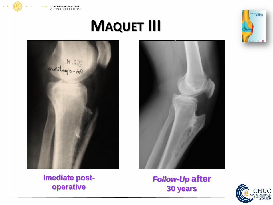
MAQUET III
Imediate post-
operative Follow-Up after
30 years
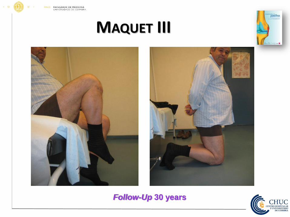
MAQUET III
Follow-Up 30 years
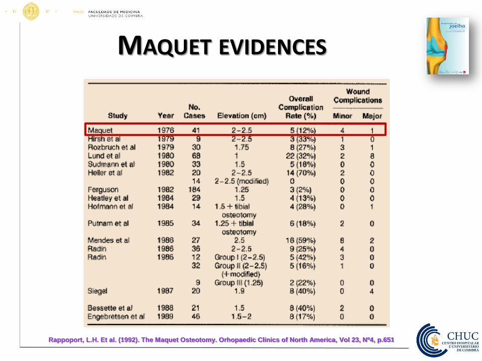
MAQUET EVIDENCES
Rappoport, L.H. Et al. (1992). The Maquet Osteotomy. Orhopaedic Clinics of North America, Vol 23, Nº4, p.651
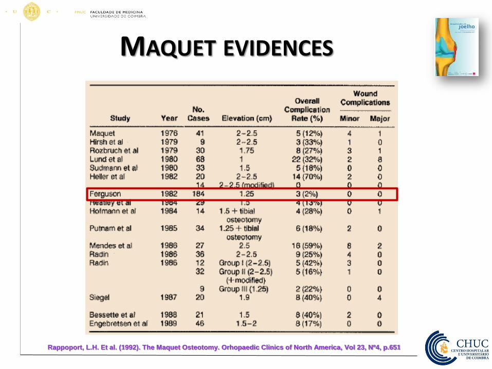
MAQUET EVIDENCES
Rappoport, L.H. Et al. (1992). The Maquet Osteotomy. Orhopaedic Clinics of North America, Vol 23, Nº4, p.651

MAQUET EVIDENCES
Rappoport, L.H. Et al. (1992). The Maquet Osteotomy. Orhopaedic Clinics of North America, Vol
23, Nº4, p.651
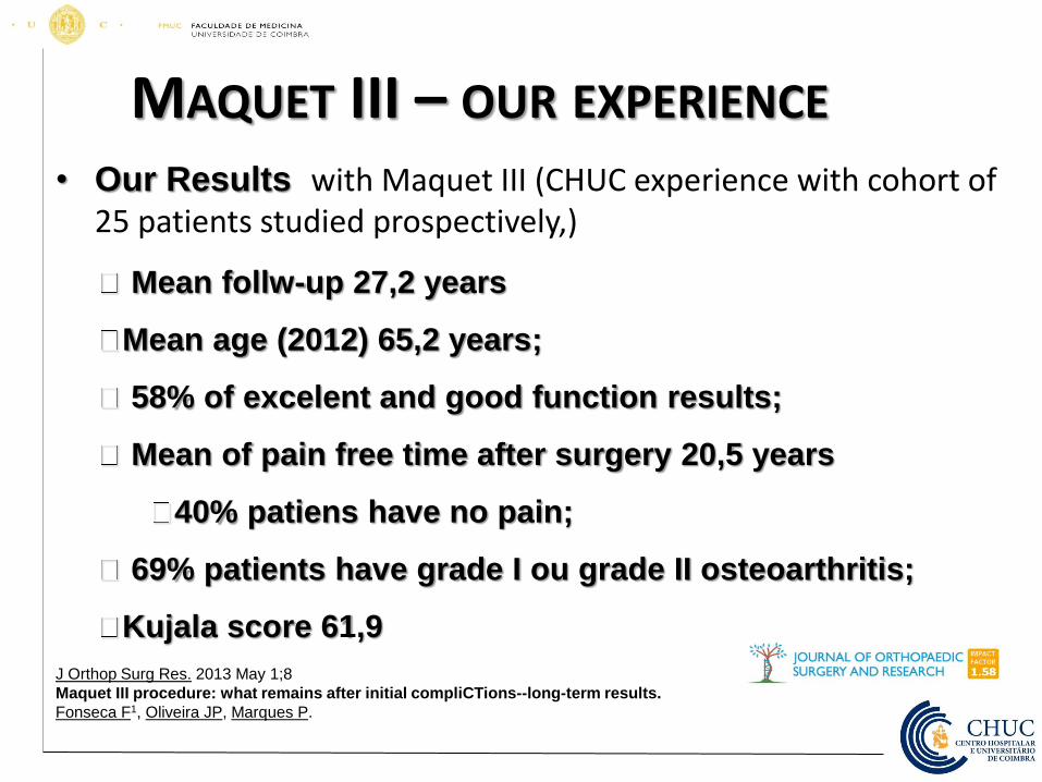
MAQUET III – OUR EXPERIENCE
• Our Results with Maquet III (CHUC experience with cohort of 25 patients studied prospectively,)
Mean follw-up 27,2 years
Mean age (2012) 65,2 years;
58% of excelent and good function results;
Mean of pain free time after surgery 20,5 years
40% patiens have no pain;
69% patients have grade I ou grade II osteoarthritis;
Kujala score 61,9
J Orthop Surg Res. 2013 May 1;8
Maquet III procedure: what remains after initial compliCTions--long-term results.
Fonseca F1, Oliveira JP, Marques P.
Literature revision
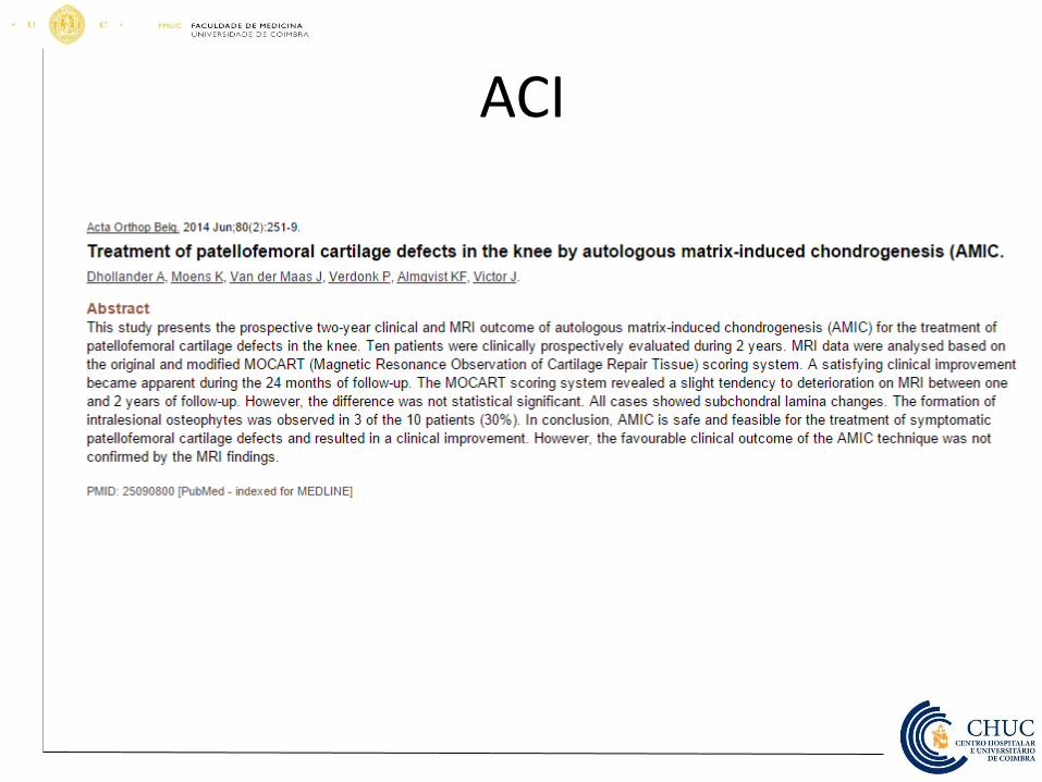
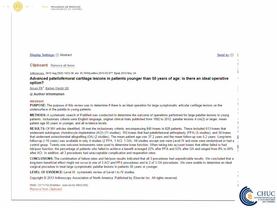
ACI
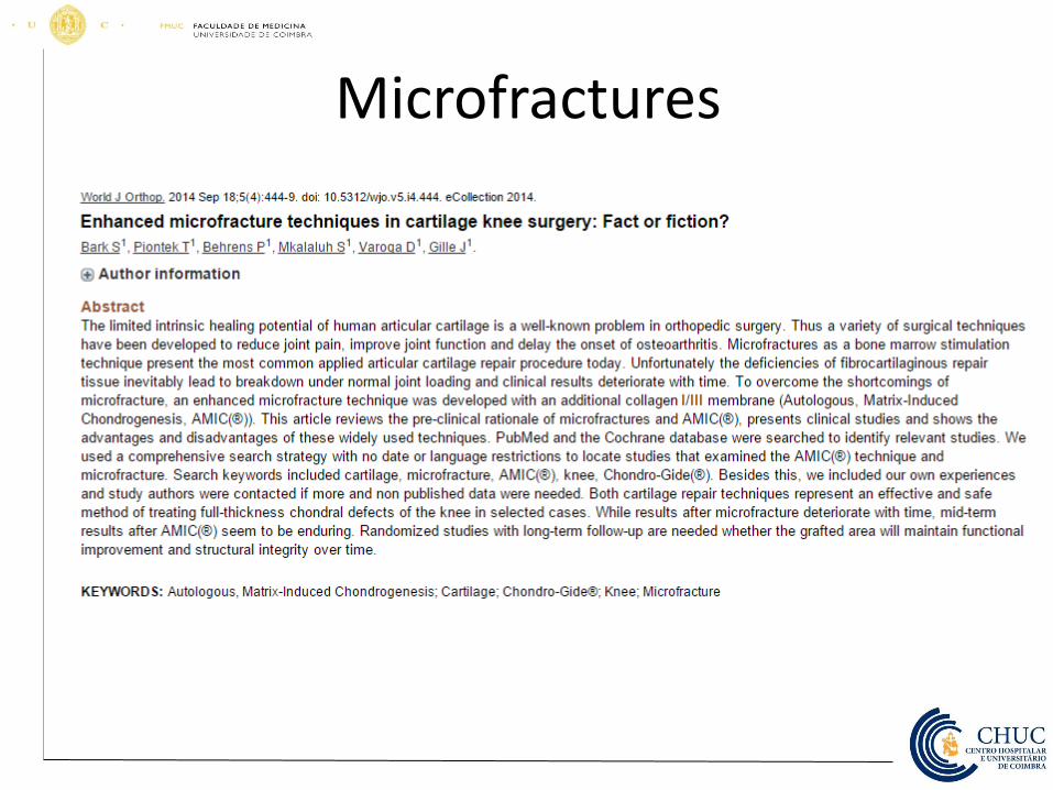
Microfractures
Lesson learned in25 years ?
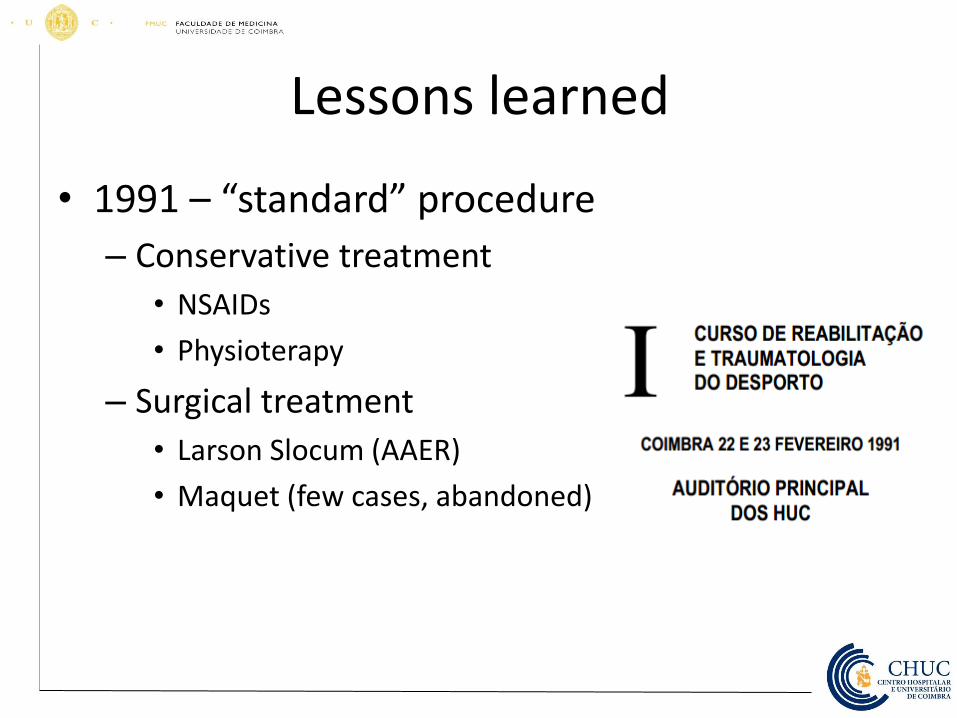
Lessons learned
• 1991 – “standard” procedure
– Conservative treatment
• NSAIDs
• Physioterapy
– Surgical treatment
• Larson Slocum (AAER)
• Maquet (few cases, abandoned)

My options(2015) - 1
• Have a excellent patient history, evaluate carefully patient’s syntoms
– Listen patient complaints is a corner stone
• Carefully physical examination
– Rule out secondary causes of knee pain
• If there are secondary causes for pain …
– Treat secondary causes!
• Remember !!
– Like at low back pain, hernia may not be the origin of the pain!
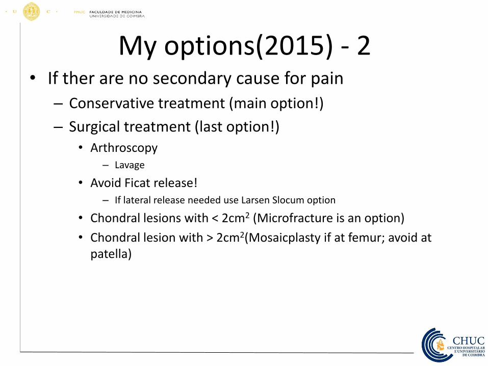
My options(2015) - 2 • If ther are no secondary cause for pain
– Conservative treatment (main option!)
– Surgical treatment (last option!) • Arthroscopy
– Lavage
• Avoid Ficat release! – If lateral release needed use Larsen Slocum option
• Chondral lesions with < 2cm2 (Microfracture is an option)
• Chondral lesion with > 2cm2(Mosaicplasty if at femur; avoid at patella)
Thank you