bone metastases métastases osseuses interest of bone scan ... · bone metastases métastases...
TRANSCRIPT

Bone metastasesMétastases osseusesBotmetastasen
Interest of bone scan in malignancy:
-Patient with high probability of bone metastases
-follow up when specific tumor marker levels rise
-evaluation of bone pain
-further study in solitary radiographic lesion (or equivocal)
-monitoring of therapy

Skeleton is a frequent site of metastatic spread for many neoplasms
Mechanism of uptake includes:-active blood flow-enhanced ostoblastic activity :primarily lytic lesions ->stimulation of
osteoblasts->concentration of ->Hot area radiolabeled phosphateson hydroxyapatite crystal
->if lytic component ->Cold area predominant
Bone scintigraphy: more sensitive than radiography
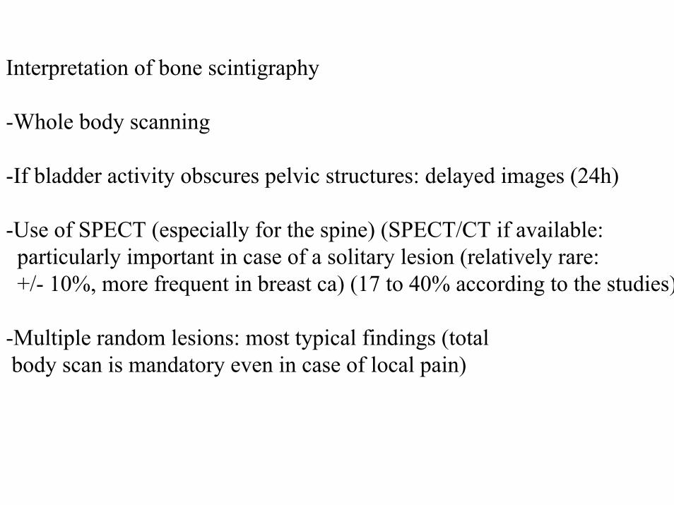
Interpretation of bone scintigraphy
-Whole body scanning
-If bladder activity obscures pelvic structures: delayed images (24h)
-Use of SPECT (especially for the spine) (SPECT/CT if available:particularly important in case of a solitary lesion (relatively rare:+/- 10%, more frequent in breast ca) (17 to 40% according to the studies)
-Multiple random lesions: most typical findings (totalbody scan is mandatory even in case of local pain)

Isolated lytic lesion of L1 in a case of myeloma. Left: planar bone scintigraphy, right:SPECT/CT: note the lytic lesion of right part of vertebra on CT and osteoblastic response ofthe rest of the vertebra. If osteoblastic response is minimal, osteolysis is predominant witha cold area

Drug interaction: increased renal uptake may be observed shortlyafter administration of chemotherapeutic drugs (this finding can alsooccur in nephrocalcinosis, myoglobinuria, etc…)

10 200404 2004
Patient suffering from a breast cancer. Random placed lesionsTreatment: zometa. Multiple metastases, evolution between april and october 04Bone metastases frequently occur in the axial skeleton by involvement of vertebral venous system(bypass of vena cava and portal system). The involvement occurs generally in the vertebral bodyand pedicle of the vertebra (posterior activity is predominantly due to degenerative process: the useof SPECT is helpful to determine the part of involved vertebra)

Increased uptake by long bones in a case of chronic myeloid leukemia: noteincreased uptake by femoral and humeral shafts. Proliferative changes in most of the bone marrow elements can produce changes in the bone scan appearance due to reactiveprocess or direct involvement. This is most commonly associated with abnormalities inthe white blood cells elements.
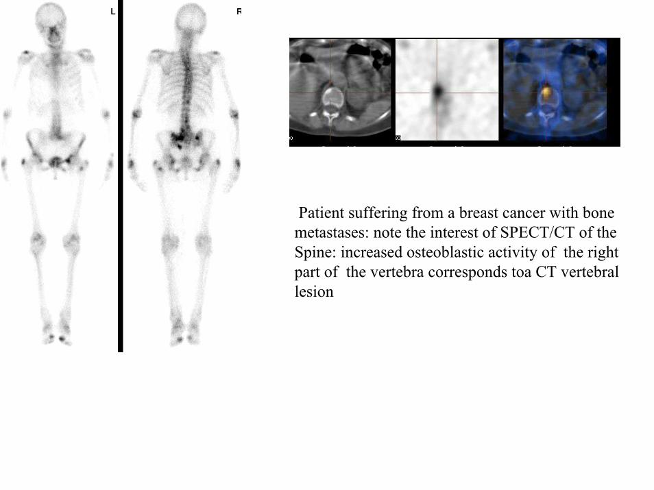
Patient suffering from a breast cancer with bonemetastases: note the interest of SPECT/CT of the Spine: increased osteoblastic activity of the rightpart of the vertebra corresponds toa CT vertebrallesion

Patient with increasedPSA level: SPECT of the spine is in favour ofa vertebral metastasis
Prostate cancerAbout 40% of patients with positive scan present with no pain->bone scan = most sensitive method for early detection ofmetastatic disease
Incidence of skeletal metastases rises with increasing bloodPSA level (In about 30% of patients receiving antiandrogen therapy,PSA is in the normal range, despite the presence of metastases)

Unknown oat cell carcinoma of upper part of left lung (See RX). Patient complained from left shoulder painand was refered for a suspicion of arthritis: Pancoast S.
Lung cancer If bone metastases present: grave prognostic sign (about 80% of patients with abnormal bone scanat presentation die within 6 months)Incidence of bone metastases in small cell and anaplastic cancer is high and, bone scan at initial workup is necessary for these patientsIncidence of appendicular metastases is higher than in other cancers (tumor cells spread by arterial route to distal extremities)

Patient suffering from a left lung carcinoma (see RX) with involvementOf first left rib: Pancoast syndrome. Note a second vertebral lesion.

Renal and bladder carcinomaLow incidence of bone metastases (10% of the metastases are lyticwith photopenic images)
Thyroid cancerDetection of metastases in well differnetiated ca can be performedusing 131 scan. However, bone scan can be of interest in medullarycarcinoma (which does not concentrate iodine)
Hemoproliferative disordersLymphomas, leukemias: Bone marrow expansion often cause diffuseUptake of the tracer in appendicular skeletonNote that in myeloma, lytic lesions are often observed

Child presenting with a metastatic neuroblastoma: metastases in the skeleton: osseous metastasesin the calvarium, metaphyseal location, proximal humerus (left), distal femora, proximal tibiae, ribsRight: bone scintigraphy, left: 123I MIBG scan: most bone scan abnormalities demonstrateMIBG uptake