webinar 1 slides 2
TRANSCRIPT

Chemical Exposures & Life-Long Reproductive Health Impacts
Suzanne E. Fenton, Ph.D.
National Institute of Environmental
Health Sciences
Energy Development and
Reproductive Health
April 6, 2015

1 in 8

In the Beginning……
Mailleux AA et al. 2002. Development. 2002 Jan;129(1):53-60.

Mammary Development over the Life Span

Endocrine/Paracrine Influences Paracrine/Autocrine Influences
Adipose tissue Blood vessels Stromal cells Inflammatory
Response
Developing mammary
terminal end bud
‘epithelium’
Target Site
Complicated Tissue

Hormonal control – Version A

Brisken C , and O’Malley B Cold Spring Harb Perspect Biol
2010;2:a003178
Hormonal control – Version B

Developmental Event Human Rodent
milk streak evident EW4-6 GD10-11 (mice)
mammary epithelial bud forms EW10-13 GD12-14 (mice), GD 14-16
(rat)
female nipple and areola form EW12-16 GD18 (mice)/GD20 (rat)
branching and canalization of epithelium EW20-32 GD16 to birth (mice), GD
18 to birth (rat)
secretion is possible EW32-40 (ability
lost postnatally)
at birth, with hormonal
stimuli
isometric development of ducts birth to puberty birth to puberty
TEBs present (peri-pubertal) 8-13 year old girls 23 to 60 days old
(rodents)
formation of lobular units EW32-40, or within
1-2 yr. of first
menstrual cycle
puberty and into
adulthood
TEB=terminal end bud, EW=embryonic week, GD=gestational day
taken from S.E. Fenton, 2006 Endocrinology 147(Supplement):S18-S24.
Developmental Events in Human
and Rodent Mammary Tissue

The Terminal End Bud (TEB):
• Tear-drop shaped structure that is
present before vaginal opening and
until young adulthood in mammary
whole mounts and sections
• Multiple epithelial cells thick and
stains intensely with hemotoxylin &
eosin
• Measures > 100 mm in rat gland
• Highly mitogenic, invading end of
epithelium, often seen dividing into
new ducts
• Contains stem cells
• Target for carcinogens

Childhood Puberty Infancy In utero
First week of life 10-20 days old 3-6 weeks of age
6-7 days before birth First week of life 15-25 days old 4-6 weeks of age
End of first trimester First years of life 4-8 years old 8-16 years old
Hu
man
M
ou
se
Rat

Mammary Cancer: A look at carcinogens
• As of February 2014, there were about 84,000 chemicals registered for
commercial use on the market [Toxic Substances Control Act (TSCA)
Chemical Substances Inventory]
• Chemical specific health hazards have been identified for somewhere
between 1-2% (about 600 at NTP) of those chemicals using the traditional
2 yr cancer bioassay in adult rats or mice (little or no early life exposure
data). NTP estimates suggest about 250 of those compounds cause
cancer.
• In a 2007 report by Silent Spring Institute, 216 chemicals have been
identified to cause mammary tumors in a rodent bioassay (in at least one
lab) (Rudel et al., 2007. Cancer 109(S12):2635-66).
• About 60 of those chemicals cause mammary cancer in one or more
species in a 2 yr bioassay, but may also affect other sites.
• There are 6 substances in the Report on Carcinogens that cause or may
cause breast cancer in humans. These include the drug diethylstilbestrol
(DES), steroid hormones used for menopausal therapy, some forms of
radiation, alcohol, tobacco smoke and the sterilizing agent, ethylene
oxide.
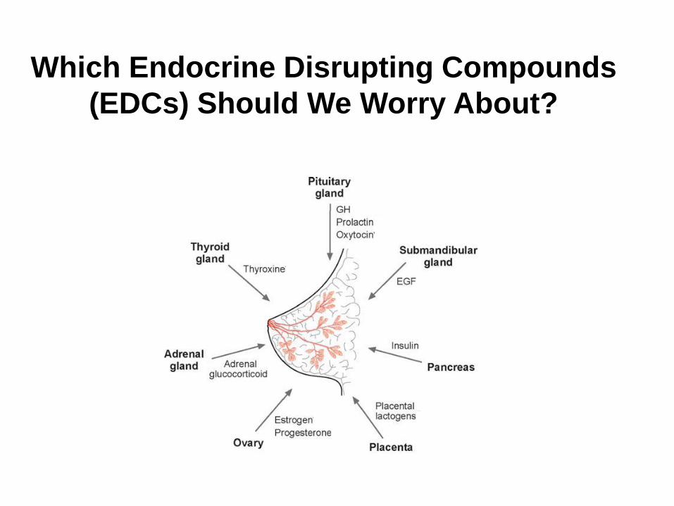
Which Endocrine Disrupting Compounds
(EDCs) Should We Worry About?
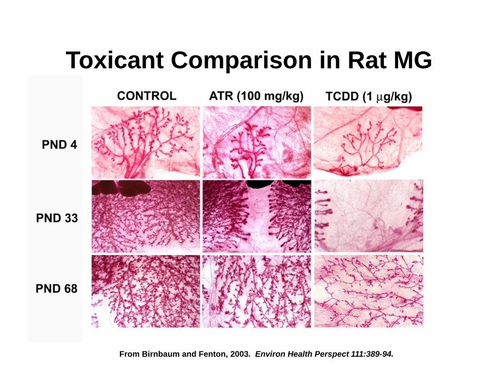
Toxicant Comparison in Rat MG
From Birnbaum and Fenton, 2003. Environ Health Perspect 111:389-94.

Puberty
Gestation
Altered growth and development;
altered carcinogen susceptibility
Adulthood
Pregnancy/
lactation
Breast cancer
Early life
N
orm
al
MG
(ra
t)
• Fat pad and
bud form
• Epithelium
forms ductal
tree
• Isometric
epithelial growth
• Branching ducts
and budding
• TEBs develop
• Exponential
epithelial growth
• TEBs
differentiate
• Epithelium is
predominant
• Lobulo-alveolar
development
Altered hormone/growth factor levels and responsiveness; effects may be
systemic or localized to MG
• Static resting
state, changing
with cyclicity
• Responsive to
hormonal changes
MG
aft
er
earl
y E
DC
exp
osu
re
Lactational
impairment
Exposure during MG development
Fetal Basis of Adult Disease

EDCs shown to cause abnormal mammary gland development in rodents using either sectioned tissue or whole mount analyses:
Atrazine and its degradates* Bisphenol A* Cadmium Dieldrin *most sensitive of the Dioxin or TCDD reproductive tissues DES evaluated DDT/DDE* Flame retardant mixture DE-71 Genistein* Nonylphenol Organochlorine mixtures Phthalates PCBs PhIP Perfluorooctanoic acid (PFOA) N-3 and N-6 PUFA Resveratrol Zearalenone Ziracin
Many associated with increased incidence of tumors in carcinogen- induced models 19 of >84,000 chems

Compound Study Species; Dev Stage of Exposure
MG Dev Effect LOEL Basis for Conclusion
FEMALE STUDIES Atrazine metabolite mixture
Enoch 2007 Rat; Prenatal 0.09 mg/kg/d Effects on pup body wt and age of VO were observed at a higher doses in this study (0.87 mg/kg/d).
Atrazine Rayner et al., 2005 Rat; Prenatal 100 mg/kg/d Effects on age of VO and body wt occurred at the same dose but only with a longer duration of exposure than for the MG dev effects.
BPA Jenkins et al., 2009 Rat; Postnatal (lact) 250 μg/kg/d In this single dose study, effects on age of VO, body wt, serum P, or serum estradiol were not observed at 250 μg /kg/d.
Murray et al., 2007 Rat; Prenatal 2.5 μg /kg/d Effects on body wt, age of VO;, litter size, or sex ratio were not observed at this or higher doses (2.5 – 1000 μg /kg/d).
Munoz-de-Toro et al., 2005
Mouse; Perinatal 250 ng/kg/d Effects on plasma estradiol at 1st proestrus was not observed at 250 ng/kg/d (highest dose tested).
DDT (o-p) Brown and Lamartiniere 1995
Rat; Peripubertal 50 μg/kg/d In this single dose study, effects on body wt or uterine-ovarian wt were not observed at 50 μg/kg/d.
Genistein Fritz et al., 1998 Rat; Pre- to Postnatal 25 mg/kg/d In this single dose study, effects on body wt, uterine wt, or AGD were not observed at 25 mg/kg/d.
Padilla-Banks et al., 2006
Mouse; Neonatal 0.5 mg/kg/d Effects on pup mortality occurred at a higher dose (50 mg/kg/d).
MALE STUDIES Genistein Delclos et al., 2001 Rat; Pre- to post-natal 25 ppm Effects on decreased ventral prostate wt, age of
eye opening, and age of ear unfolding occurred at a higher dose (1250 ppm).
Examples of EDC developmental exposures for which mammary gland developmental effects were more sensitive than other endpoints of
endocrine disruption: Within-study comparisons of rodent studies1 1 Only statistically significant effects were included in this table. BPA, Bisphenol A; DDT, dichlorodiphenyltrichloroethane; LOEL, Lowest
observed effect level, indicates the lowest tested dose where effects were statistically significant; VO, vaginal opening.
Mammary is Sensitive Tissue to EDCs

Male Breast Cancer
• < 1% of breast cancer cases are in men
• It is estimated that about 2,000 new cases are diagnosed annually in the U.S.
• Breast cancer typically appears in men in their 70’s and older with a family history of breast cancer and carry a BRCA mutation
• Many men from Camp Lejuene developed cancer in their 30-40s and have no family histories of breast cancer
http://www.nlm.nih.gov/medlineplus/malebreastcancer.html
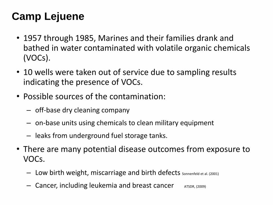
Camp Lejuene
• 1957 through 1985, Marines and their families drank and bathed in water contaminated with volatile organic chemicals (VOCs).
• 10 wells were taken out of service due to sampling results indicating the presence of VOCs.
• Possible sources of the contamination:
– off-base dry cleaning company
– on-base units using chemicals to clean military equipment
– leaks from underground fuel storage tanks.
• There are many potential disease outcomes from exposure to VOCs.
– Low birth weight, miscarriage and birth defects Sonnenfeld et al. (2001)
– Cancer, including leukemia and breast cancer ATSDR, (2009)

Human Examples of Early Life Environmental
Exposures Linked to Breast Outcomes
1. Dioxin:
Delayed breast development in adolescents with the highest circulating dioxin levels (Seveso, Italy; denHond et al., 2002. EHP 110:771-6) or prenatal/lactational dioxin levels (The Netherlands; Leijs et al., 2008. Chemosphere 73:999-1004).
Early reports of doubled risk (2.1) of breast cancer with 10-fold increase in serum dioxin (95% confidence interval, 1.0-4.6) (Warner et al., 2002. EHP 110:625-8) and in recent follow-up, all cancers combined was significantly increased [adjusted HR = 1.80; 95% confidence interval: 1.29-2.52] (Warner et al., 2011. EHP 119(12):1700-1705). Breast cancer alone did not reach significance.
2. DDT: (Cohn et al., 2007 EHP 115:1406-14)
Exposure increased from 1945 through 1959, when DDT use peaked (with dietary exposure peaking in 1965)
Breast cancer odds ratio risk estimates by period of birth for the highest tertile of p,p′-DDT exposure were
– 0.6 for women born in 1931 or earlier (i.e., ≥ 14 years of age in 1945)
– 3.9 for women born in 1932–1937 (i.e., 8–13 years of age in 1945)
– 9.6 for women born in 1938–1941 (i.e., 4–7 years of age in 1945)
– 11.5 for women born in 1942 or later (i.e., < 4 years of age in 1945)
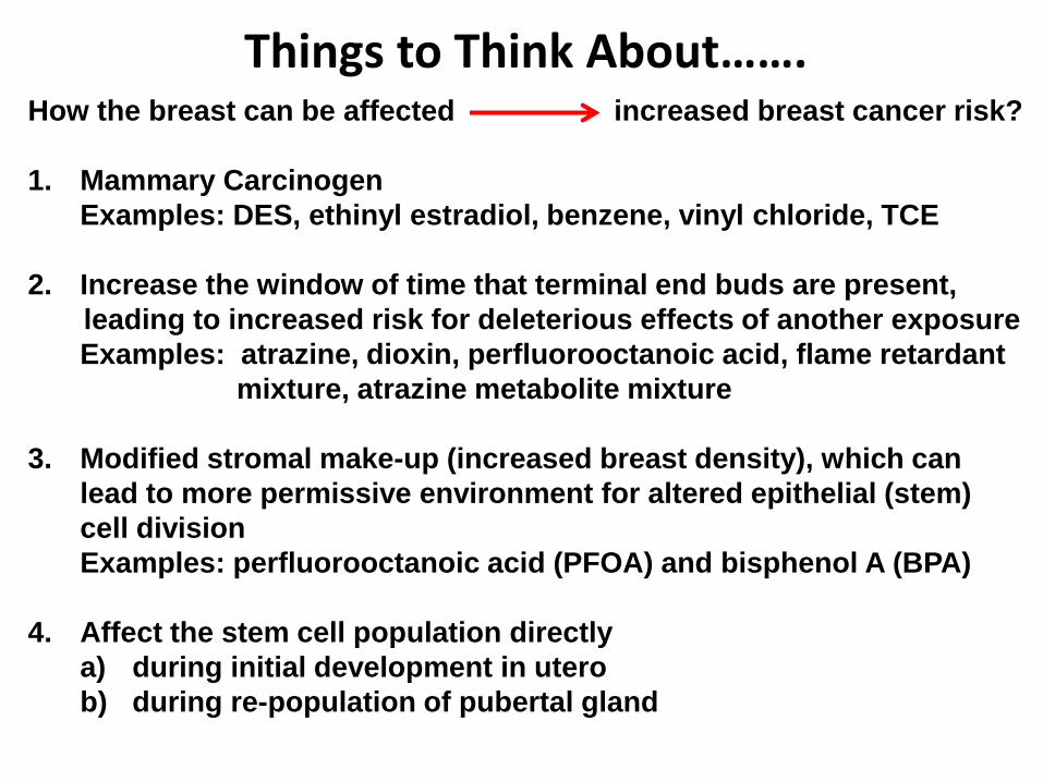
How the breast can be affected increased breast cancer risk?
1. Mammary Carcinogen
Examples: DES, ethinyl estradiol, benzene, vinyl chloride, TCE
2. Increase the window of time that terminal end buds are present,
leading to increased risk for deleterious effects of another exposure
Examples: atrazine, dioxin, perfluorooctanoic acid, flame retardant
mixture, atrazine metabolite mixture
3. Modified stromal make-up (increased breast density), which can
lead to more permissive environment for altered epithelial (stem)
cell division
Examples: perfluorooctanoic acid (PFOA) and bisphenol A (BPA)
4. Affect the stem cell population directly
a) during initial development in utero
b) during re-population of pubertal gland
Things to Think About…….

Thank you…..
NIEHS Campus