wake-up stroke and stroke of unknown onset: a critical review · of stroke (ischemic, hemorrhagic,...
TRANSCRIPT
REVIEW ARTICLEpublished: 12 August 2014
doi: 10.3389/fneur.2014.00153
Wake-up stroke and stroke of unknown onset: a criticalreviewAnke Wouters1,2,3*, Robin Lemmens1,2,4,5, Patrick Dupont 3,6,7 and VincentThijs1,2,4,5
1 KU Leuven Department of Neurosciences and Experimental Neurology, KU Leuven, Leuven, Belgium2 Department of Neurology, University Hospital Leuven, Leuven, Belgium3 Medical Imaging Research Center, UZ Leuven, Leuven, Belgium4 Leuven Research Institute for Neuroscience and Disease (LIND), KU Leuven, Leuven, Belgium5 Laboratory of Neurobiology, Vesalius Research Center, Leuven, Belgium6 Laboratory for Epilepsy Research, KU Leuven, Leuven, Belgium7 Laboratory for Cognitive Neurology, KU Leuven, Leuven, Belgium
Edited by:Argye Hillis, Johns Hopkins Medicine,USA
Reviewed by:Edward C. Jauch, Medical Universityof South Carolina, USASara E. Hocker, Mayo Clinic, USA
*Correspondence:Anke Wouters, Laboratory forNeurosciences and ExperimentalNeurology, KU Leuven, Herestraat 49,Leuven 3000, Belgiume-mail: [email protected]
Patients, who wake up with an ischemic stroke, account for a large number of the totalstroke population, due to circadian morning predominance of stroke. Currently, this sub-set of patients is excluded from revascularization-therapy since no exact time of onset isknown. A large group of these patients might be eligible for therapy. In this review, weassessed the current literature about the hypothesis that wake-up-strokes occur just prioron awakening and if this subgroup differs in characteristics compared to the overall strokepopulation. We looked at the safety and efficacy of thrombolysis and interventional tech-niques in the group of patients with unknown stroke-onset. We performed a meta-analysisof the diagnostic accuracy of the diffusion-FLAIR mismatch in identifying stroke within 3and 4.5 h. The different imaging-selection criteria that can be used to treat these patientsare discussed. Additional research on imaging findings associated with recent stroke andpenumbral imaging will eventually lead to a shift from a rigid time-frame based therapy toa tissue-based individualized treatment approach.
Keywords: wake-up-stroke, unknown-onset stroke, thrombolysis, thrombectomy, circadian rhythm
INTRODUCTIONAbout one in six persons older than 45 will suffer from stroke intheir remaining lifetime (1). The only Food and Drug Adminis-tration approved medical therapy for stroke is intravenous tissueplasminogen activator (alteplase or IV tPA), which should beadministered preferably as quickly as possible within 4 h 30 min(2). Despite the efficacy of IV tPA, the narrow therapeutic win-dow precludes wide scale use of the therapy mainly because a largemajority of patients arrive too late in the hospital. Additionally,observational studies indicate that between 8 and 25% of patientscome to the emergency room with an unknown time of symptomonset (3–7). This includes patients who were sleeping and woke upwith stroke-symptoms or patients who are unable to state the timeof symptom onset and in whom no witness is available. Theoret-ically, if patterns on cerebral imaging could serve as a substitutefor the time since the stroke had occurred, then this subset ofstroke patients would not be excluded from thrombolytic therapy.Another approach could be to rely on the presence of imaging char-acteristics indicative of large areas of viable tissue, regardless of theindividual’s time since symptom onset, and use this informationto use thrombolysis or other treatment strategies.
A large randomized controlled trial is currently being con-ducted in Europe to test MRI-based thrombolysis in strokes ofunknown onset (8). The approach is to select patients based on animaging pattern that appears to substitute reasonably well for timesince stroke onset, the so-called diffusion/FLAIR (DWI/FLAIR)mismatch.
Here, we review the literature on “strokes of unknown onset”or that occur on awakening. We moreover provide a detailedoverview of treatment studies that have already been completed inwake-up-strokes.
MATERIALS AND METHODSIn this paper, we review the evidence supporting the hypothe-sis that strokes discovered on awakening are recent. We assessthe current literature on the efficacy and safety of thrombolysisin wake-up-stroke patients and patients with unknown onset ofstroke. Finally, we review the imaging techniques that are proposedto determine if a patient with unknown onset of stroke will benefitfrom thrombolytic therapy.
A single author searched for articles in the Pubmed and Embasebibliographic databases using the following search terms: “wake-up-stroke,”“unknown-onset stroke,”and both these terms with theadditional term ‘treatment,’ “circadian variation and stroke,” and“diffusion-flair mismatch.” We restricted our search on articlesbetween 1990 and May 2014. We reviewed articles in the refer-ence lists of included articles. We restricted our search to articlespublished in English. The final reference list was generated onthe basis of relevance to the topics covered in this review. Weperformed a meta-analysis of diagnostic studies that assessed theDWI/FLAIR mismatch pattern in relationship to time since onsetin unselected patients with precisely known symptom onset. Weexcluded articles that only focused on posterior circulation strokeor in whom sensitivity and specificity could not be determined
www.frontiersin.org August 2014 | Volume 5 | Article 153 | 1
Wouters et al. Wake-up stroke: critical review
from the provided information. Two of the authors (AW,VT) inde-pendently extracted the number of true positives, false positives,true negatives, and false negatives within the first 3 h and withinthe first 4.5 h after symptom onset of each of the included studies.Discrepancies between the two authors were resolved by consen-sus. Data analysis was conducted using the statistical programStata (Version 12.0, StataCorp, College Station, TX, USA), andthe user-written command-midas, a module for meta-analyticalintegration of diagnostic test accuracy studies (author Ben Dwa-mena, Division of Nuclear Medicine, Department of Radiology,University of Michigan Health System, Ann Arbor, USA).
RESULTSCIRCADIAN VARIATION IN STROKE ONSETSimilar to acute myocardial infarction and sudden cardiac death,there is a diurnal variation in the onset of stroke, with a higherfrequency of strokes occurring in the morning. The incidence ofearly-morning strokes rises with around 50% compared to thenightly incidence (9). This variation is seen regardless of the typeof stroke (ischemic, hemorrhagic, and transient ischemic attacks)in some publications (10), but other studies suggest a tendency toa bimodal curve in hemorrhagic strokes, with a second peak in theafternoon (11).
The mechanisms underlying this diurnal variation in cere-brovascular events are not exactly known. Endogenous factorsmay play a role in this early-morning dominance in cardiovas-cular events. An increase in blood pressure, an increase in plateletaggregation, and a peak in prothrombotic factors are thought tobe contributing factors (12–14). Blood pressure is typically lowerduring the night and increases upon awakening (12). This phe-nomenon is prone to individual variation with some people havingan exaggerated response (15). This so-called “morning surge”in blood pressure, is an independent risk factor for stroke. It isspeculated that the blood pressure leads to an increase in the like-lihood of the rupture of a fragile atherosclerotic plaque. Timingthe administration of anti-hypertensive medication in the eveninghas been proposed as strategy to circumvent the early-morning rise(16). A morning increase in platelet aggregation is mainly seen onarising and standing, and is probably due to an increase in cate-cholamine levels, platelet count, and hemo-concentration in themorning (14). An increase in the platelet adhesiveness in morn-ing hours has been reported instead of increased platelet countsbut this may be caused by different measurement-techniques (17).Furthermore, Kozinski et al. (18) examined the diurnal effect ofclopidogrel on the inhibition of platelet aggregation. They foundless inhibition in the morning hours. A small study (n= 11)showed an increase in Lp(a) and fibrinogen, during the morninghours (19). It is not well understood how these molecules con-tribute to acute cardiovascular events, apart from their effect onchronic atherosclerosis. A matutinal endothelial dysfunction hasalso been reported. Using high-resolution ultrasound of brachialartery flow-mediated dilatation, a blunting of endothelial functionin the morning was found (20). Integrity of endothelial functionis important for several homeostatic mechanisms that influencecardiovascular risk and in this way might contribute to acute cere-brovascular events. But again, no consistent results were found(21, 22).
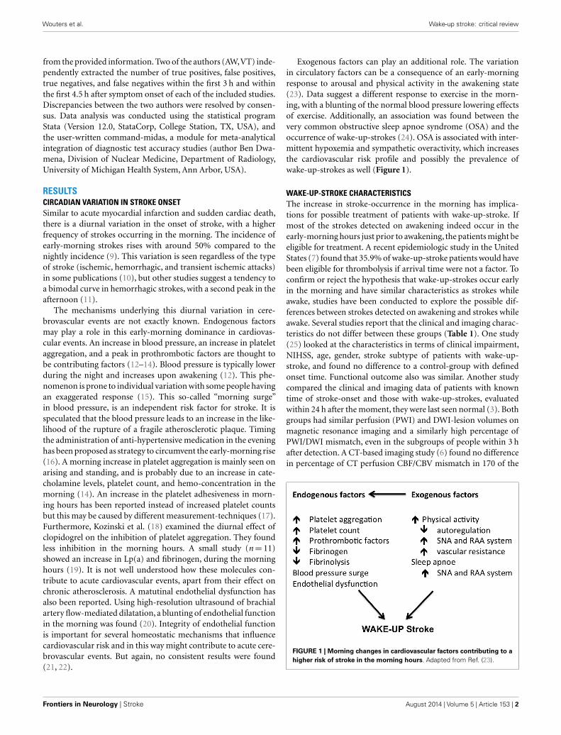
Exogenous factors can play an additional role. The variationin circulatory factors can be a consequence of an early-morningresponse to arousal and physical activity in the awakening state(23). Data suggest a different response to exercise in the morn-ing, with a blunting of the normal blood pressure lowering effectsof exercise. Additionally, an association was found between thevery common obstructive sleep apnoe syndrome (OSA) and theoccurrence of wake-up-strokes (24). OSA is associated with inter-mittent hypoxemia and sympathetic overactivity, which increasesthe cardiovascular risk profile and possibly the prevalence ofwake-up-strokes as well (Figure 1).
WAKE-UP-STROKE CHARACTERISTICSThe increase in stroke-occurrence in the morning has implica-tions for possible treatment of patients with wake-up-stroke. Ifmost of the strokes detected on awakening indeed occur in theearly-morning hours just prior to awakening, the patients might beeligible for treatment. A recent epidemiologic study in the UnitedStates (7) found that 35.9% of wake-up-stroke patients would havebeen eligible for thrombolysis if arrival time were not a factor. Toconfirm or reject the hypothesis that wake-up-strokes occur earlyin the morning and have similar characteristics as strokes whileawake, studies have been conducted to explore the possible dif-ferences between strokes detected on awakening and strokes whileawake. Several studies report that the clinical and imaging charac-teristics do not differ between these groups (Table 1). One study(25) looked at the characteristics in terms of clinical impairment,NIHSS, age, gender, stroke subtype of patients with wake-up-stroke, and found no difference to a control-group with definedonset time. Functional outcome also was similar. Another studycompared the clinical and imaging data of patients with knowntime of stroke-onset and those with wake-up-strokes, evaluatedwithin 24 h after the moment, they were last seen normal (3). Bothgroups had similar perfusion (PWI) and DWI-lesion volumes onmagnetic resonance imaging and a similarly high percentage ofPWI/DWI mismatch, even in the subgroups of people within 3 hafter detection. A CT-based imaging study (6) found no differencein percentage of CT perfusion CBF/CBV mismatch in 170 of the
FIGURE 1 | Morning changes in cardiovascular factors contributing to ahigher risk of stroke in the morning hours. Adapted from Ref. (23).
Frontiers in Neurology | Stroke August 2014 | Volume 5 | Article 153 | 2
Wouters et al. Wake-up stroke: critical review
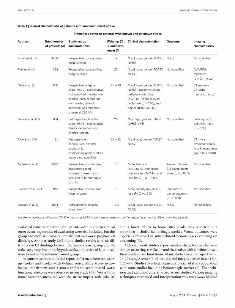
Table 1 | Clinical characteristic of patients with unknown-onset stroke.
Differences between patients with known and unknown stroke
Authors Total number
of patients (n)
Study set up
and limitations
Wake-up (%)
+ unknown
onset (%)
Clinical characteristics Outcome Imaging
characteristics
Koton et al. (25) 4408 Prospective, consecutive,
hospital based
19 N.s.d. (age, gender, TOAST,
NIHSS)
N.s.d. Not specified
Fink et al. (3) 364 Prospective, consecutive,
hospital based
27 N.s.d. (age, gender, TOAST,
NIHSS)
Not specified DWI/PWI
mismatch
(p=0.4): n.s.d.
Silva et al. (6) 676 Prospective, hospital
based (n=2), consecutive.
Not specified if reader was
blinded, each center had
own reader, time of
detection was randomly
chosen at 7.30 AM
20+18 N.s.d. (age, gender, TOAST,
NIHSS); Unknown-onset
patients were older
(p=0.08), more likely to
be female (p=0.04), and
higher NIHSS (p < 0.01)
Not specified CT perfusion
CBF/CBV
mismatch: n.s.d.
Serena et al. (27) 654 Retrospective, hospital
based (n=6), consecutive,
three independent and
blinded readers
24 Nsd. (age, gender, TOAST,
NIHSS, aHT)
Not specified Early signs of
ischemia: n.s.d
(p=0.35)
Todo et al. (26) 81 Retrospective,
consecutive, hospital
based, only
cardioemboligenic strokes,
readers not specified
21+22 N.s.d (age, gender, TOAST,
NIHSS)
Not specified CT: more
hypodens zones
in unknown-onset
group (p > 0.001)
Nadeau et al. (5) 2585 Prospective, consecutive,
population based,
informed consent, also
inclusion of hemorrhagic
strokes
13 More smokers
(p=0.0016), high blood
pressure (p=0.0144), and
less tPa (2.1 vs. 13.5%)
Worse outcome:
SIS seven points
lower (p=0.0012)
Not specified
Jimenez et al. (28) 813 Prospective, consecutive,
hospital based
16 More obesity (p=0.058),
less tPa (0 vs. 3%)
Tendency to
worse outcome
(p=0.038)
Not specified
Mackey et al. (7) 1854 Retrospective, hospital
based (n=7)
14.3 N.s.d. (age, gender, TOAST,
NIHSS)
N.s.d. Not specified
N.s.d=no significant difference, TOAST= trial of org 10172 in acute stroke treatment, aHT= arterial hypertension, SIS= stroke impact scale.
evaluated patients. Interestingly, patients with unknown time ofonset occurring outside of awakening were also included, but thisgroup had more neurological impairment and worse prognosis atdischarge. Another study (26) found similar results with no dif-ferences in CT-findings between the known onset group and thewake-up group, but more hypodensities, indicative of later onset,were found in the unknown-onset group.
In contrast, some studies did report differences between wake-up strokes and strokes with defined onset. More severe neuro-logical impairment and a non-significant trend toward worsefunctional outcome were observed in one study (28). Worse func-tional outcome, measured with the stroke impact scale (SIS-16)
and a lower return to home after stroke was reported in astudy that included hemorrhagic strokes. Worse outcomes wereespecially observed in subarachnoid hemorrhages occurring onawakening (5).
Although most studies report similar characteristics betweenstrokes occurring at wake up and the strokes with a defined onset,these studies have limitations. Many studies were retrospective (7,26, 27), single center (3, 25, 26, 28), and not population based (3, 6,7, 25–28). Studies were heterogeneous in term of study populationwith some studies including hemorrhagic strokes (5). The inclu-sion and exclusion criteria varied across studies. Various imagingtechniques were used and interpretation was not always blinded
www.frontiersin.org August 2014 | Volume 5 | Article 153 | 3
Wouters
etal.
Wake-up
stroke:criticalreview
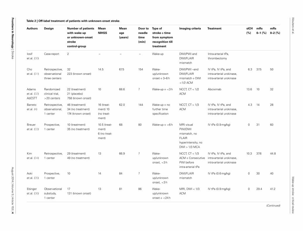
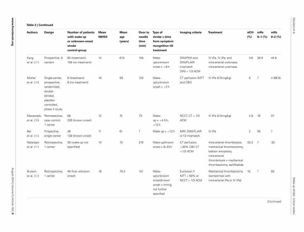
Table 2 | Off-label treatment of patients with unknown-onset stroke.
Authors Design Number of patients
with wake up
or unknown-onset
stroke
control-group
Mean
NIHSS
Mean
age
(years)
Door to
needle
time
(min)
Type of
stroke + time
from symptom
recognition till
treatment
Imaging criteria Treatment sICH
(%)
mRs
0–1 (%)
mRs
0–2 (%)
Iosif
et al. (30)
Case-report 2 – – – Wake-up DWI/PWI and
DWI/FLAIR
mismatch
Intra-arterial tPa,
thrombectomy
Cho
et al. (31)
Retrospective,
observational
three centers
32
223 (known onset)
14.5 67.5 154 Wake-
up/unknown
onset+3–6 h
DWI/PWI –and
DWI/FLAIR
mismatch+DWI
<1/2 ACM
IV tPa, IV tPa, and
intra-arterial urokinase,
intra-arterial urokinase
6.3 37.5 50
Adams
et al. (32)
AbESTT
Randomized
clinical trial,
>20 centers
22 (treatment)
21 (placebo)
758 (known onset)
10 68.6 ? Wake-up+<3 h NCCT, CT < 1/2
ACM
Abciximab 13.6 10 32
Barreto
et al. (4)
Retrospective,
observational,
1 center
46 (treatment)
34 (no treatment)
174 (known onset)
16 (treat-
ment) 10
(no treat-
ment)
62.0 144 Wake-up+no
further time
specification
NCCT, CT > 1/3
ACM
IV tPa, IV tPa, and
intra-arterial urokinase,
intra-arterial urokinase
4.3 14 28
Breuer
et al. (33)
Prospective,
1 center
10 (treatment)
35 (no treatment)
10.5 (treat-
ment)
6 (no treat-
ment)
68 80 Wake-up+<6 h MRI visual
PWI/DWI
mismatch, no
FLAIR
hyperintensity, no
DWI > 1/3 MCA
IV tPa (0.9 mg/kg) 0 31 60
Kim
et al. (34)
Retrospective,
1 center
29 (treatment)
49 (no treatment)
13 66.9 ? Wake-
up/unknown
onset, <3 h
NCCT, CT < 1/3
ACM+Consecutive
PWI before
intra-arterial tPa
IV tPa, IV tPa, and
intra-arterial urokinase,
intra-arterial urokinase
10.3 37.6 44.8
Aoki
et al. (35)
Prospective,
1 center
10 14 84 ? Wake-
up/unknown
onset, <3 h
DWI/FLAIR
mismatch
IV tPa (0.6 mg/kg) 0 30 40
Ebinger
et al. (36)
Observational
substudy,
1 center
17
131 (known onset)
13 81 86 Wake-
up/unknown
onset+<24 h
MRI, DWI < 1/3
ACM
IV tPa (0.9 mg/kg) 0 29.4 41.2
(Continued)
Fron
tiersin
Neu
rolo
gy|S
trokeA
ugust2014
|Volume
5|A
rticle153
|4
Wouters
etal.
Wake-up
stroke:criticalreview
Table 2 | Continued
Authors Design Number of patients
with wake up
or unknown-onset
stroke
control-group
Mean
NIHSS
Mean
age
(years)
Door to
needle
time
(min)
Type of
stroke + time
from symptom
recognition till
treatment
Imaging criteria Treatment sICH
(%)
mRs
0–1 (%)
mRs
0–2 (%)
Kang
et al. (37)
Prospective, 6
centers
83 (treatment)
156 (no treatment)
14 67.5 155 Wake-
up/unknown
onset+<6 h
DWI/PWI and
DWI/FLAIR
mismatch
DWI < 1/3 ACM
IV tPa, IV tPa, and
intra-arterial urokinase,
intra-arterial urokinase
3.6 28.9 44.6
Michel
et al. (38)
Single-center,
prospective,
randomized,
double-
blinded,
placebo-
controlled,
phase II study
6 (treatment)
6 (no treatment)
16 59 122 Wake-
up/unknown
onset+<2 h
CT perfusion (MTT
and CBV)
IV tPa (0.9 mg/kg) 0 ? 4 (66.6)
Manawadu
et al. (39)
Retrospective,
case control,
1 center
68
326 (known onset)
12 74 73 Wake-
up+>4.5 h,
<12 h
NCCT, CT < 1/3
ACM
IV tPa (0.9 mg/kg) 2.9 16 37
Bai
et al. (40)
Prispective,
single center
48
138 (known onset)
11 61 ? Wake up+<12 h MRI: DWI/FLAIR
or T2 mismatch.
IV tPa 2 55 ?
Natarajan
et al. (41)
Retrospective,
1 center
30 (wake-up not
specified)
13 72 210 Wake-up/known
onset+8–23 h
CT perfusion,
>30% CBV CT
<1/3 ACM
Intra-arterial thrombolysis,
mechanical thrombectomy,
balloon anioplasty,
intra-arterial
thrombolysis+mechanical
thrombectomy, eptifibatide
33.3 ? 20
Burkart
et al. (42)
Retrospective,
1 center
40 (five unknown
onset)
18 75.4 151 Wake-
up/unknown
onset/known
onset+ timing
not further
specified
Exclusion if
MTT > 50% or
NCCT > 1/3 ACM
Mechanical thrombectomy
(sometimes with
intra-arterial tPa or IV tPa)
10 ? 50
(Continued)
ww
w.fro
ntiersin
.org
August
2014|Volum
e5
|Article
153|5
Wouters et al. Wake-up stroke: critical review
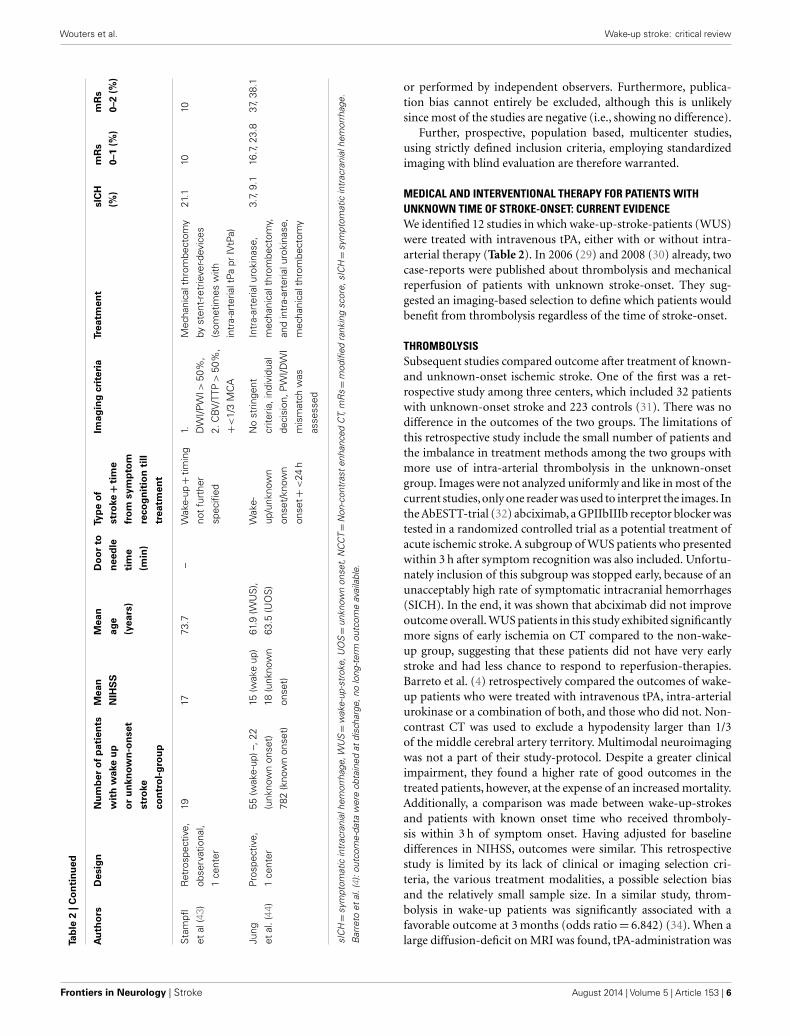
Tab
le2
|Co
nti
nu
ed
Au
tho
rsD
esig
nN
um
ber
of
pat
ien
ts
wit
hw
ake
up
or
un
know
n-o
nse
t
stro
ke
con
tro
l-g
rou
p
Mea
n
NIH
SS
Mea
n
age
(yea
rs)
Do
or
to
nee
dle
tim
e
(min
)
Typ
eo
f
stro
ke+
tim
e
fro
msy
mp
tom
reco
gn
itio
nti
ll
trea
tmen
t
Imag
ing
crit
eria
Trea
tmen
tsI
CH
(%)
mR
s
0–1
(%)
mR
s
0–2
(%)
Sta
mpfl
etal
(43)
Ret
rosp
ectiv
e,
obse
rvat
iona
l,
1ce
nter
1917
73.7
–W
ake-
up+
timin
g
not
furt
her
spec
ified
1. DW
I/PW
I>50
%,
2.C
BV
/TTP
>50
%,
+<
1/3
MC
A
Mec
hani
calt
hrom
bect
omy
byst
ent-
retr
ieve
r-dev
ices
(som
etim
esw
ith
intr
a-ar
teria
ltPa
prIV
tPa)
21.1
1010
Jung
etal
.(44
)
Pros
pect
ive,
1ce
nter
55(w
ake-
up)–
,22
(unk
now
non
set)
782
(kno
wn
onse
t)
15(w
ake
up)
18(u
nkno
wn
onse
t)
61.9
(WU
S),
63.5
(UO
S)
Wak
e-
up/u
nkno
wn
onse
t/kn
own
onse
t+<
24h
No
strin
gent
crite
ria,i
ndiv
idua
l
deci
sion
,PW
I/DW
I
mis
mat
chw
as
asse
ssed
Intr
a-ar
teria
luro
kina
se,
mec
hani
calt
hrom
bect
omy,
and
intr
a-ar
teria
luro
kina
se,
mec
hani
calt
hrom
bect
omy
3.7,
9.1
16.7
,23.
837
,38.
1
sIC
H=
sym
ptom
atic
intr
acra
nial
hem
orrh
age,
WU
S=
wak
e-up
-str
oke,
UO
S=
unkn
own
onse
t,N
CC
T=
Non
-con
tras
ten
hanc
edC
T,m
Rs=
mod
ified
rank
ing
scor
e,sI
CH=
sym
ptom
atic
intr
acra
nial
hem
orrh
age.
Bar
reto
etal
.(4)
:out
com
e-da
taw
ere
obta
ined
atdi
scha
rge,
nolo
ng-t
erm
outc
ome
avai
labl
e.
or performed by independent observers. Furthermore, publica-tion bias cannot entirely be excluded, although this is unlikelysince most of the studies are negative (i.e., showing no difference).
Further, prospective, population based, multicenter studies,using strictly defined inclusion criteria, employing standardizedimaging with blind evaluation are therefore warranted.
MEDICAL AND INTERVENTIONAL THERAPY FOR PATIENTS WITHUNKNOWN TIME OF STROKE-ONSET: CURRENT EVIDENCEWe identified 12 studies in which wake-up-stroke-patients (WUS)were treated with intravenous tPA, either with or without intra-arterial therapy (Table 2). In 2006 (29) and 2008 (30) already, twocase-reports were published about thrombolysis and mechanicalreperfusion of patients with unknown stroke-onset. They sug-gested an imaging-based selection to define which patients wouldbenefit from thrombolysis regardless of the time of stroke-onset.
THROMBOLYSISSubsequent studies compared outcome after treatment of known-and unknown-onset ischemic stroke. One of the first was a ret-rospective study among three centers, which included 32 patientswith unknown-onset stroke and 223 controls (31). There was nodifference in the outcomes of the two groups. The limitations ofthis retrospective study include the small number of patients andthe imbalance in treatment methods among the two groups withmore use of intra-arterial thrombolysis in the unknown-onsetgroup. Images were not analyzed uniformly and like in most of thecurrent studies,only one reader was used to interpret the images. Inthe AbESTT-trial (32) abciximab, a GPIIbIIIb receptor blocker wastested in a randomized controlled trial as a potential treatment ofacute ischemic stroke. A subgroup of WUS patients who presentedwithin 3 h after symptom recognition was also included. Unfortu-nately inclusion of this subgroup was stopped early, because of anunacceptably high rate of symptomatic intracranial hemorrhages(SICH). In the end, it was shown that abciximab did not improveoutcome overall. WUS patients in this study exhibited significantlymore signs of early ischemia on CT compared to the non-wake-up group, suggesting that these patients did not have very earlystroke and had less chance to respond to reperfusion-therapies.Barreto et al. (4) retrospectively compared the outcomes of wake-up patients who were treated with intravenous tPA, intra-arterialurokinase or a combination of both, and those who did not. Non-contrast CT was used to exclude a hypodensity larger than 1/3of the middle cerebral artery territory. Multimodal neuroimagingwas not a part of their study-protocol. Despite a greater clinicalimpairment, they found a higher rate of good outcomes in thetreated patients, however, at the expense of an increased mortality.Additionally, a comparison was made between wake-up-strokesand patients with known onset time who received thromboly-sis within 3 h of symptom onset. Having adjusted for baselinedifferences in NIHSS, outcomes were similar. This retrospectivestudy is limited by its lack of clinical or imaging selection cri-teria, the various treatment modalities, a possible selection biasand the relatively small sample size. In a similar study, throm-bolysis in wake-up patients was significantly associated with afavorable outcome at 3 months (odds ratio= 6.842) (34). When alarge diffusion-deficit on MRI was found, tPA-administration was
Frontiers in Neurology | Stroke August 2014 | Volume 5 | Article 153 | 6
Wouters et al. Wake-up stroke: critical review
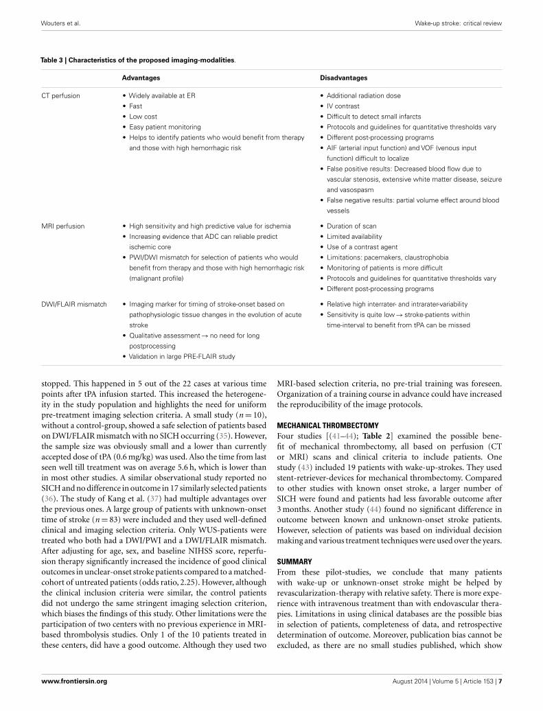
Table 3 | Characteristics of the proposed imaging-modalities.
Advantages Disadvantages
CT perfusion • Widely available at ER
• Fast
• Low cost
• Easy patient monitoring
• Helps to identify patients who would benefit from therapy
and those with high hemorrhagic risk
• Additional radiation dose
• IV contrast
• Difficult to detect small infarcts
• Protocols and guidelines for quantitative thresholds vary
• Different post-processing programs
• AIF (arterial input function) and VOF (venous input
function) difficult to localize
• False positive results: Decreased blood flow due to
vascular stenosis, extensive white matter disease, seizure
and vasospasm
• False negative results: partial volume effect around blood
vessels
MRI perfusion • High sensitivity and high predictive value for ischemia
• Increasing evidence that ADC can reliable predict
ischemic core
• PWI/DWI mismatch for selection of patients who would
benefit from therapy and those with high hemorrhagic risk
(malignant profile)
• Duration of scan
• Limited availability
• Use of a contrast agent
• Limitations: pacemakers, claustrophobia
• Monitoring of patients is more difficult
• Protocols and guidelines for quantitative thresholds vary
• Different post-processing programs
DWI/FLAIR mismatch • Imaging marker for timing of stroke-onset based on
pathophysiologic tissue changes in the evolution of acute
stroke
• Qualitative assessment→no need for long
postprocessing
• Validation in large PRE-FLAIR study
• Relative high interrater- and intrarater-variability
• Sensitivity is quite low→ stroke-patients within
time-interval to benefit from tPA can be missed
stopped. This happened in 5 out of the 22 cases at various timepoints after tPA infusion started. This increased the heterogene-ity in the study population and highlights the need for uniformpre-treatment imaging selection criteria. A small study (n= 10),without a control-group, showed a safe selection of patients basedon DWI/FLAIR mismatch with no SICH occurring (35). However,the sample size was obviously small and a lower than currentlyaccepted dose of tPA (0.6 mg/kg) was used. Also the time from lastseen well till treatment was on average 5.6 h, which is lower thanin most other studies. A similar observational study reported noSICH and no difference in outcome in 17 similarly selected patients(36). The study of Kang et al. (37) had multiple advantages overthe previous ones. A large group of patients with unknown-onsettime of stroke (n= 83) were included and they used well-definedclinical and imaging selection criteria. Only WUS-patients weretreated who both had a DWI/PWI and a DWI/FLAIR mismatch.After adjusting for age, sex, and baseline NIHSS score, reperfu-sion therapy significantly increased the incidence of good clinicaloutcomes in unclear-onset stroke patients compared to a matched-cohort of untreated patients (odds ratio, 2.25). However, althoughthe clinical inclusion criteria were similar, the control patientsdid not undergo the same stringent imaging selection criterion,which biases the findings of this study. Other limitations were theparticipation of two centers with no previous experience in MRI-based thrombolysis studies. Only 1 of the 10 patients treated inthese centers, did have a good outcome. Although they used two
MRI-based selection criteria, no pre-trial training was foreseen.Organization of a training course in advance could have increasedthe reproducibility of the image protocols.
MECHANICAL THROMBECTOMYFour studies [(41–44); Table 2] examined the possible bene-fit of mechanical thrombectomy, all based on perfusion (CTor MRI) scans and clinical criteria to include patients. Onestudy (43) included 19 patients with wake-up-strokes. They usedstent-retriever-devices for mechanical thrombectomy. Comparedto other studies with known onset stroke, a larger number ofSICH were found and patients had less favorable outcome after3 months. Another study (44) found no significant difference inoutcome between known and unknown-onset stroke patients.However, selection of patients was based on individual decisionmaking and various treatment techniques were used over the years.
SUMMARYFrom these pilot-studies, we conclude that many patientswith wake-up or unknown-onset stroke might be helped byrevascularization-therapy with relative safety. There is more expe-rience with intravenous treatment than with endovascular thera-pies. Limitations in using clinical databases are the possible biasin selection of patients, completeness of data, and retrospectivedetermination of outcome. Moreover, publication bias cannot beexcluded, as there are no small studies published, which show
www.frontiersin.org August 2014 | Volume 5 | Article 153 | 7
Wouters et al. Wake-up stroke: critical review
unfavorable results. Only two studies (32, 38) testing thromboly-sis or thrombectomy were randomized. One was the AbESTT-trial(32), discussed previously and the other one was a small pilot-study with only 12 patients included (38). The imaging selectioncriteria used to select patients for treatment were not uniform,since most centers used individual decision making to treat thissubset of patients. A large clinical randomized trial is thereforeneeded to confirm these preliminary results.
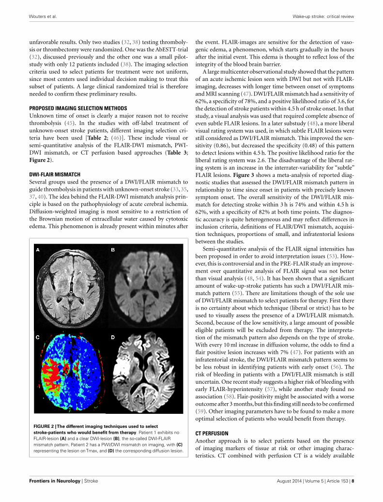
PROPOSED IMAGING SELECTION METHODSUnknown time of onset is clearly a major reason not to receivethrombolysis (45). In the studies with off-label treatment ofunknown-onset stroke patients, different imaging selection cri-teria have been used [Table 2; (46)]. These include visual orsemi-quantitative analysis of the FLAIR-DWI mismatch, PWI-DWI mismatch, or CT perfusion based approaches (Table 3;Figure 2).
DWI-FLAIR MISMATCHSeveral groups used the presence of a DWI/FLAIR mismatch toguide thrombolysis in patients with unknown-onset stroke (33, 35,37, 40). The idea behind the FLAIR-DWI mismatch analysis prin-ciple is based on the pathophysiology of acute cerebral ischemia.Diffusion-weighted imaging is most sensitive to a restriction ofthe Brownian motion of extracellular water caused by cytotoxicedema. This phenomenon is already present within minutes after
FIGURE 2 |The different imaging techniques used to selectstroke-patients who would benefit from therapy. Patient 1 exhibits noFLAIR-lesion (A) and a clear DWI-lesion (B), the so-called DWI-FLAIRmismatch pattern. Patient 2 has a PWI/DWI mismatch on imaging, with (C)representing the lesion onTmax, and (D) the corresponding diffusion lesion.
the event. FLAIR-images are sensitive for the detection of vaso-genic edema, a phenomenon, which starts gradually in the hoursafter the initial event. This edema is thought to reflect loss of theintegrity of the blood brain barrier.
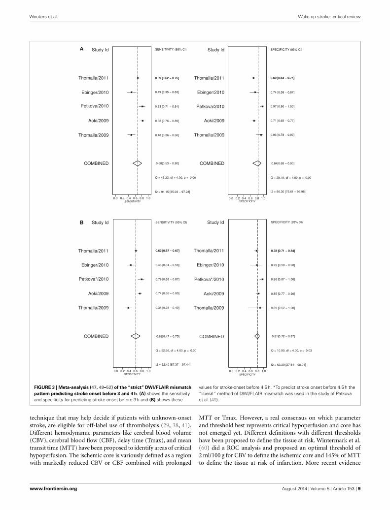
A large multicenter observational study showed that the patternof an acute ischemic lesion seen with DWI but not with FLAIR-imaging, decreases with longer time between onset of symptomsand MRI scanning (47). DWI/FLAIR mismatch had a sensitivity of62%, a specificity of 78%, and a positive likelihood ratio of 3.6, forthe detection of stroke patients within 4.5 h of stroke onset. In thatstudy, a visual analysis was used that required complete absence ofeven subtle FLAIR lesions. In a later substudy (48), a more liberalvisual rating system was used, in which subtle FLAIR lesions werestill considered as DWI/FLAIR mismatch. This improved the sen-sitivity (0.86), but decreased the specificity (0.48) of this patternto detect lesions within 4.5 h. The positive likelihood ratio for theliberal rating system was 2.6. The disadvantage of the liberal rat-ing system is an increase in the interrater-variability for “subtle”FLAIR lesions. Figure 3 shows a meta-analysis of reported diag-nostic studies that assessed the DWI/FLAIR mismatch pattern inrelationship to time since onset in patients with precisely knownsymptom onset. The overall sensitivity of the DWI/FLAIR mis-match for detecting stroke within 3 h is 74% and within 4.5 h is62%, with a specificity of 82% at both time points. The diagnos-tic accuracy is quite heterogeneous and may reflect differences ininclusion criteria, definitions of FLAIR/DWI mismatch, acquisi-tion techniques, proportions of small, and infratentorial lesionsbetween the studies.
Semi-quantitative analysis of the FLAIR signal intensities hasbeen proposed in order to avoid interpretation issues (53). How-ever, this is controversial and in the PRE-FLAIR study an improve-ment over quantitative analysis of FLAIR signal was not betterthan visual analysis (48, 54). It has been shown that a significantamount of wake-up-stroke patients has such a DWI/FLAIR mis-match pattern (55). There are limitations though of the sole useof DWI/FLAIR mismatch to select patients for therapy. First thereis no certainty about which technique (liberal or strict) has to beused to visually assess the presence of a DWI/FLAIR mismatch.Second, because of the low sensitivity, a large amount of possibleeligible patients will be excluded from therapy. The interpreta-tion of the mismatch pattern also depends on the type of stroke.With every 10 ml increase in diffusion volume, the odds to find aflair positive lesion increases with 7% (47). For patients with aninfratentorial stroke, the DWI/FLAIR mismatch pattern seems tobe less robust in identifying patients with early onset (56). Therisk of bleeding in patients with a DWI/FLAIR mismatch is stilluncertain. One recent study suggests a higher risk of bleeding withearly FLAIR-hyperintensity (57), while another study found noassociation (58). Flair-positivity might be associated with a worseoutcome after 3 months,but this finding still needs to be confirmed(59). Other imaging parameters have to be found to make a moreoptimal selection of patients who would benefit from therapy.
CT PERFUSIONAnother approach is to select patients based on the presenceof imaging markers of tissue at risk or other imaging charac-teristics. CT combined with perfusion CT is a widely available
Frontiers in Neurology | Stroke August 2014 | Volume 5 | Article 153 | 8
Wouters et al. Wake-up stroke: critical review
A
B
FIGURE 3 | Meta-analysis (47, 49–52) of the “strict” DWI/FLAIR mismatchpattern predicting stroke onset before 3 and 4 h. (A) shows the sensitivityand specificity for predicting stroke-onset before 3 h and (B) shows these
values for stroke-onset before 4.5 h. *To predict stroke onset before 4.5 h the“liberal” method of DWI/FLAIR mismatch was used in the study of Petkovaet al. (49).
technique that may help decide if patients with unknown-onsetstroke, are eligible for off-label use of thrombolysis (29, 38, 41).Different hemodynamic parameters like cerebral blood volume(CBV), cerebral blood flow (CBF), delay time (Tmax), and meantransit time (MTT) have been proposed to identify areas of criticalhypoperfusion. The ischemic core is variously defined as a regionwith markedly reduced CBV or CBF combined with prolonged
MTT or Tmax. However, a real consensus on which parameterand threshold best represents critical hypoperfusion and core hasnot emerged yet. Different definitions with different thresholdshave been proposed to define the tissue at risk. Wintermark et al.(60) did a ROC analysis and proposed an optimal threshold of2 ml/100 g for CBV to define the ischemic core and 145% of MTTto define the tissue at risk of infarction. More recent evidence
www.frontiersin.org August 2014 | Volume 5 | Article 153 | 9
Wouters et al. Wake-up stroke: critical review
suggests that relative CBF might be better to define infarct corethen CBV (61, 62). The more recent literature suggests a thresholdof CT-Tmax of >6 s to define the tissue at risk (63). Despite thatno real consensus exists about the thresholds that should be used,the speed and wide availability make perfusion CT an interestingalternative compared to other perfusion-modalities.
PWI/DWI MISMATCHPerfusion and diffusion based MR imaging techniques have beenadvocated as an imaging selection method in wake-up strokes(Figure 2). The disadvantage of the latter technique is the longerimaging time required and the limited availability of MRI com-pared to CT. Dynamic susceptibility contrast enhanced MRI isthe most widely used technique. Arterial spin labeling is a newermethod and has no need for contrast, but requires, in general,longer imaging times. MRI does have the advantage of a reliableprediction of the ischemic core with ADC-maps (64). However,the possible reversibility of the diffusion lesion, questions theparadigm that diffusion lesions represent the ischemic core. Analy-sis of the EPITHET-data showed that true DWI-lesion reversalis uncommon and if present would rarely alter treatment deci-sion making (65). As with CT, difficulties arise in determining theoptimal thresholds to differentiate ischemic core from salvageablebrain tissue (66). Also the selection of the most optimal parame-ter or combination of parameters is still a matter of debate (67).An ADC-threshold of 600× 10−6 mm2/s seems a fairly robustparameter in predicting ischemic core tissue (64). The mismatchbetween an area that has a Tmax > 6 s and is below this ADC-threshold is currently considered in several clinical trials as anoperational definition of the tissue at risk. A substudy of DEFUSE2 (68) supported this hypothesis by showing that in patients witha strong reperfusion, there is a high correlation between base-line DWI-volume and final infarct and in patients with minimalor no reperfusion, there is a high correlation between the base-line PWI-volume and final infarct. To determine the tissue atrisk the PWI-DWI mismatch is useful, with 120% most com-monly used to define a mismatch, although more stringent criteriahave been advocated, with studies now advocating a perfusion-diffusion ratio that is larger than 180%, dependent on the para-meter that is used to define the perfusion abnormality. MRI canalso detect the so-called “ malignant” profile. The DEFUSE-data(69) showed that patients with a baseline DWI-lesion bigger than100 ml and/or a PWI lesion of 100 ml or more with 8 s or longerof Tmax delay, suffered more intracranial hemorrhages after earlyreperfusion. The major drawback in the clinical use of MRI per-fusion is that the extent of perfusion abnormalities varies amongperfusion parameters, software packages, and various algorithmsand that upon today no consensus is reached. Other techniquesto define tissue at risk, like FDG-PET or SPECT, are less eas-ily used in clinical practice. Two large trials to treat wake-uppatients are currently ongoing, one based on DWI/FLAIR mis-match (WAKE-UP) (8) and one based on penumbral imaging(EXTEND) (70).
CONCLUSIONWake-up-stroke and stroke with unknown time of onset are fre-quent. These patients are at present excluded from thrombolytic
therapy. Evidence suggests that these strokes occur closely onawakening and most observational studies did not find differencesin terms of clinical features or outcome after therapy, suggestingthat at least a subset of these patients could benefit of throm-bolysis or endovascular treatment. Selection of eligible patientsis preferably done using neuro-imaging, but the optimal imag-ing selection strategy for treating these patients has not yet beendefined. The proposed selection modalities have not been properlyevaluated in randomized trials, therefore, inclusion in these trialsis primordial. In case randomization is not possible, advocatingtreatment on the basis of the presence of a DWI/FLAIR mismatchor PWI/DWI mismatch can generally not be recommended. Inthis scenario, treatment must remain an individualized decision.Ongoing randomized controlled trials testing these strategies arethe WAKE-UP trial (8) and the EXTEND-trial (70). Identifying asafe and efficacious selection strategy will not only benefit patientswith WUS, but also allow moving away from a rigid time windowbased approach for all patients.
ACKNOWLEDGMENTSAnke Wouters receives a research grant from the European Union.Rahmenprogramm der Europäischen Union [FP7/2007–2013]gefördert, Grant Agreement Nr. 278276 (WAKE-UP).
REFERENCES1. Seshadri S, Wolf PA. Lifetime risk of stroke and dementia: current concepts, and
estimates from the Framingham Study. Lancet Neurol (2007) 6(12):1106–14.doi:10.1016/S1474-4422(07)70291-0
2. Hacke W, Kaste M, Bluhmki E, Brozman M, Dávalos A, Guidetti D, et al. Throm-bolysis with alteplase 3 to 4.5 hours after acute ischemic stroke. N Engl J Med(2008) 13:1317–29. doi:10.1056/NEJMoa0804656
3. Fink JN, Kumar S, Horkan C, Linfante I, Selim MH, Caplan LR, et al. The strokepatient who woke up: clinical and radiological features, including diffusion andperfusion MRI. Stroke (2002) 33(4):988–93. doi:10.1161/01.STR.0000014585.17714.67
4. Barreto AD, Martin-Schild S, Hallevi H, Morales MM, Abraham AT, GonzalesNR, et al. Thrombolytic therapy for patients who wake-up with stroke. Stroke AJ Cereb Circ (2009) 40:827–32. doi:10.1161/STROKEAHA.108.528034
5. Nadeau JO, Fang J, Kapral MK, Silver FL, Hill MD. Outcome after stroke uponawakening. Can J Neurol Sci Le J Can des Sci Neuro (2005) 32(2):232–6.
6. Silva GS, Lima FO, Camargo ECS, Smith WS, Singhal AB, Greer DM, et al. Wake-up stroke: clinical and neuroimaging characteristics. Cerebrovasc Dis Basel SwitzS (2010) 29(4):336–42. doi:10.1159/000278929
7. Mackey J, Kleindorfer D, Sucharew H, Moomaw CJ, Kissela BM, Alwell K,et al. Population-based study of wake-up strokes. Neurology (2011) 76:1662–7.doi:10.1212/WNL.0b013e318219fb30
8. Thomalla G, Fiebach JB, Ostergaard L, Pedraza S, Thijs V, Nighoghossian N, et al.A multicenter, randomized, double-blind, placebo-controlled trial to test effi-cacy and safety of magnetic resonance imaging-based thrombolysis in wake-upstroke (WAKE-UP). Int J Stroke (2013) 278276:1–8. doi:10.1111/ijs.12011
9. Marler JR, Price TR, Clark GL, Muller JE, Robertson T, Mohr JP, et al. Morningincrease in onset of ischemic stroke. Stroke (1989) 20(4):473–6.
10. Elliott WJ. Circadian variation in the timing of stroke onset: a meta-analysis.Stroke (1998) 29(5):992–6. doi:10.1161/01.STR.29.5.992
11. Omama S, Yoshida Y, Ogawa A, Onoda T, Okayama A. Differences in circa-dian variation of cerebral infarction, intracerebral haemorrhage and subarach-noid haemorrhage by situation at onset. J Neurol Neurosurg Psychiatry (2006)77(12):1345–9. doi:10.1136/jnnp.2006.090373
12. Redon J. The normal circadian pattern of blood pressure: implications fortreatment. Int J Clin Pract Suppl (2004) 145:3–8. doi:10.1111/j.1742-1241.2004.00403.x
13. Kario K, Yano Y, Matsuo T, Hoshide S, Asada Y, Shimada K. Morning bloodpressure surge, morning platelet aggregation, and silent cerebral infarction
Frontiers in Neurology | Stroke August 2014 | Volume 5 | Article 153 | 10
Wouters et al. Wake-up stroke: critical review
in older Japanese hypertensive patients. J Hypertens (2011) 29(12):2433–9.doi:10.1097/HJH.0b013e32834cf1c0
14. Andrews NP, Gralnick HR, Merryman P, Vail M, Quyyumi AA. Mechanismsunderlying the morning increase in platelet aggregation: a flow cytometry study.J Am Coll Cardiol (1996) 28:1789–95. doi:10.1016/S0735-1097(96)00398-1
15. Kario K. Morning surge in blood pressure as a predictor of silent and clinicalcerebrovascular disease in elderly hypertensives: A Prospective Study. Circulation(2003) 107(10):1401–6. doi:10.1161/01.CIR.0000056521.67546.AA
16. Ashizawa N, Seto S, Shibata Y, Yano K. Bedtime administration of cilnidipinecontrols morning hypertension. Int Heart J (2007) 48:597–603. doi:10.1536/ihj.48.597
17. Haus E. Chronobiology of hemostasis and inferences for the chronotherapy ofcoagulation disorders and thrombosis prevention. Adv Drug Deliv Rev (2007)59(9–10):966–84. doi:10.1016/j.addr.2006.11.002
18. Kozinski M, Bielis L, Wisniewska-Szmyt J, Boinska J, Stolarek W, Marciniak A,et al. Diurnal variation in platelet inhibition by clopidogrel. Platelets (2011)22(8):579–87. doi:10.3109/09537104.2011.582900
19. Bremner W, Sothern RB, Kanabrocki EL, Ryan M, McCormick JB, Connors ES,et al. Relation between circadian patterns in levels of circulating lipoprotein(a), fibrinogen, platelets, and related lipid variables in men. Am Heart J (2000)139:164–73. doi:10.1016/S0002-8703(00)90324-7
20. Otto ME, Svatikova A, Barretto RBDM, Santos S, Hoffmann M, KhandheriaB, et al. Early morning attenuation of endothelial function in healthy humans.Circulation (2004) 109(21):2507–10. doi:10.1161/01.CIR.0000128207.26863.C4
21. Kawano H, Motoyama T, Yasue H, Hirai N, Waly HM, Ogawa H. Endothe-lial function fluctuates with diurnal variation in the frequency of ischemicepisodes in patients with variant angina. J Am Coll Cardiol (2002) 40(2):266–70.doi:10.1016/S0735-1097(02)01956-3
22. Shaw JA, Chin-dusting JPF, Kingwell BA, Dart AM. Diurnal variation inendothelium-dependent vasodilatation is not apparent in coronary artery dis-ease. Circulation (2001) 103:806–12. doi:10.1161/01.CIR.103.6.806
23. Atkinson G, Jones H,Ainslie PN. Circadian variation in the circulatory responsesto exercise: relevance to the morning peaks in strokes and cardiac events. Eur JAppl Physiol (2010) 108(1):15–29. doi:10.1007/s00421-009-1243-y
24. Hsieh S-W, Lai C-L, Liu C-K, Hsieh C-F, Hsu C-Y. Obstructive sleep apnealinked to wake-up strokes. J Neurol (2012) 259(7):1433–9. doi:10.1007/s00415-011-6370-9
25. Koton S, Tanne D, Bornstein NM. Ischemic stroke on awakening: patients’ char-acteristics, outcomes and potential for reperfusion therapy. Neuroepidemiology(2012) 39(3–4):149–53. doi:10.1159/000341242
26. Todo K, Moriwaki H, Saito K, Tanaka M, Oe H, Naritomi H. Early CT find-ings in unknown-onset and wake-up strokes. Cerebrovasc Dis Basel Switz (2006)21(5–6):367–71. doi:10.1159/000091545
27. Serena J, Dávalos A, Segura T, Mostacero E, Castillo J. Stroke on awakening:looking for a more rational management. Cerebrovasc Dis (2003) 16(2):128–33.doi:10.1159/000070592
28. Jiménez-Conde J, Ois A, Rodríguez-Campello A, Gomis M, Roquer J. Does sleepprotect against ischemic stroke? Less frequent ischemic strokes but more severeones. J Neurol (2007) 254(6):782–8. doi:10.1007/s00415-006-0438-y
29. Hellier KD, Hampton JL, Guadagno JV, Higgins NP, Antoun NM, Day DJ,et al. Perfusion CT helps decision making for thrombolysis when there isno clear time of onset. J Neurol Neurosurg Psychiatry (2006) 77(3):417–9.doi:10.1136/jnnp.2005.067363
30. Iosif C, Oppenheim C, Trystram D, Domigo V, Méder J-F. MR imaging-baseddecision in thrombolytic therapy for stroke on awakening: report of 2 cases.AJNR Am J Neuroradiol (2008) 29:1314–6. doi:10.3174/ajnr.A1069
31. Cho A-H, Sohn S-I, Han M-K, Lee DH, Kim JS, Choi CG, et al. Safety and effi-cacy of MRI-based thrombolysis in unclear-onset stroke. A preliminary report.Cerebrovasc Dis Basel Switz (2008) 25(6):572–9. doi:10.1159/000132204
32. Adams HP,Leira EC,Torner JC,Barnathan E,Padgett L,Effron MB,et al. Treatingpatients with “wake-up” stroke: the experience of the AbESTT-II trial. Stroke AJ Cereb Circ (2008) 39(12):3277–82. doi:10.1161/STROKEAHA.107.508853
33. Breuer L, Schellinger PD, Huttner HB, Halwachs R, Engelhorn T, Doerfler A,et al. Feasibility and safety of magnetic resonance imaging-based thrombolysisin patients with stroke on awakening: initial single-centre experience. Int J Stroke(2010) 5(2):68–73. doi:10.1111/j.1747-4949.2010.00410.x
34. Kim J-T, Park M-S, Nam T-S, Choi S-M, Kim B-C, Kim M-K, et al.Thrombolysis as a factor associated with favorable outcomes in patients with
unclear-onset stroke. Eur J Neurol (2011) 18(7):988–94. doi:10.1111/j.1468-1331.2011.03351.x
35. Aoki J, Kimura K, Iguchi Y, Shibazaki K, Iwanaga T, Watanabe M, et al. Intra-venous thrombolysis based on diffusion-weighted imaging and fluid-attenuatedinversion recovery mismatch in acute stroke patients with unknown onset time.Cerebrovasc Dis (2011) 31(5):435–41. doi:10.1159/000323850
36. Ebinger M, Scheitz JF, Kufner A, Endres M, Fiebach JB, Nolte CH. MRI-basedintravenous thrombolysis in stroke patients with unknown time of symp-tom onset. Eur J Neurol (2012) 19(2):348–50. doi:10.1111/j.1468-1331.2011.03504.x
37. Kang D-W, Sohn S-I, Hong K-S, Yu K-H, Hwang Y-H, Han M-K, et al. Reper-fusion therapy in unclear-onset stroke based on MRI evaluation (RESTORE):a prospective multicenter study. Stroke a J Cereb Circ (2012) 43(12):3278–83.doi:10.1161/STROKEAHA.112.675926
38. Michel P, Ntaios G, Reichhart M, Schindler C, Bogousslavsky J, Maeder P, et al.Perfusion-CT guided intravenous thrombolysis in patients with unknown-onsetstroke: a randomized, double-blind, placebo-controlled, pilot feasibility trial.Neuroradiology (2012) 54(6):579–88. doi:10.1007/s00234-011-0944-1
39. Manawadu D, Bodla S, Jarosz J, Keep J, Kalra L. A case-controlled comparisonof thrombolysis outcomes between wake-up and known time of onset ischemicstroke patients. Stroke (2013) 44(8):2226–31. doi:10.1161/STROKEAHA.111.000757
40. Bai Q, Zhao Z, Fu P, Sui H, Xie X, Chen J, et al. Clinical outcomes of fast MRI-based thrombolysis in wake-up strokes compared to superacute ischemic strokeswithin 12 hours. Neurol Res (2013) 35(5):492–7. doi:10.1179/1743132813Y.0000000208
41. Natarajan SK, Snyder KV, Siddiqui AH, Ionita CC, Hopkins LN, Levy EI.Safety and effectiveness of endovascular therapy after 8 hours of acute ischemicstroke onset and wake-up strokes. Stroke (2009) 40(10):3269–74. doi:10.1161/STROKEAHA.109.555102
42. Burkart DJ, Day JS, Henderson K, Borsa JJ. Efficacy of peripheral interventionalradiologists performing endovascular stroke therapy guided by CT perfusiontriage of patients. J Vasc Interv Radiol (2013) 24(9):1267–72. doi:10.1016/j.jvir.2013.05.002
43. Stampfl S, Ringleb PA, Haehnel S, Rocco A, Herweh C, Hametner C, et al.Recanalization with Stent-Retriever Devices in Patients with Wake-Up Stroke.AJNR Am J Neuroradiol (2012) 34:1040–3. doi:10.3174/ajnr.A3357
44. Jung S, Gralla J, Fischer U, Mono M-L, Weck A, Lüdi R, et al. Safety of endovas-cular treatment beyond the 6-h time window in 205 patients. Eur J Neurol (2013)20(6):865–71. doi:10.1111/ene.12069
45. Kleindorfer D, Kissela B, Schneider A, Woo D, Khoury J, Miller R, et al.Eligibility for recombinant tissue plasminogen activator in acute ischemicstroke: a population-based study. Stroke (2004) 35(2):e27–9. doi:10.1161/01.STR.0000109767.11426.17
46. Rimmele DL, Thomalla G. Wake-up stroke: clinical characteristics, imag-ing findings, and treatment option – an update. Front Neurol (2014) 5:35.doi:10.3389/fneur.2014.00035
47. Thomalla G, Cheng B, Ebinger M, Hao Q, Tourdias T, Wu O, et al. DWI-FLAIRmismatch for the identification of patients with acute ischaemic stroke within4·5 h of symptom onset (PRE-FLAIR): a multicentre observational study. Lancet(2011) 10(11):978–86. doi:10.1016/S1474-4422(11)70192-2
48. Cheng B, Brinkmann M, Forkert ND, Treszl A, Ebinger M, Köhrmann M,et al. Quantitative measurements of relative fluid-attenuated inversion recov-ery (FLAIR) signal intensities in acute stroke for the prediction of time fromsymptom onset. J Cereb Blood Flow Metab (2013) 33(1):76–84. doi:10.1038/jcbfm.2012.129
49. Petkova M, Rodrigo S, Larny C, Oppenheim G, Touzé E, Mas JL, et al. MRImaging helps predict time from symptom onset in patients with acute stroke.Neuroradiology (2010) 257(3):782–92. doi:10.1148/radiol.10100461
50. Ebinger M, Galinovic I, Rozanski M, Brunecker P, Endres M, Fiebach J. Fluid-attenuated inversion recovery evolution within 12 hours from stroke onset: areliable tissue clock? Stroke (2010) 41:250–5. doi:10.1161/STROKEAHA.109.568410
51. Aoki J, Kimura K, Iguchi Y, Shibazaki K, Sakai K, Iwanga T. Flair can estimate theonset time in acute ischemic stroke patients. J Neurol Sci (2010) 293(1–2):39–44.doi:10.1016/j.jns.2010.03.011
52. Thomalla G, Rossbach P, Rosenkranz M, Siemonsen S, Krützelmann A, FiehlerJ, et al. Negative fluid-attenuated inversion recovery imaging identifies acute
www.frontiersin.org August 2014 | Volume 5 | Article 153 | 11
Wouters et al. Wake-up stroke: critical review
ischemic stroke at 3 hours or less. Ann Neurol (2009) 65:724–32. doi:10.1002/ana.21651
53. Song SS, Latour LL, Ritter CH, Wu O, Tighiouart M, Hernandez D, et al. A prag-matic approach using magnetic resonance imaging to treat ischemic strokesof unknown onset time in a thrombolytic trial. Stroke (2012) 43(9):2331–5.doi:10.1161/STROKEAHA.111.630947
54. Galinovic I, Puig J, Neeb L, Guibernau J, Kemmling A, Pedraza S, et al. Visual andregion of interest-based inter-rater agreement in the assessment of the diffusion-weighted imaging-fluid-attenuated inversion recovery mismatch. Stroke (2014)45:1170–2. doi:10.1161/STROKEAHA.113.002661
55. Kim BJ, Kim H-J, Lee DH, Kwon SU, Kim SJ, Kim JS, et al. Diffusion-weighted image and fluid-attenuated inversion recovery image mismatch:unclear-onset versus clear-onset stroke. Stroke (2014) 45(2):450–5. doi:10.1161/STROKEAHA.113.002830
56. Grosse-Dresselhaus F, Galinovic I, Villringer K, Audebert HJ, Fiebach JB. Diffi-culty of MRI based identification of lesion age by acute infra-tentorial ischemicstroke. PLoS One (2014) 9(3):e92868. doi:10.1371/journal.pone.0092868
57. Kufner A, Galinovic I, Brunecker P, Cheng B, Thomalla G, Gerloff C, et al. Earlyinfarct FLAIR hyperintensity is associated with increased hemorrhagic trans-formation after thrombolysis. Eur J Neurol (2013) 20(2):281–5. doi:10.1111/j.1468-1331.2012.03841.x
58. Campbell BCV, Costello C, Christensen S, Ebinger M, Parsons MW, DesmondPM, et al. Fluid-attenuated inversion recovery hyperintensity in acute ischemicstroke may not predict hemorrhagic transformation. Cerebrovasc Dis (2011)32(4):401–5. doi:10.1159/000331467
59. Ebinger M, Kufner A, Galinovic I, Brunecker P, Malzahn U, Nolte CH, et al.Fluid-attenuated inversion recovery images and stroke outcome after thrombol-ysis. Stroke (2012) 43(2):539–42. doi:10.1161/STROKEAHA.111.632026
60. Wintermark M, Flanders AE, Velthuis B, Meuli R, van Leeuwen M, Gold-sher D, et al. Perfusion-CT assessment of infarct core and penumbra: receiveroperating characteristic curve analysis in 130 patients suspected of acute hemi-spheric stroke. Stroke (2006) 37(4):979–85. doi:10.1161/01.STR.0000209238.61459.39
61. Campbell BCV, Christensen S, Levi CR, Desmond PM, Donnan G. Davis SM,et al. Cerebral blood flow is the optimal CT perfusion parameter for assessinginfarct core. Stroke (2011) 42(12):3435–40. doi:10.1161/STROKEAHA.111.618355
62. Bivard A, Levi C, Spratt N, Parsons M. Perfusion CT in acute stroke: a com-prehensive analysis of infarct and penumbra. Radiology (2013) 267(2):543–50.doi:10.1148/radiol.12120971
63. Campbell BCV, Christensen S, Levi CR, Desmond PM, Donnan G, Davis SM,et al. Comparison of computed tomography perfusion and magnetic reso-nance imaging perfusion-diffusion mismatch in ischemic stroke. Stroke (2012)43(10):2648–53. doi:10.1161/STROKEAHA.112.660548
64. Purushotham A, Campbell BCV, Straka M, Mlynash M, Olivot J-M, Bammer R,et al. Apparent diffusion coefficient threshold for delineation of ischemic core.Int J Stroke (2013) 13:1–6. doi:10.1111/ijs.12068
65. Chemmanam T, Campbell BCV, Christensen S, Nagakane Y, Desmond PM,Bladin CF, et al. Ischemic diffusion lesion reversal is uncommon and rarelyalters perfusion-diffusion mismatch. Neurology (2010) 75(12):1040–7. doi:10.1212/WNL.0b013e3181f39ab6
66. Dani K, Thomas RGR, Chappell FM, Shuler K, MacLeod MJ, Muir KW,et al. Computed tomography and magnetic resonance perfusion imaging inischemic stroke: definitions and thresholds. Ann Neurol (2011) 70(3):384–401.doi:10.1002/ana.22500
67. Wintermark M, Albers GW, Broderick JP, Demchuk AM, Fiebach JB, Fiehler J,et al. Acute Stroke Imaging Research Roadmap II. Stroke (2013) 44(9):2628–39.doi:10.1161/STROKEAHA.113.002015
68. Wheeler HM, Mlynash M, Inoue M, Tipirneni A, Liggins J, Zaharchuk G,et al. Early diffusion-weighted imaging and perfusion-weighted imaging lesionvolumes forecast final infarct size in DEFUSE 2. Stroke (2013) 44(3):681–5.doi:10.1161/STROKEAHA.111.000135
69. Albers GW, Thijs VN,Wechsler L, Kemp S, Schlaug G, Skalabrin E, et al. MagneticResonance Imaging Profiles Predict Clinical Response to Early Reperfusion?: TheDiffusion and Perfusion Imaging Evaluation for Understanding Stroke Evolu-tion (DEFUSE) Study. Ann Neurol (2006) 60(5):508–17. doi:10.1002/ana.20976
70. Ma H, Parsons MW, Christensen S, Campbell BCV, Churilov L, Connelly A,et al. A multicentre, randomized, double-blinded, placebo-controlled Phase IIIstudy to investigate EXtending the time for thrombolysis in emergency neuro-logical deficits (EXTEND). Int J Stroke (2012) 7(1):74–80. doi:10.1111/j.1747-4949.2011.00730.x
Conflict of Interest Statement: The authors declare that the research was conductedin the absence of any commercial or financial relationships that could be construedas a potential conflict of interest.
Received: 01 July 2014; accepted: 27 July 2014; published online: 12 August 2014.Citation: Wouters A, Lemmens R, Dupont P and Thijs V (2014) Wake-upstroke and stroke of unknown onset: a critical review. Front. Neurol. 5:153. doi:10.3389/fneur.2014.00153This article was submitted to Stroke, a section of the journal Frontiers in Neurology.Copyright © 2014 Wouters, Lemmens, Dupont and Thijs. This is an open-access arti-cle distributed under the terms of the Creative Commons Attribution License (CCBY). The use, distribution or reproduction in other forums is permitted, provided theoriginal author(s) or licensor are credited and that the original publication in thisjournal is cited, in accordance with accepted academic practice. No use, distribution orreproduction is permitted which does not comply with these terms.
Frontiers in Neurology | Stroke August 2014 | Volume 5 | Article 153 | 12