validation of gazepoint low-cost eye-tracking and
TRANSCRIPT
Validation of Gazepoint low-cost eye-trackingand psychophysiology bundle
Hélio Clemente Cuve1 & Jelka Stojanov1 & Xavier Roberts-Gaal1 & Caroline Catmur2 & Geoffrey Bird1,3
Accepted: 15 June 2021# The Author(s) 2021
AbstractEye-tracking and recording of physiological signals are increasingly used in research within cognitive science and human–computerinteraction. For example, gaze position and measures of autonomic arousal, including pupil dilation, skin conductance (SC), and heartrate (HR), provide an indicator of cognitive and physiological processes. The growing popularity of these techniques is partially drivenby the emergence of low-cost recording equipment and the proliferation of open-source software for data collection and analysis ofsuch signals. However, the use of new technology requires investigation of its reliability and validationwith respect to real-world usageand against established technologies. Accordingly, in two experiments (total N = 69), we assessed the Gazepoint GP3-HD eye-trackerand Gazepoint Biometrics (GPB) system from Gazepoint. We show that the accuracy, precision, and robustness of the eye-tracker arecomparable to competing systems. While fixation and saccade events can be reliably extracted, the study of saccade kinematics isaffected by the low sampling rate. The GP3-HD is also able to capture psychological effects on pupil dilation in addition to the well-defined pupillary light reflex. Finally, moderate-to-strong correlations between physiological recordings and derived metrics of SC andHR between the GPB and the well-established BIOPAC MP160 support its validity. However, low amplitude of the SC signalobtained from the GPB may reduce sensitivity when separating phasic and tonic components. Similarly, data loss in pulse monitoringmay pose difficulties for certain HR variability analyses.
Keywords Eye-tracking . Psychophysiology . Gaze position . Pupillometry . Skin conductance . Heart rate . Validation .
Gazepoint GP3-HD
Introduction
Eye-tracking and psychophysiological recording1 have gainedpopularity in recent years as a way to gain insight into cogni-tive processes, particularly the time course of those processes(Cacioppo et al., 2016; Holmqvist et al., 2011). The use of
these techniques for research is not new; attempts to trackhuman gaze and link physiological signals (e.g., heart rate,skin conductance, and pupillary change) to cognition, al-though highly expensive and invasive, can be found even inthe 19th century (see, Buswell, 1935; Cacioppo et al., 2016;Dodge & Cline, 1901 for a review).
While of interest to researchers for decades, this technologywas until recently, mostly limited to research groups thatcould afford their high cost along with the proprietary soft-ware necessary to analyze the data (Funke et al., 2016).However, the proliferation of technology companies workingon virtual reality, human–computer interaction, and market-ing, has diversified research into, and applications of, eye-tracking and psychophysiology technologies.
Manufacturers are now starting to offer low-cost eye-trackers (e.g., GP3 and GP3-HD from Gazepoint; Tobii EyeTracker 4C and Tobii Eye Tracker 5 from Tobii or the nowdiscontinued EyeTribe), and there is increased availability ofopen-source data acquisition and analysis software (e.g.,OpenSesame - Mathôt et al., 2012; PsychoPy - Peirce et al.,2019; PyGaze - Dalmaijer et al., 2014; GazeR - Geller et al.,
1 Note that although eye-tracking is also considered to be a psychophysiologytechnique, we use the terms separately in this paper simply to aid exposition ofthe validation of the eye-tracking device and the biometrics bundle (for skinconductance and heart rate).
* Hélio Clemente [email protected]
1 Department of Experimental Psychology, University of Oxford,Oxford, UK
2 Department of Psychology, Institute of Psychiatry, Psychology andNeuroscience, King’s College London, London, UK
3 Social, Genetic and Developmental Psychiatry Centre, Institute ofPsychiatry, Psychology and Neuroscience, King’s College London,London, UK
Behavior Research Methodshttps://doi.org/10.3758/s13428-021-01654-x
2020). Similarly, there are several psychophysiology devicesfor skin conductance and heart rate measurement targeted atboth consumers, e.g., Fitbit bracelets, Apple Watch, andsmartphone apps (Mühlen et al., 2021), and researchers(e.g., Shimmer by Tobii, the E4 wristband by Empatica),alongside the more conventional (and more expensive) de-vices traditionally used for scientific research. A number ofestablished open-source tools for analyses of signals like SCR(Ledalab, Benedek & Kaernbach, 2010; PSPM - Bach &Staib, 2015; EDAExplorer - Taylor et al., 2015) and heart rateand variability (ArtiFact, Kaufmann et al., 2011; Kubios -Tarvainen et al., 2014; RapidHRV; Kirk et al., 2021) havemade it easier to automate often cumbersome pre-processingprocedures.
These inexpensive eye-tracking solutions represent a veryattractive option, not only for researchers operating on a lim-ited budget, but also for those interested in more portable andless cumbersome eye-tracking devices that can be easilymoved and retrofitted according to specific study purposesand environments. There are also several potential advantagesprovided by the newer and simpler devices to measure SC andHR. For instance, traditional psychophysiological recordingtakes time to set up and can be invasive (e.g., attaching spe-cialized ECG sensors to the participant’s chest and torso, oftenrequiring the removal of clothing), which can add burdenparticularly to special participant populations (e.g., clinicalgroups).
While the diversification of eye-tracking and psychophys-iology solutions provides considerable opportunities, it canalso represent a risk to research validity and reproducibilityif researchers are not adequately informed about the limita-tions of the low-cost devices available on the market (Orquin& Holmqvist, 2018; Society for PsychophysiologicalResearch Ad, 2012). For eye-tracking applications, manufac-turers commonly specify spatial accuracy—the average dis-tance between a known target in space and the gaze positionestimated by the eye-tracker; and spatial precision—the aver-age distance between consecutive gaze position data pointswhere gaze is assumed to have remained relatively stationary(Holmqvist et al., 2012). However, manufacturers’ perfor-mance evaluations are usually conducted under optimal con-ditions with trained participants using chinrests, or even usingartificial eyes (Hessels, Cornelissen, et al., 2015b). As a result,relying solely on performance estimates provided by the man-ufacturers can yield unjustified optimism when evaluating thesuitability of low-cost eye-tracking devices to answer certainresearch questions. Aware of the need for performance eval-uations in more realistic experimental conditions, eye-trackingresearchers have conducted extensive validation and compar-ison studies for some of the most frequently used eye-trackingdevices (see, Funke et al., 2016; Hessels, Andersson, et al.,2015a; Janthanasub & Meesad, 2015; Leube et al., 2017;Mannaru, Balasingam, Pattipati, Sibley, & Coyne, 2017b;
Niehorster et al., 2018). A common observation across thesestudies is that, even under ideal conditions, there is still a greatdeal of variability in how well different eye-tracking systemsperform.
In addition to gaze position, eye-movement researchersoften study saccades. However, systematic analysis of sac-cades in validation studies is often overlooked. While systemaccuracy and precision can inform saccadometry research,there aren’t many established baselines for saccade metricsand most research has relied on direct comparison of differenteye-trackers (e.g., Dalmaijer, 2014; Nyström, Niehorster,Andersson & Hooge, 2021). Nonetheless, it is possible toassess saccade parameters descriptively, for example, bylooking at known regular relationships between saccade pa-rameters (e.g., duration, amplitude, velocity) known as thesaccadic ‘main sequence’ (Bahill et al., 1975; Gibaldi &Sabatini, 2021). The shape of the main sequence is wellknown—for small to medium saccades (between 10 and 20degrees of visual angle in size), one should expect the rela-tionship between these saccade metrics to be approximatelylinear (Gibaldi & Sabatini, 2021).
Similarly, for a given task where the size of the expectedsaccade is known, researchers could use the actual observedsaccades of typical participants to assess undershooting orovershooting, as well the degree of saccade curvature (vanLeeuwen & Belopolsky, 2018).
In this study, we aimed to assess the performance of a newrelatively low-cost eye-tracker, the GP3-HD (Gazepoint),with a sampling rate of 150 Hz and incorporating a high-definition machine vision-powered camera. The GP3-HD re-places the previous model, the GP3, which recorded at 60 Hzand for which independent validations exist (Brand et al.,2020; Mannaru, Balasingam, Pattipati, & Sibley, 2017a).
In addition to the GP3-HD, Gazepoint recently launched aBiometrics system (GPB) for the measurement of autonomicresponses, specifically, skin conductance (SC) and heart rate(HR). SC and HR provide an indication of the degree of anindividual’s physiological arousal, and the physio-anatomicalmechanisms underlying changes in SC and HR are relativelywell understood. As is the case with the GP3-HD, however,the reliability and validity of the GPB is currently unknown. Acomparison of raw and derived SC and HR metrics obtainedfrom the GPB and from a well-established device would pro-vide a useful insight into the potential of the GPB to providevalid measurements. Therefore, this study also aimed to vali-date the GPB.
In Experiment 1, common data quality indicators (calibra-tion quality, data loss, accuracy, precision) were obtained forthe GP3-HD eye-tracker. We also provide information onsampling rate variability and fixation and saccade metrics(Holmqvist et al., 2011). In addition to gaze position and sac-cade analyses, we provide pupillometry analyses tracking thepupillary light reflex (PLR), a physiological process in which
Behav Res
the pupil constricts in response to increased light intensity anddilates in response to reduced light intensity (Mathôt, 2018).In Experiment 2, we provide a second validation of the GP3-HD system that enabled us to study its performance under theconditions encountered in a typical psychological experiment,as well as to further test measurement of pupillary responses.Additionally, in Experiment 2, data were collected simulta-neously from a well-established psychophysiological record-ing system (BIOPAC-MP160) and from the GPB system toassess the validity of SC and HR data, and derived metrics,recorded from the GPB system. Finally, recommendations forresearchers planning to use this technology are provided.
Experiment 1
Method
Participants
A total of 13 university students (seven women, 13 right-handed) took part in Experiment 1 after exclusion of one par-ticipant due to a failure to calibrate, and one for excessive dataloss and difficulties tracking. They ranged in age from 19 to28 years (M = 22.08, SD = 2.40). All participants had normalor corrected-to-normal vision and reported being able to com-plete the study without relying on vision correction. Hence, noparticipants wore glasses or contact lenses throughout the ex-periment. Finally, no participants wore make-up during theexperiment.
Apparatus and task environment
The experiment was run on a Dell computer (Intel Core i7-3610QM @ 2.30 GHz, 16 GB RAM, Windows 10) and thetask stimuli were presented on a monitor (53 x 30 cm, 60 Hzrefresh rate, 1920 x 1080 pixels, 45.99 x 27.01 degrees ofvisual angle). The experiment was completed in a dimly lit,sound-proof testing room.
Eye-tracking
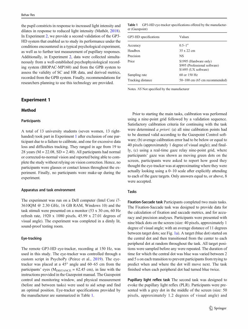
The remote GP3-HD eye-tracker, recording at 150 Hz, wasused in this study. The eye-tracker was controlled through acustom script in PsychoPy (Peirce et al., 2019). The eye-tracker was placed at a 45° angle and 60–65 cm from theparticipants’ eyes (MDISTANCE = 62.45 cm), in line with theinstructions provided in the Gazepoint manual. The Gazepointcontrol and monitoring window, and physical measurement(before and between tasks) were used to aid setup and findan optimal position. Eye-tracker specifications provided bythe manufacturer are summarized in Table 1.
Prior to starting the main tasks, calibration was performedusing a nine-point grid followed by a validation sequence.Satisfactory calibration criteria for continuing with the taskwere determined a priori: (a) all nine calibration points hadto be deemed valid according to the Gazepoint Control soft-ware; (b) average calibration error had to be below or equal to40 pixels (approximately 1 degree of visual angle); and final-ly, (c) using a real-time gaze relay nine-point grid, whereparticipants’ gaze was shown as moving green dots on thescreen, participants were asked to report how good theythought the eye-tracker was at approximating where they wereactually looking using a 0–10 scale after explicitly attendingto each of the gaze targets. Only answers equal to, or above, 8were accepted.
Tasks
Fixation-Saccade task Participants completed two main tasks.The Fixation-Saccade task was designed to provide data forthe calculation of fixation and saccade metrics, and for accu-racy and precision analyses. Participants were presented withnine black dots on the screen (size: 40 pixels, approximately 1degree of visual angle; with an average distance of 11 degreesbetween target dots; see Fig. 1a). A target (blue dot) started onthe central dot and then transitioned from the center to eachperipheral dot at random throughout the task. All target posi-tions were sampled before any were repeated. The duration oftime for which the central dot was blue was varied between 2and 5 s on each transition to prevent participants from trying topredict when and where the dot will move next. The taskfinished when each peripheral dot had turned blue twice.
Pupillary light reflex task The second task was designed toevoke the pupillary light reflex (PLR). Participants were pre-sented with a grey dot in the middle of the screen (size: 50pixels, approximately 1.2 degrees of visual angle) and
Table 1 GP3-HD eye-tracker specifications offered by the manufactur-er (Gazepoint)
GP3-HD specifications Values
Accuracy 0.5–1o
Headbox 35 x 22 cm
Precision NS
Price $1995 (Hardware only)$995 (Professional software)$1495 (UX software)
Sampling rate 60 or 150 Hz
Tracking distance 50–100 cm (65 cm recommended)
Notes. NS Not specified by the manufacturer
Behav Res

instructed to fixate on it while black and white backgrounds2
interchanged every 5 s. Each screen was presented 12 times(24 changes of color, see Fig. 1b).
Procedure
Participants completed the setup and the calibration procedure,followed by the tasks detailed above. Each task was completedtwice, once with the participants’ heads placed on the chinrest to
limit their headmovements, and oncewithout. In both conditionsparticipants were asked to avoid head and bodymovements. Theorder of tasks and conditions (chinrest vs. no-chinrest) wascounterbalanced. The calibration procedure was performed priorto each task. After the experiment participants were debriefed.All experimental procedures were conducted in accordance withthe revised 2013 Declaration of Helsinki and were approved bythe local research ethics committee.
Pre-processing
Eye-tracking data were pre-processed using custom code in R(version: 3.6.1). Gaze samples falling outside screen
2 Please note that the screenwas not exactlywhite as the change from a completelyblack to a completely white screen would have been straining for participants andwould have caused excessive blinks and data loss during this transition period. Theexact color of the screen was [0, 0, 0] in the RGB color space.
Fig. 1 a Schematic of the Fixation-Saccade task. A blue target appearedand switched from the central dot to each of the peripheral dots. bSchematic of the Pupillary Light Reflex task. Each screen appeared 12times. Both tasks were repeated twice, once with, and once without, achinrest. c Illustration of the concepts of accuracy and precision for eye-tracking data. The blue center dot represents the known gaze target, andthe pink smaller circles represent gaze locations estimated by the eye-
tracker. d Three types of eye-tracker accuracy calculations. AH =Horizontal accuracy for a particular gaze sample. AV = Vertical accuracyfor a particular gaze sample. AG/EUCL = Global/Euclidian accuracy for aparticular gaze sample. e Illustration of saccade metrics. SE = Startingerror. LE = Landing error. Curvature = Median of all individual αi anglesbetween each sample point and the straight line connecting the start andend point of the saccade
Behav Res

coordinates were eliminated, as well as the samples labeled asinvalid by the eye-tracker (in total, 2.72% of the samples wereexcluded, out of which 0.74% fell outside the screen bound-aries, and 1.98%were labeled as invalid by the eye-tracker). Asimple implementation of the adaptive velocity-based algo-rithm proposed by Engbert and Kliegl (2003) was used todetect fixations and saccades in the ‘Fixation-Saccade’ task.Saccades were defined as periods of at least 20 ms (the dura-tion of three adjacent gaze samples) where velocity exceededan adaptive threshold set for each participant and condition(chinrest and no-chinrest) based on the level of noise in thedata. The velocity threshold was defined as 5 median absolutedeviations above the median velocity for each participant andcondition. Finally, to prevent artificial improvements in accu-racy and precision, no smoothing, filtering, or interpolationwas applied to gaze position coordinates.
For the PLR task, pupil data were first cleaned by removingpupil sizes outside the range of 2–10mm. Pupil data were thenpre-processed using functions from the R package GazeR(Geller et al., 2020). Data loss (e.g., blinks) up to 150 ms induration were imputed using linear interpolation. Finally, asubtractive baseline correction was applied in line with therecommendations in Mathôt et al. (2018). Median pupil sizeduring the last 20 samples of the preceding trial and the first 20samples of the current trial (approximately 240 ms, incorpo-rating an equal duration of light and dark screens) was taken asbaseline pupil size, from which all individual pupil sizes weresubtracted on each trial.
Metrics and analyses
Calibration quality To assess the calibration quality, two met-rics were used based on the manufacturer’s calibration proce-dure: 1) the number of calibration attempts it took until theexperimenter accepted the calibration, and 2) the average errorof the accepted calibration. All calibrations needed to havenine valid calibration points to be accepted so the number ofvalid calibration points was not considered in further analyses.
Sampling rate variability As the GP3-HD eye-tracker has asampling frequency of 150 Hz, the expected average inter-sample time is approximately 0.0067 s (6.7 ms). Samplingrate variability was assessed by calculating the mean and thestandard deviation of inter-sample time as well as their robustequivalents (median and median absolute deviation), for boththe chinrest and no-chinrest condition. Sampling rate variabil-ity was assessed across both the Fixation-Saccade and thePLR task.
Data loss Data loss occurs when the eye-tracker cannot detectthe position of the eyes, and individual samples where thishappens are labeled as invalid by the device. Proportion of
lost gaze was computed for each trial and participant andcompared between conditions (chinrest and no-chinrest).
Accuracy Prior to computing accuracy and precision, the firstand last 250 ms of each trial were removed to give participantstime to fixate on the new target dot and to limit the extent towhich participants’ anticipatory saccades influenced thesemetrics. This interval was chosen after calculation and visualinspection of saccade latency. Accuracy was computed as theerror between the estimated gaze location and the location of aknown target (Holmqvist et al., 2012). Horizontal and verticalaccuracy were calculated for each gaze sample in the Fixation-Saccade task by subtracting estimated x and y gaze coordi-nates from the pre-defined x and y coordinates of each targetdot location. Sample-level global accuracies were calculatedas Euclidian distances between estimated x and y gaze coor-dinates and pre-defined x and y coordinates of target dot lo-cations (see Fig. 1c, d).
Having calculated all three types of sample-level accura-cies, outlier samples were removed if they were greater than 4median absolute deviations from the median respective accu-racy for each participant, condition, and trial (2.1% of thesamples were excluded for vertical accuracy, 3% for horizon-tal accuracy, and 2.9% for global accuracy—note that thesevalues are not independent). These outliers correspondedmostly to saccades, with the size of the error matching theexpected saccade sizes during the task (note that analyseswith outliers yielded consistent results, see Tables S1 and S2in Supplementary materials). This was performed to avoidbiasing the accuracy calculation by including gaze sampleswhere the participant was likely to have clearly moved theireyes away from the target dot. Finally, mean vertical, horizon-tal, and global accuracy were calculated for each participant,condition, and trial.
Following the calculation of descriptive statistics, linearmixed models in lme4 (Bates et al., 2014) were fitted to testwhether global accuracy differed between conditions (chinrestand no-chinrest) and target dot locations (central and periph-eral) while accounting for participant and trial random effects.Maximal models were always fitted first (Barr et al., 2013),and convergence and singularity warnings were resolved bysimplifying the random structure using principal componentanalysis to determine the most relevant random components(Bates et al., 2015).
Finally, we decided to compare accuracy on the central dotlocation against accuracy on all the peripheral dot locationsgrouped together for two reasons: (a) in psychological re-search, stimuli are commonly presented at the center of thescreen, and it might be useful for researchers to know whetheraccuracy at this location is superior to accuracy at any periph-eral location; (b) since trials with different target dot locationsvaried in frequency and duration (central target was presentedmore frequently and for a longer period of time in comparison
Behav Res
to peripheral targets), grouping all peripheral locationsallowed us to increase statistical power. Additionally, onlysample-level accuracies within the first 2 s of the central trialswere used in this comparison in order to match their durationwith the duration of peripheral trials. For analyses, accuracywas log-transformed to correct the violation of the assumptionthat the residuals of the model are normally distributed.
Precision Precision is a measure of the spatial variance inaccuracy when the eye is assumed to be relatively stationary(Holmqvist et al., 2012). Therefore, gaze samples were firstparsed into fixations and saccades based on the adaptive ve-locity threshold described above. Only fixations longer than80 ms were used for calculating precision to avoid includingsmall saccadeswhichmay be inaccurately labeled as fixations.Horizontal and vertical precision were calculated on a trial-by-trial basis for each participant by computing the root meansquare from successive gaze samples for each fixation to thetarget dot. Global precision was calculated by first estimatingthe Euclidian distances between pairs of adjacent gaze sam-ples and then computing the root mean square over thesedistances. After calculating descriptive statistics, linear mixedmodels were fitted to test whether the three types of precision(horizontal, vertical, global) varied between conditions(chinrest and no-chinrest) and target locations (central andperipheral) while controlling for participant and trial variabil-ity. Comparison between central and peripheral target loca-tions as well as the choice ofmodel’s random structure follow-ed the same logic as the analysis of accuracy. Precision datawas also log-transformed for analysis due to non-normality ofresiduals.
PLR Following pupil pre-processing and baseline correction,the degree of PLR elicited by the changing stimulus was esti-mated (i.e., pupil constriction in response to light and pupildilation in response to darkness). More specifically, linearmixed models were fitted for each condition (chinrest andno-chinrest) to compare pupil size changes in response tothe two stimuli (black vs. white).
Saccade metrics Saccade starting and landing error, ampli-tude, gain, curvature, latency, mean, and peak velocity werecalculated (see Fig. 1). These metrics are provided to allowresearchers to judge howwell saccade parameters are reflectedin gaze data from the GP3-HD, as there are no standard normsto which these values can be judged against, other than directcomparisons with other systems.
Saccade starting error was calculated as the Euclidian dis-tance between the gaze sample labeled as the saccade onsetand the center of the starting target, while saccade landingerror was calculated as the Euclidian distance between thegaze sample labeled as the saccade end and the target center(Dalmaijer, 2014). After calculating saccade amplitude (size
of the saccade in degrees of visual angle), we proceeded tocompute gain, the ratio between the observed saccade ampli-tude and the expected saccade amplitude, actual distance be-tween the two consecutive target locations (Noto & Robinson,2001). Saccades with gains less than 1 were too small(hypometric), while saccades with gains higher than 1 weretoo large (hypermetric). Gain provides an approximation ofover- or under-estimation of the expected saccade amplitudes.
In order to capture saccade trajectories, curvature was cal-culated as the median angle between each gaze point in asaccade and an imaginary straight line connecting the startand the end of the saccade, following the strategy of vanLeeuwen and Belopolsky (2018). Saccade latency is the timefrom target onset until the initiation of the saccade to thattarget.
Finally, velocity was calculated for each gaze sample mak-ing up a saccade by dividing inter-sample distance (in degreesof visual angle) by inter-sample time (in seconds), after whichmean and peak velocity were computed for each saccade as awhole.
Results
Calibration quality
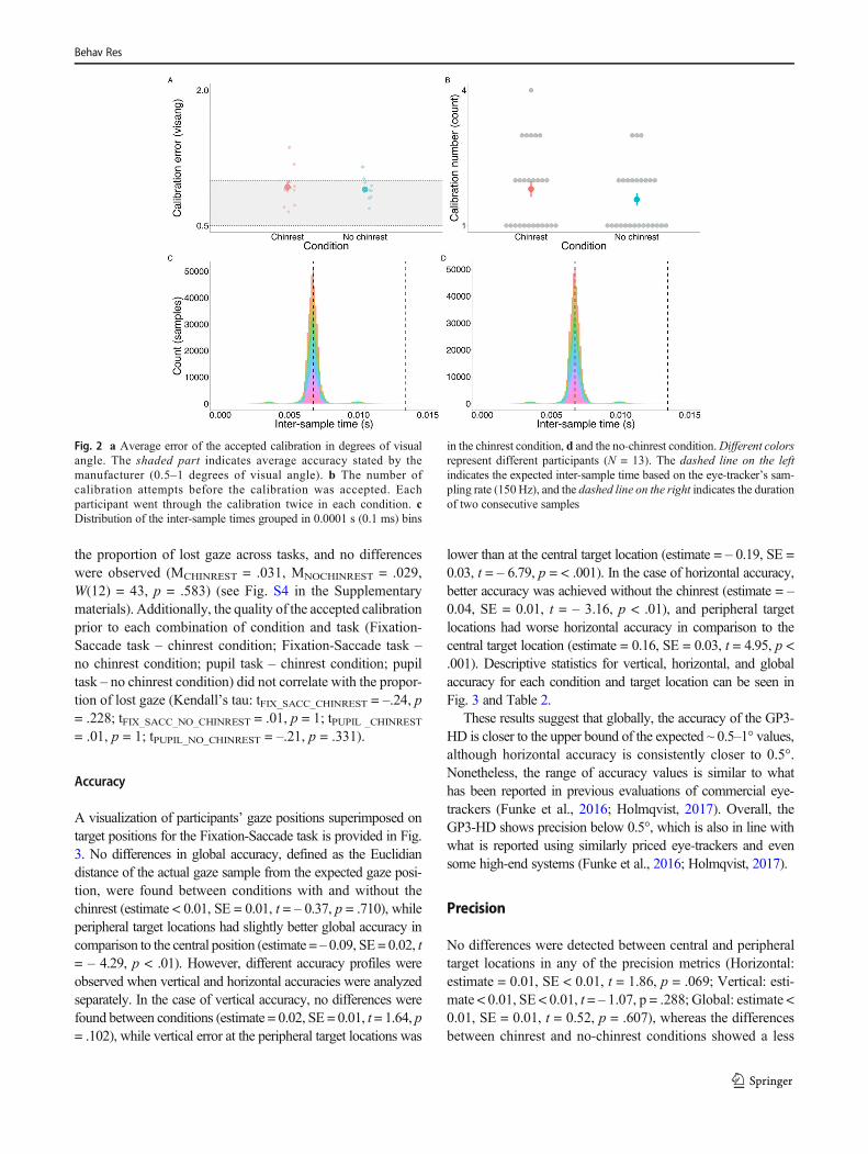
Calibration metrics included the number of attempts it took toachieve an acceptable calibration, and the average error of theaccepted calibration. Due to the violation of normality(Shapiro–Wilk test: W(12) = 0.72, p < .001), a Wilcoxonsigned-rank test was performed to examine differences be-tween the chinrest and no-chinrest conditions on both calibra-tion quality metrics. No differences were detected in the num-ber of calibration attempts (MCHINREST = 1.81, MNOCHINREST
= 1.58, W(12) = 30, p = .402) nor in the average error of theaccepted calibration (MCHINREST = 0.933, MNOCHINREST =0.99, W(12) = 47, p = .946) (see Fig. 2).
Sampling rate variability
As expected, the observed average inter-sample time was6.7 ms and the standard deviation of the inter-sample timewas 0.79 ms (robust descriptives: Mdn = 6.68 ms; MAD =35.29 ms). Only 0.0003% of the inter-sample times weregreater than the duration of two consecutive samples (13.4ms). Distribution of inter-sample time is shown in Fig. 2.
Data loss
A Wilcoxon signed-rank test was performed to comparewhether the chinrest and no-chinrest condition differed in
3 Average error of the accepted calibration is measured in degrees of visualangle.
Behav Res
the proportion of lost gaze across tasks, and no differenceswere observed (MCHINREST = .031, MNOCHINREST = .029,W(12) = 43, p = .583) (see Fig. S4 in the Supplementarymaterials). Additionally, the quality of the accepted calibrationprior to each combination of condition and task (Fixation-Saccade task – chinrest condition; Fixation-Saccade task –no chinrest condition; pupil task – chinrest condition; pupiltask – no chinrest condition) did not correlate with the propor-tion of lost gaze (Kendall’s tau: tFIX_SACC_CHINREST = –.24, p= .228; tFIX_SACC_NO_CHINREST = .01, p = 1; tPUPIL _CHINREST
= .01, p = 1; tPUPIL_NO_CHINREST = –.21, p = .331).
Accuracy
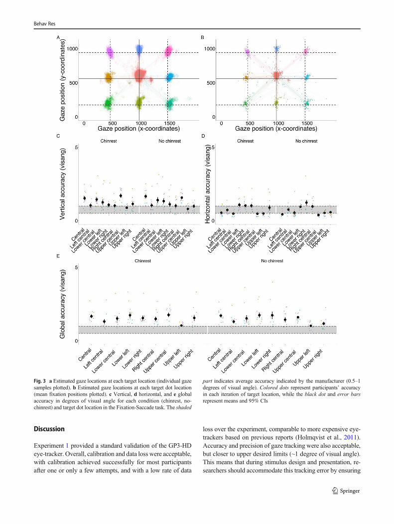
A visualization of participants’ gaze positions superimposed ontarget positions for the Fixation-Saccade task is provided in Fig.3. No differences in global accuracy, defined as the Euclidiandistance of the actual gaze sample from the expected gaze posi-tion, were found between conditions with and without thechinrest (estimate < 0.01, SE = 0.01, t = – 0.37, p = .710), whileperipheral target locations had slightly better global accuracy incomparison to the central position (estimate = – 0.09, SE = 0.02, t= – 4.29, p < .01). However, different accuracy profiles wereobserved when vertical and horizontal accuracies were analyzedseparately. In the case of vertical accuracy, no differences werefound between conditions (estimate = 0.02, SE = 0.01, t = 1.64, p= .102), while vertical error at the peripheral target locations was
lower than at the central target location (estimate = – 0.19, SE =0.03, t = – 6.79, p = < .001). In the case of horizontal accuracy,better accuracy was achieved without the chinrest (estimate = –0.04, SE = 0.01, t = – 3.16, p < .01), and peripheral targetlocations had worse horizontal accuracy in comparison to thecentral target location (estimate = 0.16, SE = 0.03, t = 4.95, p <.001). Descriptive statistics for vertical, horizontal, and globalaccuracy for each condition and target location can be seen inFig. 3 and Table 2.
These results suggest that globally, the accuracy of the GP3-HD is closer to the upper bound of the expected ~ 0.5–1° values,although horizontal accuracy is consistently closer to 0.5°.Nonetheless, the range of accuracy values is similar to whathas been reported in previous evaluations of commercial eye-trackers (Funke et al., 2016; Holmqvist, 2017). Overall, theGP3-HD shows precision below 0.5°, which is also in line withwhat is reported using similarly priced eye-trackers and evensome high-end systems (Funke et al., 2016; Holmqvist, 2017).
Precision
No differences were detected between central and peripheraltarget locations in any of the precision metrics (Horizontal:estimate = 0.01, SE < 0.01, t = 1.86, p = .069; Vertical: esti-mate < 0.01, SE < 0.01, t = – 1.07, p = .288; Global: estimate <0.01, SE = 0.01, t = 0.52, p = .607), whereas the differencesbetween chinrest and no-chinrest conditions showed a less
Fig. 2 a Average error of the accepted calibration in degrees of visualangle. The shaded part indicates average accuracy stated by themanufacturer (0.5–1 degrees of visual angle). b The number ofcalibration attempts before the calibration was accepted. Eachparticipant went through the calibration twice in each condition. cDistribution of the inter-sample times grouped in 0.0001 s (0.1 ms) bins
in the chinrest condition, d and the no-chinrest condition.Different colorsrepresent different participants (N = 13). The dashed line on the leftindicates the expected inter-sample time based on the eye-tracker’s sam-pling rate (150Hz), and the dashed line on the right indicates the durationof two consecutive samples
Behav Res

consistent pattern. The no-chinrest condition yielded in-creased vertical precision (estimate = – 0.01, SE < 0.01, t =– 2.66, p < .01), whereas no differences were observed inhorizontal (estimate < 0.01, SE < 0.01, t = 1.53, p = .126)and global precision (estimate < 0.01, SE < 0.01, t = – 0.80,p = .426). Descriptive statistics for vertical, horizontal, andglobal precision for each condition and target location canbe seen in Table 2.
Bayesian equivalents for condition comparisons are pro-vided in the Supplementary materials – see Bayesian analysesfor condition comparisons.
Pupillary light reflex
The PLR was reliably detected in both the chinrest (estimate =– 19.55, SE = 0.26, t = – 74.50, p < .001) and no-chinrestconditions (estimate = – 20.75, SE = 0.30, t = – 69.79, p <.001), see Fig. 4a, b). As expected, the PLR effect is verylarge, accounting for 95% of the variance in both conditions.
Saccadometry
Descriptive statistics for different saccade metrics are provid-ed in Table 3. The relationships between saccade amplitude,duration and peak velocity are visualized in Fig. 4c, d.
As expected, since the majority of the saccades detected inthe Fixation-Saccade task would be classified as small usingthe guidelines of Gibaldi and Sabatini (2021), the relationshipbetween these metrics was approximately linear.
Finally, we examined each participant’s saccade trajec-tories from the central target to each peripheral target (seeFig. 5). Individual saccade trajectories plotted in Fig. 5 arenot smooth, but appear broken and edgy, which is a sign ofunder-sampling (Dalmaijer, 2014). While clear identifica-tion of saccade events is possible, this suggests that theGP3-HD eye-tracker is less suitable for saccadometryresearch.
In summary, accuracy measures are comparable tothe range reported in the existing eye-tracking literaturefor similar grade devices. In both ideal (chinrest) andnon-ideal (no-chinrest) conditions, overall accuracy wasat the upper limit or even higher than the values statedby the manufacturer (1 degree of visual angle). Whilethis degree of accuracy is perfectly capable of capturinggaze behavior reliably across most experimental tasks,tracking targets smaller than this error could be prob-lematic. While clear separation of saccades and fixationsis possible using the GP3-HD, study of the properties ofsaccade kinematics is compromised by the low samplingrate.
Table 2 Descriptive statistics for accuracy and precision
Accuracy Precision
Target dot Vertical Horizontal Global Vertical Horizontal Global
Chinrest
Central 1.60 0.60 1.74 0.28 0.24 0.36
Left 0.99 0.77 1.31 0.27 0.25 0.36
Lower central 1.63 0.68 1.75 0.33 0.28 0.43
Lower left 1.27 1.21 1.84 0.25 0.28 0.37
Lower right 1.20 1.29 1.89 0.31 0.31 0.43
Right 1.08 1.27 1.76 0.3 0.25 0.38
Upper central 1.67 0.49 1.72 0.28 0.23 0.35
Upper left 0.88 0.49 1.04 0.28 0.24 0.36
Upper right 1.15 0.86 1.54 0.27 0.22 0.34
No chinrest
Central 1.71 0.46 1.76 0.28 0.25 0.36
Left 1.00 0.81 1.33 0.26 0.26 0.36
Lower central 1.45 0.55 1.56 0.25 0.25 0.35
Lower left 1.29 1.00 1.74 0.25 0.29 0.38
Lower right 1.19 1.21 1.75 0.24 0.27 0.36
Right 1.06 0.96 1.47 0.24 0.25 0.35
Upper central 1.65 0.41 1.68 0.28 0.23 0.35
Upper left 0.87 0.56 1.08 0.27 0.23 0.35
Upper right 1.04 0.58 1.22 0.28 0.24 0.36
Notes. Vertical, horizontal and global accuracy and precision in degrees of visual angle averaged across participants for each target dot location and condition
Behav Res
Discussion
Experiment 1 provided a standard validation of the GP3-HDeye-tracker. Overall, calibration and data loss were acceptable,with calibration achieved successfully for most participantsafter one or only a few attempts, and with a low rate of data
loss over the experiment, comparable to more expensive eye-trackers based on previous reports (Holmqvist et al., 2011).Accuracy and precision of gaze tracking were also acceptable,but closer to upper desired limits (~1 degree of visual angle).This means that during stimulus design and presentation, re-searchers should accommodate this tracking error by ensuring
Fig. 3 a Estimated gaze locations at each target location (individual gazesamples plotted). b Estimated gaze locations at each target dot location(mean fixation positions plotted). c Vertical, d horizontal, and e globalaccuracy in degrees of visual angle for each condition (chinrest, no-chinrest) and target dot location in the Fixation-Saccade task. The shaded
part indicates average accuracy indicated by the manufacturer (0.5–1degrees of visual angle). Colored dots represent participants’ accuracyin each iteration of target location, while the black dot and error barsrepresent means and 95% CIs
Behav Res

that target locations are separated by 3 degrees of visual angleor more, so that if a participant was looking exactly at themiddle of two targets, the estimated gaze locations (even ac-counting for error) would not fall onto either of the targets. It isworth noting, however, that better accuracy values wereachieved (0.5 degrees of visual angle) particularly for the
horizontal dimension. This is useful to know, as it is possibleto calibrate how gaze is assigned to areas of interest given thetracking error associated with a particular participant at a par-ticular point in time (Hessels & Hooge, 2019; Orquin &Holmqvist, 2018). Similarly, individualized accuracy and pre-cision profiles can be used to calibrate gaze parsing filters (see,Feit et al., 2017).
With regard to saccade metrics, while the identification ofsaccades appears good, the calculation of kinematic parame-ters appears to be affected by the low sampling rate. As aresult, the study of the properties of saccades using the GP3-HD may lead to inaccuracies depending on the specific pa-rameters being studied. For example, it appears that the am-plitude of saccades is underestimated, as the gain value was <1, indicating hypometric saccades, that is, saccades where theamplitude was smaller than expected. Looking at the plots inFig. 5, it is apparent that saccade patterns show breaks as aresult of the sampling rate. This is consistent with Nyquist'stheorem, which specifies that a signal must be sampled atmore than twice the highest frequency component of the sig-nal. Note, however, that saccade detection as well as peak
Fig. 4 a, b Pupillary light reflex in both chinrest and no-chinrest condi-tions. Dark-colored time section (from – 5 to 0 s) and dark boxplotindicate pupil dilation during the black screen, while light-colored timesection (from 0 to 5 s) and light boxplot indicate pupil constriction duringthe white screen. Dashed line in graph A indicates the switching pointfrom black towhite screen, while the dotted linesmark the baseline period
used for calculating baseline pupil size. c, d Main sequence of saccadiceye movements (r = .288, p < .001; r = .420, p < .001). Each colored linerepresents a slope for each individual participant, while the black linerepresents the slope across participants. Jittered points presented in thebackground represent individual saccades
Table 3 Saccade metrics calculated using GP3-HD data
Saccade index GP3-HDM(SD)
Unit
Amplitude 8.87 (3.97) Degrees of visual angle
Curvature 30.74 (12.94) Degrees
Duration 48.80 (25.03) Milliseconds
Gain 0.73 (0.21) -
Latency 163.38 (369.75) Milliseconds
Mean velocity 315.83 (95.73) Degrees per second
Peak velocity 756 (322.48) -
Starting error 3.56 (1.96) Degrees of visual angle
Landing error 2.16 (2.28) -
Behav Res

velocity approximations are reliable with even 60 Hz sam-pling, and there are saccade reconstruction techniques thatcan improve the approximation of the “true” parameters ofsaccades (Wierts et al., 2008).
The lack of consistent significant differences betweenchinrest and no-chinrest conditions suggests that the algorithmfor gaze estimation used by GP3-HD is relatively robust tosmall head movements. However, increased infra-red‘bounce’ was observed during the sessions with a chinrest,where small and temporary reflections from the chinrestwould cause it to be mistakenly detected as a part of the eye.Improvements in tracking accuracy expected from use of thechinrest may have been lost as a result. While this problem issolvable by eliminating infra-red reflections, a possibility fordevelopment would be for Gazepoint to offer a user interven-tion enabling the operator to manually correct themisidentified reflections during calibration, such that theGazepoint algorithm would subsequently underweight thoseregions when estimating gaze location. Similarly, providingthe stream of estimated distances from the screen, in additionto the gaze coordinates and pupil samples, would be useful forresearchers to accommodate changes in distance from thescreen in non-chinrest conditions.
Experiment 2
Experiment 1 provided a standard validation of the GP3-HDeye-tracker in terms of its accuracy and precision, degree ofdata loss and the benefits of head stabilization. While thisprovides a useful benchmark assessment of the GP3-HD, sucheye-tracking validation studies do not reflect typical experi-ments in which task demands are usually greater and there aremore limited checks on the eye-tracker’s performance im-posed by study design (Niehorster et al., 2018). Therefore,in Experiment 2 we provide data from a real-world typicalpsychological experiment to assess GP3-HD performance.Importantly, as typical behavioral experiments may vary froma few minutes to hours, we investigated how data qualityparameters changed over time, across a 1-h-long experiment.
Experiment 1 also demonstrated that GP3-HD is able tocapture pupil changes such as the PLR. In most cognitiveand behavioral research, however, researchers are typicallyinterested in how pupil changes are modulated by cognitiveand affective factors rather than low-level visual properties.This can range from quantifying cognitive effort (Papesh &Goldinger, 2012; Piquado et al., 2010) to emotional arousal(Bradley et al., 2008). Considering that cognitive and
Fig. 5 Saccade trajectories from the central target to each peripheral target. Each graph shows saccade trajectories of a different participant (N = 13; pix = pixel)
Behav Res
emotional effects on pupil diameter are much smaller in mag-nitude than luminance effects (Mathôt, 2018), it is unclearhow well the GP3-HD is able to capture such effects. InExperiment 2 we used data from a paradigm that allowed usto explore how pupil size is modulated by emotional factors.Specifically, the effect of viewing emotionally arousing im-ages on pupil diameter was investigated. We predicted a pos-itive correlation between self-rated arousal and pupil size.Since this task makes use of naturalistic visual stimuli varyingin low-level properties, the luminance of the stimuli can alsobe regressed onto pupil size to model modulation of pupil sizedue to the PLR. Stimulus brightness should have a negativecorrelation with pupil size, such that brighter stimuli shouldlead to decreased pupil size (pupil constriction) whereasdarker stimuli should predict increased pupil size (pupildilation).
Additionally, in Experiment 2, SC and HR data was col-lected from participants simultaneously using both the GPBand a well-validated physiological recording system(BIOPAC-MP160). Strong correlations between deviceswould indicate the capability of the GPB to capture physio-logical signals.
Method
Participants
A total of 46 healthy participants with no recent history ofmental health problems (28 female, 18 male, M = 23, SD =5.54, 18 to 65 years old), with normal or corrected to normalvision and without makeup, took part in this study.Additionally, ten participants with an independent diagnosisof autism were recruited at a later stage and their data used fora subset of the heart rate analyses (four female, six male, M =38, SD = 15.73, 18–65 years old). Participants took part in apsychological experiment where they had to view and rateemotional pictures, while their gaze and physiological signalswere monitored. Participants were reimbursed £10 per hour or3 course credits for their time.
Stimuli and task
Picture stimuli were 50 images from the InternationalAffective Picture System (IAPS, Lang et al., 2005) designedto elicit emotional responses in observers (e.g., open lungsurgery, naked bodies, etc.). The stimuli were chosen to covera wide range of valence (M = 5.07, SD = 1.92, range = 1.46 –8.19 – on a 1 to 9 scale) and arousal (M = 4.66, SD = 1.19,range = 2.63 – 7.21) scores based on population norms.Participants viewed each stimulus and provided ratings forvalence using a slider (‘How did the image make you feel’)from ‘–10 (Extremely negative)’ to ‘+10 (Extremely posi-tive)’; and arousal (‘How intense was your emotional
response’) from ‘–10 (Extremely calm and relaxed)’ to ‘+10(Extremely intense)’. Each trial started with a fixation cross ofvariable duration (ranging from 7 to 15s), followed by presen-tation of the image for 6 s, after which participants providedtheir valence and arousal ratings (see Fig. 6).
Apparatus and task environment
The experiment was displayed on a computer monitor mea-suring 1920 x 1200 pixels with a fixed refresh rate of 60 Hzrunning on an Intel Core i9 Windows 10 computer (32GBRAM).
Eye tracking
The sameGP3-HD eye tracker fromExperiment 1 was used totrack eye-movements and pupil size with a sampling rate of150 Hz. Setup and calibration were similar to Experiment 1.Recalibration was repeated after 25 trials (approximately 20min). Participants were asked to keep their head and body stilland a chinrest was used. However, they could move their eyesfreely to explore the images.
Accuracy and precision
Accuracy and precision were computed in a similar manner asin Experiment 1, however here computations were performedduring the fixation screen that preceded each trial. The datawere trimmed to 6 s and the first 250 ms after the start offixation cross were removed. Outlier precision and accuracyvalues were removed as in Experiment 1.
Physiological recordings
Physiological recordings of SC, pulse and heartbeat datawere obtained from two systems: the GPB and theBIOPAC M160 with EDA100C and ECG100C modules(details below).
Skin conductance – GPB The GPB measures both heart rateand skin conductance via a sensor attached to two fingers(index and middle finger), without the need for extensive skinpreparation or specialized electrodes. To record SC, the GPBuses an exosomatic recording method which applies directcurrent with a constant voltage source. The applied voltageis 5 V through high impedance voltage division. The condi-tioning method uses an analog low pass filtered at 10 Hz witha sampling frequency of 150 Hz. The sensors use gold-platedsteel electrodes strapped to distal phalanges of the fingers.
Heart rate – GPB The SCR system described above allowsdetection of heartbeats at the middle or index finger usinga photoplethysmography (PPG) method where a light
Behav Res

beam is transmitted to the tissue and heartbeats are detect-ed via intensity modulation of the reflected light. Notethat the standard GPB system reports only heart rate data,however, through the provided API it is possible to storethe raw pulse data using a custom script. This data wasonly available for a third of participants. Heart rate iscomputed using a moving average with a window of threebeats. For all measures, live monitoring was achieved viathe Gazepoint control application.
Skin conductance – BIOPAC MP160 To validate the GPB,physiological data were also collected using a BIOPACMP160 system sampling at 2000 Hz. SC data were re-corded using the EDA100C module and TSR203 trans-ducers. The skin where the GSR electrodes were placedwas cleaned with water and dried with a cotton tissue.The EDA100C uses a constant voltage (0.5 V) techniqueto measure skin conductance. SC was collected via twoelectrodes that were placed on the inside of the left foot tomeasure GSR (see Fig. S6 in Supplementary materials).Foot (rather than hand) placement was used to avoid cre-ating interference between the two devices. The foot andfingers have been shown to be the best locations to mea-sure SC and provide largely similar results (van Doorenet al., 2012).
ECG - BIOPAC MP160 The ECG signal was recorded via theECG100C electrocardiogram amplifier, which records electri-cal activity generated by the heart. Two electrodes wereplaced on participants (see Fig. S7 in Supplementarymaterials). One electrode was placed on the right collarboneand one on the lower left torso to measure HR. All sensorswere well secured with surgical tape to prevent loss or disrup-tion of signal. The skin where the ECG electrodes were placedwas cleaned with Signagel Electrode Gel before attachingsensors.
Procedure
Following informed consent and the opportunity to ask ques-tions, physiological recordings were prepared. A 2-min restperiod was allowed for recordings to stabilize before prepara-tions continued. The task started with a calibration, followedby another 2-min rest period, during which participants fo-cused on a fixation cross in the center of the screen, and fivepractice trials. After 25 trials, participants took a short breakfor recalibration. After the task they were debriefed and com-pensated. All research was conducted in accordance with therevised 2013 Declaration of Helsinki and was approved by thelocal Research Ethics Committee.
Fig. 6 Schematics of Experiment 2
Behav Res
Data pre-processing
Eye-tracking
Eye-tracking data were pre-processed following the samesteps as in Experiment 1. For pupil analysis, pupil responsewas baseline corrected using an interval corresponding to 1 sbefore and 1 s after the stimulus, using the same method as inExperiment 1.
Skin conductance and heart rate
SC data from both the GPB and BIOPAC systems were ana-lyzed using a continuous decomposition analysis (CDA) al-gorithm implemented in the open-source software Ledalab(Kaernbach, 2005). For analysis, data were first downsampledto 10 Hz and inspected for artifacts. Adaptive smoothing wasused prior to analysis. All optimizations used the defaultvalues in Ledalab recommended for SC measurement andanalysis (Boucsein et al., 2012). The global mean of the SCsignal as well as the event-related phasic signal was computed.
Heart rate data
Established HR data-processing pipelines were used to pro-cess the heartbeat and ECG signal from the GPB andBIOPAC, respectively. Processing was accomplished via theArtiiFact toolbox (Kaufmann et al., 2011). Artifact detectionwas achieved using the Berntson, Quigley, Jang, and Boysen(1990) algorithm based on individual thresholds derived frominter-beat-interval (IBI, also known as RR interval)
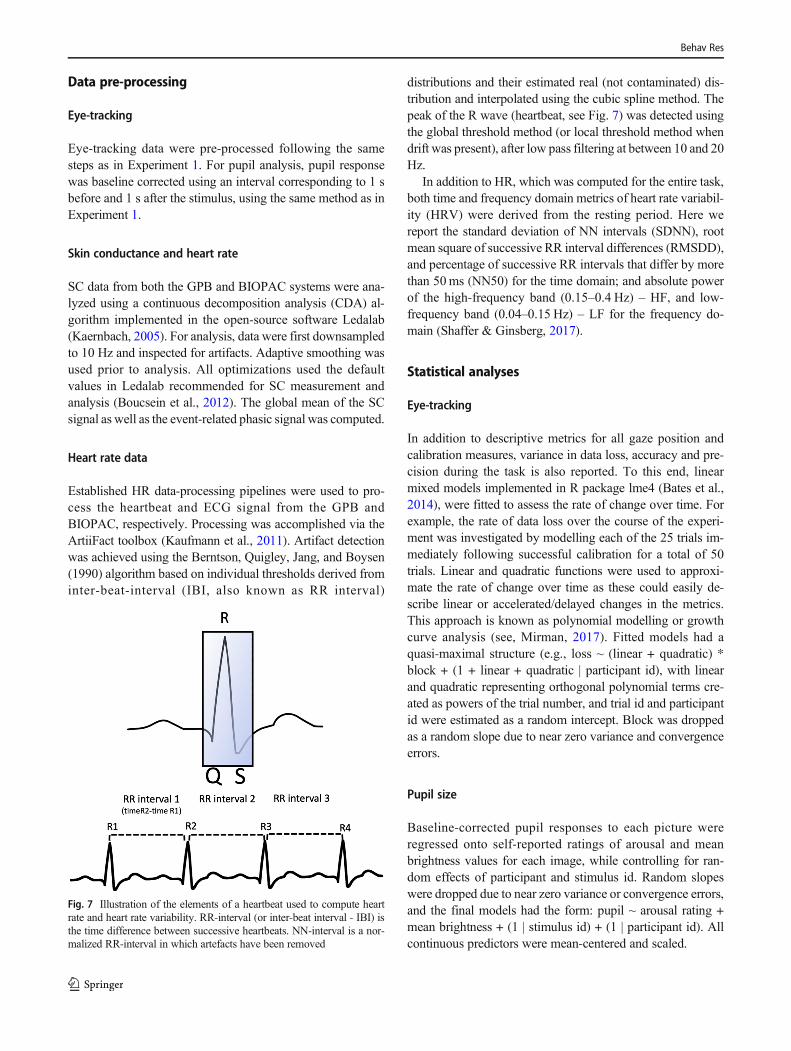
distributions and their estimated real (not contaminated) dis-tribution and interpolated using the cubic spline method. Thepeak of the R wave (heartbeat, see Fig. 7) was detected usingthe global threshold method (or local threshold method whendrift was present), after low pass filtering at between 10 and 20Hz.
In addition to HR, which was computed for the entire task,both time and frequency domain metrics of heart rate variabil-ity (HRV) were derived from the resting period. Here wereport the standard deviation of NN intervals (SDNN), rootmean square of successive RR interval differences (RMSDD),and percentage of successive RR intervals that differ by morethan 50ms (NN50) for the time domain; and absolute powerof the high-frequency band (0.15–0.4 Hz) – HF, and low-frequency band (0.04–0.15 Hz) – LF for the frequency do-main (Shaffer & Ginsberg, 2017).
Statistical analyses
Eye-tracking
In addition to descriptive metrics for all gaze position andcalibration measures, variance in data loss, accuracy and pre-cision during the task is also reported. To this end, linearmixed models implemented in R package lme4 (Bates et al.,2014), were fitted to assess the rate of change over time. Forexample, the rate of data loss over the course of the experi-ment was investigated by modelling each of the 25 trials im-mediately following successful calibration for a total of 50trials. Linear and quadratic functions were used to approxi-mate the rate of change over time as these could easily de-scribe linear or accelerated/delayed changes in the metrics.This approach is known as polynomial modelling or growthcurve analysis (see, Mirman, 2017). Fitted models had aquasi-maximal structure (e.g., loss ~ (linear + quadratic) *block + (1 + linear + quadratic | participant id), with linearand quadratic representing orthogonal polynomial terms cre-ated as powers of the trial number, and trial id and participantid were estimated as a random intercept. Block was droppedas a random slope due to near zero variance and convergenceerrors.
Pupil size
Baseline-corrected pupil responses to each picture wereregressed onto self-reported ratings of arousal and meanbrightness values for each image, while controlling for ran-dom effects of participant and stimulus id. Random slopeswere dropped due to near zero variance or convergence errors,and the final models had the form: pupil ~ arousal rating +mean brightness + (1 | stimulus id) + (1 | participant id). Allcontinuous predictors were mean-centered and scaled.
Fig. 7 Illustration of the elements of a heartbeat used to compute heartrate and heart rate variability. RR-interval (or inter-beat interval - IBI) isthe time difference between successive heartbeats. NN-interval is a nor-malized RR-interval in which artefacts have been removed
Behav Res

Skin conductance and heart rate
The average SC signal during each trial, event-related phasicSC responses and HR metrics were correlated between theGPB and BIOPAC systems. HRV measures were only com-puted for the subsample of participants for whom the rawtimeseries of ECG/Pulse data was available for both theGPB and BIOPAC. This subsample had 20 participants withraw pulse measurements (ten neurotypical individuals includ-ed in the other analyses, and an additional ten autistic partic-ipants). For this subsample, all correlations are Kendal rankcorrelations due to small sample sizes.
Results
Eye-tracking
Calibration Calibration quality and other descriptive statisticsare provided in Table 4. The average calibration error acrossall three accepted calibrations during the 1-h session was with-in the expected range, with a mean of .98° (SD = .45°), and ittook an average of 1.66 attempts to achieve an acceptablecalibration.
Data loss On average, less than 10% of gaze data were lost,which is consistent with the typical rate of loss reported in theliterature and even lower than some more expensive systems(e.g., Tobi TX300 based on (Holmqvist, 2017). During thetask less than 1% of the data fell outside screen bounds.
Data loss increased at a linear rate (estimate = .004, SE –.002, t = 2.05, p = .04) across the experiment. There was also amain effect of block, with trials after the recalibration showingreduced data loss (estimate = –.12, SE = .02, t = – 5.08, p <.001). The only significant interaction was between the
quadratic term and block (estimate = .05, SE = .02, t = 2.43,p = .02): post hoc tests indicated that the rate of loss in the firstblock additionally followed an ‘inverted-U’ shape, that is, itwas characterized by rapid increase in data loss in the first fewtrials until the rate of loss stabilized and reduced in the last fewtrials (estimate = .01, SE = .002, t = 5.07, p < .001).
Accuracy
Accuracy values were in the expected range, averaging be-tween 0.5° and 1.33°, with better accuracy on the horizontaldimension with values consistently ~ 0.5° (see Fig. 8). Overall,the global accuracy changed at a linear rate such that for everytrial, tracking accuracy deteriorated by 0.04° (estimate = .04, SE= .01, t = 3.21, p = .001). There were also block differencessuch that the second block showed better accuracy, with re-calibration improving accuracy by .06° compared to the previ-ous trial (estimate = .06, SE = .02, t = 2.17, p = .01). Resultswere consistent for vertical and horizontal accuracy.
With respect to horizontal accuracy, for every trial after cal-ibration tracking accuracy deteriorated by .03° (estimate = .03,SE = .009, t = 3.21, p = .001) and recalibration after every 25trials improved accuracy by on average .05° (estimate = .05, SE= .02, t = 2.49, p = .01). For vertical accuracy there was aninteraction between block and the linear parameter (estimate =.05, SE = .02, t = 2.79, p < .001), such that in the first blockaccuracy declined more linearly than in the second block.
Precision
All precision values were within an acceptable range < 0.5°,with the horizontal dimensions showing better tracking preci-sion. There was no effect of trial on precision nor interactionswith block. There was only a main effect of block, with thesecond block showing better precision (estimate = .007, SE =.002, t = 2.93, p = .002). Both vertical and horizontal precisionshowed the same effect.
Pupil size
Experiment 2 aimed to explore whether the modulation ofpupil size by emotional arousal is detectable with the GP3-HD, considering that such effects are orders of magnitudesmaller than the PLR detected in Experiment 1. A main effectwas observed for self-reported arousal in the predicted direc-tion (estimate = .24, SE = .05, t = 4.35, p < .001), whichremained significant after statistically controlling for bright-ness. This shows that self-perceived arousal is positively re-lated to pupil change. The predicted effect of brightness onpupil size was also significant, and larger (estimate = – 1.02,SE = .12, t = – 8.59, p < .001), demonstrating again thatbrightness is negatively related to pupil change. As predicted,the effect of arousal on pupil size was less than 1% of the size
Table 4 Descriptive measures for eye tracking data
Variable Mean SD Range
Calibration count 1.66 0.98 1–5
Calibration error 0.98 0.45 0.49–4.92
Gaze loss 8.3% 15% 0–100
Accuracy
Global 0.77 0.70 0.23–13.53
Vertical 1.33 0.73 0.27–9.50
Horizontal 0.54 0.50 0.30–6.34
Precision
Global 0.27 0.11 0.15–2.14
Vertical 0.30 0.13 0.11–.95
Horizontal 0.24 0.09 0.09–.60
Notes. Calibration error, accuracy, and precision data is reported in de-grees of visual angle (range is reported at the trial level)
Behav Res

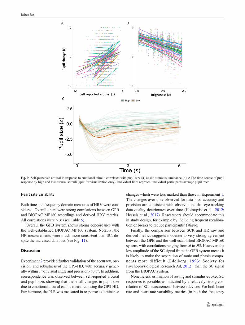
of the brightness effect and showed more individual variabil-ity in comparison to the PLR (see Fig. 9).
The model explained 54% of the variance in pupil size, ofwhich 23% was explained by the fixed effects (discountingthe random effects of participants and trial). Thus, Experiment2 provides support for the use of the GP3-HD eye trackingsystem for the study of gaze position and pupil size in typicalpsychological experiments. Tracking capability is better hori-zontally than vertically, but the vertical accuracy alsoremained acceptable range (~ 1°).
A second goal of this study was to provide a validation ofthe GPB system, by comparing SC and HR obtained using theGPB with data collected from the well-validated BIOPACMP160 (see Fig. 10).
Skin conductance
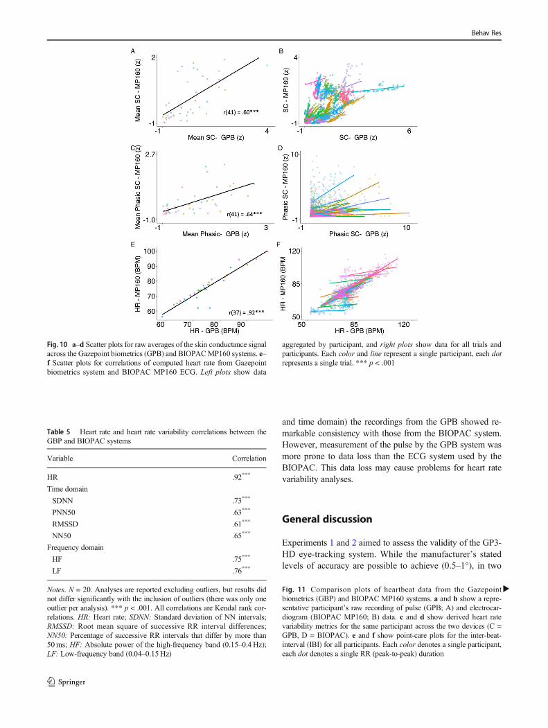
The SC measurement capacity of the GPB showed excellentrobustness, with less than .06% of loss compared to no loss ofsignal in the BIOPAC-MP160. The average SC signal fromthe GPB and BIOPAC showed a strong correlation (r(37) =.60, p < .001). This was also consistent when looking at spe-cific derived measures of skin conductance from Ledalab,such as a decomposed phasic signal (r(41) = .64, p < .001),with measurements across both devices sharing 40% of thevariance. However, as illustrated in Fig. 10, there is a signif-icant degree of difference in howwell these signals correlate atan individual level. This may be related to differences in howsimilar the physiological properties are in the measured loca-tions for each participant (foot vs. palm), as well as the factthat the SC signal from the GPB is smaller in range comparedto the BIOPAC signal.
A sample of the SC signal from an example participant isshown in Fig. S5 in the Supplementary materials for both theGPB and BIOPAC systems. One obvious difference is that thesignal from the BIOPAC is much larger in magnitude com-pared to the GPB signal. This results in more signal beingpreserved after removal of tonic data in BIOPAC comparedto GPB data.
Heart rate
Quality checks indicated that the GPB lost on average 19%(SD = .37) of data (impossible HR values or loss of signal)compared to no obvious data loss for the BIOPAC system(i.e., peaks were still present even in periods of relative noise).Importantly, however, participants were not more likely tolose data as the task progressed (estimate = .004. SE = .005,t = .69, p = .45). This was a concern as the strap of the sensorsmay be thought to limit the blood flow to the finger, system-atically affecting heartbeat tracking over time. HR recordedfrom the GPB and BIOPAC were, however, very stronglycorrelated (r(37) = .92, p < .001, Fig. 10), demonstrating thatthe GPB provides valid measurement of heart rate, corre-sponding to systems which are much more expensive.Results were similar whether using interpolated or non-interpolated GPB heart rate data. Notably, these correlationsare much larger than the correlations between the systems forSC signals, however the loss of HR data is much greater thanthe loss of SC signals for the GPB system.
Another ECG metric of interest for many researchers isheart rate variability (HRV). Notably, HRV measures requiremore sensitive and less noisy recordings than HR, which isrelatively stable.
Fig. 8 Density plots for accuracy (a), precision (b), and data loss (c). Frequency of fixations, saccades, and data loss for each trial and participant (d).Data represents the density of every single observation for every participant for each trial
Behav Res
Heart rate variability
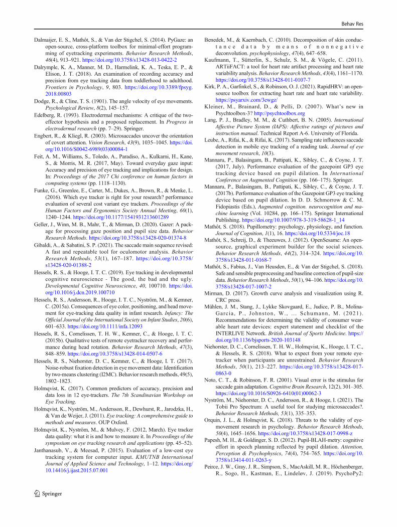
Both time and frequency domain measures of HRVwere con-sidered. Overall, there were strong correlations between GPBand BIOPAC MP160 recordings and derived HRV metrics.All correlations were > .6 (see Table 5).
Overall, the GPB system shows strong concordance withthe well-established BIOPAC MP160 system. Notably, theHR measurements were much more consistent than SC, de-spite the increased data loss (see Fig. 11).
Discussion
Experiment 2 provided further validation of the accuracy, pre-cision, and robustness of the GP3-HD, with accuracy gener-ally within 1° of visual angle and precision < 0.5°. In addition,correspondence was observed between self-reported arousaland pupil size, showing that the small changes in pupil sizedue to emotional arousal can be measured using the GP3-HD.Furthermore, the PLRwas measured in response to luminance
changes which were less marked than those in Experiment 1.The changes over time observed for data loss, accuracy andprecision are consistent with observations that eye-trackingdata quality deteriorates over time (Holmqvist et al., 2012;Hessels et al., 2017). Researchers should accommodate thisin study design, for example by including frequent recalibra-tion or breaks to reduce participants’ fatigue.
Finally, the comparison between SCR and HR raw andderived metrics suggests moderate to very strong agreementbetween the GPB and the well-established BIOPAC MP160system, with correlations ranging from .6 to .95. However, thelow amplitude of the SC signal from the GPB system means itis likely to make the separation of tonic and phasic compo-nents more difficult (Edelberg, 1993; Society forPsychophysiological Research Ad, 2012), than the SC signalfrom the BIOPAC system.
Nonetheless, estimation of resting and stimulus-evoked SCresponses is possible, as indicated by a relatively strong cor-relation of SC measurements between devices. For both heartrate and heart rate variability metrics (in both the frequency
Fig. 9 Self-perceived arousal in response to emotional stimuli correlated with pupil size (a) as did stimulus luminance (b). c The time course of pupilresponse by high and low arousal stimuli (split for visualization only). Individual lines represent individual participants average pupil trace
Behav Res
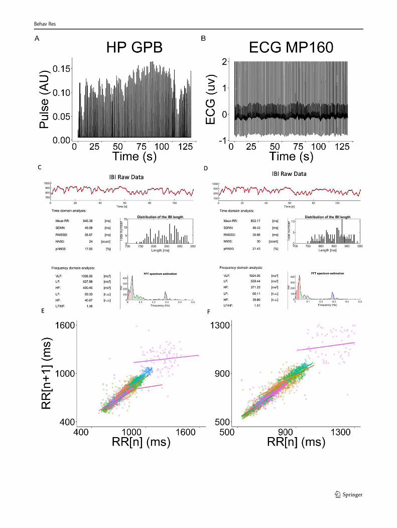
and time domain) the recordings from the GPB showed re-markable consistency with those from the BIOPAC system.However, measurement of the pulse by the GPB system wasmore prone to data loss than the ECG system used by theBIOPAC. This data loss may cause problems for heart ratevariability analyses.
General discussion
Experiments 1 and 2 aimed to assess the validity of the GP3-HD eye-tracking system. While the manufacturer’s statedlevels of accuracy are possible to achieve (0.5–1°), in two
Fig. 10 a–d Scatter plots for raw averages of the skin conductance signalacross the Gazepoint biometrics (GPB) and BIOPACMP160 systems. e–f Scatter plots for correlations of computed heart rate from Gazepointbiometrics system and BIOPAC MP160 ECG. Left plots show data
aggregated by participant, and right plots show data for all trials andparticipants. Each color and line represent a single participant, each dotrepresents a single trial. *** p < .001
Table 5 Heart rate and heart rate variability correlations between theGBP and BIOPAC systems
Variable Correlation
HR .92***
Time domain
SDNN .73***
PNN50 .63***
RMSSD .61***
NN50 .65***
Frequency domain
HF .75***
LF .76***
Notes. N = 20. Analyses are reported excluding outliers, but results didnot differ significantly with the inclusion of outliers (there was only oneoutlier per analysis). *** p < .001. All correlations are Kendal rank cor-relations. HR: Heart rate; SDNN: Standard deviation of NN intervals;RMSSD: Root mean square of successive RR interval differences;NN50: Percentage of successive RR intervals that differ by more than50ms; HF: Absolute power of the high-frequency band (0.15–0.4 Hz);LF: Low-frequency band (0.04–0.15Hz)
�Fig. 11 Comparison plots of heartbeat data from the Gazepointbiometrics (GBP) and BIOPAC MP160 systems. a and b show a repre-sentative participant’s raw recording of pulse (GPB; A) and electrocar-diogram (BIOPAC MP160; B) data. c and d show derived heart ratevariability metrics for the same participant across the two devices (C =GPB, D = BIOPAC). e and f show point-care plots for the inter-beat-interval (IBI) for all participants. Each color denotes a single participant,each dot denotes a single RR (peak-to-peak) duration
Behav Res
Behav Res

studies the average accuracy of the system was closer to 1°,with the horizontal accuracy and precision matching the stated~ 0.5 and < 0.5, respectively. This is similar to what has beenreported for similar grade (e.g., EyeTribe, Tobii EyeX) as wellas higher grade devices (e.g., Tobii TX 300) in large-scaleeye-tracking comparison studies, where the measured accura-cy averaged around 1o (Funke et al., 2016; Holmqvist, 2017).Data loss is also a good indicator of the capabilities of a sys-tem and in this regard, the GP3-HD also performs quite well,with discarded data after cleaning making up on average lessthan 10% of the collected data, compared to reported data lossin different systems which ranges from less than 2% to up to20% (Holmqvist, 2017).
It is important to note that in behavioral studies, reducedaccuracy can also result from participant behavior rather thanhardware limitations, as the computation of accuracy and pre-cision is reliant on participants attending to the targets.Nonetheless, this type of validation represents the most likelyscenario under which most eye-tracking systems will be usedwith human participants. The lack of any major effects on thequality of gaze estimation between chinrest and no-chinrestconditions in Experiment 1 suggests that the gaze estimationalgorithm for the GP3-HD is robust. It is worth noting, how-ever, that in both conditions participants were instructed toavoid body and head movements. Other studies have shownthat body and head position can severely affect the quality ofdata obtained from remote eye-trackers (Niehorster et al.,2018), and that infants and participants with neuropsychiatricconditions are more likely to show poor data quality, in termsof calibration, accuracy, precision and data loss (Dalrympleet al., 2018; Hessels & Hooge, 2019; Holmqvist et al., 2012).Therefore, we recommend that experimenters systematicallyassess data quality parameters when using GP3-HD for exper-imental research.
One issue to consider when using the GP3-HD is that track-ing participants’ gaze when using chinrests or with additionalobjects near their head (e.g., headphones, glasses, masks) maycause infrared bounce. We observed this during some record-ings, where transient reflections from headphones (which wasrequired for a separate task) or the chinrest would cause gazeestimation failures. While these data samples are typicallyflagged as invalid by the GP3-HD algorithm, it can increasedata loss in some cases sufficiently to invalidate a full trial ifno correction is made. Simple solutions, such as coveringareas that are likely to be reflective with non-reflective tape,are usually enough to solve this issue.
Analyses of gaze position metrics show that detection offixations and saccades is reliable, yet measurement of the ki-nematics of saccades are negatively impacted by the low sam-pling rate. Similarly, analyses of velocity profiles show thatknown relationships between saccade parameters, e.g., sac-cade velocity and amplitude, or saccade duration and ampli-tude, recorded from the GP3-HD only approximate the
expected relationships. Nonetheless, any inaccuracy in theestimation of fixation and saccade metrics is likely systematic,such that comparisons between different conditions, individ-uals or groups should be possible, if all other factors are takeninto account (e.g., differences in accuracy or precision).
Finally, in terms of the software for collection and analysisof data, the GP3-HD software is unlikely to meet the demandsof most experimental research. However, using the GazepointAPI, a number of popular experimental software libraries nowsupport Gazepoint eye-trackers, such as PsychoPy (Peirceet al., 2019), PyGaze (Dalmaijer et al., 2014), OpenSesame(Mathôt et al., 2012), and Psychtoolbox (Kleiner et al., 2007).Similarly, for analyses, the output generated by Gazepoint canbe imported into third party open-source software like Pythonand R (or proprietary software such as MATLAB) for furtherprocessing.
Gazepoint biometrics system
Overall, the GBP system showed a high degree of consistencywith the well-established (and considerably more expensive)BIOPAC system, which is often considered to be the ‘gold-standard’ for physiological recording. However, specializedpre-processing of pulse data obtained from the GBP systemis necessary for calculation of HRV metrics. Similarly, likeother PPG measures, the study of properties of the pulse (orheartbeat) signal, such the QRS complex, is likely to be chal-lenging (although see Chiu et al., 2020). While the SC signalis more robust to data loss, the low amplitude of the signal islikely to make the separation of phasic and tonic measures ofSC more dif f icul t (Edelberg , 1993; Socie ty forPsychophysiological Research Ad, 2012). Another problemrelates to motion and respiration artifacts. Irregular respirationand deep breaths cause fluctuations in the SCR signal, whichmay lead to inaccurate SCR detection (Posada-Quintero &Chon, 2020). In systems like the BIOPAC MP160, it is pos-sible to also collect respiration (with additional modules) andusing this information to remove artefactual SCRs caused byrespiration. Such automation is impossible with the GPB. Thisalso means that tasks involving physical activity cannot beused when measuring SC and HR with the GPB. The GPBdesign is also optimized for use in the right hand, while itworks on the left hand the positioning is less ideal, whichmay be a problem in reaction-time tasks where the use of adominant right-hand is needed.
In terms of software integration, at the time of writing, rawrecordings of SC, HR, and pulse are not accessible by defaultin the implementation of Gazepoint systems in PsychoPy,OpenSesame or Psychtoolbox. However, it is possible to ac-cess these data through the provided Gazepoint API.Similarly, the current iteration of Gazepoint’s collection andanalysis software does not include the raw pulse rate, althoughit is likely to be included in future releases. As with the eye-
Behav Res

tracking data, however, output from the GPB can be exportedto be used in open-source toolboxes for analyses of SC such asLedalab (Kaernbach, 2005) or PSPM (Bach& Staib, 2015), orheart rate variability such as Artifact (Kaufmann et al., 2011)or Kubios (Tarvainen et al., 2014). Based on our tests, simplek-means clustering on the raw timeseries of pulse data fromthe GPB provided acceptable classification of heart beats,which means that basic processing pipelines can be used rel-atively easily.
Conclusions
Two experiments assess the validity of a new relatively low-cost eye-tracking and psychophysiology system fromGazepoint. We show that the GP3-HD eye-tracker shows ac-ceptable accuracy and precision, with only the study of sac-cade kinematics likely to be problematic. The GP3-HD wasalso shown to reliably capture the PLR and arousal effects onpupil size. Measurement of SC, HR and HRV from the GPBshow a high degree of consistency with the well-establishedBIOPAC MP160 system. However, the low amplitude of SCsignal may make it difficult to parse small phasic responses,and the relatively high degree of pulse rate loss in some par-ticipants may render pulse data unsuitable for HRV analyseswithout extensive pre-processing.
Supplementary Information The online version contains supplementarymaterial available at https://doi.org/10.3758/s13428-021-01654-x.
Acknowledgements We would like to thank Lucy Johnson Perrett, forthe assistance with recruiting and testing participants, as well as all thevolunteers who took part in this study. Finally, we would like to thank theGazepoint team for their correspondence, clarifications and support pro-vided during this study.
Author statement HCC: Conception and design, data acquisition, anal-ysis and interpretation, visualization, drafting and revising themanuscript.
JS: Conception and design, data acquisition, analysis and interpreta-tion, visualization, drafting and revising the manuscript.
XMG: Conception and design, data acquisition, analysis, revising themanuscript.
CC: Revising the manuscript.GB: Revising the manuscript.
Funding HCCuve is supported by aMedical Sciences Division GraduateStudentship awarded by the Clarendon Fund (SFF1819_CB2_MSD_1152472).
J Stojanov is supported by a Dulverton Scholarship (SFF1920_DVS_1241286) as well as a Rotary D1090 Scholarship awarded by the RotaryFoundation.
G Bird is supported by the Baily Thomas Charitable Trust and by theEconomic and Social Research Council (ES/R007527/1).
Declarations All research procedures were in accordance with therevised 2013 Declaration of Helsinki.
Competing interests None.
Open Access This article is licensed under a Creative CommonsAttribution 4.0 International License, which permits use, sharing, adap-tation, distribution and reproduction in any medium or format, as long asyou give appropriate credit to the original author(s) and the source, pro-vide a link to the Creative Commons licence, and indicate if changes weremade. The images or other third party material in this article are includedin the article's Creative Commons licence, unless indicated otherwise in acredit line to the material. If material is not included in the article'sCreative Commons licence and your intended use is not permitted bystatutory regulation or exceeds the permitted use, you will need to obtainpermission directly from the copyright holder. To view a copy of thislicence, visit http://creativecommons.org/licenses/by/4.0/.
References
Bach, D. R., & Staib, M. (2015). A matching pursuit algorithm for infer-ring tonic sympathetic arousal from spontaneous skin conductancefluctuations. Psychophysiology, 52(8), 1106–1112. https://doi.org/10.1111/psyp.12434
Bahill, A. T., Clark, M. R., & Stark, L. (1975). The main sequence, a toolfor studying human eye movements. Mathematical biosciences,24(3–4), 191–204.
Barr, D. J., Levy, R., Scheepers, C., & Tily, H. J. (2013). Random effectsstructure for confirmatory hypothesis testing: Keep it maximal.Journal of memory and language, 68(3), 255–278.
Bates, D., Mächler, M., Bolker, B., & Walker, S. (2014). Fitting linearmixed-effects models using lme4. ArXiv Preprint ArXiv:1406.5823.
Bates, D., Kliegl, R., Vasishth, S., & Baayen, H. (2015). Parsimoniousmixed models. arXiv preprint arXiv:1506.04967
Benedek, M., & Kaernbach, C. (2010). A continuous measure of phasicelectrodermal activity. Journal of neuroscience methods, 190(1),80–91. https://doi.org/10.1016/j.jneumeth.2010.04.028
Boucsein, W., Fowles, D. C., Grimnes, S., Ben-Shakhar, G., Roth, W. T.,Dawson, M. E., … Society for Psychophysiological Research AdHoc Committee on Electrodermal Measures. (2012). Publicationrecommenda t ions fo r e l ec t rode rma l measu remen t s .Psychophysiology, 49(8), 1017–1034. https://doi.org/10.1111/j.1469-8986.2012.01384.x
Bradley, M. M., Miccoli, L., Escrig, M. A., & Lang, P. J. (2008). Thepupil as a measure of emotional arousal and autonomic activation.Psychophysiology, 45(4), 602–607. https://doi.org/10.1111/j.1469-8986.2008.00654.x
Brand, J., Diamond, S. G., Thomas, N., & Gilbert-Diamond, D. (2020).Evaluating the data quality of the Gazepoint GP3 low-cost eyetracker when used independently by study participants. BehaviorResearch Methods. https://doi.org/10.3758/s13428-020-01504-2
Buswell, G. T. (1935). How people look at pictures: a study of the psy-chology and perception in art. University of Chicago Press.
Cacioppo, J. T., Tassinary, L. G., & Berntson, G. G. (Eds.). (2016).Handbook of Psychophysiology. Cambridge University Press.https://doi.org/10.1017/9781107415782
Chiu, H. Y., Shuai, H. H., & Chao, P. C. P. (2020). Reconstructing QRScomplex from PPG by transformed attentional neural networks.IEEE Sensors Journal, 20, 12374-12383
Dalmaijer, E. (2014). Is the low-cost EyeTribe eye tracker any good forresearch? https://doi.org/10.7287/peerj.preprints.585v1
Behav Res
Dalmaijer, E. S., Mathôt, S., & Van der Stigchel, S. (2014). PyGaze: anopen-source, cross-platform toolbox for minimal-effort program-ming of eyetracking experiments. Behavior Research Methods,46(4), 913–921. https://doi.org/10.3758/s13428-013-0422-2
Dalrymple, K. A., Manner, M. D., Harmelink, K. A., Teska, E. P., &Elison, J. T. (2018). An examination of recording accuracy andprecision from eye tracking data from toddlerhood to adulthood.Frontiers in Psychology, 9, 803. https://doi.org/10.3389/fpsyg.2018.00803
Dodge, R., & Cline, T. S. (1901). The angle velocity of eye movements.Psychological Review, 8(2), 145–157.
Edelberg, R. (1993). Electrodermal mechanisms: A critique of the two-effector hypothesis and a proposed replacement. In Progress inelectrodermal research (pp. 7–29). Springer.
Engbert, R., & Kliegl, R. (2003). Microsaccades uncover the orientationof covert attention. Vision Research, 43(9), 1035–1045. https://doi.org/10.1016/S0042-6989(03)00084-1
Feit, A. M., Williams, S., Toledo, A., Paradiso, A., Kulkarni, H., Kane,S., & Morris, M. R. (2017, May). Toward everyday gaze input:Accuracy and precision of eye tracking and implications for design.In: Proceedings of the 2017 Chi conference on human factors incomputing systems (pp. 1118–1130).
Funke, G., Greenlee, E., Carter, M., Dukes, A., Brown, R., & Menke, L.(2016). Which eye tracker is right for your research? performanceevaluation of several cost variant eye trackers. Proceedings of theHuman Factors and Ergonomics Society Annual Meeting, 60(1),1240–1244. https://doi.org/10.1177/1541931213601289
Geller, J., Winn, M. B., Mahr, T., & Mirman, D. (2020). Gazer: A pack-age for processing gaze position and pupil size data. BehaviorResearch Methods. https://doi.org/10.3758/s13428-020-01374-8
Gibaldi, A., & Sabatini, S. P. (2021). The saccade main sequence revised:A fast and repeatable tool for oculomotor analysis. BehaviorResearch Methods, 53(1), 167–187. https://doi.org/10.3758/s13428-020-01388-2
Hessels, R. S., & Hooge, I. T. C. (2019). Eye tracking in developmentalcognitive neuroscience - The good, the bad and the ugly.Developmental Cognitive Neuroscience, 40, 100710. https://doi.org/10.1016/j.dcn.2019.100710
Hessels, R. S., Andersson, R., Hooge, I. T. C., Nyström, M., & Kemner,C. (2015a). Consequences of eye color, positioning, and head move-ment for eye-tracking data quality in infant research. Infancy: TheOfficial Journal of the International Society on Infant Studies, 20(6),601–633. https://doi.org/10.1111/infa.12093
Hessels, R. S., Cornelissen, T. H. W., Kemner, C., & Hooge, I. T. C.(2015b). Qualitative tests of remote eyetracker recovery and perfor-mance during head rotation. Behavior Research Methods, 47(3),848–859. https://doi.org/10.3758/s13428-014-0507-6
Hessels, R. S., Niehorster, D. C., Kemner, C., & Hooge, I. T. (2017).Noise-robust fixation detection in eye movement data: Identificationby two-means clustering (I2MC). Behavior researchmethods, 49(5),1802–1823.
Holmqvist, K. (2017). Common predictors of accuracy, precision anddata loss in 12 eye-trackers. The 7th Scandinavian Workshop onEye Tracking.
Holmqvist, K., Nyström, M., Andersson, R., Dewhurst, R., Jarodzka, H.,& Van deWeijer, J. (2011). Eye tracking: A comprehensive guide tomethods and measures. OUP Oxford.
Holmqvist, K., Nyström, M., & Mulvey, F. (2012, March). Eye trackerdata quality: what it is and how to measure it. In Proceedings of thesymposium on eye tracking research and applications (pp. 45–52).
Janthanasub, V., & Meesad, P. (2015). Evaluation of a low-cost eyetracking system for computer input. KMUTNB InternationalJournal of Applied Science and Technology, 1–12. https://doi.org/10.14416/j.ijast.2015.07.001
Benedek, M., & Kaernbach, C. (2010). Decomposition of skin conduc-t a n c e d a t a b y m e a n s o f n o n n e g a t i v edeconvolution. psychophysiology, 47(4), 647–658.
Kaufmann, T., Sütterlin, S., Schulz, S. M., & Vögele, C. (2011).ARTiiFACT: a tool for heart rate artifact processing and heart ratevariability analysis. Behavior ResearchMethods, 43(4), 1161–1170.https://doi.org/10.3758/s13428-011-0107-7
Kirk, P. A., Garfinkel, S., & Robinson, O. J. (2021). RapidHRV: an open-source toolbox for extracting heart rate and heart rate variability.https://psyarxiv.com/3ewgz/
Kleiner, M., Brainard, D., & Pelli, D. (2007). What’s new inPsychtoolbox-3? http://psychtoolbox.org
Lang, P. J., Bradley, M. M., & Cuthbert, B. N. (2005). InternationalAffective Picture System (IAPS): Affective ratings of pictures andinstruction manual. Technical Report A-6. University of Florida.
Leube, A., Rifai, K., & Rifai, K. (2017). Sampling rate influences saccadedetection in mobile eye tracking of a reading task. Journal of eyemovement research, 10(3).
Mannaru, P., Balasingam, B., Pattipati, K., Sibley, C., & Coyne, J. T.(2017, July). Performance evaluation of the gazepoint GP3 eyetracking device based on pupil dilation. In InternationalConference on Augmented Cognition (pp. 166–175). Springer.
Mannaru, P., Balasingam, B., Pattipati, K., Sibley, C., & Coyne, J. T.(2017b). Performance evaluation of the Gazepoint GP3 eye trackingdevice based on pupil dilation. In D. D. Schmorrow & C. M.Fidopiastis (Eds.), Augmented cognition. neurocognition and ma-chine learning (Vol. 10284, pp. 166–175). Springer InternationalPublishing. https://doi.org/10.1007/978-3-319-58628-1_14
Mathôt, S. (2018). Pupillometry: psychology, physiology, and function.Journal of Cognition, 1(1), 16. https://doi.org/10.5334/joc.18
Mathôt, S., Schreij, D., & Theeuwes, J. (2012). OpenSesame: An open-source, graphical experiment builder for the social sciences.Behavior Research Methods, 44(2), 314–324. https://doi.org/10.3758/s13428-011-0168-7
Mathôt, S., Fabius, J., Van Heusden, E., & Van der Stigchel, S. (2018).Safe and sensible preprocessing and baseline correction of pupil-sizedata. Behavior ResearchMethods, 50(1), 94–106. https://doi.org/10.3758/s13428-017-1007-2
Mirman, D. (2017). Growth curve analysis and visualization using R.CRC press.
Mühlen, J. M., Stang, J., Lykke Skovgaard, E., Judice, P. B., Molina-Garcia , P . , Johnston, W., … Schumann, M. (2021) .Recommendations for determining the validity of consumer wear-able heart rate devices: expert statement and checklist of theINTERLIVE Network. British Journal of Sports Medicine. https://doi.org/10.1136/bjsports-2020-103148
Niehorster, D. C., Cornelissen, T. H. W., Holmqvist, K., Hooge, I. T. C.,& Hessels, R. S. (2018). What to expect from your remote eye-tracker when participants are unrestrained. Behavior ResearchMethods, 50(1), 213–227. https://doi.org/10.3758/s13428-017-0863-0
Noto, C. T., & Robinson, F. R. (2001). Visual error is the stimulus forsaccade gain adaptation.Cognitive Brain Research, 12(2), 301–305.https://doi.org/10.1016/S0926-6410(01)00062-3
Nyström, M., Niehorster, D. C., Andersson, R., & Hooge, I. (2021). TheTobii Pro Spectrum: A useful tool for studying microsaccades?.Behavior Research Methods, 53(1), 335–353.
Orquin, J. L., & Holmqvist, K. (2018). Threats to the validity of eye-movement research in psychology. Behavior Research Methods,50(4), 1645–1656. https://doi.org/10.3758/s13428-017-0998-z
Papesh, M. H., & Goldinger, S. D. (2012). Pupil-BLAH-metry: cognitiveeffort in speech planning reflected by pupil dilation. Attention,Perception & Psychophysics, 74(4), 754–765. https://doi.org/10.3758/s13414-011-0263-y
Peirce, J. W., Gray, J. R., Simpson, S., MacAskill, M. R., Höchenberger,R., Sogo, H., Kastman, E., Lindeløv, J. (2019). PsychoPy2:
Behav Res
experiments in behavior made easy. Behavior Research Methods.https://doi.org/10.3758/s13428-018-01193-y
Piquado, T., Isaacowitz, D., & Wingfield, A. (2010). Pupillometry as ameasure of cognitive effort in younger and older adults.Psychophysiology, 47(3), 560–569. https://doi.org/10.1111/j.1469-8986.2009.00947.x
Posada-Quintero, H. F., & Chon, K. H. (2020). Innovations in electroder-mal activity data collection and signal processing: A systematicreview. Sensors (Basel, Switzerland), 20(2). https://doi.org/10.3390/s20020479
Shaffer, F., & Ginsberg, J. P. (2017). An overview of heart rate variabilitymetrics and norms. Frontiers in Public Health, 5, 258. https://doi.org/10.3389/fpubh.2017.00258
Society for Psychophysiological Research Ad Hoc Committee onElectrodermal Measures, Boucsein, W., Fowles, D. C., Grimnes,S., Ben‐Shakhar, G., Roth, W. T., ... & Filion, D. L. (2012).P u b l i c a t i o n r e c ommen d a t i o n s f o r e l e c t r o d e rm a lmeasurements. Psychophysiology, 49(8), 1017–1034.
Tarvainen, M. P., Niskanen, J. P., Lipponen, J. A., Ranta-Aho, P. O., &Karjalainen, P. A. (2014). Kubios HRV–heart rate variability anal-ysis software. Computer Methods and Programs in Biomedicine,113(1), 210–220. https://doi.org/10.1016/j.cmpb.2013.07.024
Taylor, S., Jaques, N., Chen,W., Fedor, S., Sano, A., & Picard, R. (2015).Automatic identification of artifacts in electrodermal activity data. In2015 37th Annual International Conference of the IEEEEngineering in Medicine and Biology Society (EMBC) (pp. 1934–1937). IEEE
van Dooren, M., de Vries, J. J. G. G.-J., & Janssen, J. H. (2012).Emotional sweating across the body: comparing 16 different skinconductance measurement locations. Physiology & Behavior,106(2), 298–304. https://doi.org/10.1016/j.physbeh.2012.01.020
Van Leeuwen, J., & Belopolsky, A. V. (2018). Distractor displacementsduring saccades are reflected in the time-course of saccade curva-ture. Scientific Reports, 8(1), 1–12. https://doi.org/10.1038/s41598-018-20578-9
Wierts, R., Janssen, M. J. A., & Kingma, H. (2008). Measuring saccadepeak velocity using a low-frequency sampling rate of 50 Hz. IEEETransactions on Bio-Medical Engineering, 55(12), 2840–2842.https://doi.org/10.1109/TBME.2008.925290
Publisher’s note Springer Nature remains neutral with regard to jurisdic-tional claims in published maps and institutional affiliations.
Behav Res