tissuecontrolsfor ihc background-selection interpretation · 2 ihc –biomarker controls whatis an...
TRANSCRIPT
Tissue controls for IHC
Background - SelectionInterpretation
Søren Nielsen
Scheme Manager
NordiQC
Aalborg Hospital, Denmark
2
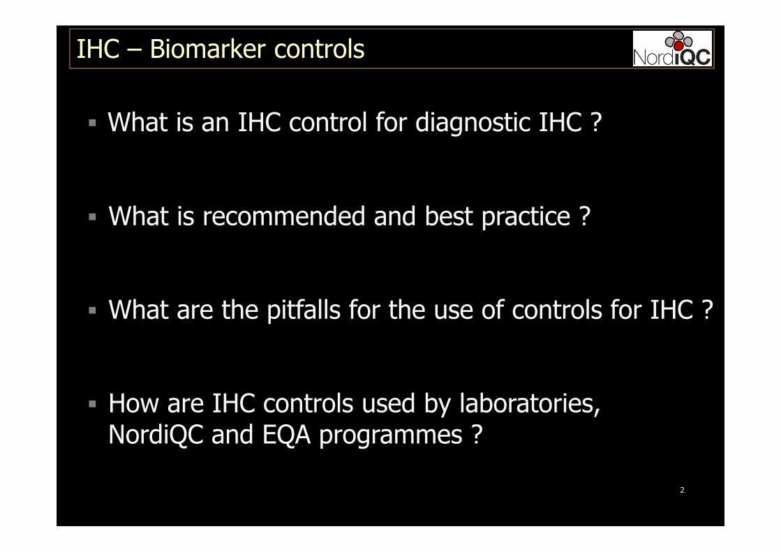
IHC – Biomarker controls
� What is an IHC control for diagnostic IHC ?
� What is recommended and best practice ?
� What are the pitfalls for the use of controls for IHC ?
� How are IHC controls used by laboratories, .NordiQC and EQA programmes ?
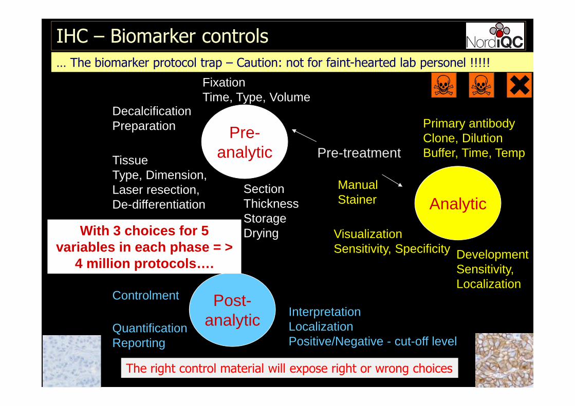
DecalcificationPreparation Pre-
analytic
Analytic
Post-analytic
TissueType, Dimension,Laser resection,De-differentiation
FixationTime, Type, Volume
SectionThicknessStorageDrying Visualization
Sensitivity, Specificity
Primary antibodyClone, Dilution Buffer, Time, Temp
DevelopmentSensitivity,Localization
InterpretationLocalizationPositive/Negative - cut-off level
QuantificationReporting
Controlment
Pre-treatment
With 3 choices for 5 variables in each phase = >
4 million protocols….
ManualStainer
IHC – Biomarker controls
The right control material will expose right or wrong choices
… The biomarker protocol trap – Caution: not for faint-hearted lab personel !!!!!
4
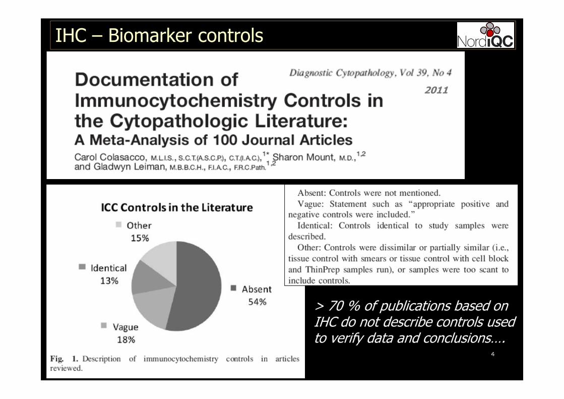
IHC – Biomarker controls
2011
> 70 % of publications based onIHC do not describe controls usedto verify data and conclusions….

5
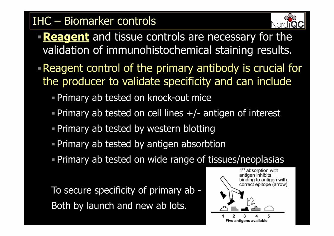
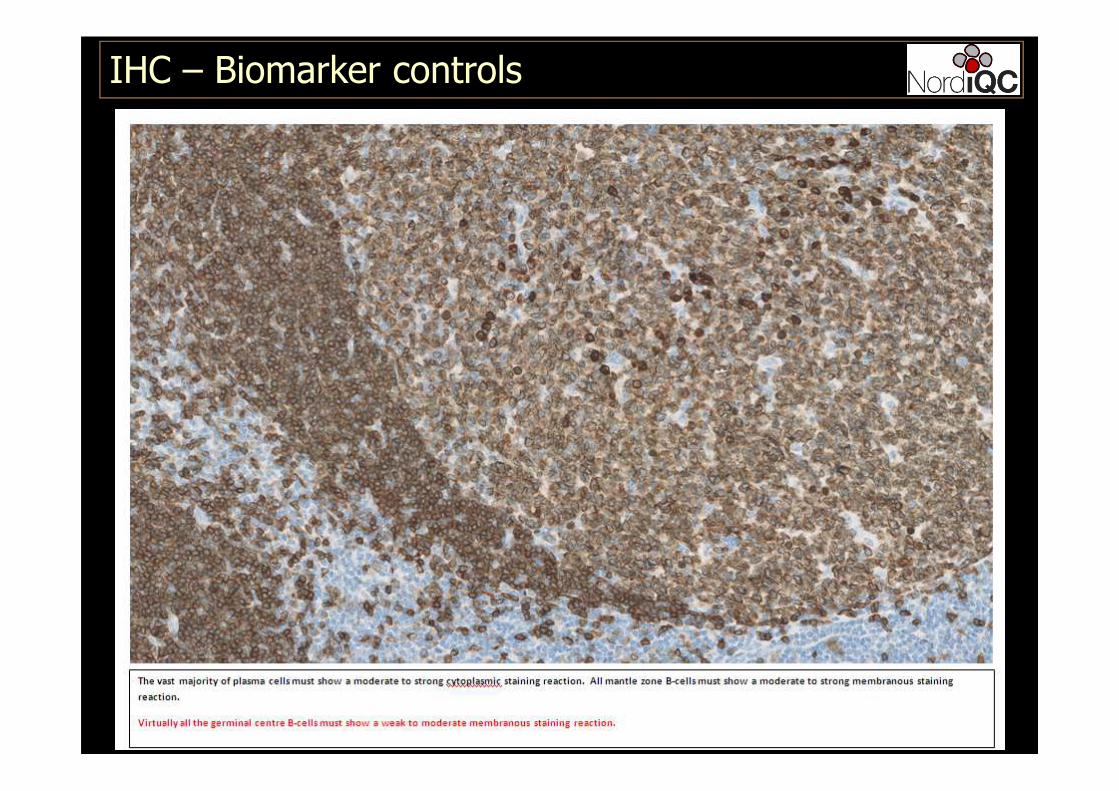
�Reagent and tissue controls are necessary for the validation of immunohistochemical staining results.
�Without their use, interpretation of staining wouldbe haphazard and the results of doubtful value. More specifically, controls determine if the stainingprotocols were followed correctly, whether day-to-day and worker-to-worker variations have occurred, and that reagents remain in good working order.
IHC – Biomarker controls
6
�Reagent and tissue controls are necessary for the validation of immunohistochemical staining results.
�Reagent controls typically used to validate specificty of the primary and secondary antibodies – to show that the antibody-antigen reaction is due to expression of the targetof interest.
�Often referred as negative controls
� Tissue controls typically used to show that the IHC stainingwas successful and capeable to demonstrate the target of interest
�Often referred as positive controls
IHC – Biomarker controls
7
�Reagent and tissue controls are necessary for the validation of immunohistochemical staining results.
�Reagent control of the primary antibody is crucial for the producer to validate specificity and can include
� Primary ab tested on knock-out mice
� Primary ab tested on cell lines +/- antigen of interest
� Primary ab tested by western blotting
� Primary ab tested by antigen absorbtion
� Primary ab tested on wide range of tissues/neoplasias
To secure specificity of primary ab -
Both by launch and new ab lots.
IHC – Biomarker controls
8
�Reagent and tissue controls are necessary for the validation of immunohistochemical staining results.
�Negative reagent control is for the laboratories of limited use and ”impossible” to perform correctly.
� Primary ab control – negative reagent control
�Each primary ab must have its own negative control serum, and thus all the IHC slides performed will be doubled
IHC – Biomarker controls
9
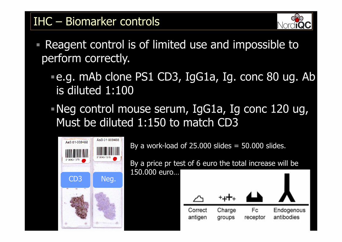
� Reagent control is of limited use and impossible to perform correctly.
�e.g. mAb clone PS1 CD3, IgG1a, Ig. conc 80 ug. Ab is diluted 1:100
�Neg control mouse serum, IgG1a, Ig conc 120 ug, Must be diluted 1:150 to match CD3
IHC – Biomarker controls
CD3 Neg.
By a work-load of 25.000 slides = 50.000 slides.
By a price pr test of 6 euro the total increase will be150.000 euro…

10
�Reagent and tissue controls are necessary for the validation of immunohistochemical staining results.
�Negative reagent control is for the laboratories of limited use and ”impossible” to perform correctly.
� Primary ab control – negative reagent control
�Each primary ab must have its own negative control serum, and thus all the IHC slides performed will be doubled
� WILL NOT EXPOSE IF WRONG OR CONTAMINATED PRIMARY AB HAS BEEN APPLIED!!!!!
IHC – Biomarker controls
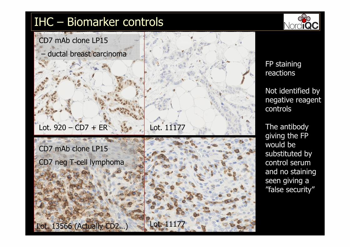
IHC – Biomarker controls
CD7 mAb clone LP15
– ductal breast carcinoma
Lot. 920 – CD7 + ER Lot. 11177
FP stainingreactions
Not identified by negative reagent controls
The antibodygiving the FP would besubstituted by control serum and no stainingseen giving a ”false security”
CD7 mAb clone LP15
CD7 neg T-cell lymphoma
Lot. 11177 Lot. 13566 (Actually CD2…)
12
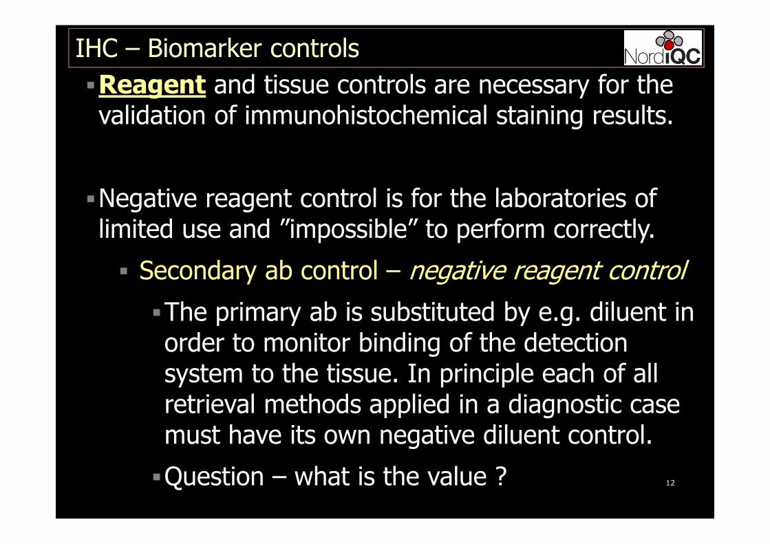
�Reagent and tissue controls are necessary for the validation of immunohistochemical staining results.
�Negative reagent control is for the laboratories of limited use and ”impossible” to perform correctly.
� Secondary ab control – negative reagent control
�The primary ab is substituted by e.g. diluent in order to monitor binding of the detectionsystem to the tissue. In principle each of all retrieval methods applied in a diagnostic case must have its own negative diluent control.
�Question – what is the value ?
IHC – Biomarker controls
13
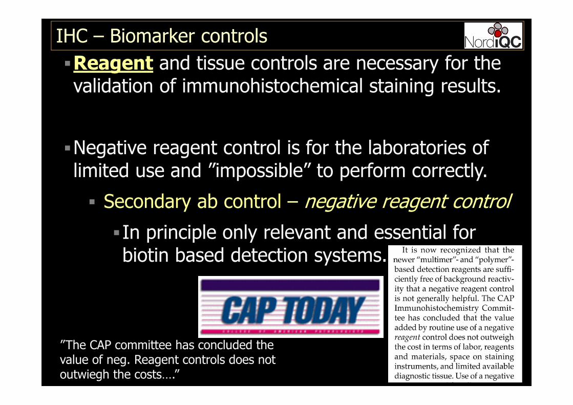
�Reagent and tissue controls are necessary for the validation of immunohistochemical staining results.
�Negative reagent control is for the laboratories of limited use and ”impossible” to perform correctly.
� Secondary ab control – negative reagent control
�In principle only relevant and essential for biotin based detection systems.
IHC – Biomarker controls
”The CAP committee has concluded the value of neg. Reagent controls does not outwiegh the costs….”
14
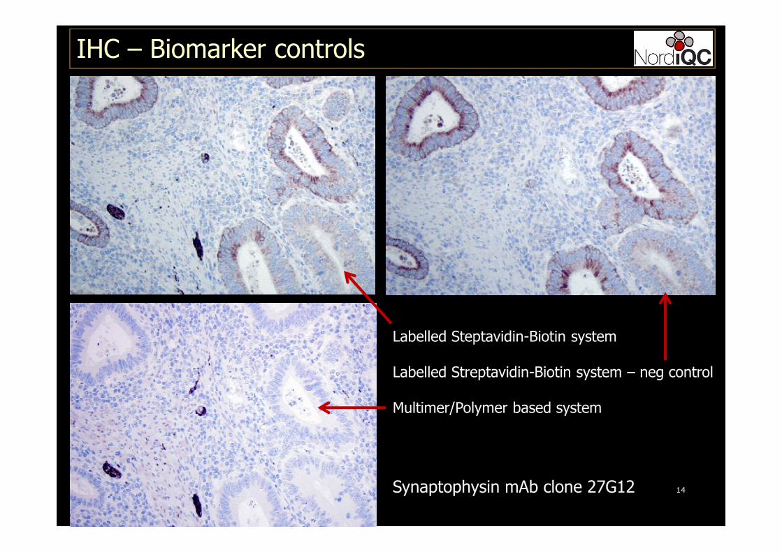
IHC – Biomarker controls
Labelled Steptavidin-Biotin system
Labelled Streptavidin-Biotin system – neg control
Multimer/Polymer based system
Synaptophysin mAb clone 27G12
15
�Reagent and tissue controls are necessary for the validation of immunohistochemical staining results.
�Tissue controls are the most valueable tool to monitor the specificity and sensitivity for IHC
� Internal positive and negative tissue control
� Cells/structures within the patient slide
� External positive and negative tissue control
� Slide next to patient slide
IHC – Biomarker controls
16
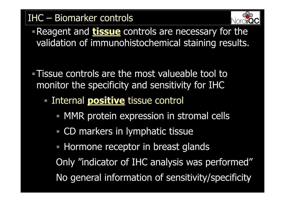
�Reagent and tissue controls are necessary for the validation of immunohistochemical staining results.
�Tissue controls are the most valueable tool to monitor the specificity and sensitivity for IHC
� Internal positive tissue control
� MMR protein expression in stromal cells
� CD markers in lymphatic tissue
� Hormone receptor in breast glands
Only ”indicator of IHC analysis was performed”
No general information of sensitivity/specificity
IHC – Biomarker controls
17
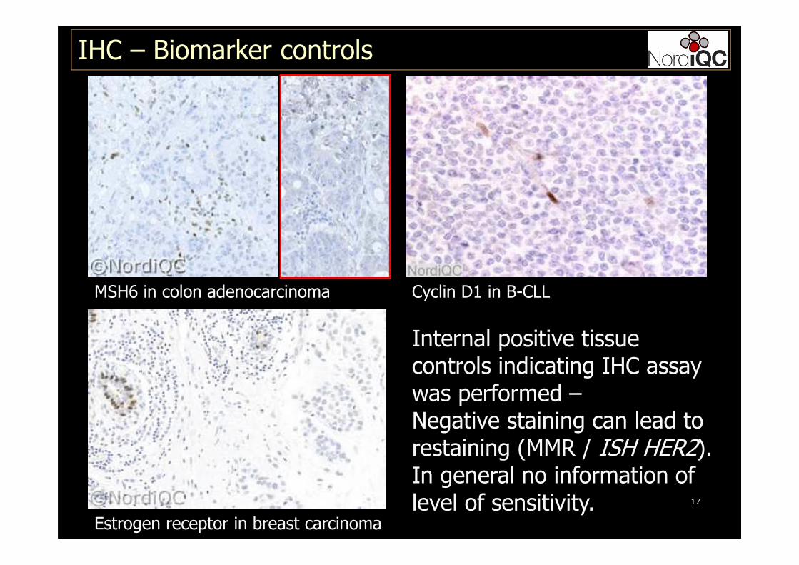
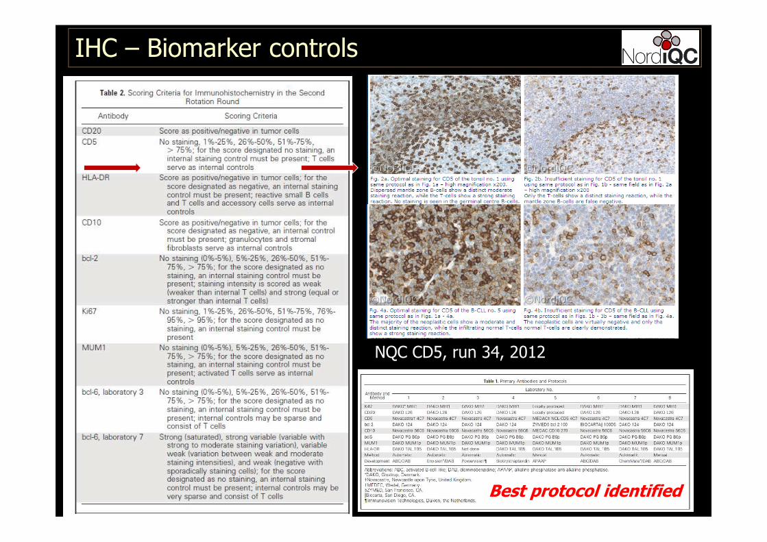
IHC – Biomarker controls
MSH6 in colon adenocarcinoma Cyclin D1 in B-CLL
Estrogen receptor in breast carcinoma
Internal positive tissuecontrols indicating IHC assaywas performed –Negative staining can lead to restaining (MMR / ISH HER2).In general no information of level of sensitivity.
IHC – Biomarker controls
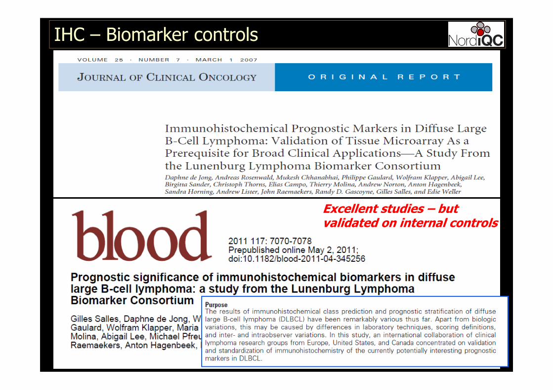
Excellent studies – but validated on internal controls
IHC – Biomarker controls
NQC CD5, run 34, 2012
Best protocol identified

20
�Reagent and tissue controls are necessary for the validation of immunohistochemical staining results.
�Tissue controls are the most valueable tool to monitor the specificity and sensitivity for IHC
� Internal negative tissue control
� Cells / structures to be negative
� E.g. T-cells for CD19, CD20, CD79a…
� Mantle zone B-cells for Ki67, Bcl-6…
� Epithelial cells for CD3, CD5, MUM1,…
Information of primary ab specificity
IHC – Biomarker controls

21
IHC – Biomarker controls
NordiQC run 35, CD19
mAb clone LE-CD19
Dako: B-cells positive, T-cells negativeSerotec: B-cells positive, T-cells false positive
IHC – Biomarker controls
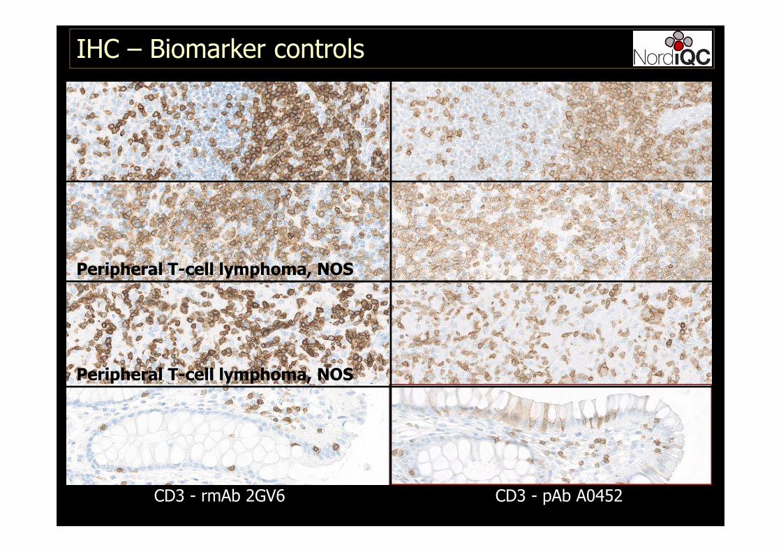
CD3 - pAb A0452CD3 - rmAb 2GV6 SP54
Peripheral T-cell lymphoma, NOS
Peripheral T-cell lymphoma, NOS
IHC – Biomarker controls
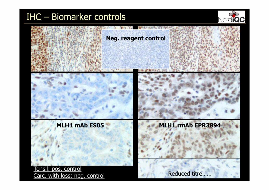
MLH1 rmAb EPR3894
Reduced titre……
MLH1 mAb ES05
Tonsil: pos. controlCarc. with loss: neg. control
Neg. reagent control
24
�Reagent and tissue controls are necessary for the validation of immunohistochemical staining results.
�Conclusions – Internal tissue controls
� Internal positive tissue control
� Indicative of ”successful ” IHC result
� Cannot be recommended as generally reliablefor evaluation of appropriate sensitivity
� Internal negative tissue control
� Can provide valueable information of specificity of the primary antibody/protocol
IHC – Biomarker controls
25
�Reagent and tissue controls are necessary for the validation of immunohistochemical staining results.
�Tissue controls are the most valueable tool to monitor the specificity and sensitivity for IHC
� External positive and negative tissue control
� Appropriate sensitivity of the IHC assay
� Appropriate specificity of the IHC assay
The central tool to monitor the IHC qualityand consistency
IHC – Biomarker controls
26
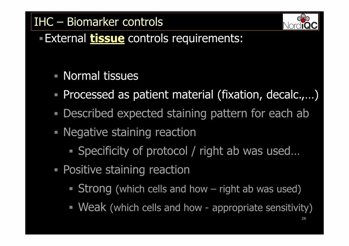
�External tissue controls requirements:
� Normal tissues
� Processed as patient material (fixation, decalc.,…)
� Described expected staining pattern for each ab
� Negative staining reaction
� Specificity of protocol / right ab was used…
� Positive staining reaction
� Strong (which cells and how – right ab was used)
� Weak (which cells and how - appropriate sensitivity)
IHC – Biomarker controls
27
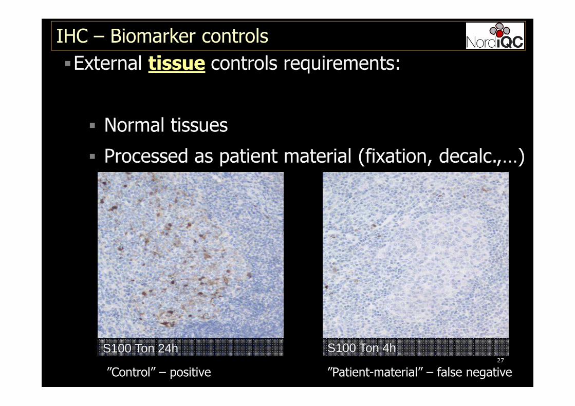
�External tissue controls requirements:
� Normal tissues
� Processed as patient material (fixation, decalc.,…)
IHC – Biomarker controls
S100 Ton 24h S100 Ton 4h
”Control” – positive ”Patient-material” – false negative

28
�External tissue controls requirements:
� Normal tissues
� Processed as patient material (fixation, decalc.,…)
IHC – Biomarker controls
Ki67 10% NBF 24 h → Form acid. Ki67 NBF + Form Acid simultan.
”Control” – positive ”Patient-material” – false negative
29
�External tissue controls requirements:
� Normal tissues
� Processed as patient material (fixation, decalc.,…)
� Described expected staining pattern for each ab
� Negative staining reaction
� Specificity of protocol / right ab was used…
� Positive staining reaction
� Strong (which cells and how – right ab was used)
� Weak (which cells and how - appropriate sensitivity)
IHC – Biomarker controls
�Low antigen expressors
�Critical Stain Quality Indicators (CSQI)
� essential to evaluate consistency
� essential to evaluate sensitivity
� normal tissue (easy to compare)
� 90 % of insufficient staining results in EQA are
caused by weak/false negative results and often
related to the use of inappropriate positive tissue
controls......
IHC – Biomarker controls
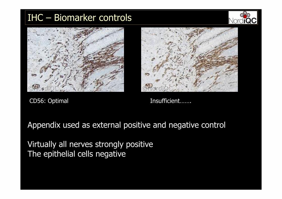
IHC – Biomarker controls
CD56: Optimal Insufficient…….
Appendix used as external positive and negative control
Virtually all nerves strongly positiveThe epithelial cells negative
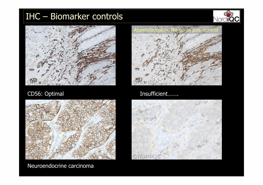
IHC – Biomarker controls
CD56: Optimal Insufficient…….
Neuroendocrine carcinoma
Appendix/colon: No-go as pos. control
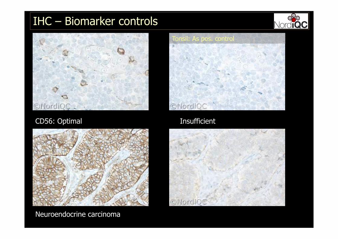
IHC – Biomarker controls
CD56: Optimal Insufficient
Neuroendocrine carcinoma
Tonsil: As pos. control

34
� The NordiQC focus areas
� Central protocol elements for an optimal staining� Antibody selected
� Antibody dilution range / Ready-To-Use
� Epitope retrieval
� IHC detection system & stainer platforms
� Recommendable control and identification of
critical quality stain indicators
(Which tissue ? Which cells ?, How must they look ?)
IHC – Biomarker controls
IHC – Biomarker controls
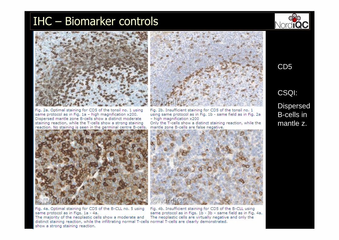
CD5
CSQI:
Dispersed B-cells in mantle z.
36
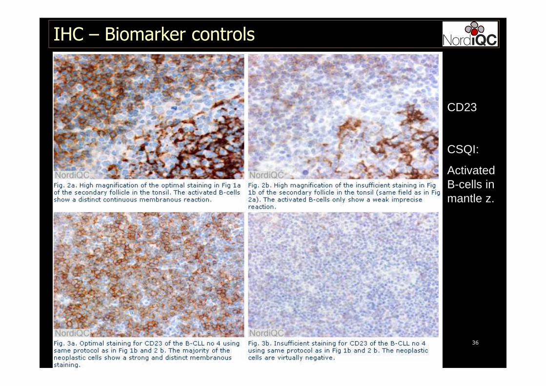
CD23
CSQI:
Activated B-cells in mantle z.
IHC – Biomarker controls
37
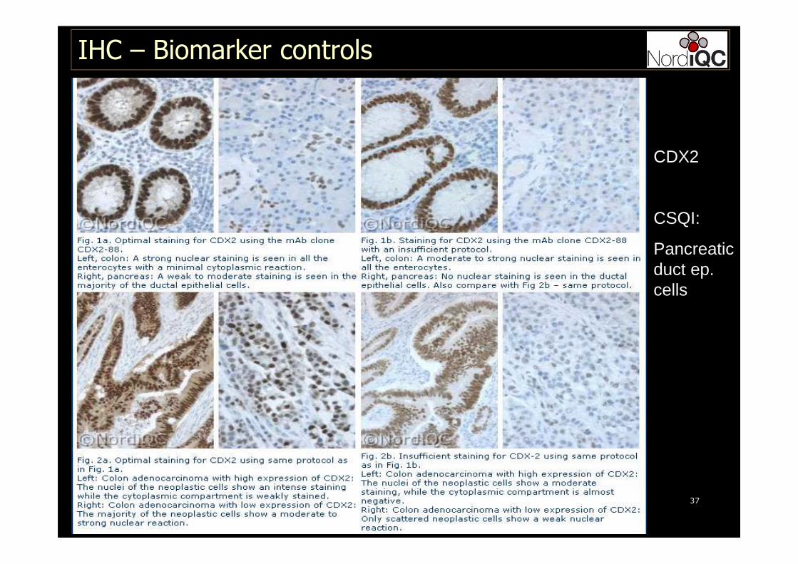
CDX2
CSQI:
Pancreatic duct ep. cells
IHC – Biomarker controls
IHC – Biomarker controls
IHC – Biomarker controls
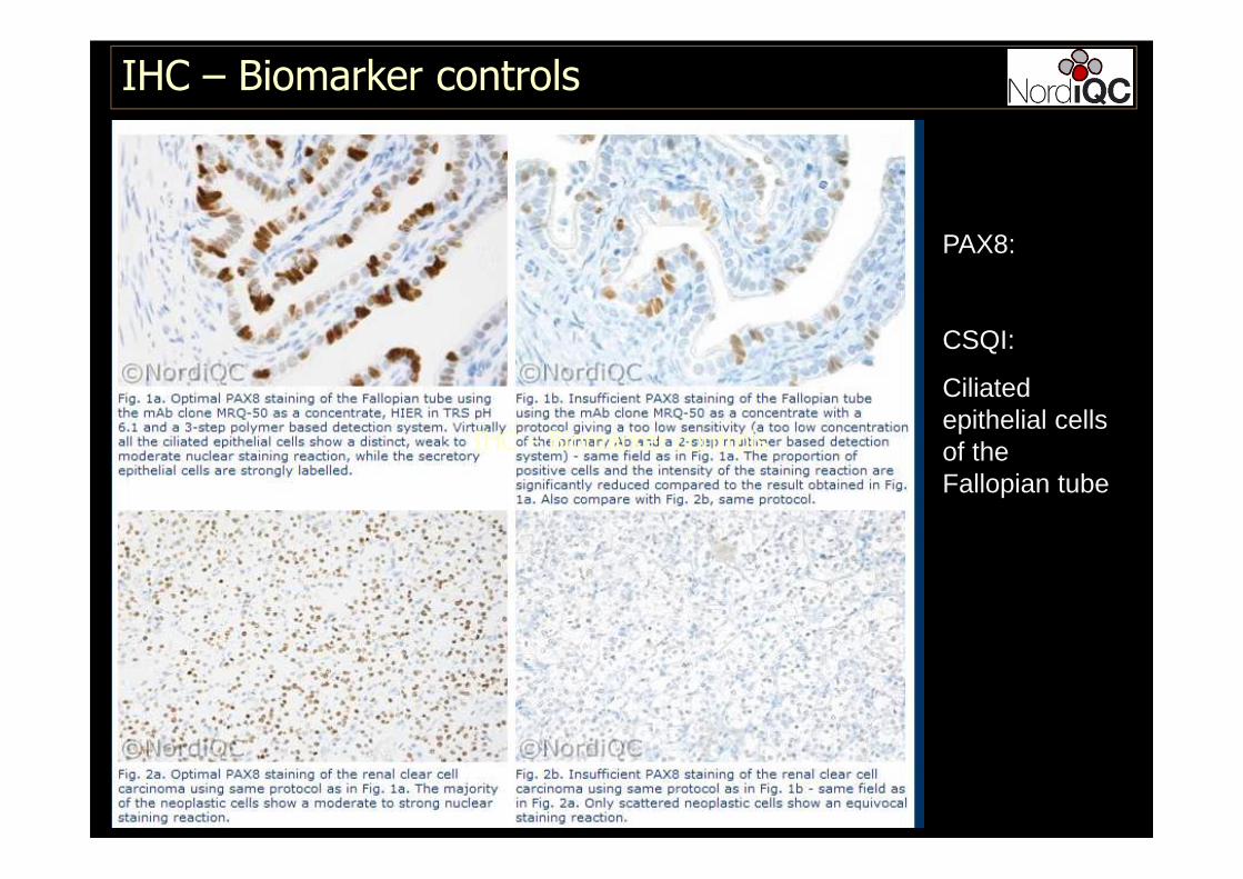
PAX8:
CSQI:
Ciliated epithelial cells of the Fallopian tube
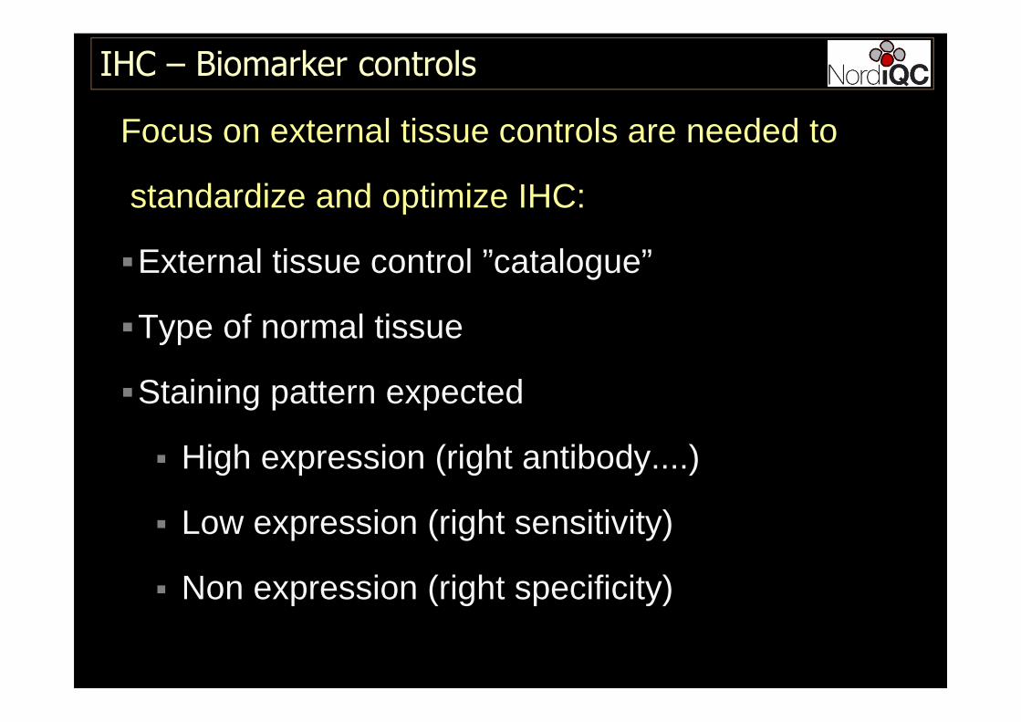
Focus on external tissue controls are needed to
standardize and optimize IHC:
�External tissue control ”catalogue”
�Type of normal tissue
�Staining pattern expected
� High expression (right antibody....)
� Low expression (right sensitivity)
� Non expression (right specificity)
IHC – Biomarker controls
40
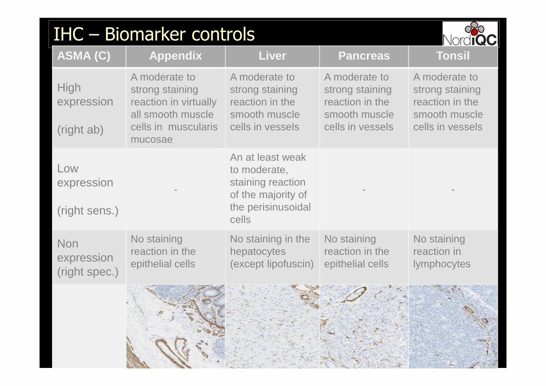
IHC – Biomarker controlsASMA (C) Appendix Liver Pancreas Tonsil
Highexpression
(right ab)
A moderate to strong stainingreaction in virtuallyall smooth musclecells in muscularismucosae
A moderate to strong stainingreaction in the smooth musclecells in vessels
A moderate to strong stainingreaction in the smooth musclecells in vessels
A moderate to strong stainingreaction in the smooth musclecells in vessels
Lowexpression
(right sens.)
-
An at least weak to moderate, staining reaction of the majority of the perisinusoidalcells
- -
Non expression(right spec.)
No stainingreaction in the epithelial cells
No staining in the hepatocytes(except lipofuscin)
No stainingreaction in the epithelial cells
No stainingreaction in lymphocytes
41
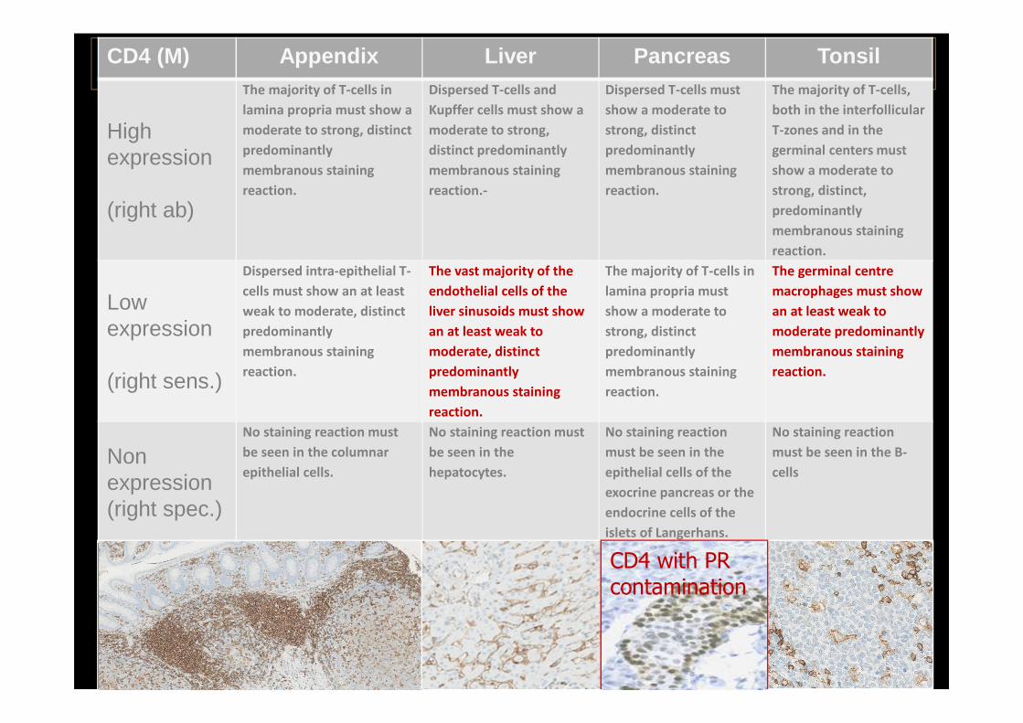
IHC – Biomarker controlsCD4 (M) Appendix Liver Pancreas Tonsil
Highexpression
(right ab)
The majority of T-cells in
lamina propria must show a
moderate to strong, distinct
predominantly
membranous staining
reaction.
Dispersed T-cells and
Kupffer cells must show a
moderate to strong,
distinct predominantly
membranous staining
reaction.-
Dispersed T-cells must
show a moderate to
strong, distinct
predominantly
membranous staining
reaction.
The majority of T-cells,
both in the interfollicular
T-zones and in the
germinal centers must
show a moderate to
strong, distinct,
predominantly
membranous staining
reaction.
Lowexpression
(right sens.)
Dispersed intra-epithelial T-
cells must show an at least
weak to moderate, distinct
predominantly
membranous staining
reaction.
The vast majority of the
endothelial cells of the
liver sinusoids must show
an at least weak to
moderate, distinct
predominantly
membranous staining
reaction.
The majority of T-cells in
lamina propria must
show a moderate to
strong, distinct
predominantly
membranous staining
reaction.
The germinal centre
macrophages must show
an at least weak to
moderate predominantly
membranous staining
reaction.
Non expression(right spec.)
No staining reaction must
be seen in the columnar
epithelial cells.
No staining reaction must
be seen in the
hepatocytes.
No staining reaction
must be seen in the
epithelial cells of the
exocrine pancreas or the
endocrine cells of the
islets of Langerhans.
No staining reaction
must be seen in the B-
cells
CD4 with PR contamination
42
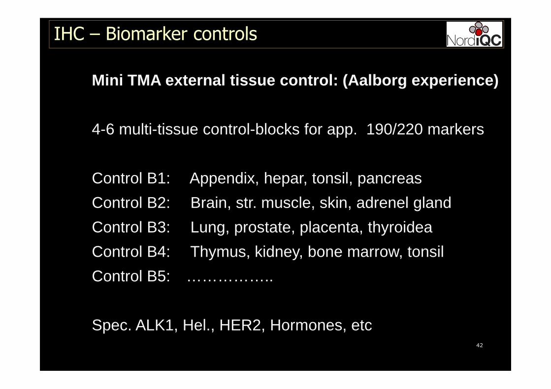
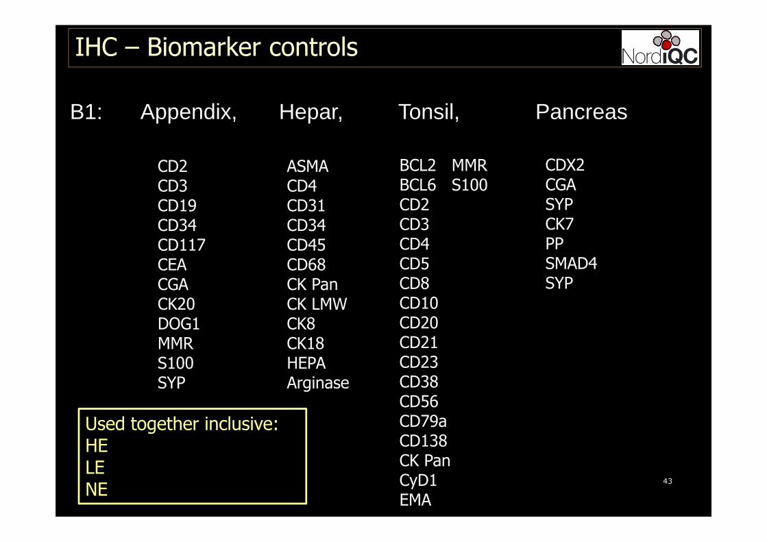
Mini TMA external tissue control: (Aalborg experien ce)
4-6 multi-tissue control-blocks for app. 190/220 markers
Control B1: Appendix, hepar, tonsil, pancreas
Control B2: Brain, str. muscle, skin, adrenel gland
Control B3: Lung, prostate, placenta, thyroidea
Control B4: Thymus, kidney, bone marrow, tonsil
Control B5: ……………..
Spec. ALK1, Hel., HER2, Hormones, etc
IHC – Biomarker controls
43
B1: Appendix, Hepar, Tonsil, Pancreas
IHC – Biomarker controls
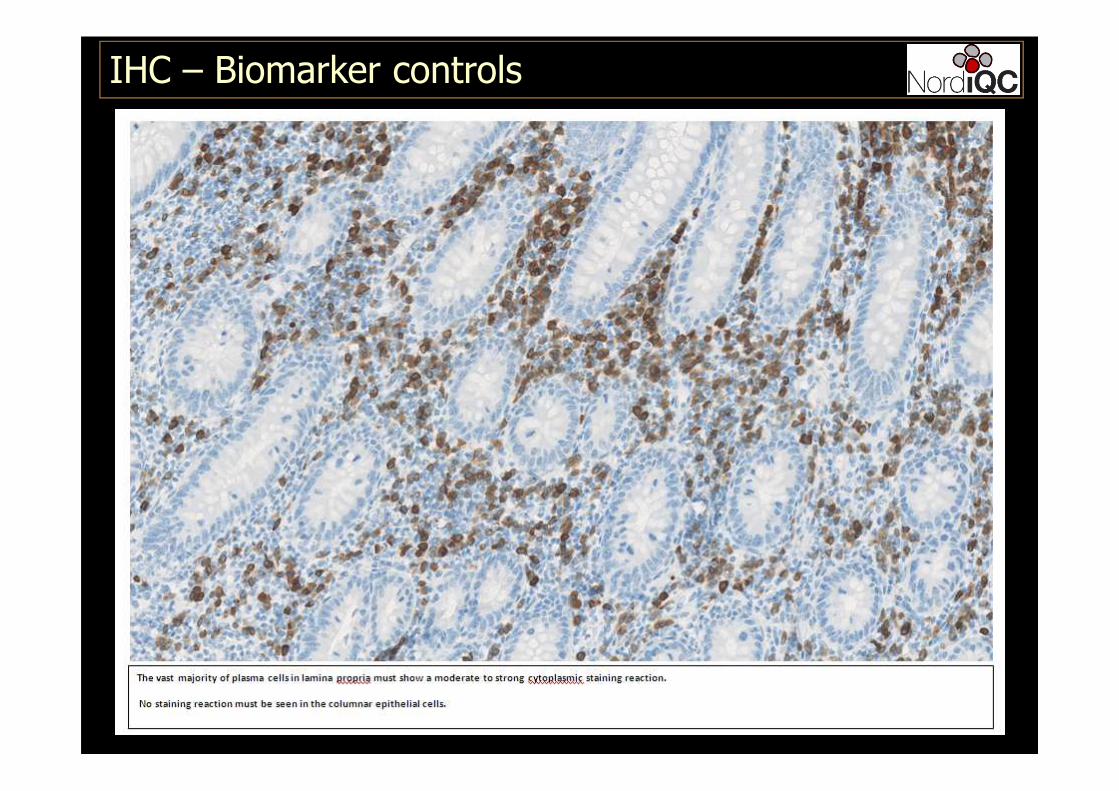
CD2CD3CD19CD34CD117CEACGACK20DOG1MMRS100SYP
ASMACD4CD31CD34CD45CD68CK PanCK LMWCK8CK18HEPAArginase
BCL2 MMRBCL6 S100 CD2CD3CD4CD5CD8CD10CD20CD21CD23CD38CD56CD79aCD138CK PanCyD1EMA
CDX2CGASYPCK7PPSMAD4SYP
Used together inclusive:HELENE
44
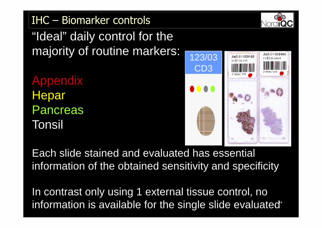
“Ideal” daily control for the majority of routine markers:
AppendixHeparPancreasTonsil
Each slide stained and evaluated has essentialinformation of the obtained sensitivity and specificity
In contrast only using 1 external tissue control, no information is available for the single slide evaluated
123/03CD3
IHC – Biomarker controls

45
IHC – Biomarker controls
TMA control onall slides
One batch control
Remarks
Missing reagentFN in patient test
YesNo – only control
slide
Potential internalpos. control onlyindicator of protocol performed
Wrong antibodyFP in patient test Yes
No – only controlslide
Inappropriateprotocolperformance- Drying out etcFN / FP in patient test
YesNo – only control
slide
Errors seen for all IHC automated and semi-automated IHC platforms
46
IHC – Biomarker controls
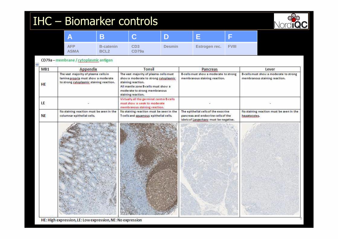
A B C D E FAFPASMA
B-cateninBCL2
CD3CD79a
Desmin Estrogen rec. FVIII
47
IHC – Biomarker controls
48
IHC – Biomarker controls
49
IHC – Biomarker controls
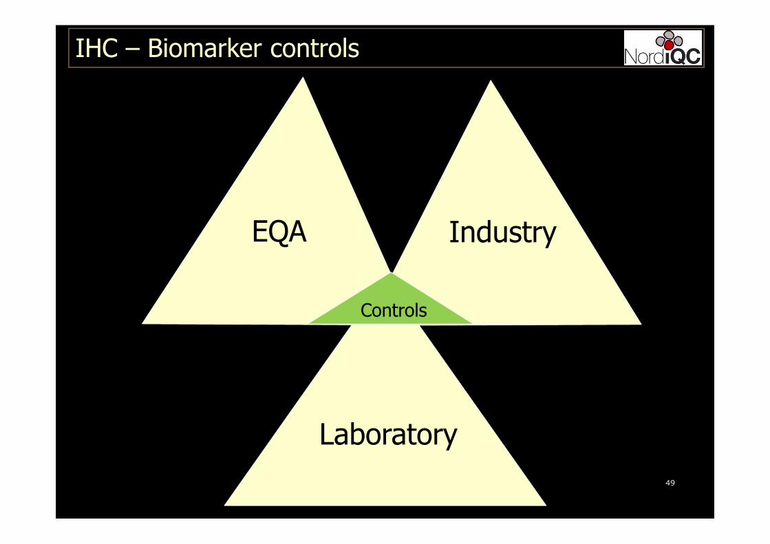
EQA Industry
Laboratory
Controls
50
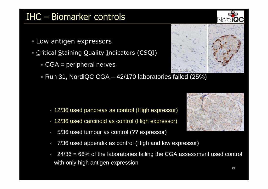
� Low antigen expressors
� Critical Staining Quality Indicators (CSQI)
� CGA = peripheral nerves
� Run 31, NordiQC CGA – 42/170 laboratories failed (25%)
� 12/36 used pancreas as control (High expressor)
� 12/36 used carcinoid as control (High expressor)
� 5/36 used tumour as control (?? expressor)
� 7/36 used appendix as control (High and low expressor)
� 24/36 = 66% of the laboratories failing the CGA assessment used control
with only high antigen expression
IHC – Biomarker controls
51
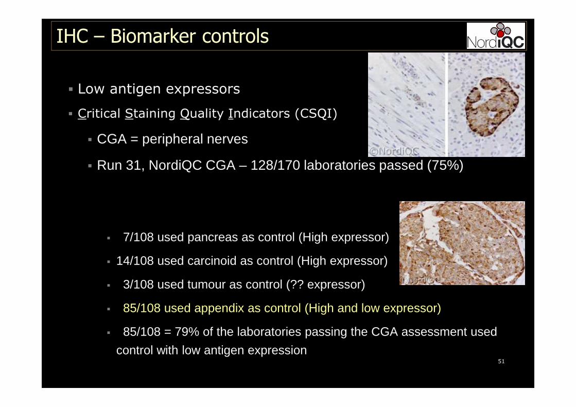
� Low antigen expressors
� Critical Staining Quality Indicators (CSQI)
� CGA = peripheral nerves
� Run 31, NordiQC CGA – 128/170 laboratories passed (75%)
� 7/108 used pancreas as control (High expressor)
� 14/108 used carcinoid as control (High expressor)
� 3/108 used tumour as control (?? expressor)
� 85/108 used appendix as control (High and low expressor)
� 85/108 = 79% of the laboratories passing the CGA assessment used
control with low antigen expression
IHC – Biomarker controls
52
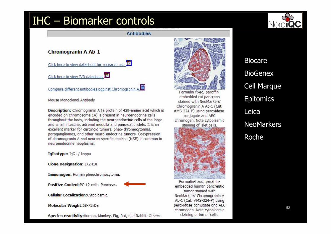
IHC – Biomarker controls
Biocare
BioGenex
Cell Marque
Epitomics
Leica
NeoMarkers
Roche

Conclusions:
Focus on external tissue controls are central to
standardize and optimize IHC:
�On-slide TMA controls are preferable to 1 bacth control
� Internal tissue controls are of limited value
�Negative reagent controls are only essential for biotin-
based detection systems
�Negative reagent controls can be valueable for non-
biotin based systems e.g. If pigment, frozen sections..
IHC – Biomarker controls
Conclusions:
Focus on external tissue controls are central to
standardize and optimize IHC:
� External tissue control ”catalogue” (normal preferable)
with describtions of HE, LE and NE
� Accepted and developed by KOL, EQA, Industry, Labs
� Used to validate/verify IHC studies and publications
� Used for both internal and external IHC QC
IHC – Biomarker controls
Conclusions:
Focus on external tissue controls are central to
standardize and optimize IHC:
� Usefull to monitor IHC consistency and thus quality
indicator of technical precision of individual IHC tests
� Each day all controls are evaluated and categorized
� All controls and slides that do not pass are classified with respect to
source of error
� Do we see any errors related to retrieval (morphology), background
(drying out at stainer), weak signal (antibody) etc....
IHC – Biomarker controls
IHC – Biomarker controls
IHC is a challenge, technical complex but not mission impossible and rests on 5 legs
� Use proper controls
� Use a robust and specific detection system
� Use efficient HIER
� Use Ab clones, optimal for the IHC platform
� Harmonize and standardize tissue processing
Begin at the beginning,' the King said gravely, and go on till you come to the end: then stop.'
Alice in Wonderland
For IHC: begin at the end, tune in your protocol: then stop.