the use of hybrid radiofrequency device for the treatment of
TRANSCRIPT

The Use of Hybrid Radiofrequency Device for the Treatment ofRhytides and Lax Skin
DAVID J. FRIEDMAN, MD,� AND LEON T. GILEAD, MD, PHD�y
BACKGROUND Recently, radiofrequency (RF) devices have been introduced and commercialized fornonablative procedures in dermatology and plastic surgery for the treatment of age-related rhytides andlax skin.
OBJECTIVE The objective was to assess the efficacy and safety of a novel RF device (Accent, AlmaLasers, Ltd, Caesarea, Israel) for the treatment of rhytides and lax skin.
METHODS AND MATERIALS Sixteen female patients (age range, 29–66 years; mean, 47 7 6 years; skinphototypes II to IV) were treated with Accent system. Patients were treated on the chin (n = 5), forehead(n = 8), cheeks (n = 12), jowl lines (n = 9), periorbital area (n = 7), marionette line (n = 3), and nasolabialfolds (n = 6) for wrinkles (n = 27 cases) and skin laxity (n = 23 cases). Patients received two to six treat-ments (mean, 4.3 7 1.1), with the time interval of 2 to 3 weeks. Photographs were assessed 1 month afterthe last treatment.
RESULTS For wrinkles and skin laxity, in 5 patients (42%), the cheeks (n = 12) scored 51% to 75% (sig-nificant improvement), and 2 patients (17%), 76% to 100% (excellent improvement). For the jowl lines(n = 9), 4 patients (44%) scored 51% to 75% (significant improvement), and 1 patient scored 76% to 100%(excellent improvement) for lax skin. For wrinkles on the periorbital (n = 7) and forehead areas (n = 8),three patients (37%) scored 51% to 75% (significant improvement).
CONCLUSION The Accent system is an effective and safe modality for the improvement of age-relatedrhytides and lax skin.
The radiofrequency device used in this study was loaned by Alma Lasers, Ltd.
One of the significant advances in facial cuta-
neous laser surgery in the past few years has
been the proliferation of radiofrequency (RF) tech-
nology to provide nonsurgical, nonablative means
for the improvement of age-related rhytides and lax
skin and acne vulgaris.1 Although proven effective,
dermabrasion, chemical peels, and laser resurfacing
(CO2 and erbium) techniques waned significantly
because of pain, substantial posttreatment ‘‘down-
time,’’ and prolonged recovery period. In fact, the
potential risks associated with ablative laser therapy
paved the way to the nonablative RF technologies.2
RF energy is a form of electromagnetic energy rang-
ing from 300 MHz to 3 kHz. RF affects skin by
emitting high-frequency radio waves that mimic the
thermal effects of lasers and intense pulsed light
sources. RF is similar to optical energy in that it
interacts with the tissue to produce a thermal change.
Unlike lasers, however, which induce heat by select-
ively targeting particular chromopheres, nonablative
RF devices generate heat as a result of tissue resistance
to the movement of electrons within the RF field.3–5
The delivery of RF energy is thought to induce der-
mal heating to the critical temperature of B651C,
causing collagen to shrink and allowing wound
healing with a subsequent contraction. In the skin,
RF radiation provokes significant thermal effects at a
particular depth based on the electrode configurationF
monopolar (deep) and bipolar (superficial). With a
controlled delivery of RF energy to the dermal and
subdermal layers, the RF technology (monopolar
and bipolar) has demonstrated an ability to stimulate
& 2007 by the American Society for Dermatologic Surgery, Inc. � Published by Blackwell Publishing �ISSN: 1076-0512 � Dermatol Surg 2007;33:543–551 � DOI: 10.1111/j.1524-4725.2007.33112.x
5 4 3
�LaseOhr Dermatology Cosmetic Laser Institute, Jerusalem, Israel; yDepartment of Dermatology, Hadassah UniversityMedical Center, Ein Kerem, Jerusalem, Israel

collagen production and remodeling, resulting in
softening wrinkles.6 Most RF energy delivery de-
vices, however, have slow coverage rates, need ag-
gressive cooling, or require a lengthy patient
preparation that includes local anesthesia due to
significant pain.7
The Accent system (Alma Lasers, Ltd., Caesarea,
Israel) is a new RF system that uses two RF
configured handpieces on a single platform:
unipolar, which permits volumetric (deep), and
bipolar, superficial tissue heating with a predictable
depth of penetration and without the need for
an aggressive cooling or local anesthesia.
The purpose of this study is to report the clinical
experience with the device in the treatment of facial
rhytides and lax skin.
Methods and Materials
Study Design
The data for 16 volunteer women patients were ana-
lyzed retrospectively and are the subject of this report.
The treatment was confined only to the facial area.
The facial areas were defined as follows: upper face
areaFfrom the forehead to the periorbital and crow’s
feet borders; the middle face areaFfrom the naso-
labial folds to the preauricular area and mandibular
angle; and the lower faceFextending from the man-
dible to the middle of the neck. Patients received be-
tween 4 and 6 (mean, 4.371.1) treatments, spaced
every 2 to 3 weeks. Patients returned for a follow-up
visit 1 month after the last treatment. Each treatment
cycle consisted of two phases: a nontherapeutic phase
(Phase I, two passes/20 seconds each) and a thera-
peutic phase (Phase II, three passes/20 seconds each).
The total energy [energy (W)� time (seconds)] deliv-
ered during each pass was expressed in kilojoules.
Patients were photographed in a standardized
method using high-resolution digital photography
(D70, Nikon, Japan) at baseline, before each treat-
ment, and 1 month after the last treatment. Replicate
photographs were taken from 01, 451 (right), and
451 (left) using a standardized ratio of f/16 for full
facial photographs. Pre- and posttreatment photo-
graphs and specific anatomic landmarks were
assessed and graded on a 5-point scale by two
physicians blinded to the study and the patients:
0% change from the baseline was graded as no
improvement; 1% to 25% change as a mild im-
provement; 26% to 50% change as a moderate
improvement; 51% to 75% as a significant
improvement; and 76% to 100% as most significant
improvement. The patient satisfaction score was
obtained at each visit and 1 month after the last
treatment according to the following scoring system:
1 = not satisfied; 2 = somewhat satisfied; 3 = satisfied;
4 = very satisfied; 5 = excellent.
Patients
Sixteen women patients (age range, 29–66 years;
mean, 47.67 10.9; Fitzpatrick skin phototype II-IV;
mean Fitzpatrick wrinkles score I-III) were treated in
our dermatology clinic with the Accent system.
Demographics, skin characteristics, and individual
patient treatment areas are presented in Table 1. The
patient volunteers are women who had visited the
physician’s office at least once in the 3 months before
enrollment in the study. All patients were in good
health and had undergone no surgical or nonsurgical
facial procedure [i.e., laser skin resurfacing, derma-
brasion, phenol peel, nonablative laser, or temporary
filler (e.g., collagen, fat, hyaluronic acid injections)],
nor did they have a history of pacemaker deploy-
ment or permanent fillers in the facial area at least
12 months before the enrollment. No topical anes-
thesia was used on any of the treatment areas.
Patients were educated regarding the treatment
regimen, result expectations, discomfort, and possi-
ble adverse side effects.
Device
The Accent system operation is based on the RF-
heating technology. The RF generator operating
frequency is 40.68 MHz. The device is designed for
contact operation using two handpieces: a unipolar
D E R M AT O L O G I C S U R G E RY5 4 4
R F T R E AT M E N T O F R H Y T I D E S A N D L A X S K I N
and a bipolar, for volumetric and surface heating,
respectively. Each handpiece contains an RF-reson-
ant system, a thermoelectric cooler, and a trigger
push-button (on the handle). Figure 1 shows the
system’s unipolar and bipolar handpieces. The
output power is pulse-width-modulation
controlled. This feature provides constant output
RF-power amplitude and enables the mean
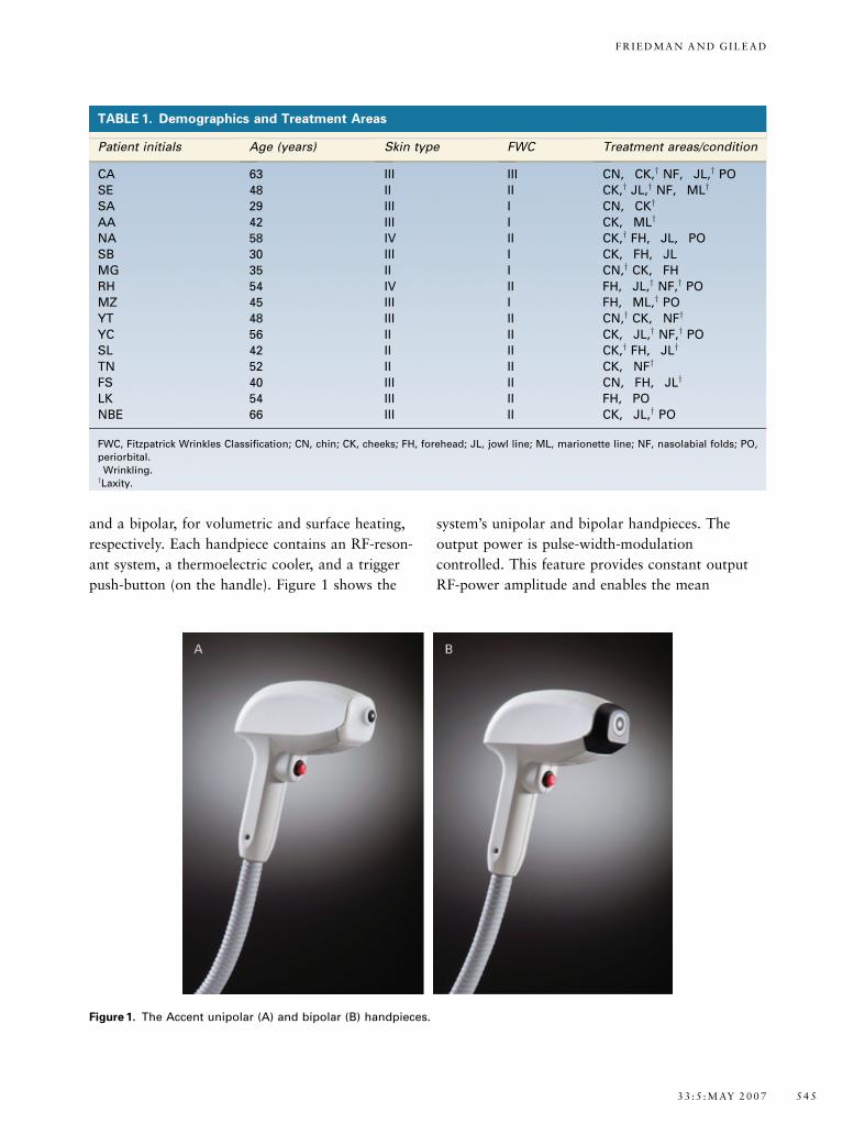
TABLE 1. Demographics and Treatment Areas
Patient initials Age (years) Skin type FWC Treatment areas/condition
CA 63 III III CN,� CK,y NF,� JL,y PO�
SE 48 II II CK,y JL,y NF,� MLy
SA 29 III I CN,� CKy
AA 42 III I CK,� MLy
NA 58 IV II CK,y FH,� JL,� PO�
SB 30 III I CK,� FH,� JL�
MG 35 II I CN,y CK,� FH�
RH 54 IV II FH,� JL,y NF,y PO�
MZ 45 III I FH,� ML,y PO�
YT 48 III II CN,y CK,� NFy
YC 56 II II CK,� JL,y NF,y PO�
SL 42 II II CK,y FH,� JLy
TN 52 II II CK,� NFy
FS 40 III II CN,� FH,� JLy
LK 54 III II FH,� PO�
NBE 66 III II CK,� JL,y PO�
FWC, Fitzpatrick Wrinkles Classification; CN, chin; CK, cheeks; FH, forehead; JL, jowl line; ML, marionette line; NF, nasolabial folds; PO,
periorbital.�Wrinkling.yLaxity.
Figure 1. The Accent unipolar (A) and bipolar (B) handpieces.
3 3 : 5 : M AY 2 0 0 7 5 4 5
F R I E D M A N A N D G I L E A D
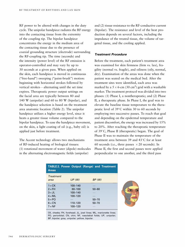
RF power to be altered with changes in the duty
cycle. The unipolar handpiece radiates the RF energy
into the contacting tissue from the extremity
of the coupling tip. The bipolar handpiece
concentrates the energy in the medium area of
the contacting tissue due to the presence of
coaxial grounding structure (electrode) surrounding
the RF-coupling tip. The time (seconds) and
the intensity (power level) of the RF emission is
operator-controlled and may vary by up to
30 seconds at a given pass. When applied on
the skin, each handpiece is moved in continuous
(‘‘free-hand’’) sweeping (‘‘paint-brush’’) motion:
beginning with horizontal strokes followed by
vertical strokesFalternating until the set time
expires. Therapeutic power output settings on
the facial area are typically between 80 and
140 W (unipolar) and 60 to 80 W (bipolar), and
the handpiece selection is based on the treatment
area anatomic location (Table 2). The unipolar
handpiece utilizes a higher energy level, since it
heats a greater tissue volume compared to the
bipolar handpiece. To ease the handpiece movement,
on the skin, a light coating of oil (e.g., baby oil) is
applied just before treatment.
The Accent technology allows two mechanisms
of RF-induced heating of biological tissues:
(1) rotational movement of water (dipole) molecules
in the alternating electromagnetic fields (unipolar)
and (2) tissue resistance to the RF-conductive current
(bipolar). The resistance and level of the heat pro-
duction depends on several factors, including the
impedance of the treated tissue, the volume of tar-
geted tissue, and the cooling applied.
Treatment Procedure
Before the treatment, each patient’s treatment area
was examined for skin firmness (firm vs. lax), fra-
gility (normal vs. fragile), and oiliness (oily, normal,
dry). Examination of the areas was done when the
patient was seated on the medical bed. After the
treatment sites were identified, each area was
marked by a 5� 6-cm (30 cm2) grid with a washable
marker. The treatment protocol was divided into two
phases: (1) Phase I, a nontherapeutic; and (2) Phase
II, a therapeutic phase. In Phase I, the goal was to
elevate the baseline tissue temperature to the thera-
peutic level of 391C within 30 to 60 seconds by
employing two successive passes. To reach that goal
and depending on the epidermal temperature and
patient discomfort, the energy was increased by 15%
to 20%. After reaching the therapeutic temperature
of 391C, Phase II (therapeutic) began. The goal of
Phase II was to maintain the temperature of the
treatment area between 39 and 431C for at least
60 seconds (i.e., three passes �20 seconds). In
Phase II, the first and second passes were applied
perpendicular to one another, and the third pass
TABLE 2. Power Output (Range) and Treatment
Areas
Treatment
area UP (W) BP (W)
1 = CK 100–140 F2 = FH 80–100 50–60
3 = JL 100–140 F4 = ML 100–120 F5 = PO F 50–70
6 = CN 110–120 50–70
7 = NF 100–120 F
CK, cheeks; FH, forehead; JL, jowl lines; ML, marionette lines;
PO, periorbital; CN, chin; NF, nasolabial folds; UP, unipolar;
BP, bipolar; gray, unipolar; yellow, bipolar.
1
2
3 4
5
6
7
D E R M AT O L O G I C S U R G E RY5 4 6
R F T R E AT M E N T O F R H Y T I D E S A N D L A X S K I N

was performed with rotational movements. To
prevent overheating the epidermal–dermal interface,
the energy level was down-scaled by 10% to
15% from Phase I most recent pass. The mean
treatment time for each patient was 15 to 30 minutes
per visit, depending on the number of areas that
were treated. Phase I treatment parameters
averaged 120 W� 20 seconds (1.6 kJ) per pass
for the unipolar and 60 W20 seconds (1.0 kJ) per
pass for the bipolar handpieces, respectively.
Phase II treatment parameters averaged
100 W�20 seconds (2.4 kJ) per pass for the
unipolar and 50 W� 20 seconds (1.2 kJ) per pass
for the bipolar, respectively. The total energy
(W� seconds) expressed in kilojoules was recorded
at each pass.
The energy deposition delivered over
20 seconds in 30-cm2 area with power output
of 120 W (unipolar handpiece) and 60 W
(bipolar handpiece) was 80 and 40 J/cm3
(single pass), respectively. The speed of the
handpiece movement was metronomed at a
pace of approximately 10 cm/seconds;
the area was covered within 5 seconds� 4 times
for 20 seconds (one pass). Skin temperature
was measured immediately after each pass,
until end points were reached. Before and
after each pass, the epidermal skin temperature
was monitored and recorded with a
laser thermometer (Center, 350 series,
Center Technology Corp., Korea). Therapeutic
temperatures were considered as greater
than 391C and less than 441C
(mean, 421C).
The patient discomfort level was monitored
(1 = cold; 2 = natural; 3 = nice warm; 4 = getting too
warm; 5 = too hot) during each pass. Acute clinical
response was recorded after each session to assess
skin changes (edema, erythema, and blistering). For
moisturized skin or sensitive skin types, the power
level was reduced by 10 to 20 W, while increased by
10 W for oily skin.
Statistical Analysis
Mean and standard deviation were calculated for
each category. Differences between age groups were
compared using an independent-sample Student’s
t test. Statistical significance was considered when
p values were less than .05.
Results
All 16 patients completed the course of the treatment
protocol. A total of 50 areas were treated: chin
(n = 5), forehead (n = 8), cheeks (n = 12), jowl lines
(n = 9), periorbital (n = 7), marionette line (n = 3), and
nasolabial folds (n = 6; Table 1). The mean energy
level set-up for the unipolar handpiece was 120722
and 60713 W for the bipolar handpiece. No sta-
tistically significant differences were found between
the age groups and the energy delivered by each
handpiece in the various facial areas (Table 3). In all
50 areas, the therapeutic temperature level (mean
epidermal skin temperature, approx. 421C) was
reached within two to three passes. No unexpected
adverse side effects were detected or reported. As
expected, the discomfort level during the treatment
was associated with the energy level and the time of
exposure. In all patients, posttreatment erythema
(hyperemia) was detected which was resolved within
1 to 2 hours. No patients experienced burns, skin
breakdown, or scarring.
One month after the last treatment, the improvement
score in 2 patients was ‘‘excellent,’’ in 9 patients
between ‘‘satisfied/very satisfied,’’ in 9 patients
‘‘somewhat satisfied/satisfied,’’ and in 3 patients
‘‘not satisfied/somewhat satisfied.’’. Eleven patients
(69%) scored between ‘‘satisfied/excellent.’’
TABLE 3. Energy Output per Treatment for the
Unipolar and Bipolar Handpieces by Age Group
Age group (years) Unipolar (kJ) Bipolar (kJ)
25–35 (n = 3) 11.37 1.8 5.77 0.4
36–45 (n = 4) 12.17 1.3 5.47 0.3
46–55 (n = 5) 10.87 1.6 5.47 0.6
56–70 (n = 4) 10.97 1.2 5.37 0.9
3 3 : 5 : M AY 2 0 0 7 5 4 7
F R I E D M A N A N D G I L E A D
The mean patient satisfaction score was 3.0671.2
(satisfied/very satisfied), representing 9 of the 16
patients (57%). When divided into two age groups,
the younger group reported statistically significantly
(po.01) higher satisfaction scores (3.717 0.75)
when compared to the older group (2.5571.3;
Figure 2).
Photographic analysis of pre- and posttreatment
digital images showed moderate-significant im-
provement in 11 of the 16 patients (69%) and
marked improvement (475%) in 3 patients (19%).
For wrinkles and skin laxity, in 5 patients (42%), the
cheeks (n = 12) scored 51% to 75% (significant im-
provement) and 2 patients (17%) scored 76% to
100% (excellent improvement). For the jowl lines
(n = 9), 4 patients (44%) scored 51% to 75% (sig-
nificant improvement) and 1 patient scored 76% to
100% (excellent improvement) for lax skin. For
wrinkles in the periorbital (n = 7) and forehead areas
(n = 8), 3 patients (37%) scored 51% to 75% (sig-
nificant improvement; Figure 3). Figures 4 and 5
show before and after photos of 2 patients (SE and
NBE) 1 month after the last treatment.
Discussion
Nonablative skin resurfacing technologies share a
common method of inducing thermal dermal injury
while preserving epidermal integrity. The prime
consideration during the past decade for the physi-
cians and scientists regarding procedures for the
improvement of aged skin is to ensure an effective
treatment while sparing damage to the epidermis and
adjoining upper dermal layer. The data reported in
our study demonstrate that this RF device offers a
safe and effective noninvasive technique to improve
the appearance of age-related rhytides and lax skin
in women.
Numerous studies have documented that thermally
modified tissue undergoes a remodeling process
characterized by fibroplasia and increased collagen
deposition.8,9 The two main mechanisms for heat
flow inside the tissue is conduction (heat gradient)
and convection (blood perfusion). During our treat-
ment regimen, we have documented baseline epi-
dermal temperature of approximately 31 to 331C
and after Phase II temperatures of 40 to 421C. We
assume that the surrounding tissue is 101C cooler
than the dermal and subdermal temperature (B50–
551C). Although the changes that were seen clini-
cally were detected 1 month after the last treatment,
it is not known whether the thermally altered col-
lagen persists indefinitely in its new form or acts as a
matrix for new collagen formation that recapitulates
its shortened overall structure.
The clinical results of nonablative RF tissue tight-
ening were first reported in the periorbital areas.6
In this multicenter study, Fitzpatrick and his col-
0
1
2
3
4
5
6
CK JL PO FH
Anatomic Sites
Num
ber
of C
ases
1-25%
26-50%
51-75%
76-100%
Figure 3. Photography assessment of laxity (CK, cheeks; JL,jowl lines) and wrinkling (PO, periorbital; FH, forehead)before and 1 month after the last treatment. 1%–25%,mild improvement; 26%–50%, moderate improvement;51%–75%, significant improvement; 76%–100% markedimprovement.
0
1
2
3
4
5
Not Satisfied
Somewhat Satisfied
Satisfied Very Satisfied
Excellent Average Score
Num
ber
of P
atie
nts
25-45 yr. 46-66 yr.
Figure 2. Patients’ evaluation of treatment outcome 1 monthafter the last treatment by age group.
D E R M AT O L O G I C S U R G E RY5 4 8
R F T R E AT M E N T O F R H Y T I D E S A N D L A X S K I N

Figure 4. (A) Patient SE: Before (left) and 1 month after (right) four treatments. Marionette line (laxity), improvement byphotography 51% to 75%; patient satisfaction, satisfied. (B) Patient SE: before (left) and 1 month after (right) four treatments.Periorbital (wrinkles), improvement by photography 76% to 100%; patient satisfaction, excellent.
Figure 5. Patient NBE: Before (A) and 1 month after (B) four treatments. Cheek area (laxity): improvement by photography,51% to 75%; patient satisfaction, very satisfied.
3 3 : 5 : M AY 2 0 0 7 5 4 9
F R I E D M A N A N D G I L E A D

leagues demonstrated clinical improvement in
periorbital rhytides in 80% of subjects. In contrast,
in 24 patients who underwent a single RF treatment
to improve the upper third of the face, only 36% of
the patients’ self-assessment reported improve-
ment.10 In our study, the improvement in the ap-
pearance of rhytides and lax skin was reported in
56% of the patients. Our results, however, are in
agreement with other studies performed in other
facial areas.11,12 Although the temperature to which
the tissue rises cannot be established precisely, the
histopathologic findings suggest that the temperature
of 55 to 651C is most likely achieved and that heat
fibroblasts may be stimulating fibroblasts to the
produced collagen. The proposed mechanism of RF
tissue tightening through thermally induced imme-
diate collagen reorganization followed by remodel-
ing is supported by clinical observation and
ultrastructural analysis. The skin treated with the RF
monopolar device has demonstrated epidermal
preservation, thermal changes in collagen fibrils, and
increased Type I collagen messenger RNA steady-
state expression.13 It has been suggested that in near-
infrared lasers, which exclusively target the water,
these thermal-induced changes can help to improve
the appearance of the ryhtides.3 Interestingly,
Oringer and colleagues14 studied connective tissue
remodeling induced by carbon dioxide (CO2) laser
resurfacing of photodamaged human skin. In this
study, it has been proposed that at least some of the
clinical benefit seen after the CO2 skin resurfacing
may be based on increased elastin levels.14 We have
speculated that displacement of the disorganized
elastotic material, which is the hallmark of photo-
damaged skin deeper into the dermis after RF in-
duced heating of the tissue, may partially be
responsible for the cosmetic changes in some of the
patients in our study.
In our study, the patients received successive
multiple treatments (four to six treatments)
spaced 2 to 3 weeks apart. Ruiz-Esparza and
Gomez15 reported clinically evident skin tightening
in 14 of 15 patients treated with a single RF
treatment on the lower third of the face. In a
recent study, Fritz and coworkers16 compared the
effectiveness of single and double RF treatments
done with a monopolar device. Two RF treatments
yielded in the nasolabial folds a significantly better
improvement than a single treatment. Although
overall improvements were modest in both groups,
patient satisfaction was relatively high. Iyer and
colleagues17 demonstrated that approximately
70% of patients noticed a significant improvement
in the skin laxity 3 months after a single RF
treatment with a greater improvement noted after
multiple treatments.
The mean energy settings for the unipolar
handpiece in our study were found to be similar to
those reported in other studies done with
comparative RF technologies.13,15–17 Hsu and
Kaminer11 reported an association between the
energy levels and improved clinical results.
Overall, the younger patients were reported to
respond better compared to the older group.
It has been suggested that heat-labile collagen
bonds are progressively replaced by irreducible
multivalent cross-links as the tissue ages, thus
rendering older skin less amenable to heat-induced
tissue tightening.11 Our observations support this
since we found that the older group (446 years
old) reported less favorable results.
In a similar device, it has been reported that the use
of a RF device was associated with significant pain,
and in a small but significant number of cases sub-
cutaneous fat atrophy developed.7 No subcutaneous
fat atrophy was noted in our patients. In our study,
the procedures with both unipolar and bipolar
handpieces were performed without any anesthesia
and yet were regarded by all patients as pain-free and
associated with only moderate discomfort. In fact,
no patients considered the procedure intolerable at
any session.
In conclusion, the data reported in this study support
the effectiveness of the Accent system in the treat-
ment of facial rhytides and skin laxity in women.
D E R M AT O L O G I C S U R G E RY5 5 0
R F T R E AT M E N T O F R H Y T I D E S A N D L A X S K I N
References
1. Sadick N, Sorhaindo L. The radiofrequency frontier: a review
of radiofrequency and combined radiofrequency pulsed light
technology in aesthetic medicine. Facial Plast Surg 2005;21:
131–8.
2. Alam M, Dover JS, Arndt KA. Energy delivery devices for cuta-
neous remodeling: lasers, lights, and radio waves. Arch Dermatol
2003;139:1351–60.
3. Hardaway CA, Ross V. Nonablative laser skin remodeling.
Dermatol Clin 2002;20:97–111.
4. Sadick NS, Makino Y. Selective electro-thermolysis in aesthetic
medicine: a review. Lasers Surg Med 2004;34:91–7.
5. Kim KH, Geronemus RG. Nonablative laser and light therapies
for skin rejuvenation. Arch Facial Plast Surg 2004;6:398–409.
6. Fitzpatrick R, Geronemus R, Goldberg D, et al. Multicenter study
of noninvasive radiofrequency for periorbital tissue tightening.
Lasers Surg Med 2003;33:232–42.
7. Biesnman BS. Radiofrequency Devices: Monopolar vs bipolar vs
radiofrequency plus laser; indications; treatment approaches;
novel applications; results. In: Arndt KA, Dover JS, Anderson RR,
editors. Controversies and Conversations in Laser and Cosmetic
Surgery. Symposium Proceedings; 2005, Denver, CO.
8. Grema H, Raulin C, Greve B. ‘‘Skin rejuvenation’’ by non ablative
laser and light systems. literature research and overview. Hautarzt
2002;53:385–92.
9. Arnoczky SP, Aksan A. Thermal modification of connective tis-
sues: basic science considerations and clinical implications. J Am
Acad Orthop Surg 2000;8:305–13.
10. Bassichis BA, Dayan S, Thomas JR. Use of nonablative
readiofrequency device to rejuvenate the upper one-third
of the face. Otolaryngol Head Neck Surg 2004;130:
397–406.
11. Hsu TS, Kaminer MS. The use of nonablative radiofrequency
technology to tighten the lower face and neck. Semin Cutan Med
Surg 2003;22:115–23.
12. Alster TS, Tanzi E. Improvement of neck and cheek laxity with a
nonablative radiofrequency device: a lifting experience. Dermatol
Surg 2004;30(4 Pt 1):503–7.
13. Zelickson BD, Kist D, Bernstein E, et al. Histological and ultra-
structural evaluation of the effects of a radiofrequency-based
nonablative dermal remodeling device: a pilot study. Arch Der-
matol 2004;140:204–9.
14. Oringer JS, Kang S, Johnson TM, et al. Connective tissue
remodeling induced by carbon dioxide laser resurfacing of
photodamaged human skin. Arch Dermatol 2004;140:
1326–32.
15. Ruiz-Esparza J, Gomez JB. The medical face lift: a noninvasive,
nonsurgical approach to tissue tightening in facial skin using
nonablative radiofrequency. Dermatol Surg 2003;29:325–32.
16. Fritz M, Counters JT, Zelickson BD. Radiofrequency treatment
for middle and lower face laxity. Arch Facial Plast Surg
2004;6:370–3.
17. Iyer S, Suthamajariya K, Fitzpatrick RE. Using a radiofrequency
energy device to treat the lower face: a treatment paradigm for a
non-surgical facelift. Cosmet Dermatol 2003;16:37–40.
Address correspondence and reprint requests to: D. J.Friedman, MD, LaseOhr, 60 Diskin Street, Jerusalem,96440, Israel, or e-mail: [email protected]
3 3 : 5 : M AY 2 0 0 7 5 5 1
F R I E D M A N A N D G I L E A D