the history of lifethe eukaryota include the organisms that most people are most familiar with
TRANSCRIPT

8/4/2019 The History of LifeThe Eukaryota Include the Organisms That Most People Are Most Familiar With
http://slidepdf.com/reader/full/the-history-of-lifethe-eukaryota-include-the-organisms-that-most-people-are 1/5
The History of life The Eukaryota include the organisms that mostpeople are most familiar with - all animals, plants, fungi, and protists. They alsoinclude the vast majority of the organisms that paleontologists work with. Althoughthey show unbelievable diversity in form, they share fundamental characteristics of cellular organization, biochemistry, and molecular biology. Shown here, clockwisefrom upper left: a dinoflagellate, a single-celled photosynthetic protist; a palm treerepresenting the plants; a spider, one of the animals; and a cluster of mushroomsrepresenting the fungi.
Eukaryotic Kingdoms
CHROMISTA (Kelps, diatoms, haptophytes)
FUNGI (Fungi)
METAZOA (Animals)
PLANTAE (Plants)
PROTISTA (Protists)
Cell biology (formerly cytology, from the Greek kytos, "container") is ascientific discipline that studies cells – their physiological properties, their structure, the organelles they contain, interactions with their environment, their life cycle, division and death. This is done both on a microscopic and molecular level.Cell biology research encompasses both the great diversity of single-celledorganisms like bacteria and protozoa, as well as the many specialized cells inmulticellular organisms such as humans.
Knowing the components of cells and how cells work is fundamental to all
biological sciences. Appreciating the similarities and differences between celltypes is particularly important to the fields of cell and molecular biology as well asto biomedical fields such as cancer research and developmental biology. Thesefundamental similarities and differences provide a unifying theme, sometimesallowing the principles learned from studying one cell type to be extrapolated andgeneralized to other cell types. Hence, research in cell biology is closely related togenetics, biochemistry, molecular biology, immunology, and developmental biology.
Understanding cells in terms of their molecular components.
Processes
Movement of proteins

8/4/2019 The History of LifeThe Eukaryota Include the Organisms That Most People Are Most Familiar With
http://slidepdf.com/reader/full/the-history-of-lifethe-eukaryota-include-the-organisms-that-most-people-are 2/5
Endothelial cells under the microscope. Nuclei are stained blue with DAPI,microtubles are marked green by an antibody and actin filaments are labelled redwith phalloidin.
Each type of protein is usually sent to a particular part of the cell. An important partof cell biology is the investigation of molecular mechanisms by which proteins aremoved to different places inside cells or secreted from cells.
Most proteins are synthesized by ribosomes in the cytoplasm. This process isknown as protein biosynthesis. Biosynthesis (also called biogenesis) is anenzyme-catalyzed process in cells of living organisms by which substrates areconverted to more complex products (also simply known as protein translation).Some proteins, such as those to be incorporated in membranes (known asmembrane proteins), are transported into the "rough" endoplasmic reticulum (ER)during synthesis. This process can be followed by transportation and processing inthe Golgi apparatus. From the Golgi, membrane proteins can move to the plasma membrane, to other sub-cellular compartments, or they can be secreted from thecell. The ER and Golgi can be thought of as the "membrane protein synthesiscompartment" and the "membrane protein processing compartment", respectively.
There is a semi-constant flux of proteins through these compartments. ER andGolgi-resident proteins associate with other proteins but remain in their respectivecompartments. Other proteins "flow" through the ER and Golgi to the plasmamembrane. Motor proteins transport membrane protein-containing vesicles alongcytoskeletal tracks to distant parts of cells such as axon terminals.
Some proteins that are made in the cytoplasm contain structural features thattarget them for transport into mitochondria or the nucleus. Some mitochondrialproteins are made inside mitochondria and are coded for by mitochondrial DNA. Inplants, chloroplasts also make some cell proteins.
Extracellular and cell surface proteins destined to be degraded can move back intointracellular compartments upon being incorporated into endocytosed vesiclessome of which fuse with lysosomes where the proteins are broken down to their individual amino acids. The degradation of some membrane proteins begins whilestill at the cell surface when they are cleaved by secretases. Proteins that functionin the cytoplasm are often degraded by proteasomes.
Other cellular processes
• Active transport and Passive transport - Movement of molecules into and outof cells.
• Autophagy - The process whereby cells "eat" their own internal components or microbial invaders.• Adhesion - Holding together cells and tissues.• Reproduction - Made possible by the combination of sperm made in thetesticuli(contained in some male cells nucleus') and the egg made in theovary(contained in the nucleus of a female cell). When the sperm breaks throughthe hard outer shell of the egg a new cell embryo is formed, which, in humans,grows to full size in 9 months.• Cell movement: Chemotaxis, Contraction, cilia and flagella.• Cell signaling - Regulation of cell behavior by signals from outside.• DNA repair and Cell death
8/4/2019 The History of LifeThe Eukaryota Include the Organisms That Most People Are Most Familiar With
http://slidepdf.com/reader/full/the-history-of-lifethe-eukaryota-include-the-organisms-that-most-people-are 3/5
• Metabolism: Glycolysis, respiration, Photosynthesis• Transcription and mRNA splicing - gene expression.
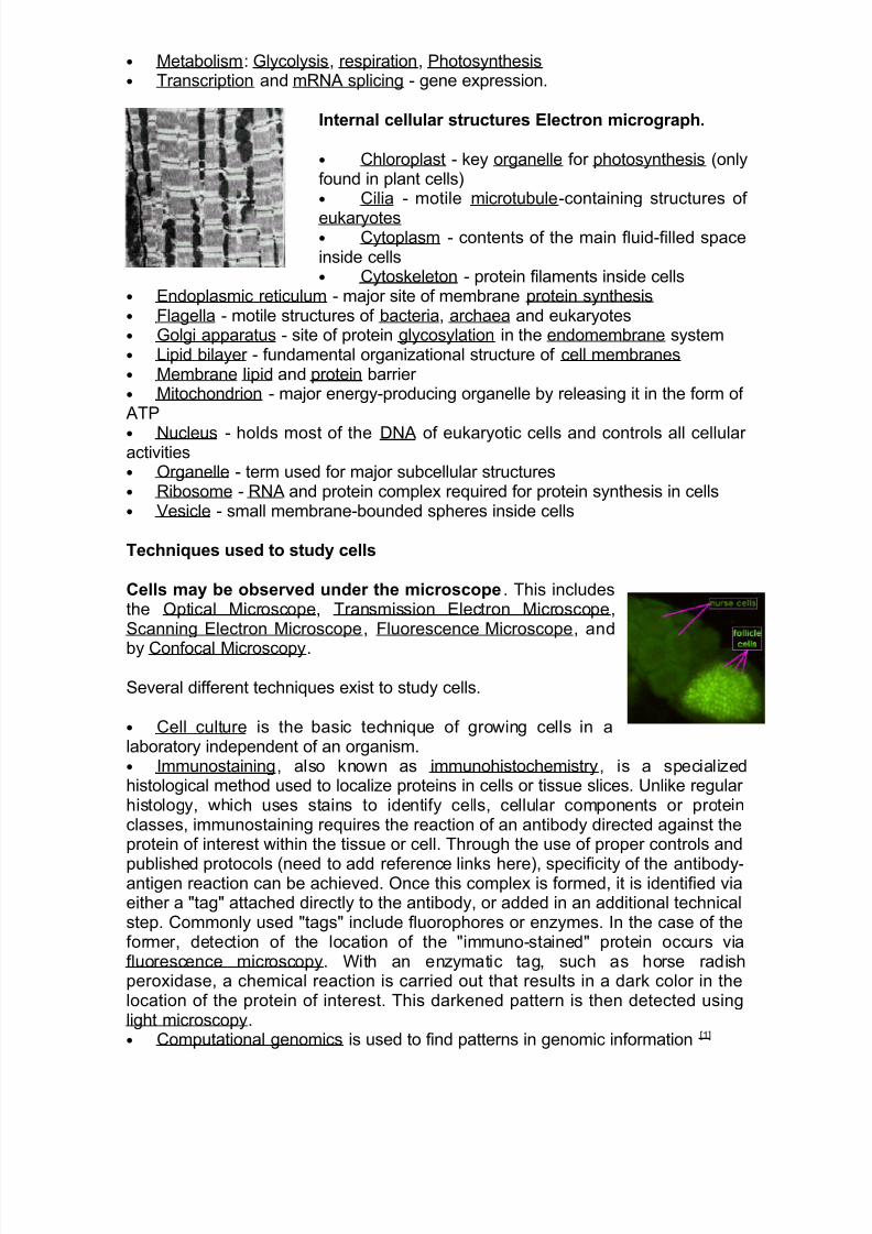
Internal cellular structures Electron micrograph.
• Chloroplast - key organelle for photosynthesis (onlyfound in plant cells)• Cilia - motile microtubule-containing structures of
eukaryotes• Cytoplasm - contents of the main fluid-filled spaceinside cells• Cytoskeleton - protein filaments inside cells
• Endoplasmic reticulum - major site of membrane protein synthesis• Flagella - motile structures of bacteria, archaea and eukaryotes• Golgi apparatus - site of protein glycosylation in the endomembrane system• Lipid bilayer - fundamental organizational structure of cell membranes• Membrane lipid and protein barrier • Mitochondrion - major energy-producing organelle by releasing it in the form of ATP
• Nucleus - holds most of the DNA of eukaryotic cells and controls all cellular activities• Organelle - term used for major subcellular structures• Ribosome - RNA and protein complex required for protein synthesis in cells• Vesicle - small membrane-bounded spheres inside cells
Techniques used to study cells
Cells may be observed under the microscope. This includesthe Optical Microscope, Transmission Electron Microscope,Scanning Electron Microscope, Fluorescence Microscope, and
by Confocal Microscopy.
Several different techniques exist to study cells.
• Cell culture is the basic technique of growing cells in alaboratory independent of an organism.• Immunostaining, also known as immunohistochemistry, is a specializedhistological method used to localize proteins in cells or tissue slices. Unlike regular histology, which uses stains to identify cells, cellular components or proteinclasses, immunostaining requires the reaction of an antibody directed against theprotein of interest within the tissue or cell. Through the use of proper controls and
published protocols (need to add reference links here), specificity of the antibody-antigen reaction can be achieved. Once this complex is formed, it is identified viaeither a "tag" attached directly to the antibody, or added in an additional technicalstep. Commonly used "tags" include fluorophores or enzymes. In the case of theformer, detection of the location of the "immuno-stained" protein occurs viafluorescence microscopy. With an enzymatic tag, such as horse radishperoxidase, a chemical reaction is carried out that results in a dark color in thelocation of the protein of interest. This darkened pattern is then detected usinglight microscopy.• Computational genomics is used to find patterns in genomic information [1]

8/4/2019 The History of LifeThe Eukaryota Include the Organisms That Most People Are Most Familiar With
http://slidepdf.com/reader/full/the-history-of-lifethe-eukaryota-include-the-organisms-that-most-people-are 4/5
• DNA microarrays identify changes in transcript levels between differentexperimental conditions.• Gene knockdown mutates a selected gene.• In situ hybridization shows which cells are expressing a particular RNAtranscript.• PCR can be used to determine how many copies of a gene are present in acell.• Transfection introduces a new gene into a cell, usually an expression
construct
Purification of cells and their parts Purification may be performed using thefollowing methods:
• Cell fractionationo Release of cellular organelles by disruption of cells.
o Separation of different organelles by centrifugation.
• Flow cytometry• Immunoprecipitation• Proteins extracted from cell membranes by detergents and salts or other kinds
of chemicals.
The bacterial outer membrane is found in Gram-negative bacteria. Its composition isdistinct from that of the cytoplasmic membrane - among other things, the outer leaflet of the membrane includes a complex lipopolysaccharide whose lipid portion acts as anendotoxin - and it is linked to the cell's peptidoglycan by Braun's lipoprotein.
Porins can be found in this layer.
The cell envelope is the cell membrane and cell wall plus an outer membrane, if one ispresent.
Most bacterial cell envelopes fall into two major categories: Gram positive and Gramnegative. These are differentiated by their Gram staining characteristics.
Gram-negative bacteria (or negibacteria) are bacteria that do not retain crystal violetdye in the Gram staining protocol. In a Gram stain test, a counterstain (commonlysafranin) is added after the crystal violet, coloring all Gram-negative bacteria with a redor pink color. The test itself is useful in classifying two distinct types of bacteria basedon the structural differences of their bacterial cell walls. Gram-positive bacteria willretain the crystal violet dye when washed in a decolorizing solution.
Gram-negative bacterial infection refers to a disease caused by Gram-negativebacteria. One example is E. coli.
It is important to recognize that this class is defined morphologically (by the presence of a bacterial outer membrane), and not histologically (by a pink appearance whenstained), though the two usually coincide.
One reason for this division is that the outer membrane is of major clinical significance:it can play a role in the reduced effectiveness of certain antibiotics,and it is the source of endotoxin.

8/4/2019 The History of LifeThe Eukaryota Include the Organisms That Most People Are Most Familiar With
http://slidepdf.com/reader/full/the-history-of-lifethe-eukaryota-include-the-organisms-that-most-people-are 5/5
The gram status of some organisms is complex or disputed:
* Mycoplasma are sometimes considered gram negative, but because of its lack of acell wall and unusual membrane composition, it is sometimes considered separatelyfrom other gram negative bacteria.
* Gardnerella is often considered gram negative, but it is classified in MeSH as bothgram positive and gram negative. It has some traits of gram positive bacteria,but has a
gram negative appearance. It has been described as a "gram-variable rod"
The periplasmic space or periplasm is a space between the inner cytoplasmicmembrane and external outer membrane of Gram-negative bacteria or the equivalentspace outside the inner membrane of Gram-positive bacteria. It may constitute up to40% of the total cell volume in Gram-negative species, and is drastically smaller inGram-positive species. The space contains a loose network of murein (peptidoglycan)chains, as well as a gel containing hydrolytic and degradative enzymes.[vague] Other enzymes in the gel are involved in various biochemical pathways includingpeptidoglycan synthesis, electron transport, and alteration of substances toxic to the cell(xenobiotic metabolism). In some species, the gel also contains beta-lactamase, an
enzyme responsible for degrading penicillin. This can be of clinical importance whenconsidering antibiotic resistance.
Az endotoxin („belső méreg”) egyes baktériumok testében található, de a testükönkívülre ki nem választott mérgező anyag. Külünösen gyakori az élősködő életmódototfolytató, patogén baktériumfajok esetében.
A Gram-negatív baktériumok külső membránjában található lipopoliszacharidok (vagylipooligoszacharidok) jellemzően mérgezőek (toxikusak). A baktériumok e toxinokatnem választják ki, tehát ezek nem szabadulnak ki a gazda testébe, amíg a baktérium él.
A Listeria monocytogenes egy emberben igen elterjedt kórokozó Gram-pozitívbaktérium, amely szintén endotoxint tartalmaz, de ennek kémiai jellege nemlipopoliszacharid.