squalus acanthias. taxonomy domain: eukaryota kingdom: animalia phylum: chordata subphylum:...
TRANSCRIPT

DOGFISH SHARK DISSECTION
Squalus acanthias
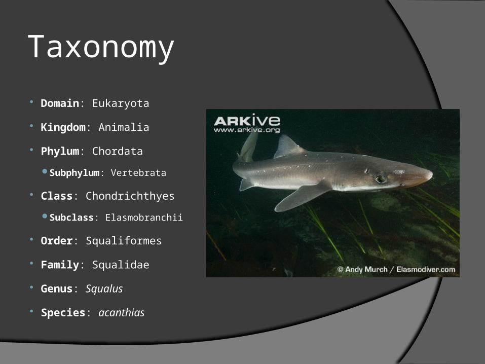
Taxonomy
Domain: Eukaryota
Kingdom: Animalia
Phylum: Chordata
Subphylum: Vertebrata
Class: Chondrichthyes
Subclass: Elasmobranchii
Order: Squaliformes
Family: Squalidae
Genus: Squalus
Species: acanthias

Sharks and Fish
Sharks are fish. But unlike the bony fish
they do not have gas bladders.
This means that if they stop swimming,
they sink.

Myth: if a shark stop swimming it dies.
Truth: There are some sharks that live in open
water and because sharks don’t have gas
bladders if they stop swimming they sink. If
the ocean floor is too deep the pressure may
crush the shark.

Ion Balance
Sharks release urea and other solutes into
their body which keeps their internal fluids
at relatively the same salinity as the ocean.
This means that they do not need to ingest
water or find a source of salt.

Birth
Some sharks, like the dogfish shark are
ovoviviparous.
This means that they lay “eggs” inside their own
body. The babies hatch inside the mother and
continue their development until they are ready to
enter the ocean as smaller versions of their
parents.
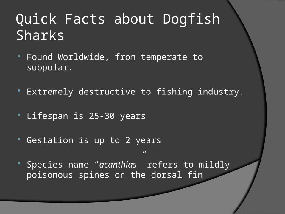
Quick Facts about Dogfish Sharks Found Worldwide, from temperate to subpolar.
Extremely destructive to fishing industry.
Lifespan is 25-30 years
Gestation is up to 2 years
Species name “acanthias” refers to mildly poisonous spines on the dorsal fin

Terminology Anterior – toward the head
Posterior – toward the tail
Dorsal – toward the backbone
Ventral – toward the belly
Lateral – toward the side

Overall Description
Fusiform – shape of the body is streamline. Built for swimming in the
sea with least possible resistance
The Head (cranial) – from the point of the rostrum to the pectoral fins.
The Trunk – from the pectoral fins to the pelvic fins.
The Tail (caudal) – from the pelvic fin to the end of the caudal fin.

Skin Made of Placoid scales, or scales that are modifications of
teeth. Their structure and development are similar to teeth.
Fun Fact: Shark skin has been used as an abrasive in furniture
building and to coat the hilt of swords and handles of tools.

Body Color
Sharks are dark above and much lighter below.
This is referred to as counter shading and is common to aquatic vertebrates.
This tends to neutralize the effects of natural lights which come from above.

Lateral Line
On the side of the body near the dorsal surface is a light-colored stripe.
This is the lateral line, below the surface is a line of nerve receptors that
have pores opening to the surface.
These receptors are sensitive to the mechanical movement of water,
disturbances in the water, and sudden changes in pressure.
This warns sharks of vibrations and movements even when visibility is
reduced.

Ampullae of Lorenzini
These are patches of pores on the head near the eyes, snout, and nostrils.
They are sense organs which are sensitive to changes in temperature, water pressure, electrical fields, and salinity

Nares
These are the openings for the external nostrils.
Located on the ventral surface of the rostrum this is where
water is drawn into the nares to moisten the sensory cells
of the olfactory sac.
Water passes into the and out of the olfactory sac,
permitting the shark to detect the odors of the water.

Jaws
A testing device, called the gnathodynamometer, was used to
measure the force exerted by the jaws of a typical eight-foot
shark.
The measured force came out to 18 tons per square inch!
Beside the visible teeth you will find several rows of flattened
teeth ready to replace the visible teeth when they are lost or
worn out.

Spiracles
These large openings posterior and dorsal to the eyes
are actually reduced first gill slits.
These serve as incurrent water passageways leading
into the mouth.
This allows water to be brought in for respiration even
when the shark’s mouth is closed or when it is feeding.

Gill Slits
Most sharks have 5 external gill slits.
Water taken in by the mouth is passed over the
internal gills, oxygen is removed and carbon
dioxide excreted.
The water is then forced out of the body by way of
the gill slits.

Dorsal Fins
Dogfish sharks have two dorsal fins.
A feature peculiar to spiny dogfish, is the presence of two spines. One anterior to each of the dorsal fins.
These spines are used when captured to inflict puncture wounds and carry poison secreted by glands at their base.

Caudal Fin
Each tail fin is divided into two lobes.
Dogfish sharks have a type of tail known
as heterocercal.
Heterocercal Homocercal

Pectoral and Pelvic Fins
Pectoral fins: located at the anterior end of
the shark these are vital for the necessary
lift to keep a shark horizontal.
Pelvic fins: located at the posterior end of
the shark. These are different in males and
females.

Cloaca
Cloaca means “sewer”
This is the chamber between the pelvic
fins. It receives the products of the
intestine, the urinary and the genital
ducts.
Claspers
Found only on the males these are extensions of the pelvic fin.
They are used during copulation.

DOGFISH SHARK DISSECTION

Dissection Procedures
During Dissection rely heavily upon your dissecting needles, probes, and fingers rather than your scalpel.
When using scissors advance with the rounded end and not the sharp pointed end.
Dissecting Needles
Dissection Procedures
Identify the following:
Pectoral Fins Pelvic Fins Caudal Fin Dorsal Fins

Dissection Procedures
1. Run your hand over the body of the
shark from head to tail.
2. Now try the other way.

Dissection Procedures
3. Look for the lateral line.
It will be a light-colored horizontal stripe on the side of the body nearer the dorsal surface than the ventral surface.
4. Use a magnifying glass and observe the pores.

Dissection Procedures
5. Locate the ampullae of Lorenzini
These will look like black heads on the nose of the
shark.
6. Press firmly upon the skin near the nares
(nostrils).
Note the jelly-like material you have squeezed out of
the pores.
Muscle7. Make a shallow incision into the skin along the spine of the dogfish just behind
the second dorsal fin
8. Continue to cut caudally for about two inches
9. At each of the ends cut the skin ventrally along the sides of the body until you
reach the middle of the belly
10. Using a blunt instrument remove the section of skin
11. Make observations about the muscleLater, if you have time, remove the skin from the gills

Opening the Body Cavity
12. Turn your specimen ventral side up
13. Make an incision anterior to the cloaca
14. Cut through the skin and muscle in an anterior direction just to
the right of the middle
15. Continue your cut until your reach the anterior portion of the
pectoral fins
16. Use your scissors and cut laterally toward the right and left
17. Return to the cloaca and cut laterally toward the right and left

The Liver
The largest organ in the cavity is the liver.
Has 3 lobes. There are 2 main lobes,the right and left and a third median lobe that is much smaller located in the middle.
The gall bladder is the green sac on the right edge of the median lobe.

Esophagus
Move the large lobes of the liver laterally.
Behind them you will find a thick muscular tube coming from the top of the cavity and extending posteriorly to the left.
This is the esophagus.

Stomach
The esophagus leads to the “J” shaped stomach and ends in
the “U” shaped duodenum.
Cut the stomach open along its axis. Open it and note its
contents.
The duodenum is the first portion of the small intestine and is
where the gall bladder and bile duct enter the digestive
system.
Pancreas and Spleen
The pancreas is the whitish glandular tissue
that partially obscures the duodenum. The
larger part is found on the dorsal side.
The spleen is found near the posterior end of
the stomach and looks like a dark triangle.

Vascular Intestine
The next part of the small intestine is easily found because it is
marked by rings.
Cut away the outer tissue to find a symmetrical spiral shape within,
called the spiral valve.
This spiral shape adds surface area to increase digestion and
absorption.
Higher vertebrates have finger-like projections called villi.

Non-Digestive Organs
Gonads: these can be found by moving the liver and digestive organs to one side and looking to the anterodorsal portion of the body cavity.
The kidneys are the elongated structures running the length of the body on either side of the mid-dorsal line.

Opening the Oral Cavity
18. Insert your scissors into the side of the shark’s mouth.
19. Begin cutting posteriorly through the angle of the jaws across the gill slits as far back as the pectoral girdle.
20. Cut across the bottom of the sharks mouth.
21. Then open the mouth and lay it flat.

Oral Cavity
Note the teeth
Find the tongue. It is not a true tongue and is practically immovable.
Find the spiracles
Find the gills
The Circulatory System
Back in the body cavity find the heart it will be located between the pectoral fins.
It may be hidden beneath a thin membrane.

Enjoy the rest of your time I suggest that you look at the eye.

Shark Dissection Analysis1. Boney fish have swim bladders that allow them
to remain buoyant. How have sharks adapted to the same situation?
2. How have sharks overcome the dangers of leaving offspring in the open ocean to develop?
3. Why is the fusiform body shape an important adaptation in sharks? Would that be beneficial in a terrestrial ecosystem?

Shark Dissection Analysis4. Why is the Lateral Line an important
adaptation for fishes?
5. The counter shading that sharks have is an important adaptation in a marine ecosystem. Would this adaptation be beneficial in terrestrial or freshwater ecosystems? Why?

Created by Scott Johnson and Pam Simmons-Brooks
Sloan Creek Middle School, October 2010