the endocrine sytem second controlling system of the body ◦ nervous system is the fast-control...
TRANSCRIPT

The Endocrine SytemSecond controlling system of the body
◦Nervous system is the fast-control systemUses chemical messengers
(hormones) that are released into the blood
Hormones control several major processes◦Reproduction◦Growth and development◦Mobilization of body defenses◦Maintenance of much of homeostasis◦Regulation of metabolism
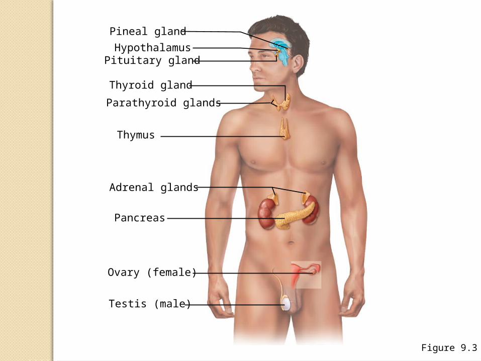
Figure 9.3
Pineal gland
HypothalamusPituitary gland
Thyroid gland
Parathyroid glands
Thymus
Adrenal glands
Pancreas
Ovary (female)
Testis (male)
Chemical Make-upHormones are classified
chemically as◦Amino acid–based, which includes
Proteins Peptides Amines
◦Steroids—made from cholesterol◦Prostaglandins—made from highly
active lipids
Mechanisms of Hormone Action
Hormones affect only certain tissues or organs (target cells or target organs)
Target cells must have specific protein receptors
Hormone-binding alters cellular activity
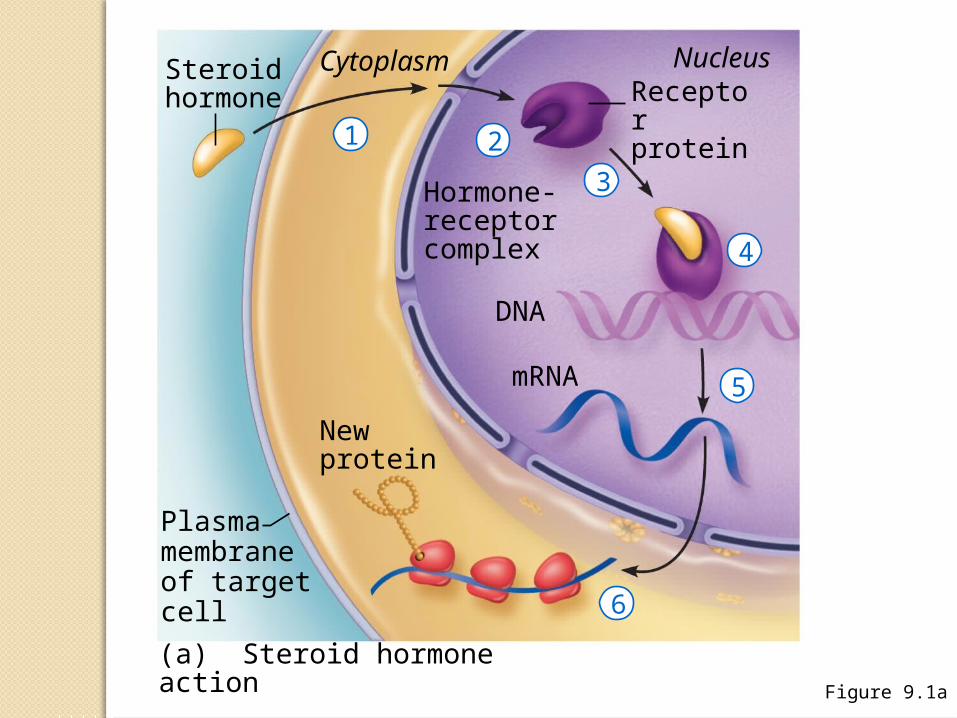
Figure 9.1a
Steroidhormone
Cytoplasm Nucleus
Receptorprotein
Hormone-receptor complex
DNA
mRNA
Newprotein
Plasmamembraneof targetcell
(a) Steroid hormone action
1 2
3
4
5
6
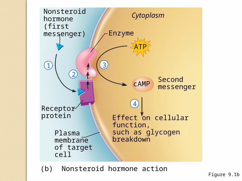
Figure 9.1b
Nonsteroidhormone (first messenger)
Cytoplasm
Enzyme
Receptorprotein
Plasma membraneof target cell
Secondmessenger
Effect on cellular function, such as glycogenbreakdown
(b) Nonsteroid hormone action
ATP
cAMP
12
3
4
Control of Hormone Release
Hormone levels in the blood are mostly maintained by negative feedback
A stimulus or low hormone levels in the blood triggers the release of more hormone
Hormone release stops once an appropriate level in the blood is reached
Hormonal Stimuli of Endocrine Glands
Most common stimuliEndocrine glands are activated by
other hormones
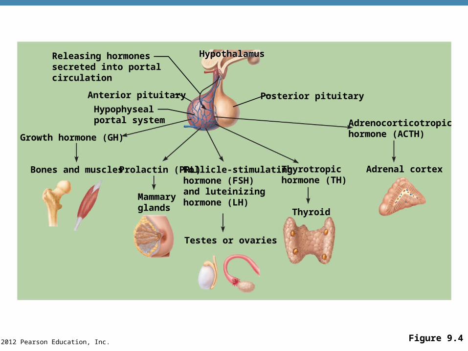
© 2012 Pearson Education, Inc. Figure 9.4
Releasing hormonessecreted into portalcirculation
Anterior pituitary
Hypophysealportal system
Growth hormone (GH)
Bones and muscles Prolactin (PRL)
Mammaryglands
Follicle-stimulatinghormone (FSH)and luteinizinghormone (LH)
Posterior pituitary
Hypothalamus
Adrenocorticotropichormone (ACTH)
Adrenal cortexThyrotropichormone (TH)
Thyroid
Testes or ovaries
HypothalamusHypothalamus
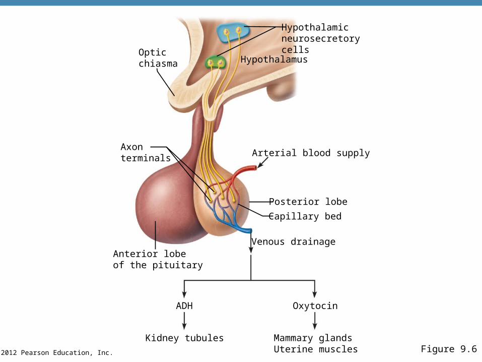
© 2012 Pearson Education, Inc. Figure 9.6
Opticchiasma
Axon terminals
Anterior lobeof the pituitary
ADH
Kidney tubules
Hypothalamicneurosecretorycells
Hypothalamus
Arterial blood supply
Posterior lobe
Capillary bed
Venous drainage
Oxytocin
Mammary glandsUterine muscles
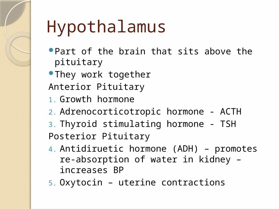
HypothalamusPart of the brain that sits above the pituitaryThey work togetherAnterior Pituitary1. Growth hormone2. Adrenocorticotropic hormone - ACTH3. Thyroid stimulating hormone - TSHPosterior Pituitary4. Antidiruetic hormone (ADH) – promotes re-
absorption of water in kidney – increases BP
5. Oxytocin – uterine contractions
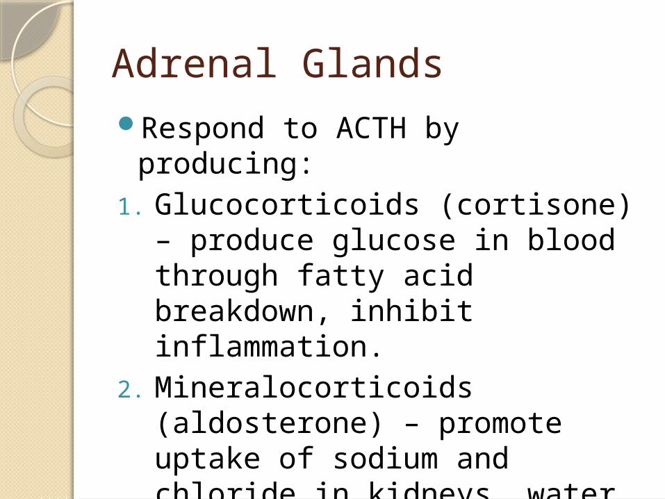
Adrenal GlandsRespond to ACTH by producing:1. Glucocorticoids (cortisone) –
produce glucose in blood through fatty acid breakdown, inhibit inflammation.
2. Mineralocorticoids (aldosterone) – promote uptake of sodium and chloride in kidneys, water follows, increase BP
3. Epinephrine - emergency hormone
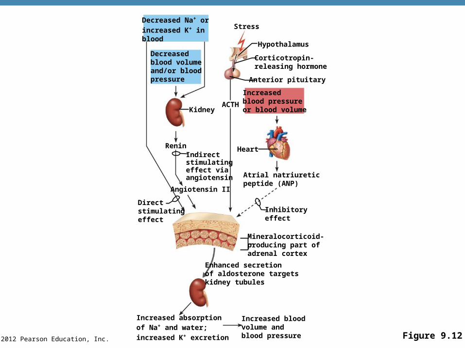
© 2012 Pearson Education, Inc. Figure 9.12
Decreased Na+ or
increased K+ in blood
Decreasedblood volumeand/or bloodpressure
Kidney
ReninIndirectstimulatingeffect viaangiotensin
Angiotensin II
Directstimulatingeffect
Increased absorptionof Na+ and water;
increased K+ excretion
Increased bloodvolume andblood pressure
Enhanced secretion of aldosterone targets kidney tubules
Mineralocorticoid-producing part of adrenal cortex
Inhibitoryeffect
Atrial natriureticpeptide (ANP)
ACTH
Heart
Increasedblood pressureor blood volume
Anterior pituitary
Corticotropin-releasing hormone
Hypothalamus
Stress
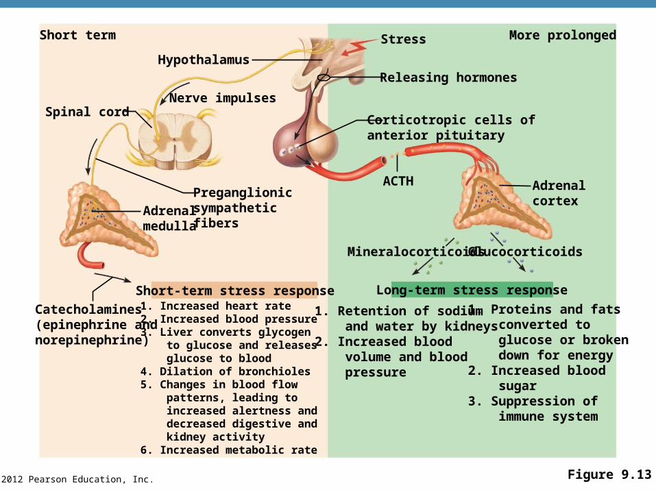
© 2012 Pearson Education, Inc. Figure 9.13
Short term
Spinal cord
Catecholamines(epinephrine and norepinephrine)
Adrenalmedulla
1. Increased heart rate2. Increased blood pressure3. Liver converts glycogen to glucose and releases glucose to blood4. Dilation of bronchioles5. Changes in blood flow patterns, leading to increased alertness and decreased digestive and kidney activity6. Increased metabolic rate
1. Retention of sodium and water by kidneys2. Increased blood volume and blood pressure
1. Proteins and fats converted to glucose or broken down for energy2. Increased blood sugar3. Suppression of immune system
Short-term stress response Long-term stress response
Preganglionicsympatheticfibers
Nerve impulses
Hypothalamus
More prolongedStress
Releasing hormones
Corticotropic cells ofanterior pituitary
ACTH Adrenalcortex
Mineralocorticoids Glucocorticoids
ThyroidStimulated by Thyrotropin releasing
hormone (TRH) from the hypothalamus which stimulates TSH from anterior pituitary.
1. Thyroxine (iodine)Maintains metabolism Hyperthyroidism – high metabolic rate,
nervous, irritableHypothyroidism – low met. Rate, slow,
sluggish, overweight2. Calcitonin – takes up calcium from blood
to bones
Disorders of the endocrine systemGoiter – iodine deficiency, thyroxin
production declinesThe hypothalamus secretes TRH, but
has no effectResults in enlargement of the thyroid Grave’s disease – antibodies in the
immune system mistakenly bind to TSH receptors on the thyroid gland stimulating more thyroxin production - hyperthyroidism
ParathyroidsRest on the thyroid1. Parathyroid hormone – releases
calcium from bone to blood Opposite of calcitonin Bone remodeling
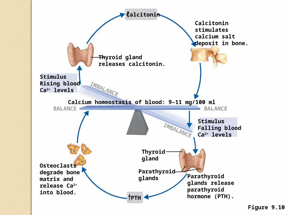
Figure 9.10
Calcitonin
Thyroid glandreleases calcitonin.
StimulusRising bloodCa2+ levels
Calcium homeostasis of blood: 9–11 mg/100 ml
Osteoclastsdegrade bonematrix andrelease Ca2+ into blood.
BALANCE
IMBALANCE
IMBALANCE
Calcitoninstimulatescalcium saltdeposit in bone.
StimulusFalling bloodCa2+ levels
BALANCE
Parathyroidglands releaseparathyroidhormone (PTH).
Thyroidgland
Parathyroidglands
PTH
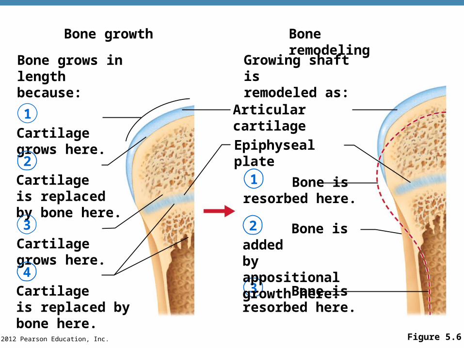
© 2012 Pearson Education, Inc. Figure 5.6
Bone growth
Bone grows inlength because:
Bone remodeling
Growing shaft isremodeled as:
Cartilagegrows here.
Cartilageis replacedby bone here.
Cartilagegrows here.
Cartilageis replaced by bone here.
1
2
3
4
1
2
3 Bone isresorbed here.
Epiphyseal plate
Articular cartilage
Bone isresorbed here.
Bone is addedby appositionalgrowth here.
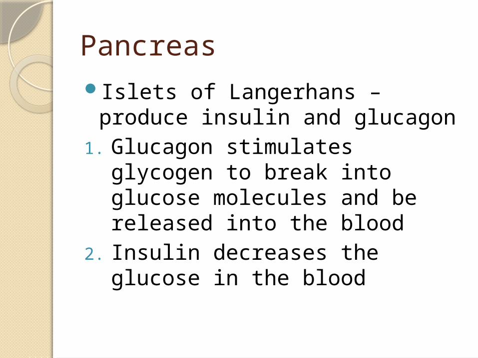
PancreasIslets of Langerhans – produce
insulin and glucagon1. Glucagon stimulates glycogen to
break into glucose molecules and be released into the blood
2. Insulin decreases the glucose in the blood
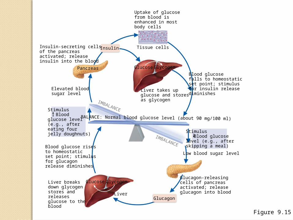
Figure 9.15
Uptake of glucosefrom blood isenhanced in mostbody cells
Tissue cells
Glucose GlycogenPancreas
InsulinInsulin-secreting cellsof the pancreasactivated; releaseinsulin into the blood
Elevated bloodsugar level
Stimulus Bloodglucose level(e.g., aftereating fourjelly doughnuts)
Blood glucose risesto homeostaticset point; stimulusfor glucagonrelease diminishes
Liver breaksdown glycogenstores andreleasesglucose to theblood
Glucose Glycogen
LiverGlucagon
Glucagon-releasingcells of pancreasactivated; releaseglucagon into blood
Low blood sugar level
Stimulus Blood glucoselevel (e.g., afterskipping a meal)
BALANCE: Normal blood glucose level (about 90 mg/100 ml)
IMBALANCE
Liver takes upglucose and storesas glycogen
Blood glucosefalls to homeostaticset point; stimulusfor insulin releasediminishes
IMBALANCE
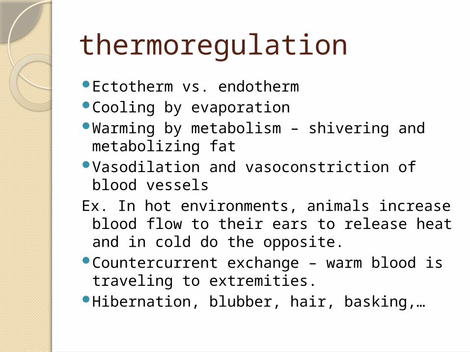
thermoregulationEctotherm vs. endothermCooling by evaporationWarming by metabolism – shivering and
metabolizing fatVasodilation and vasoconstriction of blood
vesselsEx. In hot environments, animals increase
blood flow to their ears to release heat and in cold do the opposite.
Countercurrent exchange – warm blood is traveling to extremities.
Hibernation, blubber, hair, basking,…
Reproductive SystemsFemaleOvaries – produce eggs (ova) and
secrete estrogen and progesterone
Stimulated by FSH and LHMaleFSH – stimulates sperm
productionLH – stimulates interstitial cells to
produce testosterone
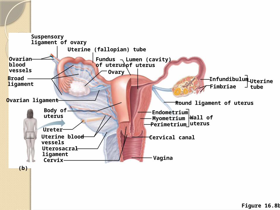
Figure 16.8b
(b)
Vagina
Cervical canal
Wall ofuterus
EndometriumMyometriumPerimetrium
Round ligament of uterus
FimbriaeInfundibulumUterine
tube
Lumen (cavity)of uterus
Fundusof uterus
Ovary
Uterine (fallopian) tube
Suspensoryligament of ovary
Ovarianbloodvessels
Broadligament
Ovarian ligament
Body ofuterus
Ureter
UterosacralligamentCervix
Uterine bloodvessels
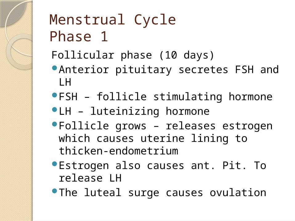
Menstrual CyclePhase 1Follicular phase (10 days)Anterior pituitary secretes FSH and LHFSH – follicle stimulating hormoneLH – luteinizing hormone Follicle grows – releases estrogen
which causes uterine lining to thicken-endometrium
Estrogen also causes ant. Pit. To release LH
The luteal surge causes ovulation
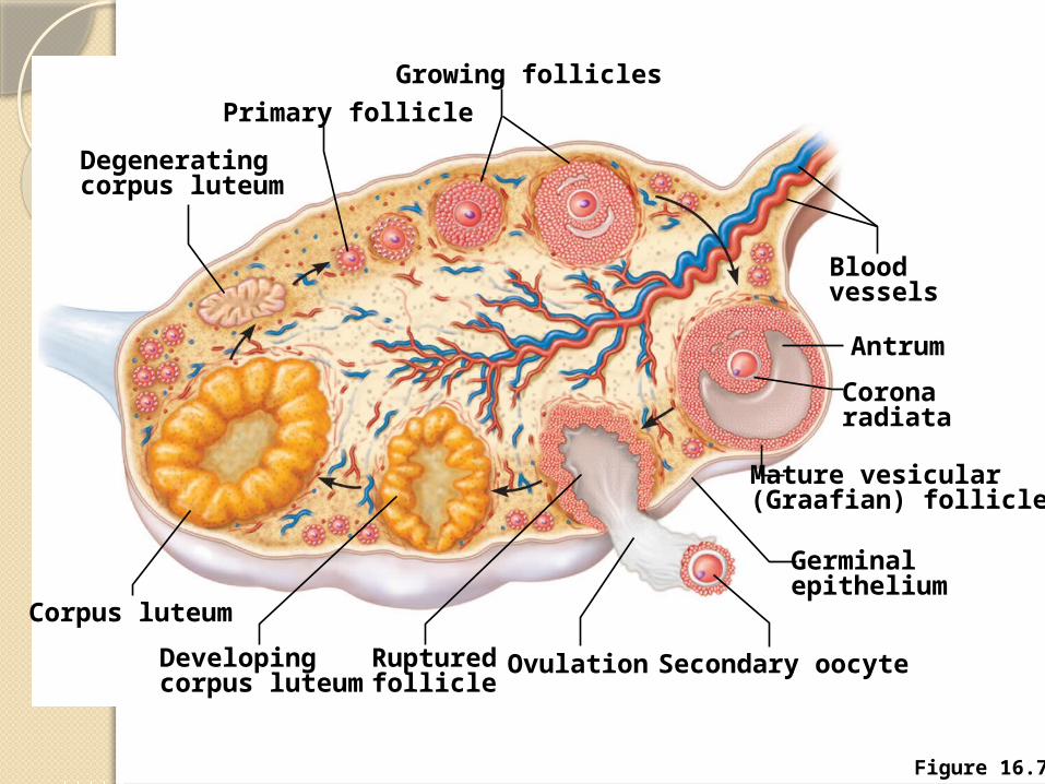
Figure 16.7
Growing follicles
Primary follicle
Degeneratingcorpus luteum
Corpus luteum
Developingcorpus luteum
Rupturedfollicle
OvulationSecondary oocyte
Germinalepithelium
Bloodvessels
Antrum
Coronaradiata
Mature vesicular(Graafian) follicle
Ovulation – release of the egg from the ovary ends the follicular phase.
Ova floats into fallopian tube (oviduct)
Phase 2Luteal phaseFollicle has released the egg and
changes into the corpus luteumCorpus luteum continues to secrete
estrogen and now begins to produce progesterone
Progesterone increases vessels and glands in uterus
After about 13-15 days, if fertilization and implantation have NOT occurred the corpus luteum shuts down
Phase 3
MenstruationUterus reabsorbs some of the
tissue created in the endometrium
The rest sheds off and is passed out of the body
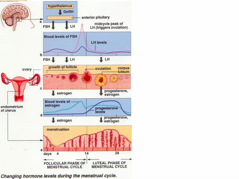
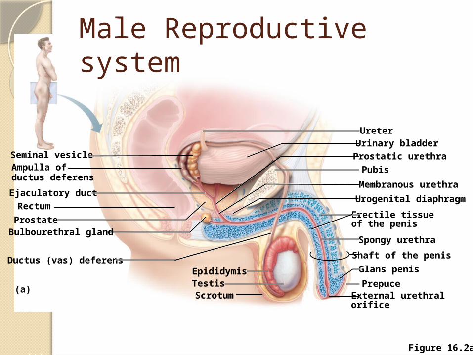
Figure 16.2a
Ampulla ofductus deferens
Ductus (vas) deferens
(a)
Bulbourethral glandProstate
RectumEjaculatory duct
Seminal vesicle
Epididymis
ScrotumTestis
Shaft of the penis
Spongy urethra
External urethral orifice
Prepuce
Glans penis
Urogenital diaphragm
Membranous urethra
UreterUrinary bladderProstatic urethraPubis
Erectile tissue of the penis
Male Reproductive system