rivm report 388802024 toxicoloby and occurrence of nivalenol
TRANSCRIPT

RIVM Report 388802024/2002
Toxicology and occurrence of nivalenol,fusarenon X, diacetoxyscirpenol, neosolaniol and3- and 15-acetyldeoxynivalenol: a review of sixtrichothecenes
M.E.J. Pronk, R.C. Schothorst, H.P. van Egmond
This investigation has been performed by order and for the account of the Inspectorate forHealth Protection and Veterinary Public Health, within the framework of project 388802,Natural Toxins.
RIVM, P.O. Box 1, 3720 BA Bilthoven, telephone: 31 - 30 - 274 91 11; telefax: 31 - 30 - 274 29 71
page 2 of 75 RIVM report 388802024
Abstract
To assess the potential health effects of the trichothecenes nivalenol (NIV), fusarenon X(FusX), diacetoxyscirpenol (DAS), neosolaniol (NeoSol) and 3- and 15-acetyldeoxynivalenol(3-Ac-DON, 15-Ac-DON), toxicology data available on these compounds were reviewed toderive, where possible, tolerable daily intakes (TDIs). Also, their occurrence in cereal grains,animal feed and human foodstuffs was investigated. The six trichothecenes are mycotoxinsproduced by Fusarium spp. NIV was the one most often found in food commodities. FusX,DAS, NeoSol and 3- and 15-Ac-DON have rarely been reported to occur, most likely becausethey were not routinely monitored for. It was therefore recommended to pay more attention toFusX, DAS, NeoSol and 3- and 15-Ac-DON in routine monitoring programs. The toxicologydata available on FusX, DAS, 3-Ac-DON, 15-Ac-DON and NeoSol were too limited toderive TDIs. Only for NIV sufficient toxicology data were available to derive a temporaryTDI of 0.7 µg/kg bw.

RIVM report 388802024 page 3 of 75
Contents
Abstract
Samenvatting 5
Summary 7
1. Introduction 9
2. Trichothecenes 13
2.1 Chemical structure 13
2.2 Physical and chemical properties 14
2.3 Analysis of trichothecenes 152.3.1 Sampling 152.3.2 Extraction 152.3.3 Clean-up 152.3.4 Detection and quantification 16
3. Sources and occurrence of trichothecenes 17
3.1 Sources 17
3.2 Plant pathogenicity 18
3.3 Contamination of cereal grains and animal feed 18
3.4 Contamination of human foodstuffs 21
3.5 Fate during processing 23
4. Toxicology 25
4.1 Trichothecenes in general 254.1.1 Toxicokinetics 254.1.2 Toxicity 25
4.2 Nivalenol (NIV) 294.2.1 Toxicokinetics 294.2.2 Toxic effects 30
4.3 Fusarenon X (FusX) 374.3.1 Toxicokinetics 374.3.2 Toxic effects 38
4.4 Diacetoxyscirpenol (DAS) 424.4.1 Toxicokinetics 424.4.2 Toxic effects 44
4.5 3-Acetyl-deoxynivalenol (3-Ac-DON) 514.5.1 Toxicokinetics 514.5.2 Toxic effects 51
4.6 15-Acetyl-deoxynivalenol (15-Ac-DON) 544.6.1 Toxicokinetics 544.6.2 Toxic effects 54
4.7 Neosolaniol (NeoSol) 55

page 4 of 75 RIVM report 388802024
4.7.1 Toxicokinetics 554.7.2 Toxic effects 56
5. Evaluation 57
5.1 NIV 57
5.2 FusX 59
5.3 DAS 60
5.4 3-Ac-DON 61
5.5 15-Ac-DON 62
5.6 NeoSol 62
6. Conclusions 63
References 65
Appendix 1 Mailing list 75
RIVM report 388802024 page 5 of 75
Samenvatting
Mycotoxinen zijn door schimmels geproduceerde, van nature voorkomende toxische stoffen.Indien deze mycotoxinen terecht komen in diervoeders of in voor mensen bestemd voedsel,dan kunnen ze toxische verschijnselen induceren en aldus een bedreiging vormen voor degezondheid van dier en mens. Trichothecenen vormen een belangrijke groep mycotoxinen, enalhoewel er meer dan 150 verschillende trichothecenen bekend zijn komt slechts een kleinaantal daarvan daadwerkelijk voor in granen, diervoeders en voedingsmiddelen. Dit zijndeoxynivalenol (DON), nivalenol (NIV), diacetoxyscirpenol (DAS), T-2 toxine (T-2) en, inmindere mate, hun derivaten 3- and 15-acetyldeoxynivalenol (3-Ac-DON, 15-Ac-DON),fusarenon X (FusX), neosolaniol (NeoSol) en HT-2 toxine (HT-2). Alle behoren tot de type Aen type B trichothecenen, worden geproduceerd door schimmels van het geslacht Fusariumen bezitten als basis dezelfde tetracyclische 12,13-epoxytrichotheceen structuur. Door detoegenomen handel in granen, diervoeders en voedingsmiddelen, de wereldwijdecontaminatie van deze producten met trichothecenen (en andere mycotoxinen) en het feit dattrichothecenen over het algemeen vrij stabiel zijn en tijdens voedselbereiding niet volledigvernietigd worden, wordt het steeds belangrijker om de eventuele gezondheidsrisico’s van demeest voorkomende trichothecenen in kaart te brengen en waar mogelijk toelaatbaredagelijkse innames (TDIs) vast te stellen. Recentelijk is dat door een aantal internationalewetenschappelijke commissies gedaan voor DON, T-2 en HT-2, en dat heeft geresulteerd in(voorlopige) TDIs voor deze stoffen. In dit rapport worden de mogelijke gezondheidseffectenvan zes andere trichothecenen, te weten NIV, FusX, DAS, NeoSol en 3- en 15-Ac-DON,onderzocht en wordt getracht ook voor deze trichothecenen (voorlopige) TDIs af te leiden(hetgeen overigens voor NIV ondertussen gedaan is door het Wetenschappelijk Comité voorde Voeding).In eerste instantie is gekeken naar het voorkomen van de zes trichothecenen in granen,diervoeders en voedingsmiddelen. FusX, DAS, NeoSol en 3- en 15-Ac-DON blijken niet ofnauwelijks in deze producten gevonden te worden. Naar alle waarschijnlijkheid zijn deze vijftrichothecenen echter tot nu toe niet routinematig meegenomen geweest in monitoringprogramma’s. NIV wordt daarentegen wel regelmatig aangetroffen, vaak tezamen met DONen zearalenon (ook een Fusarium mycotoxine). NIV werd wel gevonden in granen,diervoeders en plantaardige voedingsmiddelen, maar niet in voedingsmiddelen van dierlijkeoorsprong.In tweede instantie is de toxicologie van de zes individuele trichothecenen beschreven, en iseen kort overzicht gegeven van de toxicologie van trichothecenen in het algemeen. Alhoewelniet voor alle zes trichothecenen evenveel gegevens beschikbaar waren over huntoxicokinetische en toxische eigenschappen, lijken de effecten gevonden voor NIV, FusX,DAS, 3- en 15-Ac-DON en NeoSol op de effecten zoals die gevonden worden voortrichothecenen in het algemeen. De belangrijkste toxicologische bevindingen in dezeevaluatie van de zes trichothecenen bestaan uit haematotoxiciteit, immunotoxiciteit, embryo-
page 6 of 75 RIVM report 388802024
and foetotoxiciteit, braken en verminderde lichaamsgewicht toename en voerconsumptie.Ook lijken sommige een (intrinsiek) genotoxische potentie te hebben.Voor NIV zijn voldoende gegevens beschikbaar om een voorlopige TDI van 0,7 µg/kglichaamsgewicht af te kunnen leiden. Deze voorlopige TDI is gebaseerd op de meestgevoelige effecten zoals die voor NIV worden gevonden in proefdieren, namelijk algemenetoxiciteit en immunotoxiciteit in muizen. De TDI heeft een voorlopig karakter vanwege hetontbreken van relevante gegevens in de toxicologische dataset.Voor FusX, DAS en met name voor 3- en 15-Ac-DON en NeoSol zijn de toxicologischedatasets zodanig beperkt dat geen TDI (zelfs geen voorlopige) afgeleid kan worden. Om vastte kunnen stellen of deze vijf trichothecenen eventueel een gezondheidsrisico vormen voormens en dier wordt aanbevolen om in monitoring programma’s meer routinematig aandachtte schenken aan FusX, DAS, 3- en 15-Ac-DON en NeoSol. Er zijn analysemethoden voordeze stoffen beschikbaar. Afhankelijk van óf, en de mate waarin, deze vijf trichothecenenworden aangetroffen in granen, diervoeders en voedingsmiddelen, dient vervolgens bezien teworden hoe moet worden omgegaan met de beperkte toxicologische datasets voor dezestoffen.Omdat de meest voorkomende trichothecenen een overeenkomstige chemische structuurhebben en mogelijk ook een overeenkomstig werkingsmechanisme, verdient het aanbevelingom te bekijken of voor de groep van trichothecenen als geheel een zogenaamde groeps TDIvastgesteld zou moeten worden. Op dit moment zijn daarvoor te weinig gegevensbeschikbaar.

RIVM report 388802024 page 7 of 75
Summary
Trichothecenes are an important class of mycotoxins, i.e. naturally occurring, toxiccompounds produced by fungi. Of the fungal genera known to produce trichothecenes,Fusarium spp. are the most important ones given their global occurrence and the range oftrichothecenes they produce. When ending up in animal feed and human foodstuffs,trichothecenes can pose a threat to animal and human health by the induction of toxicsyndromes. Only a few of over 150 known trichothecenes are believed to be of importancewith respect to their actual presence in crops, feeds and foods. These are deoxynivalenol(DON), nivalenol (NIV), diacetoxyscirpenol (DAS), T-2 toxin (T-2) and, to a lesser extent,their derivatives 3- and 15-acetyldeoxynivalenol (3-Ac-DON, 15-Ac-DON), fusarenon X(FusX), neosolaniol (NeoSol) and HT-2 toxin (HT-2). They belong to the type A and type Btrichothecenes and share the same basic chemical structure, a tetracyclic 12,13-epoxy-trichothecene skeleton. Given the worldwide trade in cereals, feeds and foods that more andmore are globally contaminated with mycotoxins, including trichothecenes, it has nowbecome important to assess the potential health risks of the most commonly occurringtrichothecenes and to derive tolerable daily intakes (TDIs) where possible. Moreover sobecause trichothecenes are in general stable compounds, and most food processing operationsare not successful in complete decontamination.Recently, the potential risks of DON, T-2 and HT-2 have been assessed by severalinternational committees, which resulted in (temporary) TDIs for these compounds. In thisreport we therefore address the potential health effects of six other trichothecenes, i.e. NIV,FusX, DAS, NeoSol and 3- and 15-Ac-DON, and try to derive (temporary) TDIs for thesetrichothecenes as well, although in the mean time an international committee already did thatfor NIV.Upon investigation of the occurrence of these six trichothecenes in cereal grains, animal feedand human foodstuffs, it appeared that FusX, DAS, NeoSol and 3- and 15-Ac-DON haverarely been reported to occur in these commodities. It should be noted, though, that in mostmonitoring programs these five trichothecenes likely have not been routinely included. NIVwas reported to occur more frequently, often together with DON and zearalenone, the latteralso being a Fusarium mycotoxin. NIV was found in cereal grains, in animal feed and inhuman food of plant-origin (cereals and processed grains) but not in human food derivedfrom animals given contaminated feed.After reviewing the toxicology of trichothecenes in general, the toxicological data on the sixindividual trichothecenes subject of this report were addressed. Data on the toxicokineticsand the toxic effects were studied, although the available data package was rather limited forsome of the six trichothecenes. Overall, however, the toxic effects observed for NIV, FusX,DAS, 3- and 15-Ac-DON and NeoSol seemed to fit quite well the profile of toxic effects oftrichothecenes in general. The most important observed effects were haematotoxicity,
page 8 of 75 RIVM report 388802024
immunotoxicity, embryo- and fetotoxicity, reductions in body weight gain and foodconsumption, vomiting and, for some, (intrinsic) genotoxicity.The data available on NIV allowed us to derive a temporary TDI of 0.7 µg/kg bw. Thistemporary TDI was based on the most sensitive effects of NIV observed in the tested animalspecies, i.e. general toxicity and immunotoxicity in mice. Due to deficiences in the data set atemporary and not a full TDI was derived.In our opinion, the limited data available on the toxicology of FusX, DAS and, especially,3- and 15-Ac-DON and NeoSol, do not allow derivation of a (temporary) TDI for thesecompounds. In order to assess whether or not these five trichothecenes are a potential threatto animals or humans, it is recommended to pay more attention to FusX, DAS, 3- and15-Ac-DON and NeoSol in routine monitoring programs. Analytical methods for thesecompounds are available. When it turns out that these trichothecenes are regularly found atconsiderable levels in cereal grains, feeds or foods, it should be considered how to deal withthe limited data sets on the toxicology of these compounds.Consideration should also be given to the appropriateness of the derivation of a group TDIfor trichothecenes since the most commonly occurring trichothecenes share a common basicchemical structure and, presumably, also a common mechanism of action.
RIVM report 388802024 page 9 of 75
1. Introduction
Many species of Fusarium, Aspergillus, Penicillum and Alternaria are not only recognisedplant pathogens but are also sources of mycotoxins. Mycotoxins are naturally occurring, toxiccompounds, produced by fungi infecting agricultural crops, particularly cereals and oilseeds.Infection can occur during growth and storage of crops, but also later on in processed foodsand feeds. Upon ingestion, these mycotoxins can produce toxic syndromes (mycotoxicoses)in animals and humans.
An important class of mycotoxins are the trichothecenes. Fungal genera known to producetrichothecenes are Fusarium, Trichoderma, Trichothecium, Stachybotrys,Verticimonosporium, Cephalosporium, Myrothecium, and Cylindrocarpon. The fusaria are byfar the most important of these genera because they are widespread globally and produce thegreatest range of trichothecenes. However, of over 150 known trichothecenes, only a fewappear to be of importance with respect to their actual presence in crops intended for humanor animal use. These are: deoxynivalenol (DON; also known as vomitoxin), nivalenol (NIV),diacetoxyscirpenol (DAS) and T-2 toxin (T-2), and, less frequently, certain derivatives(3-acetyldeoxynivalenol (3-Ac-DON), 15-acetyldeoxynivalenol (15-Ac-DON), fusarenon X(FusX), neosolaniol (NeoSol) and HT-2 toxin (HT-2)).
There is increasing evidence of global contamination of cereals and animal feeds withFusarium mycotoxins, and trade in these commodities may contribute to the worldwidedispersal of the mycotoxins. It has therefore become increasingly important to assess thepotential health risks associated with exposure to the different Fusarium mycotoxins. Fromthe point of view of animal health and productivity the trichothecenes (the focus of thisreport), zearalenone, moniliformin and the fumonisins are the most important Fusariummycotoxins. However, information to base a risk assessment on is scarce, despite the fact thatmycotoxicoses have been known to occur since ancient times.
Although since a few years analytical methods are available for the nine most occurringtrichothecenes, up till now in monitoring programs most attention has been focussed onDON. Hence, compared to other trichothecenes, relatively a lot of data are reported on theoccurrence of DON in feeds and foods. Also the toxicology of DON is relatively wellinvestigated, like that of T-2 and HT-2. For these three trichothecenes risk assessments haverecently been performed by the Scientific Committee on Food (SCF) of the EuropeanCommission (SCF, 1999, 2001, 2002), the Joint FAO/WHO Expert Committee on FoodAdditives (JECFA; WHO/FAO, 2001) and the Nordic Working Group (Eriksen andAlexander, 1998). Each established Tolerable Daily Intakes (TDI) for DON, T-2 and HT-2.Whereas the SCF and Eriksen and Alexander (1998) also evaluated NIV, only the SCFestablished a TDI for NIV (SCF, 2000, 2002).

page 10 of 75 RIVM report 388802024
For DON, a temporary TDI (tTDI) of 1 µg/kg bw was established by the SCF (SCF, 1999),based on a 2-year feeding study in mice. In this study reduced growth was the most sensitiveparameter, with a no-observed-adverse-effect-level (NOAEL) of 0.1 mg/kg bw. Anuncertainty factor of 100 was applied to this NOAEL, in order to extrapolate from rodents tohumans (factor 10) and to cover for (human) interindividual differences (factor 10). ThistTDI would also protect against other toxic effects of DON, including the acute vomitingeffect. The tTDI set by the SCF in 1999 is in line with the tTDI established for DON by theNordic Working Group (Eriksen and Alexander, 1998) and Pieters et al. (1999) and with theprovisional maximum TDI (PMTDI) established by JECFA (WHO/FAO, 2001). The SCFmade the TDI temporary pending a group evaluation. This because DON belongs to a groupof trichothecenes sharing a common basic chemical structure and common mechanisms oftoxic action and because most Fusarium species are capable of producing severaltrichothecenes. In 2002, after having evaluated four of the most commonly occurringtrichothecenes, the SCF performed a group evaluation of DON, NIV, T-2 and HT-2 andconsidered the appropriateness of a group TDI. SCF concluded that the available data, whilelimited, did not support the establishing of a group TDI for the four trichothecenes evaluatedand, with respect to DON, changed the tTDI of 1 µg/kg bw into a full TDI (SCF, 2002). TheSCF, as well as JECFA, expressed the need for further studies.
In 2001, the SCF established a tTDI of 0.06 µg/kg bw for the sum of T-2 and HT-2 (SCF,2001). This because in vivo T-2 is rapidly metabolised to HT-2. Hence, the toxic effects ofT-2 and its metabolite HT-2 cannot be differentiated, and the toxicity of T-2 in vivo might bedue at least partly to HT-2. The tTDI was based on haematotoxic and immunotoxic effectsobserved in a 3-week feeding study with T-2 in swine. The lowest-observed-adverse-effect-level (LOAEL) for these effects was 0.03 mg/kg bw/day, a dose level at which also reducedfeed intake was seen. The uncertainty factor used was 500: an extra uncertainty factor of 5was applied to account for some deficiencies in the data set and the use of a LOAEL, whichwas presumably close to the NOAEL. The TDI was made temporary pending the evaluationof the group of trichothecenes as a whole, and also because of gaps in the database. JECFA(WHO/FAO, 2001) came to the same value of 0.06 µg/kg bw as PMTDI for the sum of T-2and HT-2, and both SCF and JECFA expressed the need for further studies. The NordicWorking Group set a different tTDI of 0.2 µg/kg bw. Because of concern on the seriousnessof the possible carcinogenic effect of T-2, they applied an uncertainty factor of 1000 to thehighest level without tumourigenic effect (approximately 0.1-0.2 mg/kg bw). This tTDI wasalso for the sum of T-2 and HT-2 (Eriksen and Alexander, 1998).After having concluded that the available data, while limited, did not support the establishingof a group TDI for DON, NIV, T-2 and HT-2, the SCF in 2002 confirmed the combined tTDIof 0.06 µg/kg bw for T-2 and HT-2 and recommended that further studies should fill the datagaps (SCF, 2002).

RIVM report 388802024 page 11 of 75
The SCF also recently evaluated the toxicity of NIV. They set a tTDI of 0.7 µg/kg bw basedon a LOAEL of 0.7 mg/kg bw/day found in long-term dietary studies with mice. For NIVreduced growth and haematotoxicity/immunotoxicity were the most critical effects. The SCFapplied a large uncertainty factor of 1000 because of the use of a LOAEL and the limiteddatabase. The TDI was made temporary pending a group evaluation of the trichothecenes,and also because of gaps in the database. A need for further studies was expressed (SCF,2000). NIV was also evaluated by the Nordic Working Group (Eriksen and Alexander, 1998),but they did not find the available toxicity data sufficient to set a (t)TDI for NIV.In 2002, the SCF performed a group evaluation of four trichothecenes and concluded that theavailable data, while limited, did not support the establishing of a group TDI for DON, NIV,T-2 and HT-2. They therefore confirmed the tTDI of 0.7 µg/kg bw for NIV andrecommended that further studies should fill the data gaps (SCF, 2002).
When in the near feature other trichothecenes than just DON will be more systematically androutinely included in monitoring programs, more insight is needed in the toxicology of thetrichothecenes that up till now only received little or no attention. Hence, in this report weaddress the potential health effects of NIV, 3-Ac-DON, 15-Ac-DON, DAS, FusX andNeoSol, and try to derive (t)TDIs for these trichothecenes as well.

page 12 of 75 RIVM report 388802024

RIVM report 388802024 page 13 of 75
2. Trichothecenes
2.1 Chemical structure
Trichothecenes are a family of closely related sesquiterpenoids produced by several plantpathogenic fungi. They have a tetracyclic 12,13-epoxytrichothecene skeleton in common, andcan be divided into four categories (WHO, 1990):Type A: characterized by a functional group other than a ketone at C-8Type B: characterized by a carbonyl function at C-8Type C: characterized by a second epoxide group at C-7,8 or C-9,10Type D: characterized by a macrocyclic ring system between C-4 and C-15 with two esterlinkages.The trichothecenes that are subject of this report belong to the type A and type Btrichothecenes (see below). Examples of type C trichothecenes are crotocin and baccharin,and of type D trichothecenes roridin A, satratoxin H and verrucarin A.
O
HO
R5
CH3
H
CH2
CH3 H
R2
R1
H
R4R3
Figure 1 Structural formula of type A trichothecenes
Table 1 Examples of type A trichothecenes
Cas nr. Mol.formula
Mol.wt
R1 R2 R3 R4 R5
DAS a 2270-40-8 C19H26O7 366 OH OAc OAc H HNeoSol b 36519-25-2 C19H26O8 382 OH OAc OAc H OHT-2 c 21259-20-1 C24H34O9 466 OH OAc OAc H OCOCH2CH(CH3)2
HT-2 d 26934-87-2 C22H32O8 424 OH OH OAc H OCOCH2CH(CH3)2
a diacetoxyscirpenol (trichothec-9-ene-3,4,15-triol, 12,13-epoxy-, 4,15-diacetate; anguidine)b neosolaniol (trichothec-9-ene-3,4,8,15-tetrol, 12,13-epoxy-, 4,15-diacetate)c T-2 toxin (trichothec-9-ene-3,4,8,15-tetrol, 12,13-epoxy-, 4,15-diacetate 8-(3-methylbutanoate))d HT-2 toxin (trichothec-9-ene-3,4,8,15-tetrol, 12,13-epoxy-, 15-acetate 8-(3-methylbutanoate))

page 14 of 75 RIVM report 388802024
O
HO
O
CH3
H
CH2
CH3 H
R2
R1
H
R4R3
Figure 2 Structural formula of type B trichothecenes
Table 2 Examples of type B trichothecenes
Name Cas nr. Mol.formula
Mol.wt
R1 R2 R3 R4
NIV a 23282-20-4 C15H20O7 312 OH OH OH OHFusX b 23255-69-8 C17H22O8 354 OH OAc OH OHDON c 51481-10-8 C15H20O6 296 OH H OH OH3-Ac-DON d 50722-38-8 C17H22O7 338 OAc H OH OH15-Ac-DON e 88337-96-6 C17H22O7 338 OH H OAc OH
a nivalenol (trichothec-9-en-8-one, 12,13-epoxy-3,4,7,15-tetrahydroxy-)b fusarenon X (trichothec-9-en-8-one, 4-(acetyloxy)-12,13-epoxy-3,7,15-trihydroxy-; fusarenon;4-acetyl nivalenol; nivalenol 4-O-acetate; nivalenol monoacetate)c deoxynivalenol (trichothec-9-en-8-one, 12,13-epoxy-3,7,15-trihydroxy-; dehydronivalenol;4-deoxynivalenol; Rd toxin; vomitoxin)d 3-acetyl-deoxynivalenol (trichothec-9-en-8-one, 3-(acetyloxy)-12,13-epoxy-7,15-dihydroxy-)e 15-acetyl-deoxynivalenol (trichothec-9-en-8-one, 15-(acetyloxy)-12,13-epoxy-3,7-dihydroxy-)
2.2 Physical and chemical properties
The trichothecenes are colourless, mostly crystalline solids. Type A trichothecenes aresoluble in moderately polar solvents, such as chloroform, diethyl ether, ethyl acetate, andacetone, whereas type B trichothecenes require higher polarity solvents, such as aqueousmethanol or aqueous acetonitrile.Type A and B trichothecenes lack conjugated unsaturation in their structures with aconsequent absence of UV absorption, except for end absorption due to unsaturation atC9-C10. They also exhibit no fluorescence under UV light.
When trichothecenes containing an ester group are treated with a base, they are hydrolysed totheir corresponding parent alcohol. Free hydroxyl groups are readily acylated.

RIVM report 388802024 page 15 of 75
The 12,13-epoxy group is extremely stable to nucleophilic attack. However, prolongedheating under highly acidic conditions causes an intramolecular rearrangement of thetrichothecene skeleton to the apotrichothecene ring.
The trichothecenes are generally stable. They remain unaffected when refluxed with variousorganic solvents and also under mildly acidic conditions (WHO, 1990; Ueno, 1987b).
2.3 Analysis of trichothecenes
Langseth and Rundberget (1998) and Krska et al. (2001) amongst many others, providedexhaustive reviews of quantitative and qualitative methods for the determination oftrichothecenes.In general four major steps are necessary for the determination of trichothecenes; sampling(including sample preparation), extraction, clean-up and detection and quantification of thetoxins.
2.3.1 Sampling
In general, mycotoxins are inhomogeneously distributed in the commodities to be inspected,which makes it difficult to obtain a representative sample (Van Egmond and Speijers, 1999).To reduce the variance, clearly defined sampling plans are required, including in generallarge sample sizes. To prepare a representative test portion for analysis, the sample is groundand thoroughly mixed.
2.3.2 Extraction
Test portions of 20 to 50 g of sample are normally used for extraction. Lower quantities canbe used, but require great care in order to obtain a homogeneous test portion.Various combinations of solvents have been used for the extraction of the trichothecenesfrom the test portion, including methanol-water, acetonitrile-water and ethyl acetate-acetonitrile in various ratios (Mateo et al., 2001).Nowadays acetonitrile/water (84+16, v/v) is the most extensively used extraction medium fortrichothecene analysis (Langseth and Rundberget, 1998).The extraction time is strongly dependent on the particle size, the used shaker and extractionflask and has to be optimised for every combination.
2.3.3 Clean-up
Various procedures for clean-up of the extracts have been published. Most proceduresinclude purification of the extract by means of columns packed with adsorbents like silica,
page 16 of 75 RIVM report 388802024
Florisil or charcoal-alumina. A modified charcoal-alumina based column, the MycoSep
column, is commercially available and showed very good results (Radová et al., 1998;Weingaertner et al., 1997).Recently immunoaffinity columns for DON and T-2/HT-2 became commercially available.(Cahill et al., 1999). Drawbacks are that these columns are only applicable for a single toxinand that recoveries in general are poor.
2.3.4 Detection and quantification
Methods that have been applied to identify and quantify the trichothecenes are• Thin-layer chromatography (TLC),• High-performance liquid chromatography (HPLC) with post or pre-column derivatization
and either UV detection, fluorescence detection (FLD) or mass spectrometric (MS)detection,
• Supercritical fluid chromatography (SFC),• Capillary gas chromatography (GC) with either electron-capture detection (ECD), flame
ionisation detection (FID) or mass spectrometric (MS) detection (Schothorst and Jekel,2001).
At present, gas chromatographic methods with ECD or MS detection are most commonly used(Langseth and Rundberget, 1998).Prior to GC analysis, trichothecenes require derivatization of the hydroxyl groups, which canbe performed with a number of different agents. The choice of derivatizing agent depends onthe trichothecene analysed and detection method used.For screening purposes enzyme-linked immunosorbent assays (ELISA) (Krska et al., 2001)can be used. In general, no clean-up is required after extraction of the mycotoxin. The assaycan therefore be applied directly to the crude extract and the results are quickly available.ELISA methods are very sensitive, however the uncertainty of the results is in general high.ELISA methods are not available for all trichothecenes.
In monitoring programs in the Netherlands on the presence of trichothecenes in wheat, theGC-FID method is used. In short, the procedure involves extraction of trichothecenes from thesample matrix by acetonitrile/water (84/16, v/v). Then, two different Mycosep clean-upcolumns are used to purify the extract. The extract is evaporated to dryness and thetrichothecenes are derivatised to trimethylsilyl (TMS) ethers at room temperature. The residueis dissolved in iso-octane and washed with water. The final extract is analysed fortrichothecenes by GC with FID. Quantification is based on the internal standard α-chloralose.This GC-FID method produced good results in an intercomparison study of trichotheceneanalysis within the European Union Standards, Measurements and Testing Programme(Schothorst and Jekel, 2001).

RIVM report 388802024 page 17 of 75
3. Sources and occurrence of trichothecenes
3.1 Sources
There are several fungal genera known to produce trichothecenes: Fusarium, Trichoderma,Trichothecium, Stachybotrys, Verticimonosporium, Cephalosporium, Myrothecium,Cylindrocarpon. The fusaria are by far the most important of these genera because they arewidespread globally and produce the greatest range of trichothecenes. Production oftrichothecenes depends on many factors, including substrate, temperature, humidity etc.Besides, within one fungal species more than one toxin can be produced (depending on thestrain and/or environmental conditions), while a certain trichothecene can be produced bydifferent fungal strains/species. The identification of trichothecene-producing fusaria iscomplex because of the existence of different taxonomic systems, but F. sporotrichioides andF. graminearum (Gibberella zeae in perfect stage) are generally considered very important intrichothecene mycotoxicology. F. sporotrichioides (synonym of F. tricinctum) is mainlyassociated with the production of type A trichothecenes (T-2, HT-2, DAS, NeoSol and relatedtrichothecenes), while F. graminearum is the major producer of NIV, DON and relatedtrichothecenes (see also Table 3 below). There are two chemotypes of F. graminearum, oneproducing NIV, FusX and 4,15-diacetyl-NIV (NIV type) and the other producing DON,3- and 15-Ac-DON and 3,15-diacetyl-DON (DON type) (Ichinoe and Kurata, 1983; Ueno,1987a/b; WHO, 1990; IARC, 1993; Smith et al., 1994; Bottalico, 1998).
Table 3 Trichothecene production by Fusarium species
Type A T-2, HT-2 F. sporotrichoioides, F. acuminatum, F. poae DAS F. poae, F. equiseti, F. sambucinum, F. sporotrichioides, F. acuminatum NeoSol F. sporotrichioides, F. poae, F. acuminatum, F. equiseti
Type B DON F. graminearum, F. culmorum NIV F. crookwellense, F. poae, F. graminearum, F. culmorum, F. nivale*, F. equiseti FusX F. crookwellense, F. poae, F. graminearum, F. culmorum, F. nivale, F. equiseti 3-Ac-DON F. graminearum, F. culmorum 15-Ac-DON F. graminearum, F. culmorum
* atypical strain of F. sporotrichioides

page 18 of 75 RIVM report 388802024
3.2 Plant pathogenicity
The fusaria are considered to be field fungi, since they are primarily plant pathogens.However, if the environmental conditions are favourable they can continue to grow on cropsheld in storage. Mycotoxin-producing strains of Fusarium are a world-wide problem. Theyhave been isolated from Norway’s Arctic region, Argentina, Austria, Bulgaria, Canada,China, France, Germany, Greece, Hungary, Italy, Japan, Korea, Nepal, Poland, Portugal,Russia, Sweden, UK, Yemen, Spain, Netherlands, Brazil, Finland, Egypt, Nigeria, India,Malaysia, South Africa, Australia and New Zealand, hence in both temperate andsemitropical areas. The fusaria can contaminate a wide range of agricultural crops, especiallycereals, for which Fusarium species are major pathogens. They infect small grain cereals(wheat, barley, oats, rye, rice, sorghum, millet) and maize, and cause a number of frequentlyencountered plant diseases (wilt, blight/scab, rot), with severe reductions in crop yield.Certain strains are also capable of producing mycotoxins which can be formed in preharvestinfected plants still standing in the fields, or in stored grains. Head blight (scab) of smallcereals and ear rot in maize are of the greatest concern, the more so as maize, wheat andbarley constitute almost two-thirds of the world production of cereals and almost 80% of theEuropean grain production. Fusarium graminearum and F. culmorum are the speciespredominantly found associated with these diseases. When environmental factors such astemperature and humidity are favourable, these fungi produce mycotoxins (especially DONand NIV), which can end up as contaminants in animal feed and food (WHO, 1990; Smith etal., 1994; Bottalico, 1998).Besides cereals, mycotoxin-producing Fusarium species can infect other growing crops,grass, hay and straw, all of which are sources of animal fodder and silage. At harvest or whendirectly consumed by animals these forage crops could give rise to mycotoxicoses. Uponstorage of forage crops, most mycotoxins present will remain stable under aerobic conditions,but in time the field-derived fungi (like Fusarium) will be replaced by storage fungi (likeAspergillus and Penicillium), particularly with inadequate drying or if the moisture content isnot maintained below about 15%. Also in silage, Fusarium species are not majorcontaminants because most are aerobic and are unable to grow under the anaerobic conditionsused in silage making (Scudamore and Livesey, 1998).
3.3 Contamination of cereal grains and animal feed
An overview of the occurrence of some of the most important Fusarium mycotoxins in cerealgrains in European countries and worldwide (DON, DAS, NIV, T-2, fumonisin B1,moniliformin and zearalenone) was compiled by Bottalico (1998), utilizing mainly thereviews by Smith et al. (1994) and Eriksen and Alexander (1998). In Smith et al. (1994), alsodata on some other trichothecenes subject of this report were presented.

RIVM report 388802024 page 19 of 75
Table 4 Occurrence of Fusarium mycotoxins in cereal grains
Worldwide Europe Worldwide Europe
[no. positive/assayed samples (%)] (µg/kg)
DON 1 3491/7369 (47) 3771/6288 (60) 1-67000 4-67000DAS 1 126/2103 (6) 49/1535 (3) 1-31500 20-31500NIV 1 867/2181 (40) 745/4608 (16) 2-37900 3-7800T-2 1 350/4656 (8) 519/4383 (12) 1-38890 1-14000fumonisin B1 1 365/497 (73) 154/179 (86) 1-117520 50-4670moniliformin 1 102/288 (35) 37/148 (25) 50-11570 50-750zearalenone 1 2277/18018 (13) 845/5745 (15) 2-275800 1-17500015-Ac-DON 2 36/70 (51) - 44-7900 -3-Ac-DON 2 128/545 (23) 1/33 (3) 5-1900 50FusX 2 10/379 (3) 0/199 (0) 40-1000 -NeoSol 2 4/393 (1) 2/306 (1) 100-400 300-400
Source: 1 Bottalico (1998), 2 Smith et al. (1994)
From a review by Placinta et al. (1999) it appears that besides cereal grains also animal feedis contaminated worldwide with Fusarium mycotoxins (see Table 5). This is not surprising,since cereals, like forage crops, are important sources of animal fodder and silage.Contamination is, to a significant degree, linked with specific cereal diseases caused byFusarium pathogens. Trade in these commodities may contribute to the worldwide dispersalof mycotoxins. Of the Fusarium mycotoxins, DON, NIV, zearalenone and the fumonisins areof major concern, because of their ubiquitous distribution and effects on animal health.Concentrations of NIV and zearalenone in grains are generally low relative to those for DON(see Table 5).
The data for the Netherlands mentioned in the review by Placinta et al. (1999) originate froma 1990 survey. More recent surveys into the occurrence of trichothecenes in imported wheatin the Netherlands showed the presence of DON in 19 out of 22 harvest samples from theyear 1998 at levels ranging from 76-1654 µg/kg. In none of these 1998 harvest samples NIV,FusX, DAS, NeoSol, 3-Ac-DON, T-2 or HT-2 were detected (limit of quantification75 µg/kg) (Schothorst and Jekel, 2001). In harvest samples from the year 1999 more or lessthe same findings were observed, with DON present in 21 out of 25 samples at levels rangingfrom 81-2477 µg/kg. Furthermore, NIV was detected in 2 out of 25 samples at levels of 53and 150 µg/kg. Other trichothecenes (FusX, DAS, NeoSol, 3-Ac-DON, T-2 and HT-2) werenot found above the limit of quantification of 75 µg/kg (Schothorst, 2000).

page 20 of 75 RIVM report 388802024
Table 5 Global distribution of DON, NIV and zearalenone in cereal grains and animal feed
DON NIV Zearalenone
(mg/kg)
Germany Wheat 0.004-20.5 0.003-0.032 0.001-8.04Poland Wheat 2-40 0.01 0.01-2Poland Maize kernels
Maize cobs: axial stems4-3209-927
Bulgaria Wheat up to 1.8 up to 0.12Finland Feeds and grains
Oats0.007-0.31.3-2.6
0.022-0.095
Norway WheatBarleyOats
0.45-4.32.2-13.337.2-62.05
max 0.054max 0.77max 0.67
Netherlands WheatBarleyOatsRye
0.020-0.2310.004-0.1520.056-0.1470.008-0.384
0.007-0.2030.030-0.1450.017-0.0390.010-0.034
0.002-0.1740.004-0.0090.016-0.0290.011
South Africa Maize up to 1.83 up to 0.37South Africa Cereals/animal feed 0.05-8.0India Paspalum palidosum
StrawMixed concentrate
0.4220.843
Philippines Maize 0.018-0.102 0.059-0.505Thailand Maize 0.923Korea Barley
Maize0.005-0.361mean 0.145
0.040-2.038mean 0.168
Vietnam Maize powder 1.53-6.51 0.78-1.95China Maize 0.49-3.10 0.6Japan Wheat
Barley0.03-1.28 0.04-1.22 0.002-0.025
0.010-0.658Japan Wheat
Barley0.029-11.761-71
0.01-4.414-26
0.053-0.5111-15
New Zealand Maize max 3.4-8.5 max 4.4-7.0 max 2.7-10.5USA Wheat up to 9.3USA Wheat (winter), 1991
Wheat (spring), 1991Wheat, 1993Barley, 1993
<0.1-4.9<0.1-0.9<0.5-18<0.5-26
Canada Wheat and barley up to 0.5 up to 0.3Canada Wheat (hard)
Wheat (soft, winter)Wheat (soft, spring)Maize
0.01-10.50.01-5.670.01-1.510.02-4.09
Canada Animal feeds 0.013-0.2 0.065-0.311Argentina Wheat 0.10-9.25Brazil Wheat 0.47-0.59 0.16-0.40 0.04-0.21
Source: Placinta et al., 1999

RIVM report 388802024 page 21 of 75
There is often co-occurrence of several Fusarium mycotoxins in the same sample of grain oranimal feed. DON and NIV regularly co-occur, but also other type A and/or type Btrichothecenes (like 3-Ac-DON, T-2, HT-2, 15-Ac-DON, DAS or FusX) orzearalenone/fumonisins might occur next to DON and/or NIV. This multiple contamination isof particular concern, moreover because it is often in the presence of aflatoxin B1, one of themost notorious mycotoxins (Placinta et al., 1999).
WHO (1990) and IARC (1993) also reported the wordwide occurrence of trichothecenes incereals and animal feed, with clear regional and within-regional differences. T-2, DON andNIV are amongst the most frequently found trichothecenes, with DON being the most widelydistributed Fusarium mycotoxin, often occurring with NIV as co-contaminant. Occasionallyother trichothecenes (like DAS and 3- and 15-Ac-DON) have been found in agriculturalproducts (mostly corn). Small amounts of FusX, the acetylated precursor of NIV, can occurtogether with NIV. FusX hardly ever occurs in cereals, and if found, it is together with otherFusarium toxins produced by the same fungal species (NIV and zearalenone).
3.4 Contamination of human foodstuffs
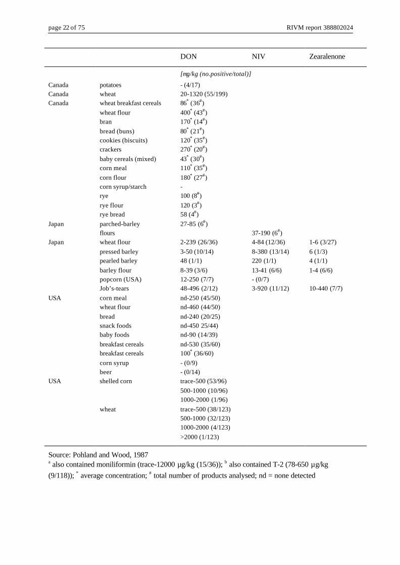
Although of all known trichothecenes only a few appear to be of importance with respect totheir actual presence in crops and animal feed (especially DON, NIV, T-2 and DAS, and to alesser extent certain derivatives thereof), they can cause problems to humans when ending upin human foodstuffs. These foodstuffs can be of plant-origin (cereals and processed grains) orof animal-origin (residues in meat, milk, eggs of animals given contaminated feed). The vastmajority of the confirmed cases of contamination of plant-based foodstuffs by trichothecenesinvolve DON in wheat or wheat products (Table 6), overall at concentrations <1 mg/kg, butoccasionally also well above 1 mg/kg. NIV has also been detected (particularly in Japan), butother type A or B trichothecenes have hardly been found. Frequently, there is concurrentexposure to more than one Fusarium mycotoxin (often DON and NIV, along withzearalenone) (Pohland and Wood, 1987; WHO, 1990).
Table 6 Global occurrence of trichothecenes in human food
DON NIV Zearalenone
[µg/kg (no.positive/total)]
Canada cornflakes 13-20 (1/1)USA corn meal 11-69 (9/10)France walnuts
sweetcorn 20000-500000 (33/33)50-450 (3/60)400-270000 (7/29)
South Africa corn a trace-820 (14/36) trace-240 (6/36) 20 (1/36)Taiwan corn (USA) b
corn (South Africa)95-312 (7/100)140 (1/5)
49-303 (7/73)503 (1/8)
page 22 of 75 RIVM report 388802024
DON NIV Zearalenone
[µg/kg (no.positive/total)]
Canada potatoes - (4/17)Canada wheat 20-1320 (55/199)Canada wheat breakfast cereals
wheat flourbranbread (buns)cookies (biscuits)crackersbaby cereals (mixed)corn mealcorn flourcorn syrup/starchryerye flourrye bread
86* (36#)400* (43#)170* (14#)80* (21#)120* (35#)270* (20#)43* (30#)110* (35#)180* (27#)-100 (8#)120 (3#)58 (4#)
Japan parched-barleyflours
27-85 (6#)37-190 (6#)
Japan wheat flourpressed barleypearled barleybarley flourpopcorn (USA)Job’s-tears
2-239 (26/36)3-50 (10/14)48 (1/1)8-39 (3/6)12-250 (7/7)48-496 (2/12)
4-84 (12/36)8-380 (13/14)220 (1/1)13-41 (6/6)- (0/7)3-920 (11/12)
1-6 (3/27)6 (1/3)4 (1/1)1-4 (6/6)
10-440 (7/7)USA corn meal
wheat flourbreadsnack foodsbaby foodsbreakfast cerealsbreakfast cerealscorn syrupbeer
nd-250 (45/50)nd-460 (44/50)nd-240 (20/25)nd-450 25/44)nd-90 (14/39)nd-530 (35/60)100* (36/60)- (0/9)- (0/14)
USA shelled corn
wheat
trace-500 (53/96)500-1000 (10/96)1000-2000 (1/96)trace-500 (38/123)500-1000 (32/123)1000-2000 (4/123)>2000 (1/123)
Source: Pohland and Wood, 1987a also contained moniliformin (trace-12000 µg/kg (15/36)); b also contained T-2 (78-650 µg/kg(9/118)); * average concentration; # total number of products analysed; nd = none detected

RIVM report 388802024 page 23 of 75
Some recent data on the occurrence of DON in plant-based foodstuffs in the Netherlands aregiven in Table 7.
Table 7 Occurrence of DON in human food in the Netherlands
Year DON
[mean/max in µg/kg(no.positive/total)]
Wheat productsWheatWheat flourMaize and by-productsMaltVarious grains and by-productsVarious grains (not wheat)
1999199919991999199920002000
134/250 (18/20)358/1900 (50/54)193/460 (20/24)110/130 (3/3)- (0/2)55/570 (80/171)40/385 (5/25)
Source: WHO/FAO, 2001
In contrast to plant-based foodstuffs, there is almost no carry-over of trichothecenes into foodof animal origin (meat and edible tissues, milk, eggs). Data available mostly concern DON,and indicate that either no or only very low residual amounts of DON can be detected intissues of poultry and swine, in eggs and in milk. DON is not transferred into milk from cattledue to de-epoxidation of DON by rumen microorganisms. This might result in low levels ofde-epoxy-DON in milk. Due to the fast and extensive metabolism and excretion it is likelythat DON and related trichothecenes will not accumulate in the exposed animals, and thattransfer into animal-derived products will be marginal (Prelusky, 1994; Spahr et al., 1999).Indeed, in a recent investigation in Germany, no DON or other type A and B trichotheceneswere detected in food from cattle, pigs and poultry. It was concluded that trichothecenes arenot important as possible contaminants in food from animal origin and as such are not ofpractical relevance from the view of food hygiene and food safety (Gareis and Wolff, 2000).
3.5 Fate during processing
The data that are available on the influence of food processing operations on the stability oftrichothecenes mostly concern DON. Cleaning and milling of wheat are not effective incompletely removing DON, which concentrates in the bran. The milling of corn also has littleeffect on DON content. In dry-milling of corn, DON concentrates in the germ meal, which isused primarily for animal feed. In wet-milling, DON concentrates in the steep liquor butsmall amounts are also retained in the starch. Baking does not result in DON-free products:depending on the baking conditions, a reduction of 0-49% in DON-content has been observed

page 24 of 75 RIVM report 388802024
(Scott, 1984). However, when baking yeast-containing products, the DON-content increased,which was attributed to enzymatic conversion of DON precursors (Charmley and Prelusky,1994; Smith et al., 1994). In baking Japanese bread, a reduction of 33-60% was reported fortrichothecenes other than DON (not specified). On cooking wheat flour into Japanese noodlesa loss of 32-59% of trichothecenes (not specified) was observed, while losses were 68-100%for Chinese noodles. In the latter process, sodium carbonate was used as food additive (Scott,1984). During tortilla fabrication, at which corn is first boiled in calcium hydroxide, DONand 15-Ac-DON were not stable: only 18-28% of DON was retained, while 15-Ac-DON wasdestroyed completely (Abbas et al., 1988).In heat stability experiments Kamimura et al. (1987) found that trichothecenes of the NIV-and T-2 type decomposed with increasing temperature, but not completely. A combination ofphysical and chemical treatment has been reported to be effective in reducing DAS in animalfeed: upon treatment with calcium hydroxide monomethylamine, a greater reduction was seenat increased temperatures and moisture content (Bauer et al., 1987).The use of gamma-irradiation to destroy preformed toxins present in grain does not appear tobe a suitable method for detoxification of low moisture grains contaminated with DON and3-Ac-DON. The high dose required for their destruction on maize (>50 kGy) makesirradiation unsuitable for practical use (O’Neill et al., 1993).As contaminated grain may contain a wide variety of mycotoxins of differing chemicalcharacteristics (including heat stability, solubility and adsorbent affinity), Charmley andPrelusky (1994) concluded that it is difficult to develop a decontamination method (eitherphysical, chemical or biological, or a combination) that will be equally effective against eachmycotoxin present. Moreover, this method should also be reliable, cost-effective andcommercially applicable, constituting an extra problem.

RIVM report 388802024 page 25 of 75
4. Toxicology
To put the available information on the toxicology of the six trichothecenes subject of thisreport into perspective, first an overview on trichothecenes in general is given in chapter 4.1.This overview is a short compilation of data published in a number of reviews and books(a.o. Ueno 1983a; Ueno, 1987a/b; Hsieh, 1987; WHO, 1990; IARC, 1993; Smith et al., 1994;Bondy and Pestka, 2000). Data specific for NIV, FusX, DAS, 3-Ac-DON, 15-Ac-DON andNeoSol are presented in chapters 4.2 - 4.7.
4.1 Trichothecenes in general
4.1.1 Toxicokinetics
Most data available on the toxicokinetics of trichothecenes concern T-2 and, to a lesserdegree, DON and DAS. In general, trichothecenes are well absorbed from the digestive tractafter which they are widely distributed in many tissues and organs. The main metabolicpathways are conjugation and de-epoxidation, while for some trichothecenes alsodeacetylation and hydroxylation play a role. For deacetylation the substituents at C3 and C8play an important role. For example, for deacetylation at C4 it is essential that there is anhydroxyl group at C3 and not an acetyl group, while at the same time the substituent at C8should be of hydrophobic (acyl, H or =O) in stead of hydrophylic nature (OH).The metabolites produced are generally less toxic than the corresponding parent toxins.
De-epoxidation is the most important step in the detoxification of trichothecenes. It is carriedout by microorganisms in the gastrointestinal tract, and in cattle also by the ruminal flora.The ability to de-epoxidate trichothecenes can be acquired either by an increase in thenumber of certain microorganisms or an increase in their capacity to metabolisetrichothecenes. Trichothecenes have been shown to change the composition of thegastrointestinal microflora (Tenk et al., 1982). It can take a few days before thegastrointestinal flora is adapted to form de-epoxide metabolites. The initial inability tode-epoxidate trichothecenes might be an explanation for the higher toxic effects often foundat the start of feeding trichothecenes. Some animal species (amongst which chickens) lack thenecessary microflora for epoxide reduction, and they are likely to be more sensitive totrichothecenes.
4.1.2 Toxicity
4.1.2.1 Toxic effects and mode of actionTrichothecene compounds produce a variety of toxic symptoms in humans and animals,including mortality, skin and gastrointestinal irritation or necrosis, haematological disorders

page 26 of 75 RIVM report 388802024
(a.o. initial leucocytosis followed by leukopenia), diarrhoea, vomiting and feed refusal,decreased body weight gain, damage to the haematopoietic systems in bone marrow, spleen,thymus and lymph nodes, and immunological alterations. As to the latter, trichothecenes canboth suppress and stimulate immune function, resulting in either impaired resistance toinfection or neoplasia, or in hypersensitivity or auto-immune like disorders.
Biochemically, trichothecenes are highly toxic at the subcellular, cellular and organ level.They are very cytotoxic to eukaryotic cells, causing cell lysis and inhibition of mitosis.Trichothecenes are also very potent inhibitors of protein and DNA and RNA synthesis, andthey can interact with the cell membrane. Toxicity at the subcellular level is largely due totheir ability to inhibit protein synthesis and to covalently bond to sulphydryl groups.The mechanism of protein inhibition can be of two types. One is inhibition of the initial stepof protein synthesis (I-type; examples are T-2, HT-2, DAS, NIV and FusX) and the otherinhibition of the elongation-termination step (ET-type; example is DON). It is considered thatpeptidyl transferase is inhibited with subsequent inhibition of peptide bond formation. Thetarget organell of trichothecene action is the 60S subunit of eukaryotic ribosomes, the proteininhibition activity correlating well with ribosome affinity.The inhibition of DNA and RNA synthesis by trichothecenes require higher toxinconcentrations than the inhibition of protein synthesis and the extent of inhibition is muchless. The suppression of nucleic acid synthesis is, however, not simply a secondary effect ofthe inhibition of protein synthesis. It is assumed that damage to membrane structure andcytoskeleton components affect macromolecule synthesis.
Being potent inhibitors of protein and DNA and RNA synthesis, trichothecenes are especiallytoxic to tissues with a high cell division rate. As the character of trichothecene-inducedlesions (necrosis, karyorrhexis) in actively dividing cells of thymus, spleen, bone marrow,ovary, testes, lymph nodes and intestinal mucosa is very similar to those induced byradiation, trichothecenes are classed as radiomimetic substances.
Given the above, leucocytes and the immune system are the primary target for trichothecenes.In vitro and in vivo studies have demonstrated that trichothecenes can affect leucocytes byderegulating cytokine production and by inducing apoptosis. The former is induced byexposure to low levels of trichothecenes, with immune stimulation as net effect. Higher levelsof trichothecenes promote apoptosis, with immune suppression as net effect.
4.1.2.2 Structure-activity relationshipsThe minimum structural feature required for biological activity of the trichothecenes is thepresence of the 12,13-epoxytrichothecene skeleton. Reduction of the epoxide leads toinactive derivatives, while hydrogenation of 9,10-double bonds and rearrangement of thetrichothecene skeleton to the apotrichothecene ring system lead to a substantial loss ofactivity. The biological activity of trichothecenes is also affected by the nature of the sidechains at specific positions.

RIVM report 388802024 page 27 of 75
Some examples of the cytotoxic and biochemical properties of trichothecenes are given inTable 8. Although there is not always a good match between the relative toxicity in vitro andin vivo, the potency ranking in vitro and in vivo is more or less the same. The potency of thetrichothecenes depends on the modifications of the side chains in the molecule. In general,the macrocyclic trichothecenes (type D) are the most potent, followed by type A, type B andtype C trichothecenes. Within the type A trichothecenes, substances with an acetyl group atR3 are the most potent. Removal of this acetyl group results in a pronounced decrease inpotency, while removal of the acetyl group at R2 and the sidechain at R5 result in a smallerloss of activity. In contrast, addition of an acetyl group at R1 also results in a loss of potency.Within the type B trichothecenes, the potency is mainly influenced by the substituent at R2,the potency decreasing in the order from acetyl > hydroxyl > hydrogen. Also for type Btrichothecenes, addition of an acetyl group at R1 results in a loss of potency (Thompson andWannemacher, 1986; Eriksen and Alexander, 1998).
Table 8 Cytotoxicity and protein synthesis inhibition of trichothecenes in cultured human a
and animal cells
HeLa 1 HEK 1 HL 1 HEp2 1 Rabbit reti-culocytes 1
Vero cells b,2 Rat spleenlymphocytes 2
(ID50 in µg/ml) [ID50 in nM (relative potency c)]
Type A T-2 0.01 0.02 0.003 0.001 0.03 14 (100) 6 (100) HT-2 0.01 0.1 0.01 0.03 65 (22) 10 (63) DAS 0.01 0.01 0.001 0.03 27 (53) 12 (53) NeoSol 0.1 0.06 0.05 0.25 273 (5.2) 127 (4.8)
Type B DON 1.0 3.0 0.5 0.250 2.0 1499 (0.95) 850 (0.72) NIV 0.3 1.0 0.3 0.225 3.0 8131 (0.18) 6835 (0.089) FusX 0.1 1.0 0.3 0.25 288 (5.0) 91 (6.7) 3-Ac-DON 10 10 10 10 26279 (0.054) 4293 (0.14) 15-Ac-DON -------------------------------------------- no data available ----------------------------------------
Type C Crotocin 0.5 0.6 2.0 0.25 1
Type D verrucarin A 0.005 0.002 0.003 0.001 0.01 12 (118) 3 (191) roridin A 0.003 0.003 0.003 0.01 12 (117) 5 (115)
a origin: uterine carcinoma (HeLa), embryonic kidney (HEK), lymphocytes (HL), epidermoidcarcinoma (HEp2); b origin: monkey kidney; c compared to T-2, set as standard having 100% potencySource: 1 Ueno (1983a,b), 2 Thompson and Wannemacher (1986)

page 28 of 75 RIVM report 388802024
Trichothecenes inhibit the protein synthesis by binding to the ribosomes according to thesubstituents at C3 (R1) and C4 (R2). Trichothecenes with substituents at both C3 and C4inhibit mainly polypeptide chain initiation while trichothecenes lacking substituents at one orboth positions mainly inhibit elongation (Eriksen and Alexander, 1998).
4.1.2.3 Animal and human mycotoxicosesFor many years, Fusarium species have been known to be associated with a number ofhuman and animal toxicoses. However, only rarely a direct connection with the mycotoxin(s)involved has been established factually.
Animal mycotoxicoses associated with trichothecene-producing Fusarium species includeamongst others the haemorrhagic syndrome (F. sporotrichioides and F. poae), Akakabi-byo(red mould disease or scabby grain intoxication; F. graminearum), feed refusal and emeticsyndromes (F. graminearum), ill-thrift, oral and other gastrointestinal lesions. Naturaloutbreaks have been reported to occur all over the world, thereby severely compromisinglivestock health, welfare and productivity (Nelson et al., 1994; D’Mello et al., 1999).
Mycotoxin outbreaks affecting humans have mainly been reported to occur in the formerSoviet Union and Asia (Japan, China, Korea), both historically and more recently. Historicaloutbreaks associated with Fusarium species include alimentary toxic aleukia(F. sporotrichioides and F. poae; closely related to the haemorrhagic syndrome in animals),Urov or Kashin-Beck disease (F. poae) and Akakabi-byo (F. graminearum). Although thereis a strong suspicion of involvement of certain type A (T-2, DAS) and/or type Btrichothecenes (DON, NIV, FusX) in these diseases, none has been positively identified.More recent outbreaks occurred in China and in India, in total affecting thousands of people.The mouldy cereals causing poisoning in China in 1984/5 contained DON and zearalenonebut no T-2 or NIV. Symptoms included nausea, vomiting, abdominal pain, diarrhoea,dizziness and headache; no death occurred. In 1987 in India, bread made from mouldy flourcaused intoxication (abdominal pain, throat irritation, diarrhoea, blood in stools and vomiting;no fatalities occurred). The mouldy flour contained DON, Ac-DON (not specified), NIV andT-2.In corn and in wheat and barley samples implicated in two poisoning outbreaks in China in1989 and 1991, DON was the predominant toxin, followed by zearalenone and NIV.3-Ac-DON and 15-Ac-DON were only detectable in highly contaminated wheat and corn,respectively. Corn contained also fumonisins. FusX, T-2 and HT-2 were not present. In the1991 outbreak about 130000 people were affected by gastrointestinal disorders, includingabdominal pain and fullness, nausea, vomiting, fatigue and fever (Li et al., 1999).
IARC (1993) described studies addressing the relationship between exposure to Fusariumtoxins and oesophageal cancer in the Transkei (South Africa) and in Linxian (China). Moststudies pointed to a co-existence of a mixture of toxins from several Fusarium species(DON, zearalenone, NIV, 3-Ac-DON and/or 15-Ac-DON) on maize, the main dietary staple.

RIVM report 388802024 page 29 of 75
According to IARC the studies suggested no correlation with the above mentionedtrichothecenes.
4.1.2.4 Co-exposureCereal grains, animal feed and human foodstuffs are quite often contaminated with more thanone mycotoxin, not only derived from Fusarium fungi but also from other fungi like forexample Aspergilli. Hence, in practice exposure of animals and humans will be to a mixtureof mycotoxins. Research, however, has mainly concentrated on the toxicity of some singlepure mycotoxins. Combined effects (antagonistic, additive and/or synergistic) of two or moremycotoxins, as well as toxicological effects at the low levels as usually found incontaminated feed and food, have received little attention.
4.2 Nivalenol (NIV)
4.2.1 Toxicokinetics
4.2.1.1 Absorption, distribution and excretionWithin 72 hrs after a single oral dose of 5 mg NIV/kg bw to male rats, 24% of the dose wasexcreted as NIV and de-epoxy-NIV via faeces and 15.5% via urine. In serum, liver andkidney the levels of NIV were lower than in urine or faeces even 1 h after dosing, and theywere rapidly lowered. No de-epoxy-nivalenol was detected in serum, liver or kidney within24 hrs after dosing (Onji, 1990).After repeated oral administration of 5 mg NIV/kg bw to male Wistar rats (12 times at 2- or3-day intervals), excretion was also mainly via faeces (>87%) (Onji et al., 1989).
In male swine given NIV in the diet at a dose of 0.05 mg/kg bw, twice daily for three days,NIV was already detected in blood samples taken 20 min after the start of feeding. During thefirst 7.5 hrs, 11-43% of the NIV dose was absorbed. Blood peak concentrations of3-6 ng NIV/ml were reached within 2.5-4.5 h after feeding. Sixteen hours after feeding, NIVwas still being absorbed from the intestine, with blood concentrations of 1-3 ng NIV/ml.Hence, absorption was rapid and extensive. NIV was mainly excreted in faeces, withconcentrations up to 3.2 mg/kg. Only 17% was excreted via urine (Hedman et al., 1997a).
4.2.1.2 MetabolismWhen incubated for 48 h with cow ruminal fluid, 80% of NIV was de-epoxidated. The sameresult was obtained for DON (Hedman and Pettersson, 1997).
After repeated oral administration, the main metabolite of NIV in the excreta of male rats wasde-epoxy-NIV (80% and 1% of the total dose in faeces and urine, respectively). The parentcompound NIV was detected at much lower levels (7% and 1% of the total dose in faeces and

page 30 of 75 RIVM report 388802024
urine, respectively). Gastrointestinal microorganisms likely participate in the de-epoxidation(Onji et al., 1989). After single oral dosing de-epoxy-NIV was excreted predominantly infaeces (18.7% vs. 5.6% in urine), and the excretion was about 24 h later than that of NIV(9.9% in urine and 5.4% in faeces) (Onji, 1990).
When male swine were fed for up to one week with NIV in the diet at a concentration of2.5 mg/kg (corresponding to a dose of 0.1 mg/kg bw/day), no metabolites of NIV could befound in plasma, urine or faeces, either as glucuronic acid or sulphate conjugates, or asde-epoxy-NIV, indicating a lack of metabolism (Hedman et al., 1997a). However, when maleswine were fed diets containing 2.5 or 5 mg/kg NIV for up to three weeks, from one week ofexposure onwards, over 90% of NIV in faeces was in the de-epoxy-NIV form. Also in bile32-44% of NIV was in the de-epoxy form. Apparently, it takes a few days for thegastrointestinal microflora to adapt itself to form de-epoxide metabolites. Once adapted, thesemicroorganisms were also able to produce de-epoxidated metabolites of DON in vitro(Hedman and Pettersson, 1997; Pettersson and Hedman, 1997).
In contrast to rats and pigs, in faeces of male broiler chickens fed NIV in the diet at 2.5 or5 mg/kg for three weeks, no de-epoxy-NIV was found. An unknown metabolite was found,though, in varying amounts. Presumably this is an acetylated metabolite of NIV (Hedman andPettersson, 1997).
4.2.2 Toxic effects
4.2.2.1 Acute toxicityIn 6-week old male ddY mice NIV appeared to be less toxic after oral administration (LD50 of38.9 mg/kg bw) than after intraperitoneal (ip), intravenous (iv) or subcutaneous (sc)administration (LD50-values of 7.4, 7.3 and 7.2 mg/kg bw, respectively). Independent ofroute, most deaths occurred within three days after administration. Histopathology revealed amarked congestion and haemorrhage in the intestines (Ryu et al., 1988). When given ip or scto male Swiss mice, the LD50-values for NIV were 9.6 and 7.3 mg/kg bw, respectively(Thompson and Wannemacher, 1986). Ueno (1984) reported LD50-values in 6-week old maleddyS mice of 4.1 and 6.3 mg/kg bw after ip and iv administration, respectively. Postmortemexamination revealed extensive haemorrhages in the intestine and diarrhoea, and cellulardestruction in the epithelial mucous membranes of intestine, in the thymus and testis. Anidentical ip LD50 of 4.1 mg/kg bw in male mice was found by Ueno et al. (1973a), with NIVinducing radiomimetic cellular injury and karyorrhexis in (amongst others) the small intestineand bone marrow.For male and female F344 rats, an oral LD50 of 19.5 mg/kg bw has been reported for NIV byKawasaki et al. (1990). Effects observed were sedation, eyelid closure, staggering gate,diarrhoea and congestion of the lungs and digestive tract. Ueno (1983a) reported an LD50 of0.9 mg/kg bw after sc administration of NIV to rats.

RIVM report 388802024 page 31 of 75
4.2.2.2 Subacute toxicityWhen administered NIV in the diet at concentrations of 0, 5, 10 or 30 mg/kg for 24 days, feedutilization efficiency and body weight gain of female C57BL/6CrSlc spf mice were dose-relatedly (but not statistically significantly) reduced at 10 and 30 mg/kg. At 30 mg/kg, micedeveloped significant erythrocytopenia and slight leukopenia, and upon ultrastructuralexamination bone marrow cells revealed polyribosomal breakdown. No marked changes wereobserved on haemoglobin and haemotocrit and on weights of the liver, spleen and thymus.No histopathological changes were found in thymus, spleen, liver, stomach, ovary, uterus,lymph node and small intestine with or without Peyer’s patches (Ryu et al., 1987).
Oral gavage administration of 0, 0.4 or 2 mg NIV/kg bw to male and female F344 rats for 15or 30 days did not affect haematological or biochemical parameters. At 2 mg/kg bw the liverand spleen weights were significantly increased, but no histopathological changes were seen(Kawasaki et al., 1990).
Groups of 5 male SD rats were administered 0, 6, 12 or 30 mg NIV/kg diet for 2-4 weeks.The highest dose caused mortality within 6-8 days due to marked feed refusal and reducedbody weight gain. Reductions in feed consumption and body weight gain were also observedat 6 and 12 mg/kg, but only in the first 1-2 weeks. After 2 weeks of treatment, absolute andrelative weights of liver and spleen were decreased at 12 mg/kg, while kidney weights werenot affected. In contrast, after 4 weeks of treatment, absolute and relative weights of liver andkidney were increased at 12 mg/kg and even more so at 6 mg/kg, while spleen weights werestill decreased at 12 mg/kg but less than after 2 weeks. Among the serum parametersinvestigated, asparate aminotransferase was dose-relatedly decreased (statisticallysignificantly only at 12 mg/kg) (Yabe et al., 1993). Effects on hepatic drug-metabolizingacitivity and aflatoxin B1 metabolism are described in 4.2.2.8.
Administration of 0, 10 or 50 mg NIV/kg diet to male rats for 8 weeks only affected the highdose animals. They showed decreased body weight gain, weak erythrocytopenia, mucosalnecrosis and disruption of the gut epithelium, and an increase of erythroid series in bonemarrow (Onji, 1990).
The feeding of NIV at a dose of 0.05 mg/kg bw, twice daily for 20 days, did not cause feedrefusal in male swine nor changes in clinical plasma parameters (Hedman et al., 1997a).When groups of 6 male swine were fed with 2.5 or 5 mg NIV/kg diet for up to three weeks(corresponding to doses of 0.1 and 0.2 mg NIV/kg bw/day), no effects were observed onbehaviour or appearance, on body weight or weight gain, on food consumption or on theweights of liver, kidney, thymus, spleen and heart. No swine vomited. However, in contrastto control swine, swine of both treated groups showed mild pathological changes in thegastrointestinal tract (changes in thickening of mucosa and haemorrhagic lesions), kidneys(pale, with narrow cortex and dilated renal pelvis, and cysts) and spleen (apex infarcts).Between control and treated swine there were no differences in total or differential blood
page 32 of 75 RIVM report 388802024
leucocyte counts, nor in the number of thymocytes or in plasma cortisol levels. Treatmentwith NIV, however, induced a dose-dependent decrease in spleen cells (statisticallysignificant only at 5 mg/kg), which was reflected in decreased numbers (but not proportions)of CD4+, CD8+ and IgM+ subpopulations in the spleen (Hedman et al., 1997b). Effects onplasma immunoglobulin levels and on immune function in vitro are described in 4.2.2.7.
Dietary treatment of 7-day old male broiler chickens with 0.5 – 12 mg NIV/kg for 20 daysresulted in decreased body weight gain (by 11%) at 6 and 12 mg/kg. At these dosages feedconsumption was also decreased (by 6-7%), while feed conversion efficiency was increased(by approximately 5%). Gizzard erosions were found in 33% of the birds fed 12 mg/kg and in8% of those fed 3 or 6 mg/kg, but not in controls. Absolute and relative liver weights weredecreased at 6 and 12 mg/kg. Although no effects were found on relative weights of bursa,spleen and gizzard, for the latter the absolute weight was decreased at 3 and 12 mg/kg. Inblood, no change compared to control was found in haematocrit or in the plasmaconcentration of glucose, calcium, cholesterol, triglycerides and uric acid, or in the plasmaactivity of aspartate amino transferase, alanine amino transferase or gamma glutamyltranspeptidase. Histopathology of liver, thymus and spleen revealed no treatment-relatedchanges (Hedman et al., 1995).
4.2.2.3 Subchronic toxicityGroups of 20 male and 20 female C57BL/6CrSlc spf mice were given diets containing0, 6, 12 or 30 mg/kg NIV for 12 weeks, with an interim kill of 10 animals/sex/group after4 weeks. Observations included body weight and feed consumption, serum biochemistry,organ weights (liver, thymus, spleen and kidneys) and histology (liver, thymus, spleen,kidneys, stomach, adrenal glands, pituitary gland, ovaries, sternum, femurs, mesenteriallymph node, brain and small intestines with or without Peyer’s patches).NIV treatment caused a dose-related reduction in body weight gain in both males andfemales: after 12 weeks the weight gain at 6, 12 and 30 mg/kg, as compared to that ofcontrols, was 98, 77 and 30% for males and 70, 55 and 45% for females, respectively. Feedconsumption was also reduced, and in particular the high dose animals showed slight feedrefusal in the first week. Absolute and relative weights of liver, kidney, spleen and thymuswere variably affected by NIV treatment, without clear trends. No gross or histopathologicalchanges were seen in the tissues and organs examined but treated groups had considerablyless fatty tissue at autopsy than controls. Among the serum parameters examined, only onewas consistently affected: in both males and females the alkaline phosphatase activity wasdose-relatedly increased at all doses after 4 and after 12 weeks (Yamamura et al., 1989).
Groups of 12 female C57BL/6CrSlc spf mice were given diets containing 0, 6, 12 or30 mg/kg NIV for 1 year (equal to 0, 0.68-0.76, 1.51-1.64 or 3.84-3.95 mg NIV/kg bw/day),with an interim kill of 6 animals/group after 6 months. Observations included body weightand feed consumption, haematology, organ weights (liver, thymus, spleen and kidneys),histology (liver, thymus, spleen, kidneys, stomach, adrenal glands, pituitary gland, ovaries,

RIVM report 388802024 page 33 of 75
sternum, bone marrow, lymph node, brain and small intestines with or without Peyer’spatches) and ultrastructural studies of the bone marrow.Body weight gain was dose-relatedly reduced (after 1 year, the weight gain at 6, 12 and30 mg/kg was 79, 69 and 40% of that of controls, respectively). Feed efficiency was alsodose-relatedly reduced. Feed refusal was only seen at 12 and 30 mg/kg in the first 3-5 weeks.Feed consumption was reduced at all doses, but only marginally (by <10%) at 6 and12 mg/kg. Both after 6 months and after 1 year of treatment, absolute weights of liver, kidneyand thymus were dose-dependently decreased (statistically significant at 30 mg/kg), whilerelative weights of these organs plus spleen were dose-dependently increased (statisticallysignificant at 12 and/or 30 mg/kg). The organ weight changes were not accompanied byhistopathological changes. NIV caused a severe leukopenia, which after 6 months was onlyobserved at 30 mg/kg but after 1 year at all doses. Other haematological parameters were notaffected. In contrast to the findings in the subacute feeding study (Ryu et al., 1987), noerythrocytopenia and no damage to the bone marrow were observed (Ryu et al., 1988).
4.2.2.4 Chronic toxicity/CarcinogenicityGroups of 42 female C57BL/6CrSlc spf mice were given diets containing 0, 6, 12 or30 mg/kg NIV for 2 years (equal to 0, 0.66, 1.38 or 3.49 mg NIV/kg bw/day). Observationsincluded body weight and feed consumption, clinical signs and mortality, haematology andserum biochemistry at autopsy, organ weights (liver, thymus, spleen, kidneys and brain) andhistology (on all tumours and on liver, thymus, spleen, kidneys and brain).During the dosing period body weight gain and feed efficiency were dose-relatedly reduced.However, due to the earlier death of the control and low dose animals (see below), the bodyweight changes were not apparent from the terminal weights. Initial feed refusal was seen at12 and 30 mg/kg. Feed consumption was reduced at all doses, but only marginally (by <10%)at 6 and 12 mg/kg. No significant changes in relative organ weights were observed.Significant decreases in absolute organ weight were found for liver (at 30 mg/kg) and kidney(at 12 and 30 mg/kg). In contrast to the 1-yr study (Ryu et al., 1988), only slight leukopeniaaccompanied by a slight decrease in lymphocytes was seen (at 12 and 30 mg/kg, but not at6 mg/kg). Dose-related increases in alkaline phosphatase activity, aspartate aminotransferaseand non-esterified fatty acids, and decreases in alanine aminotransferase, amylase, creatininephosphokinase and calcium were observed, but the changes were statistically significant onlyat 30 mg/kg. Compared to controls, NIV treatment increased the lifespan. Except foramyloidosis (especially in the small intestine) and tumour development (especiallylymphomas) no histopathological changes were observed. Controls and low dose animals hadthe highest incidence of amyloidosis, and this was a major cause of (early) death in thesegroups. Tumour development was not related to NIV treatment. Tumours occurred in similarincidences in control and treated groups, but appeared to develop later and grow slower at30 mg/kg. The low mortality rate at 30 mg/kg might be due to the lower incidence ofamyloidosis and the lower tumour incidence in the earlier part of the study (Ohtsubo et al.,1989). This study was also evaluated by IARC (1993), and they noted the limited number oftissues studied.

page 34 of 75 RIVM report 388802024
One-week old C57BL/6xC3H F1 mice received a single ip injection with 6 mg/kg bwaflatoxin B1 (AFB1), 6 weeks later followed by dietary treatment with 0, 6 or 12 mg/kg NIVfor 1 year. The mice were killed at 71 weeks of age. All male mice treated with AFB1developed liver tumours (mainly carcinomas), and feeding with NIV did not affect theincidence. The incidence of liver tumours and their degree of malignancy was much lower infemale mice treated with AFB1, and NIV treatment had a tumour-suppressing effect onAFB1-induced hepatocarcinogenesis, presumably by acting on the promotion step (Ueno etal., 1991).
In an in vivo medium-term bioassay using glutathione S-transferase placental form (GST-P)-positive liver foci as endpoint in six-week old male F344 rats initiated by diethylnitrosamine(DEN), Ueno et al. (1992) investigated whether NIV possessed hepatocarcinogenic potential,and secondly, whether AFB1-induced hepatocarcinogenicity was modulated by exposure toNIV. In the first experiment, treatment with AFB1 (1x ip 0.5 mg/kg bw) resulted in a smallformation of GST-P-positive liver foci in non-DEN-initiated rats, and this induction wasaccelerated in DEN-initiated rats (1x ip 200 mg/kg bw), confirming the hepatocarcinogenicityof AFB1. No induction of GST-P-positive liver cell foci by NIV (6 mg/kg diet for 6 weeks)was observed in rats either or not initiated with DEN, hence predicting no hepatocarcinogenicpotential for NIV. However, feeding of 6 mg/kg NIV for 6 weeks to DEN-initiated andAFB1-treated rats synergistically enhanced the formation of GST-P-positive liver cell foci(not in non-DEN initiated and AFB1-treated rats). Hence, NIV is predicted to cause anenhancing effect on AFB1-induced hepatocarcinogenesis.
4.2.2.5 GenotoxicityWhen studied for chromosomal damage, induction of sister chromatid exchanges and cellcycle delay in Chinese hamster V79-E cells in vitro, NIV showed only weak activity, more sowith metabolic activation than without. The marginal effects observed are probablyunspecific and are caused by inhibition of protein synthesis (Thust et al., 1983). An inductionof chromosomal aberrations in Chinese hamster V79 cells in vitro was also observed by Hsiaet al. (1988) when testing NIV (both pure and extracted from corn), but not by Ryu et al.(1993).In the alkaline single-cell gel electrophoresis (or COMET) assay, NIV was tested for DNAdamage in vitro in Chinese hamster ovary cells (without metabolic activation) and in vivo inmale ICR mice after oral and ip administration (using seven organs: stomach, jejunum, colon,liver, kidney, thymus and bone marrow). NIV was positive in vitro. In vivo, DNA damagewas seen in colon (both after ip and oral treatment), in kidney, bone marrow, stomach andjejunum (after oral treatment only), but not in liver and thymus. NIV showed organ specificgenotoxicity in time and intensity: in the colon DNA damage was strong yet delayed, whileDNA damage in stomach, kidney and bone marrow was strong at the onset but decreasedwith time. The in vivo findings were not secondary to cytotoxicity, as no apoptotic cells and

RIVM report 388802024 page 35 of 75
no histopathological findings including necrotic changes were detected in any organ (Tsudaet al., 1998).
4.2.2.6 Reproductive and Developmental toxicityIn a study by Ito et al. (1986) pure NIV was injected ip in pregnant ICR mice at dose levelsof 0, 0.1, 0.5 or 1.5 mg/kg bw/day on days 7-15 of gestation. The highest dose causedstillbirths after vaginal haemorrhage in 6 out of 10 animals. High embryolethality wasrecorded in the two highest dose groups (88 and 48%). No fetal malformations were observedin the treated groups. A single administration of 3 mg/kg bw on day 7 affected the embryowithin 10 h, damaged the placenta within 24 h, and caused stillbirths at 48 h.
Mice were fed diets with mouldy rice powder containing NIV at final levels of 0, 6, 12 or30 mg/kg feed throughout gestation. Additional mice were administered purified NIV bygavage at doses of 0, 1, 5, 10 or 20 mg/kg bw on days 7-15 of gestation. Four of five micegiven 20 mg/kg bw died during the dosing period. Embryotoxicity associated with maternalweight loss was observed in the groups receiving 30 mg/kg and 10 mg/kg bw. Intrauterinegrowth retardation was found in these groups, and also in the term fetuses of mice exposed to12 mg/kg and 5 mg/kg bw. NIV had no statistically significant adverse effects on theincidence of gross, skeletal and visceral malformations (Ito et al., 1988).
4.2.2.7 ImmunotoxicityNIV given in the diet at concentrations of 0, 6 or 12 mg/kg for 4 or 8 weeks reproduciblyinduced some pathological changes in mice which resemble those in human IgAnephropathy: irrespective of mice strain (C3H/HeN, C3H/HeJ or BALB/c), NIV induced IgAdeposits in the glomerular mesangium and elevated IgA levels, and the degree of thesechanges was associated with the dose and duration. As it is known that IgA production in thebody is primarily by the mucosal immune system, it is hypothesized that NIV reaches the gutof mice to dysregulate their mucosal immune system resulting in the pathogenesis of IgAN(Hinoshita et al., 1997).
Depending on dose and time, NIV and other trichothecenes like T-2, DON, 3- and15-Ac-DON, could inhibit or superinduce cytokine production and mRNA expression inmurine spleen CD4+ T cells. The rank order of superinduction was T-2 > DON, NIV >15-Ac-DON > 3-Ac-DON (Ouyang et al., 1995).
Dietary treatment of male swine with 2.5 or 5 mg NIV/kg diet for up to three weeks(corresponding to doses of 0.1 and 0.2 mg NIV/kg bw/day) did not affect IgA and IgGplasma levels at any of the samplings (after 0, 1 and 3 weeks of treatment). The in vitroproduction of both IgG and IgA by mitogen-stimulated lymphocytes was not affected byNIV, and neither was the mitogen-induced proliferation of lymphocytes from blood, spleen orthymus. Apparently, the functioning of the immune system was not affected by NIV. In the2.5 mg/kg feed group there were some time-dependent changes from the first to the third

page 36 of 75 RIVM report 388802024
week of exposure (decrease in plasma IgG and increase in IgA levels; the levels of IgA andIgG in plasma, though, did not significantly differ from controls at these samplings (seeabove)). NIV at 2.5 mg/kg feed also decreased the in vitro production of IgG by mitogen-stimulated splenocytes but not the in vitro production of IgA. However, the effects at2.5 mg/kg feed were not observed in the 5 mg/kg feed group (Hedman et al., 1997b).
NIV inhibited blastogenesis in cultured human lymphocytes. It produced a 50% inhibition of[3H]thymidine uptake in mitogen-stimulated human lymphocytes at a concentration of72 ng/ml. Other trichothecenes like T-2, HT-2, FusX, DON and 15-Ac-DON produced 50%inhibition in this system at concentrations of 1.5, 3.5, 18, 140 and 240 ng/ml, respectively.These results indicate that the lymphotoxicity of trichothecenes is related to the C4substituent: acetyl>hydroxyl>hydrogen. Acetylation at C15 slightly decreases lymphotoxicity(Forsell and Pestka, 1985; Forsell et al., 1985).
Thuvander et al. (1999) found that NIV effectively inhibited proliferation andimmunoglobulin production (IgA, IgG and IgM) in mitogen-stimulated human lymphocytesin a dose-dependent manner with limited variation in sensitivity between individuals.However, at low levels NIV exposure could also result in enhanced proliferative responses,as well as in elevated immunoglobulin production (especially IgA). NIV was about aseffective as DON, but less effective than T-2 (100-250 times more potent) and DAS (25-80times more potent). Combinations of NIV with T-2, DAS or DON resulted in additivetoxicity.
In an in vitro test with human peripheral blood mononuclear cells, NIV significantly anddose-dependently inhibited the T and B cell proliferation. This effect was also observed forT-2, FusX, DON and 3-Ac-DON, but hardly or not at all for α- and β-zearalenol. NIV alsosuppressed natural killer cell activity (dose-dependent) and inhibited antibody-dependentcellular cytotoxicity reaction. This was also the case for T-2 and DON, but was notinvestigated for the other mycotoxins (Berek et al., 2001).
When investigated in ovalbumin-specific T cell receptor αβ-transgenic mice (i.e. mice whichrespond with sensitivity to oral administration of ovalbumin as a dietary constituent byexhibiting an increase in serum IgE, the causative immunogloblin of food allergy), NIVinhibited antigen-induced total and antigen-specific IgE production in association withinhibition of IL-4 production and enhancement of IL-2 production (Choi et al., 2000).
4.2.2.8 Other effectsFor NIV, the lowest sc dose causing vomiting in ducklings was 1 mg/kg bw (Ueno, 1983a).
When applied to the shaved back skin of guinea pigs, NIV induced redness 2-10 hrs afterpainting (effective dose 10-8 mol). Compared to T-2, HT-2, DAS and type D trichothecenes(effective doses 10-10 - 10-11 mol), NIV was low in dermal toxicity (Ueno, 1984).

RIVM report 388802024 page 37 of 75
As can be seen from Table 8 in 4.1.2.2, NIV is cytotoxic to cultured cells and possessesprotein synthesis inhibiting activity. In HeLa cells, NIV inhibited multiplication at everyphase of the growth cycle. In a variety of systems (amongst which Ehrlich ascites tumourcells and HeLa cells), NIV has been found to inhibit not only protein but also DNA synthesis,while RNA synthesis was marginally affected (Saito and Ohtsubo, 1974).
After infection of human epidermoid carcinoma no.2 (HEp-2) cells with herpes simplex virustypes 1 and 2, NIV exerted antiviral activity by inhibiting plaque formation (EC50s of 120 and50 ng/ml, respectively). NIV affected virus replication after but not before or during virusadsorption to the host cells. The antiviral activity of NIV was less than that of FusX (EC50s of56 and 26 ng/ml, respectively) but greater than that of DON (EC50s of 160 and 94 ng/ml,respectively), suggesting that the potency is related to the C4 substituent:acetyl>hydroxyl>hydrogen. Possibly the inhibitory effect can be brought about by binding ofthe trichothecenes to the polyribosomes in the viral protein synthesis (Tani et al., 1995).
When given at 6 or 12 mg/kg feed for 2-4 weeks to rats, NIV induced the activities ofmicrosomal cytochrome P-450 (isozymes 2B1/2 and 1A2) and cytochrome b5, drug-metabolizing enzymes (aminopyrine-N-demethylase and aniline hydroxylase) and cytosolicglutathione S-transferase (isozyme 1-2). The induction of microsomal cytochromeP-4502B1/2 and the increase in microsomal drug metabolism was transient, as the levels at4 weeks were less than those at 2 weeks. This ability of NIV to induce both phase I and phaseII enzyme systems for drug metabolism resulted in a modulation of AFB1 adduction to DNAin vitro and in vivo. In vitro, the formation of AFB1-DNA adducts was increased usinghepatic microsomal preparations from rats fed NIV, whereas supplementation with cytosolprepared from NIV-treated rats reduced the microsomal potential for adduct formation. Invivo, the hepatic AFB1-DNA concentration was lower in NIV-treated rats than in controls(Yabe et al., 1993).
4.3 Fusarenon X (FusX)
4.3.1 Toxicokinetics
4.3.1.1 Absorption, distribution and excretionThirty minutes after sc administration of uniformly labelled 3H-FusX at 4 mg/kg bw to mice,activity was found in liver, kidneys, intestines, stomach, spleen, bile and plasma; none wasdetected in heart, brain or testis. The highest activity was found in the liver (3% of the dose).Twelve hrs after administration, no label was present in the organs, and 25% of the dose wasrecovered as metabolised forms of FusX in the urine (Ueno et al., 1971).

page 38 of 75 RIVM report 388802024
4.3.1.2 MetabolismFusX is deacetylated to NIV by rat and rabbit liver carboxy esterases (Ohta et al., 1978).
4.3.2 Toxic effects
4.3.2.1 Acute toxicityWhen given ip or sc to male Swiss mice, the LD50-values were 5.6 and 5.5 mg/kg bw,respectively (Thompson and Wannemacher, 1986). A slightly lower ip LD50 of 3.3 mg/kg bwin male mice was found by Ueno et al. (1973a), with FusX inducing radiomimetic cellularinjury and karyorrhexis in (amongst others) the small intestine and bone marrow. Ueno(1984) reported LD50-values in 6-week old male ddyS mice of 4.5, 3.4, 4.2 and 3.4 mg/kg bwafter oral, ip, sc and iv administration, respectively. Newborn mice, with an sc LD50 of0.23 mg/kg bw, were more susceptible than 6-week old mice. Postmortem examinationrevealed extensive haemorrhages in the intestine and diarrhoea, and cellular destruction in theepithelial mucous membranes of intestine, in the thymus and testis.FusX was also tested for acute toxicity in rats (LD50-values of 0.5 and 4.4 mg/kg bw for scand oral administration, respectively), guinea pigs (LD50-values of 0.5 and 0.1 mg/kg bw forip and sc administration, respectively), cats (sc LD50 <5 mg/kg bw) and ducklings(sc LD50 2 mg/kg bw) (Ueno, 1983a).
Early after injection of FusX in mice, a rapid increase of leucocytes and lymphocytes wasseen, accompanied by increased β-globulin and decreased γ-globulin. On autopsy, cellulardegeneration and karyorrhexis were marked in bone marrow and small intestine (Ueno et al.,1973b).
4.3.2.2 (Sub)Chronic toxicity/CarcinogenicitySaito and Ohtsubo (1974) reported several experiments with repeated oral and scadministration of FusX to male Donryu rats and male DDD mice. The description of theseexperiments is rather limited, which was also noted by IARC (1993).In the first experiment, 20 rats were administered weekly 0.4 mg FusX/kg bw by oralintubation for 50 weeks. Of the 12 rats that survived 50 weeks, one developed a hepatoma.Histopathology revealed intrahepatic bile duct hyperplasia and atypical hyperplasia in thegastric and intestinal mucosa in several rats. Hypoplasia and atrophy of the bone marrow,thymus and spleen were observed in about half of the animals.When given the same dose weekly by sc injection for 22 weeks, most of the 18 rats survivedmore than 1 year. Similar histopathological lesions were observed. There was one animalwith lung adenoma.Two groups of 16 or 18 mice received 10 or 20 weekly sc injections of 2.5 mg FusX/kg bw.Most of the animals survived the treatment. Local alopecia at the injection site was seen butthe hair regenerated in a few months. Pathological changes were scarce except for moderatethymus atrophy. One case of leukaemia was observed.

RIVM report 388802024 page 39 of 75
It was stated that in control animals (10 rats and 11 mice) no tumour occurred during theexperimental period of over 400 days, and that atrophy of the organs was mild and observedin a few cases only.
Male Donryu rats were given diets containing 3.5 (n=49) or 7 (n=25) mg FusX/kg for twoyears. Two additional groups of 26 animals were given the highest dose for one year followedby control diet for one year, with one group also receiving 20 oral administrations ofpenicillic acid (50 mg/animal) during the first year. There were 48 controls receiving notreatment, and 25 controls receiving 20x penicillic acid. All animals were given a restrictedvolume of feed (15 g/day), to provide 50 and 105 µg FusX per day per animal. Survivorswere killed at 24 months.The mean body weights of the treated animals were in general lower than those of the controlanimals. Body weight gain recovered after withdrawal of the experimental diet. Survival waspoor and treatment-relatedly affected: after 18 months of treatment, 50% (24/48) of thecontrols were alive, compared with 15/49 (31%) and 4/25 (16%) in the low and high dosegroup, respectively. Survival at that time in the groups receiving the high dose for one yearwas 9/52 (17%), while that for the penicillic acid controls was 9/25 (36%). After 24 monthsthe number of surviving animals was 15/48 and 7/25 for controls and 5/49, 0/25 and 6/52 fortreated groups. The major cause of death in both control and treated animals was chronicbronchopneumonia, but the incidence was higher in treated animals. No increase in theincidence of tumours was observed in treated rats (Saito et al., 1980). This study was alsoevaluated by IARC (1993), and they noted the poor survival of the treated animals.
In a skin painting study, groups of 10 female ICR mice each received topical applications(on the back) of 2 or 20 µg FusX twice a week for 25 weeks. Other groups of mice receivedtopical applications of 51.2 µg 7,8-dimethylbenz(a)anthracene (DMBA), two weeks laterfollowed by 2 or 20 µg FusX twice a week for 23 weeks. Groups of 10 positive control micereceived twice weekly a topical application of 10.5 µg phorbol-12-myristate-13-acetate(TPA), with or without DMBA pretreatment. Eight of 10 mice receiving DMBA plus TPAdeveloped skin papillomas on their backs 4-8 weeks after the start of treatment. No skinpapilloma or tumorous changes were found in mice receiving 2 or 20 µg FusX alone or20 µg FusX in combination with DMBA. One mouse treated with DMBA plus 2 µg FusXdeveloped several small skin papillomas after 21 weeks (Ueno, 1984).
4.3.2.3 GenotoxicityWhen tested in Chinese hamster V79-E cells in vitro, FusX provoked marked toxicity asevidenced by cell cycle delay. Compared to toxicity, FusX was only weakly clastogenic, byinducing chromosomal aberrations and sister chromatid exchanges. Metabolic activation hadno influence on FusX activity. The marginal effects observed are probably unspecific and arecaused by inhibition of protein synthesis (Thust et al., 1983).

page 40 of 75 RIVM report 388802024
Besides the weak clastogenic response in Chinese hamster cells in vitro, IARC (1993)reported that FusX did not induce DNA damage in the Rec-assay using Bacillus subtilis, wasnot mutagenic to Salmonella typhimurium and did not induce gene mutations in culturedmouse mammary carcinoma FM3A cells. However, at high doses FusX increased thefrequency of petite mutations in yeast and (weakly) induced DNA single-strand breaks incultured human HeLa cells. For the latter, however, a negative result was reported by WHO(1990).
4.3.2.4 Reproductive and Developmental toxicityGroups of pregnant female DDD mice received FusX by sc injection either at single doses of0.63, 1, 1.6, 2.6 or 4.1 mg/kg bw on day 10 of gestation, or at a single dose of 1.6 mg/kg bwon day 6, 8 or 13 of gestation, or at multiple doses of 0.63, 1 or 1.6 mg/kg bw on days 8-12 or8-14 of gestation. While the dams given 4.1 mg/kg bw died within 24 h of injection, all damsreceiving 2.6 mg/kg bw aborted one day after injection. With lower doses of 1.6, 1 and0.63 mg/kg bw, abortion occurred less frequently (16-20%) with longer intervals afterinjection (2-7 days), but embryotoxicity as evidenced by a dose-dependent increase in thenumber of resorbed and dead fetuses (22%, 32% and 40% at 0.63, 1 and 1.6 mg/kg bw,respectively) was seen. When compared to a single dose of 1.6 mg/kg bw on day 10 ofgestation, the same dose induced more abortions when given on day 6 (75%) or 13 (50%) butnone when given on day 8. The later the day of injection with 1.6 mg/kg bw, the more fetalabsorptions occurred. Multiple doses of 1.6 and 1 mg/kg bw caused all animals to abort,while no animal aborted at multiple doses of 0.63 mg/kg bw. Compared with control fetuses,body weight and body length of the surviving fetuses were reduced, especially when given asingle dose of 1.6 mg/kg bw on day 6 or 8 of gestation, or multiple doses of 0.63 mg/kg bw.No teratogenic effects were seen (Ito et al., 1980).
In the same experiment, pregnant female DDD mice were fed diets mixed with FusX atconcentrations of 5, 10 or 20 mg/kg diet (approximately 25, 50 or 100 µg FusX/animal/day)throughout gestation or during early, mid or late pregnancy. When given throughout gestationor during the early stages of gestation, FusX inhibited embryonal implantation and inducedabortion (in 60-100% of the animals), fetal absorption and fetal growth retardation (weight).When given during mid gestation (day 7-14), Fus X induced abortion (dose-and duration-dependent) and fetal absorption, as well as growth retardation (weight and length) insurviving fetuses. The latter was also found when mice were fed FusX during late gestation.Teratogenic effects were not apparent (Ito et al., 1980).
4.3.2.5 ImmunotoxicityRepeated daily ip administration of FusX resulted in an immunosuppressive effect in BALB/cmice. IgE and IgG1 antibody formation in vivo, as well as in vitro antibody formation bysplenic lymphocytes raised by T-dependent and independent mitogens, was suppressed(Masuda et al., 1982; IARC, 1993).

RIVM report 388802024 page 41 of 75
FusX inhibited blastogenesis in cultured human lymphocytes. It produced a 50% inhibition of[3H]thymidine uptake in mitogen-stimulated human lymphocytes at a concentration of18 ng/ml. Other trichothecenes like T-2, HT-2, NIV, DON and 15-Ac-DON produced 50%inhibition in this system at concentrations of 1.5, 3.5, 72, 140 and 240 ng/ml, respectively.These results indicate that the lymphotoxicity of trichothecenes is related to the C4substituent: acetyl>hydroxyl>hydrogen. Acetylation at C15 slightly decreases lymphotoxicity(Forsell and Pestka, 1985; Forsell et al., 1985).
In an in vitro test with human peripheral blood mononuclear cells, FusX significantly anddose-dependently inhibited the T and B cell proliferation. This effect was also observed forT-2, NIV, DON and 3-Ac-DON, but hardly or not at all for α- and β-zearalenol. For T-2,NIV and DON, the immunomodulatory effect on mononuclear cells also included asuppression in natural killer cell activity and an inhibition in antibody-dependent cellularcytotoxicity reaction, but this was not investigated for FusX (Berek et al., 2001).
4.3.2.6 Other effectsFusX induced hypothermia but did not produce significant behavioural changes in mice. Inanaesthetized rats, FusX caused a rise in blood pressure and a decrease in respiratory rate butinduced no significant change in heart beat rate. In isolated tissues such as fundus muscle, vasdeferens, tracheal muscle, ileum and atrium, no detectable effects on spontaneous activity andresponsiveness to agonists were observed in the presence of FusX, indicating that no attackon the muscle cell membrane or the nerve elements of the tissues takes place (Ueno, 1983a).
In rats, ip injection of FusX caused watery diarrhoea 36-60 h after injection, and expansion ofthe small intestine was marked 24 h after injection. Histological and functional damage to theintestinal mucosa was observed. The impairment of intestinal permeability is presumed to berelated to trichothecene-induced diarrhoea (Ueno, 1983a).
For FusX, the lowest sc dose that caused vomiting in ducklings and cats was 0.4 and1 mg/kg bw, respectively. When tested in dogs the lowest effective dose after ivadministration was 0.1 mg/kg bw. Studies with FusX in dogs revealed that the vomitingaction is possibly due to a trichothecene-induced stimulation of the chemoreceptor triggerzone in the medulla oblongata (Ueno, 1983a).
When applied to the shaved back skin of guinea pigs, FusX induced redness 2-10 hrs afterpainting (effective dose 10-9 mol). FusX was of intermediate dermal toxicity when comparedto T-2, HT-2, DAS and type D trichothecenes (effective doses 10-10 - 10-11 mol) and NIV,3-Ac-DON and DON (effective doses 10-7 – 10-8 mol). Studies with FusX into themechanism of dermal toxicity suggest that trichothecenes directly attack the capillary vesseland increase its permeability (Ueno, 1984).

page 42 of 75 RIVM report 388802024
As can be seen from Table 8 in 4.1.2.2, FusX is highly cytotoxic to cultured cells and is apotent inhibitor of protein synthesis. In a variety of systems (amongst which Ehrlich ascitestumour cells and HeLa cells), FusX has been found to inhibit not only protein but also DNAsynthesis, while RNA synthesis was not or only marginally affected (IARC, 1993).
After infection of human epidermoid carcinoma no.2 (HEp-2) cells with herpes simplex virustypes 1 and 2, FusX exerted antiviral activity by inhibiting plaque formation (EC50s of 56 and26 ng/ml, respectively). FusX affected virus replication after but not before or during virusadsorption to the host cells. The antiviral activity of FusX was greater than that of NIV(EC50s of 120 and 50 ng/ml, respectively) and DON (EC50s of 160 and 94 ng/ml,respectively), suggesting that the potency is related to the C4 substituent:acetyl>hydroxyl>hydrogen. Possibly the inhibitory effect can be brought about by binding ofthe trichothecenes to the polyribosomes in the viral protein synthesis (Tani et al., 1995).
4.4 Diacetoxyscirpenol (DAS)
4.4.1 Toxicokinetics
4.4.1.1 Absorption, distribution and excretionTissue distribution and excretion in male Fischer rats and CD-1 mice were examined at90 min, 24 hrs and 7 days following a single intragastric dose of [3H]DAS proportional tobody weight (approximately 0.66 mg/kg bw for mice and 0.55 mg/kg bw for rats). Thesedoses did not elicit visible signs of toxicity or damage to tissues. In both species, urinary andfaecal excretion were very rapid and essentially completed within the first 24 hrs, withapproximately 90% of the dose excreted. The ratio of urinary to faecal excretion wasapproximately 4.5:1 for both rat and mouse. The rapid excretion in the first 24 hrs was inboth species paralleled by a concomitant rapid decline of radiolabel in gastrointestinal tractcontents, organs and carcass, in which the radioactivity plateaued at low but significant levels(1.4-2.8% in rats, 2.8-3.4% in mice) over the remaining six days. When expressed aspercentage of dose, tissue distribution in the rat and mouse was quantitatively similar, withmost radiolabel in carcass, skin, small intestine, stomach, liver and kidney. The rank orderwas generally stable over the course of the experiment. When expressed as specificradioactivity, carcass and skin became less important and were replaced by spleen andcecum. With time, the rank order shifted to include the lympho-haematopoietic system(spleen, thymus and femur bone marrow), heart and (in mouse) testis, as well as cecum andlarge intestine. In these target organs the decrease in radioactivity with time was less than forliver, kidney, skin and carcass. Radioactivity in the brain, although low, diminished relativelyslowly.The very few interspecies differences tended to occur in target organs (with the higher levelspresent in the mouse), and in small intestine (higher levels for rats) and kidney and liver

RIVM report 388802024 page 43 of 75
(higher levels for mice). These latter findings suggest that the mouse is somewhat moreefficient than the rat in metabolizing and excreting DAS (Wang et al., 1990).
The same authors repeated the above mentioned experiment, but now with a single topicalapplication of [3H]DAS proportional to body weight (0.98 mg to 1.44 cm2 of skin for rats,0.28 mg to 0.42 cm2 of skin for mice) (Wang et al. (1996)). After topical administration, ratsand mice exhibited different patterns of absorption, excretion and tissue distribution over thetime period of 90 min to 7 days after application. In the rat, absorption and excretionincreased during this time period, while retention in tissues decreased. In mice, however,absorption from the application site occurred only during the first 24 hrs (also reflected in theamount excreted), and at 7 days posttreatment, mice retained more in tissues than did rats.Over the 7-day period, in total 57.5% of the dose was absorbed in rats compared to 13.1% inmice. Total excretion of radiolabel in rats was approximately 6-fold higher than in mice (56%vs 9%), with an excretion ratio for urine to faeces of 2:1 for rats and 3.5:1 for mice. Morethan 90% of total excretion occurred within three days after treatment, with 70-80% of totalexcretion within the first 24 hrs. Absorbed radiolabel was rapidly and widely distributed intissues, especially carcass, skin (not application site), liver, kidney, gastrointestinal tract,spleen, bladder, testes and femur, with the difference that in rats there was a shift oflocalization toward target organs (such as immune and haematopoietic tissues, gastrointesinaltract and testes) with time and in mice a shift from target tissues (spleen, thymus) andexcretory organs (kidney and bladder) to carcass and skin. In both species, despite rapidabsorption and excretion, the level of DAS or its metabolites remained relatively stable intarget and other organs.
After oral administration of 2 mg DAS/kg bw to swine, DAS was rapidly absorbed (peakwithin 1 hr) and metabolised to 15-monoacetoxyscirpenol (MAS) and scirpentriol (SCT).Measurable amounts of DAS, MAS and SCT were present in serum for only 24 hrs (Baueret al., 1985; note: only unconjugated metabolites were determined). Most of the administeredDAS was excreted in the faeces, with only low amounts in urine. The main metabolites infaeces were SCT and de-epoxy-SCT. MAS and de-epoxy-MAS were also detected, but insmaller amounts. Up to 26% of the oral dose was excreted via the vomit, either as DAS or asMAS, indicating presystemic hydrolysis in the stomach (Bauer et al., 1989).
Intravenous administration of 0.5 or 1 mg DAS/kg bw to swine resulted in half-lives of8.3-150.7 and 7.5-20.4 min, respectively, with a total body clearance of27.3-191.7 mg/min/kg. 8 Hrs after iv administration of 0.1, 0.5 or 1 mg DAS/kg bw residueswere found in liver, kidney, skeletal muscle, mesenteric lymph nodes and spleen, withoutdose-relationship. Highest concentrations were found in spleen and mesenteric lymph nodes(Coppock, 1984).

page 44 of 75 RIVM report 388802024
4.4.1.2 MetabolismDAS is deacetylated to MAS by rat and rabbit liver carboxy esterases, the latter being moreactive (Ohta et al., 1978).
After oral administration of 2.8 mg DAS/kg bw, three times at 7-day intervals, to male Wistarrats, DAS was not detected in urine and faeces. In urine, MAS, SCT, de-epoxy-MAS andde-epoxy SCT were detected, while faeces only contained the two de-epoxy metabolites.These data indicate that the liver microsomal enzymes are involved in the hydrolysis of DAS,and that the gastrointestinal microorganisms participate in the de-epoxidation reaction(Sakamoto et al., 1986).
In bile obtained from isolated rat liver perfused with DAS, the glucuronide conjugates ofMAS (the major component) and SCT were detected. No unconjugated metabolites weredetected (Gareis et al., 1986). It is assumed that DAS is metabolised biphasically: in phase Ideacetylation (first at C4 and than at C15; Bauer et al. (1985)), in phase II conjugation withglucuronic acid.
Rumen microorganisms can biotransformate DAS under anaerobic conditions to MAS, SCTand to their corresponding de-epoxides, indicating presystemic metabolism and partlydetoxicification of DAS. Direct de-epoxidation of DAS (resulting in de-epoxy-DAS) was notdetected, suggesting that C4 deacetylation occurred prior to de-epoxidation (Swanson et al.,1987a,b).
Upon anaerobic incubation with DAS, faecal microflora of rats, cattle and swine completelybiotransformed DAS, primarily to de-epoxy MAS and de-epoxy SCT. By contrast, faecalmicroflora from chickens, horses and dogs failed to reduce the epoxide group in DAS andyielded only MAS in addition to smaller amounts of unmetabolised DAS and SCT. Intestinal(caecal) microflora from rats completely biotransformed DAS to de-epoxy MAS andde-epoxy SCT, with traces of SCT. No de-epoxy metabolites were formed upon incubation ofDAS with rat faecal and intestinal microflora under aerobic conditions (Swanson et al.,1988).
4.4.2 Toxic effects
4.4.2.1 Acute toxicityWhen given ip or sc to male Swiss mice, the LD50-values for DAS were 15.3 and 19.5 mg/kgbw, respectively (Thompson and Wannemacher, 1986). Ueno et al. (1973a) reported anip LD50 of 14.5 mg/kg bw in male mice, with DAS inducing radiomimetic cellular injury andkaryorrhexis in (amongst others) the small intestine and bone marrow. After ivadministration, the LD50 in adult mice was 12 mg/kg bw. In newborn mice, the sc LD50 was0.17 mg/kg bw (Ueno, 1983a).

RIVM report 388802024 page 45 of 75
In 1-day old broiler chicks, the oral LD50 for DAS was 3.82 mg/kg bw. Clinical signsconsisted of asthenia, inappetance, diarrhoea and coma. Sublethal doses decreased bodyweight gain and feed consumption (Chi et al., 1978). DAS also produced necrosis oflymphoid tissue and bone marrow followed by rapid cell depletion. However, after 24 h cellrepletion started. Skin lesions were also seen in chicks, as well as necrosis in liver, gallbladder and gut (Hoerr et al., 1981).In swine, the iv LD50 for DAS was 0.376 mg/kg bw. The animals developed emesis, frequentdefaecation, lethargy, posterior paresis, staggering gait and prostration by 18 h leading todeath. Severe haemorrhagic necrotizing lesions and mucosal congestion involved the jejunumand ileum and large intestines, portions of which were blood-filled at necropsy. Lymphoidfollicular necrosis was present in lymph nodes and spleen (Weaver et al., 1978).DAS was also tested for acute toxicity in rats (LD50-values of 1.3, 0.75 and 7.3 mg/kg bw foriv, ip and oral administration, respectively), rabbits (iv LD50 1 mg/kg bw) and dogs (iv LD50
1.1 mg/kg bw) (Ueno, 1983a). The iv LD50s originate from an investigation by Stähelin et al.(1968) who reported that mortality was seen at the earliest 10-48 hrs after injection. Clinicalsigns appeared within a few hrs and consisted of apathy, bradypnoe, cyanosis, bloody stool,diarrhoea, vomiting, tremor and tachycardia, later followed by asthenia.
In an experiment with male CD-1 mice, the ip LD50 of DAS was 20 mg/kg bw, and theintragastric LD50 was 15.5 mg/kg bw. Most deaths occurred within 36 hrs following onset oflethargy, trembling, pasty diarrhoea and cyanosis. Morphological changes in the animals thatdied included extensive necrosis of the lympho-haematopoietic organs and intestines (oftentransmural) and degeneration of testes. Sublethal doses caused time- and dose-dependent celldepletion and necrosis in lympho-haematopoietic organs (bone marrow, thymic cortex,splenic red pulp, mesenteric lymph nodes), multifocal necrosis of intestinal epithelium andnecrosis of germinal epithelium followed by progressive tubule degeneration in the testes.There was also leukocytosis (due to lymphocytosis and neutrophilia), followed by leukopeniaand anaemia. After sublethal exposure DAS-sensitive organs rapidly recovered except fortestis where decreased weights and abnormal spermatogenesis persisted throughout the2-week observation period (Conner et al., 1986).
After dermal application to male CD-1 mice a single dose of DAS produced 0, 15, 25, 70 and95% mortality at 5, 10, 20, 30 and 40 mg/kg bw, respectively. In this test, DAS was less toxicthan T-2 (producing 100% mortality from 20 mg/kg bw onwards), but more toxic than HT-2(0, 0, 20, 40 and 90% mortality at 5, 10, 20, 30 and 40 mg/kg bw, respectively) and3-Ac-DON (not lethal at any dose). When given the doses in a combination of trichothecenes(T-2 + DAS, T-2 + 3-Ac-DON, T-2 + DAS + 3-Ac-DON, DAS + 3-Ac-DON), the time toreach 50% mortality decreased considerably for the combinations T-2 + DAS andT-2 + DAS + 3-Ac-DON as compared to the compounds given individually. Thecombinations T-2 + 3-Ac-DON and DAS + 3-Ac-DON produced similar results relative tothat seen with the individual compound. Clinical observations included piloerection (exceptwith 3-Ac-DON), conjunctivitis (with combinations), cyanosis of extremities, diarrhoea,

page 46 of 75 RIVM report 388802024
general CNS symptoms (at higher doses only). As to skin lesions, DAS caused necrosisfollowed by granulation tissue response, HT-2 caused ulceration with granulation response,and 3-Ac-DON did not produce skin lesions at all. HT-2 is the most potent in inducingnecrosis in thymus, spleen and intestine, followed in descending order by DAS, T-2 and3-Ac-DON (Schiefer et al., 1986).
Following a single dermal application of 2.625 mg of DAS to 1.44 cm2 of skin, 75%mortality was observed in male Fischer rats. In contrast, proportionate (0.75 mg to 0.42 cm2
of skin) and higher (1.875 mg to 1 cm2) doses were not lethal to CD-1 mice.Histopathological lesions in the rat consisted of severe necrosis and depletion or denuding ofcells in the bone marrow, spleen, thymus, and small intestine. Although these lesions in themouse were less severe or absent, the skin of mice was much more severely damaged thanthat of rats, with extensive edema, inflammation, epidermal sloughing, and hair folliclenecrosis present (Wang et al., 1996). The observed interspecies difference in acute toxicitywas attributable to different patterns of absorption and tissue distribution (see 4.4.1.1): thelower absorption in mice as compared to rats may be attributable to DAS-induced skindamage, which may have served as physical barrier to absorption. The minimal damage to ratskin induced by topically applied DAS (compared with mouse skin) could have resulted inelevated penetration of the toxin, and as a consequence, greater deleterious effects on targettissues.
When given a single oral dose of 2 mg/kg bw, swine showed strong salivation (afterca. 10 min), vomiting (lasting for 30-60 min), and apathy, anorexia and posterior paresis forca. 12 hrs, but no signs of intoxication after 24 hrs (Bauer et al., 1985).
Coppock et al. (1985) examined swine up to 8 hrs after iv administration of 0, 0.5 or1 mg DAS/kg bw. Observations included clinical signs, clinical chemistry andhistopathology. DAS produced clinical signs of toxicity including bruxism, emesis,mahogany flushing of the skin, diarrhoea, ataxia, anuria, muscular weakness, depression andsometimes coma and death. In serum, urea nitrogen, creatinine and alanine aminotransferasewere increased. Hyper- and hypoglycemia were observed, as well as moderate to markedliver glycogen depletion. Lesions developed in gastrointestinal tract, lymphoid tissues,pancreas, adrenal gland, brain, kidneys, liver, bone marrow and salivary glands. Thecytotoxicity induced in these proliferative and metabolically active tissues and cells whichfunction as specialized ionic pumps, pointed to DAS as a a potent radiomimetic substance.
Swine, cattle and dogs were given a single iv injection with DAS (0.5 or 1 mg/kg bw forswine, 0.5 mg/kg bw for dogs and cattle) and were observed for clinical signs and for effectson haematology and bone marrow. Clinical signs of intoxication included ptyalism, emesis(not in cattle), diarrhoea, ataxia, mahogany flushing of the skin (only in swine), muscularweakness and depression. The haemograms and morphological appearance of blood cellswere affected by DAS treatment, and in bone marrow pathological changes were found:

RIVM report 388802024 page 47 of 75
moderate to severe necrosis of bone marrow haematopoietic elements, sequential increase intype and number of abnormal cells in blood (suggesting a successive destruction ofhaematopoietic elements), a marked left shift in the neutrophil population, metarubricytes andlarge platelets in blood, and replacement of lymphocytes with immature cells. The order ofsensitivity to DAS was swine > dogs >> cattle (Coppock et al., 1989).
4.4.2.2 Subacute toxicityWhen given 1 mg DAS/kg bw by gastric intubation for 1, 4 or 8 consecutive days, maleSD rats showed signs of intoxication from the third administration onwards. Reducedspontaneous movement and peribucal necrosis were observed in animals that had receivedeight administrations. Total proteins in plasma were decreased, while aspartateaminotransferase activity was increased (only in 8-day treated rats) (Galtier et al., 1989).Effects on drug-metabolizing activity in liver, kidneys and lungs are described in 4.4.2.7.
Groups of 30 male Wistar rats received 0 or 1 mg DAS/kg bw by gastric intubation, threetimes weekly for a maximum of five weeks. Five rats per group were killed on day 0, 7, 14,21, 28 or 35, and the pathological and haematological effects of DAS were studied.Treatment with DAS did not affect body weight but resulted in roughness and discolorationof the fur. Of the haematological parameters investigated, erythrocyte counts were affectedthe most: opposite to control rats in which the erythrocyte counts increased gradually uponmaturation, in DAS-treated rats the erythrocyte counts were lower than those of controls fromday 7 onwards. Haematocrit and haemoglobin values were also lower than in controls.Although the number of platelets was not different between control and treated animals, thesize distribution showed a clear shift towards the larger platelets following DAS treatment.No effects were observed on (differential) leucocyte counts, mean cell volumes and mean cellhaemoglobin levels. The major pathological lesions were atrophy and necrosis of the activelydividing cells of the bone marrow, thymus, spleen, lymph nodes and gastrointestinal tract.They were generally mild and noted mainly after 2-4 weeks of treatment, with someregression of effects in bone marrow, thymus and lymph nodes at week 5. According to theauthors this could point to an increased detoxifying capability upon repeated exposure (Jansevan Rensburg et al., 1987).
In subacute experiments with male and female broiler breeders fed 0, 5, 10 or20 mg DAS/kg diet for 2-3 weeks, the animals were observed for effects on feedconsumption, body weight and oral lesions. Dose-related decreases in body weight (femalesonly) and feed consumption were observed, as well as an increased time to consume the feedand dose-related increases in the extent of oral lesions. Feed consumption rapidly recoveredafter ending DAS treatment, unlike body weight. The combined effects were indicative offeed refusal that was more severe in females than males. The severity of the cytotoxic effectsin the mouth increased with increased exposure duration, and the areas most sensitive to DAStreatment were associated with the salivary glands and the tip of the tongue. The lesionsrapidly disappeared upon removal of DAS (although most were severe enough to produce

page 48 of 75 RIVM report 388802024
scarring), with exception of those of the tongue tip which took a longer recovery time or (atthe higher doses) resulted in necrosis and loss of the tongue tip (Brake et al., 2000).
Stähelin et al. (1968) limitedly described a series of experiments in which DAS was given ivto rats, dogs and monkeys for a period of 4-6 weeks or 4 months (dogs only). In these studiesDAS mainly affected blood parameters (leukopenia and/or anaemia), bone marrow, spleen,liver and testes.
4.4.2.3 CarcinogenicityIn a study to investigate skin tumour induction, the skin of the back of mice was painted with0.1-1 mg DAS, twice a week, for one year. Necrosis of the skin was found, but no tumourswere detected (Lindenfelser et al., 1974).
4.4.2.4 GenotoxicityDAS was not mutagenic to Salmonella typhimurium, with and without metabolic activation(Wehner et al., 1978). In vitro, DAS did not induce sister chromatid exchanges in humanlymphocytes (Cooray, 1984). When tested in vivo in male Swiss mice after ip injection at0.5-1 mg/kg bw, DAS caused a reduction in mitotic activity in bone marrow and an increasein structural chromosal abnormalities in somatic cells (bone marrow) and in germ cells(spermatocytes). In bone marrow, the structural abnormalities included breaks, centromericattenuation, and endomitosis, and in spermatocytes breaks and X-Yunivalents. DAS alsoaffected sperm morphology, by inducing increases in head abnormalities (especiallyamorphous and small heads) and tail abnormalities (coiled tails) (Hassanane et al., 2000).
4.4.2.5 Reproductive and Developmental toxicityIn pregnant female ICR mice given a single ip injection with 0, 1, 1.5, 2, 3 or6 mg DAS/kg bw on one of gestation days 7-11, maternal toxicity was observed at 6 (deathand vaginal bleeding) and 3 (death) mg/kg bw, but not at lower doses. There was no effect ontotal number of implantations, but resorptions increased with dose and with day of injection(from 7-34% on gestation days 7-11 at 1 mg/kg bw, to 100% on all tested gestation days at6 mg/kg bw). Fetal body weight of the live fetuses was significanty depressed at all doses,and was more depressed with increasing dose. A variety of fetal malformations wereobserved, both external (a.o. exencephaly, omphalocele, hydrocephaly, short snout,protruding tongue and meningoencephalocele) and skeletal (anomalies of the skull,sternebrae, vertebrae, vertebral centra and ribs), especially when DAS was given on day 9 ofgestation. On this day external and skeletal malformations were observed at all doses (at1 mg/kg bw even 16 and 64%, respectively) (Mayura et al., 1987).
DAS is teratogenic in mice given single oral doses of 0, 1, 2, 3 or 4 mg/kg bw on gestationdays 9 to 11. At 3 mg/kg bw and above, DAS reduced resorptions, and induced grossmalformations (exencephaly, protruding tongue, short snout and missing tail), skeletal

RIVM report 388802024 page 49 of 75
abnormalities (fused ribs, missing sternum centra, missing skull bones) and soft tissue defects(cleft palate) (Gentles et al., 1993).
The testis of male Lewis rats is sensitive to treatment with DAS (1x ip 1.7 mg/kg bw), astesticular weight and sperm production were reduced and the frequency of hypocellularseminiferous tubules was increased. The hypocellular tubules had few or no germinalepithelial cells and consisted almost entirely of vacuolated Sertoli cells. There was arelatively long period between exposure and the observed testicular changes, and there wasno evidence of recovery. The effects reflect injury to germinal cells early in the maturationsequence. Other effects of DAS on the reproductive tract included alterations in epididymaltransit times, as reflected by decreased epididymal sperm reserves occurring prior to spermreduction (Conner et al., 1990).
To examine the effects of DAS on fertility and hatchability of broiler breeders, naturallymated broiler breeders received 0, 1.25, 2.5 or 5 mg DAS/kg feed for 3 weeks from weeks67-69 of age. A restricted feeding regimen of 154 g/bird/day was used when feeding DAS,and males and females were fed together. In a second experiment, female and male broilerbreeders individually received 0, 5, 10 or 20 mg DAS/kg feed for 3 weeks from weeks 25-27of age, according to a restricted feeding regimen of 114-125 g/bird/day depending upon age.After DAS treatment, semen was pooled from males within each treatment and used toinseminate females from each treatment.In the first experiment, fertility was consistently improved at 5 mg/kg feed, and intermittentlyat 1.25 and 2.5 mg/kg feed, but the effect disappeared upon removal of DAS. Hatchability offertile eggs was only slightly influenced at wk 68, not at other ages. In the secondexperiment, female-related fertility was increased at 5 and 10 mg/kg feed, and male-relatedfertility was decreased at 10 and 20 mg/kg feed (with the greater effect at 10 mg/kg feed).Female-related hatchability was not affected by DAS-treatment, but there was a decrease inhatchability attributed to a male effect at 10 mg/kg feed. Apparently, the quality of the semenat 10 mg/kg feed was poorer compared to 20 mg/kg feed, given the greater effect on male-related fertility at 10 mg/kg feed and the effect on hatchability. Upon necropsy, nodifferences in relative testes weights were found, but many treated males had small, fluid-filled cysts on the testes (Brake et al., 1999).
4.4.2.6 ImmunotoxicityIntraperitoneal treatment of mice with DAS dose-dependently and significantly affected thethymus weight (reduction) and the responsiveness to sheep red blood cells (SRBC; inhibition)(Rosenstein et al., 1979).
In an in vitro test with mitogen-stimulated murine splenic and thymic lymphocytes, DASreversibly inhibited the stimulation of both T and B cells and suppressed the ability tosynthesize anti-SRBC antibodies. At high concentrations a direct cytostatic action was seen,but at low concentrations a stimulating action (Lafarge-Frayssinet et al., 1979).
page 50 of 75 RIVM report 388802024
Thuvander et al. (1999) found that DAS effectively inhibited proliferation andimmunoglobulin production (IgA, IgG and IgM) in mitogen-stimulated human lymphocytesin a dose-dependent manner with limited variation in sensitivity between individuals.However, at low levels DAS exposure could also result in enhanced proliferative responses,as well as in elevated immunoglobulin production (especially IgA). DAS was somewhat lesseffective than T-2 (3-4 times more potent), but more effective than DON and NIV (30-100times less potent). Combinations of DAS with NIV or DON resulted in additive andantagonistic interactions, repectively, while for the combination of DAS with T-2 asynergistic effect could not be excluded.
4.4.2.7 Other effectsFor DAS, the lowest sc dose that caused vomiting in ducklings was 0.2 mg/kg bw (Ueno,1983a).
When applied to the shaved back skin of guinea pigs, DAS induced redness 2-10 hrs afterpainting (effective dose 10-10 mol). The dermal toxicity of DAS was comparable to that ofT-2, HT-2 and type D trichothecenes, and was high as compared to NIV, 3-Ac-DON andDON (effective doses 10-7 - 10-8 mol) (Ueno, 1984).
As can be seen from Table 8 in 4.1.2.2, DAS is highly cytotoxic to cultured human cells andis a potent inhibitor of protein synthesis. Also in cultured animal cells (baby hamster kidneycells, mouse fibroblasts, mouse P-815 mastocytoma cells) DAS appeared highly cytotoxic(Stähelin et al., 1968; Saito and Ohtsubo, 1974). In mitogen-stimulated human peripheralblood lymphocytes DAS has also been shown to inhibit DNA synthesis (total inhibition by8 ng/ml, 50% inhibition by 2.7 ng/ml). When metabolic activation (by isolated rat liver cells)was present, the inhibitory effect of DAS was somewhat reduced (Cooray, 1984).
DAS has undergone clinical trials as a chemotherapeutic agent in cancer patients. Toxiceffects included nausea, vomiting, hypotension, neurological symptoms, chills, fever anddiarrhoea. Also the haematopoietic system appeared extremely sensitive, showing severemyelosuppression (WHO, 1990). Because of its toxicity, this use of DAS was stopped(Eriksen and Alexander, 1998).
Mice treated orally with 3 mg DAS/kg bw for 2 days before ip inoculation with Listeriamonocytogenes showed increased mortality and splenic Listeria counts. The thymus weightwas reduced, and lymphocytes were depleted from the thymus cortex and from spleniclymphoid follicles and periarteriolar lymphoid sheaths. A single dose of 4 mg DAS/kg bw onday 6 before challenge exposure did not affect mortality. Mice treated with DAS andsubsequently inoculated with Listeria had higher levels of neutrophil populations thanListeria-infected control mice (Ziprin and Corrier, 1987).

RIVM report 388802024 page 51 of 75
After infection of human epidermoid carcinoma no.2 (HEp-2) cells with herpes simplex virustype 2, DAS inhibited virus replication (EC50 of 2.3 ng/ml) by blocking viral proteinsynthesis. The antiviral activity of DAS was greater than that of NeoSol (EC50 of 52 ng/ml)but somewhat less than that of T-2 (EC50 of 1.6 ng/ml) (Okazaki et al., 1988).
In the liver of rats, intubation with 1 mg DAS/kg bw for up to 8 days resulted in significantdecreases in microsomal and cytosolic proteins, in microsomal cytochrome P-450 contentand, consistent with the latter, in cytochrome P-450-dependent monooxygenase activities.Hepatic microsomal cytochrome b5 content was not affected, nor were the conjugatingenzymes, except for p-nitrophenol glucuronyltransferase activity which was increased.DAS-treatment had no effect on enzyme activities in kidneys and lungs (Galtier et al., 1989).
4.5 3-Acetyl-deoxynivalenol (3-Ac-DON)
4.5.1 Toxicokinetics
Very little data exist on the toxicokinetics of 3-Ac-DON. 3-Ac-DON is deacetylated to DONby rabbit liver carboxy esterases (Ohta et al., 1978).
4.5.2 Toxic effects
4.5.2.1 Acute toxicityWhen given ip or sc to male Swiss mice, the LD50-values for 3-Ac-DON were 54 and59 mg/kg bw, respectively (Thompson and Wannemacher, 1986). Ueno (1983a) reportedLD50-values for 3-Ac-DON in mice (49 and 34 mg/kg bw for ip and oral administration,respectively), dogs (sc LD50 >1 mg/kg bw), and ducklings (sc LD50 37 mg/kg bw).
To study the pathology of single oral doses of 3-Ac-DON, Schiefer et al. (1985) examinedweanling male Crl:CD1 mice up to 96 hrs after intragastric administration of 0, 5, 10, 20 or40 mg/kg bw 3-Ac-DON. Signs of toxicity were seen at 20 and 40 mg/kg bw and40 mg/kg bw produced death by 24 hrs, with all animals dying by 96 hrs. Lesions wereobserved in the duodenal crypts, thymus and spleen, but not in stomach, pancreas, liver orheart. In the affected organs and tissues, effects were seen on the dividing cells (reduction inmitotic activity in duodenum, increase in necrotic cells in duodenum, tymus and spleen, andnuclear necrosis characterized by nuclear pyknosis and karyorrhexis), with the highestintensity at 40 mg/kg bw and less at 20 and 10 mg/kg bw.
After dermal application of 3-Ac-DON to male CD-1 mice at single doses of 5, 10, 20, 30 or40 mg/kg bw, no animals died. In this test, 3-Ac-DON was less toxic than T-2 (producing100% mortality from 20 mg/kg bw onwards), DAS (0, 15, 25, 70 and 95% mortality at 5, 10,

page 52 of 75 RIVM report 388802024
20, 30 and 40 mg/kg bw, respectively) and HT-2 (0, 0, 20, 40 and 90% mortality,respectively). When given the doses in a combination of trichothecenes (T-2 + DAS,T-2 + 3-Ac-DON, T-2 + DAS + 3-Ac-DON, DAS + 3-Ac-DON), the time to reach 50%mortality decreased considerably for the combinations T-2 + DAS andT-2 + DAS + 3-Ac-DON as compared to the compounds given individually. Thecombinations T-2 + 3-Ac-DON and DAS + 3-Ac-DON produced similar results relative tothat seen with the individual compound. Clinical observations included piloerection (exceptwith 3-Ac-DON), conjunctivitis (with combinations), cyanosis of extremities, diarrhoea,general CNS symptoms (at higher doses only). As to skin lesions, 3-Ac-DON did not produceskin lesions at all, whereas DAS caused necrosis followed by granulation tissue response andHT-2 caused ulceration with granulation response. HT-2 is the most potent in inducingnecrosis in thymus, spleen and intestine, followed in descending order by DAS, T-2 and3-Ac-DON (Schiefer et al., 1986).
4.5.2.2 Subacute toxicityGroups of weanling male Crl:CD1 mice were given 3-Ac-DON in the diet at concentrationsof 0, 2.5, 5 or 10 mg/kg feed for 7, 14 or 21 days. Observations included body weight, feedconsumption, haematology, immunological function, gross examination and microscopicexamination of thymus, spleen, stomach and duodenum. Feed consumption was dose-relatedly reduced in the first week of treatment but not thereafter. Body weights were alsodepressed (in the first week at 10 mg/kg feed, in the second week at 5 and 10 mg/kg feed),but not in the third week. The primary immune response against SRBC was not affected after7 or 14 days, but was dose-relatedly enhanced after 21 days. Haematology was not affected,and no macroscopic or microscopic lesions were detected (Kasali et al., 1985).
In a 48-day feeding trial, weanling male Swiss mice were examined for the effects of 0, 2.5,5, 10 or 20 mg 3-Ac-DON/kg feed on body weight, feed and water consumption, and organweights of spleen, thymus, liver and kidney. At 20 mg/kg feed, mice became dull anddepressed-looking, with rough hair coats initially. Feed consumption and body weight gainwere reduced at 10 and 20 mg/kg bw during the first few days of treatment, but rapidlyreturned to normal. Of the organ weights, only liver weight was increased at 20 mg/kg feed(Kasali et al., 1985).
Groups of 5-6 swine were exposed for 21 days to feed containing 0 or 2 mg/kg 3-Ac-DON,with and without 6 mg DON/kg feed. DON-fed animals showed lower daily feedconsumption and daily weight gain and poorer feed efficiency than animals fed DON-freediets, particularly during the first 7 days. Over time, however, the differences decreased.There was no statistically significant effect of adding 3-Ac-DON to the diet, although with3-Ac-DON the decreases in feed consumption, weight gain and feed efficiency weresomewhat less compared with DON-free and DON-fed controls (Rotter et al., 1992).
RIVM report 388802024 page 53 of 75
4.5.2.3 Genotoxicity3-Ac-DON was not mutagenic to Salmonella typhimurium (Wehner et al., 1978) but inducedchromosomal aberrations in Chinese hamster V79 cells in vitro (Hsia et al., 1988).
4.5.2.4 ImmunotoxicityWhen tested for its in vitro effect on mitogen-induced lymphocytic blastogenesis in rat orhuman peripheral blood lymphocytes, 3-Ac-DON inhibited blastogenesis by 50% atconcentrations of 450 and 1060 ng/ml, respectively. DON was more effective, with 50%inhibitory concentrations of 90 and 220 ng/ml, respectively (Atkinson and Miller, 1984).
Depending on dose and time, 3-Ac-DON and other trichothecenes like T-2, DON, NIV and15-Ac-DON, could inhibit or superinduce cytokine production and mRNA expression inmurine spleen CD4+ T cells. The rank order of superinduction was T-2 > DON, NIV >15-Ac-DON > 3-Ac-DON (Ouyang et al., 1995).
In an in vitro test with human peripheral blood mononuclear cells, 3-Ac-DON significantlyand dose-dependently inhibited the T and B cell proliferation. This effect was also observedfor T-2, FusX, NIV and DON, but hardly or not at all for α- and β-zearalenol. For T-2, NIVand DON, the immunomodulatory effect on mononuclear cells also included a suppression innatural killer cell activity and an inhibition in antibody-dependent cellular cytotoxicityreaction, but this was not investigated for 3-Ac-DON (Berek et al., 2001).
4.5.2.5 Other effectsFor 3-Ac-DON, the lowest sc dose that caused vomiting in ducklings was 13.5 mg/kg bw.When tested in dogs the lowest effective dose after iv administration was 0.2 mg/kg bw.These effective doses were comparable to those for DON (13.5 and 0.1 mg/kg bw,respectively) (Ueno, 1983a).
When applied to the shaved back skin of guinea pigs, 3-Ac-DON induced redness 2-10 hrsafter painting (effective dose 10-8 mol). Compared to T-2, HT-2, DAS and type Dtrichothecenes (effective doses 10-10 - 10-11 mol), 3-Ac-DON was low in dermal toxicity(Ueno, 1984).
As can be seen from Table 8 in 4.1.2.2, 3-Ac-DON is cytotoxic to cultured cells andpossesses protein synthesis inhibiting activity.
page 54 of 75 RIVM report 388802024
4.6 15-Acetyl-deoxynivalenol (15-Ac-DON)
4.6.1 Toxicokinetics
No data were available on the toxicokinetics of 15-Ac-DON.
4.6.2 Toxic effects
4.6.2.1 Acute toxicityWhen given orally or ip to weanling female B6C3F1 mice, the LD50-values for 15-Ac-DONwere 34 and 113 mg/kg bw, respectively. Death occurred within 48 hrs, with 15-Ac-DONinducing extensive necrosis of the gastrointestinal tract (the crypt epithelial cells in the smalland large intestine in particular) and of the haematopoietic and myelopoietic elements in thebone marrow, lymphoid necrosis in the spleen and thymus, as well as focal lesions in thekidney (tubular necrosis). The minimum 15-Ac-DON doses required to cause these lesionswere 160 mg/kg bw ip and 40-100 mg/kg bw orally (Forsell et al., 1987).
4.6.2.2 Subacute toxicityGroups of 5-6 swine were exposed for 20 days to feed containing 0 or 2 mg/kg 15-Ac-DON,with and without 6 mg DON/kg feed. DON-fed animals showed lower daily feedconsumption and daily weight gain and poorer feed efficiency than animals fed DON-freediets, particularly during the first 7 days. Over time, however, the differences decreased.There was no effect of adding 15-Ac-DON to the diet, i.e. the effects seen in 15-Ac-DON-fedanimals mirrored those seen in the DON-free and DON-fed controls (Rotter et al., 1992).
Groups of 10 female weanling B6C3F1 mice were administered 0.5, 2 or 5 mg15-Ac-DON/kg diet for 56 days. An additional group of 20 animals served as controls.Observations included feed intake, body weight, organ weights (brain, liver, kidney, thymusand spleen) and blood clotting function. Statistically significant effects were only observed at5 mg/kg. Reduced feed intake was observed from day 44 onwards, but reduced body weightgain already from day 16 onwards. The body weight effect was associated with reduced feedconversion efficiency. Absolute liver, kidney and spleen weights were reduced, while relativespleen and kidney weights were increased. When expressed as percentages of brain weight,15-Ac-DON at 5 mg/kg caused lower liver and kidney weights than could be attributed toconcurrent losses in body weight. Treatment with 15-Ac-DON decreased bleeding times, butother measurements of clotting function indicated no effect of 15-Ac-DON (Pestka et al.,1986).
4.6.2.3 Immunotoxicity15-Ac-DON inhibited blastogenesis in cultured human lymphocytes. It produced a 50%inhibition of [3H]thymidine uptake in mitogen-stimulated human lymphocytes at a

RIVM report 388802024 page 55 of 75
concentration of 240 ng/ml. Other trichothecenes like T-2, HT-2, FusX, NIV and DONproduced 50% inhibition in this system at concentrations of 1.5, 3.5, 18, 72 and 140 ng/ml,respectively. These results indicate that the lymphotoxicity of trichothecenes is related to theC4 substituent: acetyl>hydroxyl>hydrogen. Acetylation at C15 slightly decreaseslymphotoxicity (Forsell and Pestka, 1985; Forsell et al., 1985).
Depending on dose and time, 15-Ac-DON and other trichothecenes like T-2, DON, NIV and3-Ac-DON, could inhibit or superinduce cytokine production and mRNA expression inmurine spleen CD4+ T cells. The rank order of superinduction was T-2 > DON, NIV >15-Ac-DON > 3-Ac-DON (Ouyang et al., 1995).
4.6.2.4 Other effectsGroups of three swine were given single oral or ip doses of 25, 50, 75, 100 or 200 µg/kg bw15-Ac-DON or DON. The minimum 15-Ac-DON dose at which emesis occurred was75 µg/kg bw for both routes of administration. 15-Ac-DON was, however, more effective ininducing vomiting when administered ip than orally: after ip administration more pigs wereaffected (9/15 ip compared to 3/15 orally), and increased dosage was associated withdecreased average time to vomition, increased duration of emesis and increased averagenumber of vomitions. For DON comparable results were found, with DON being slightlymore effective than 15-Ac-DON (minimum emetic dose for DON 50 µg/kg bw for bothroutes of administration) (Pestka et al., 1987).
When tested in the chick embryotoxicity screening test, in which mortality of chickenembryos was measured after hatching eggs for 22 days, 15-Ac-DON was slightly less toxicthan DON (LD50-values of 10.5 and 6.05 µg/egg, respectively). Both 15-Ac-DON and DONwere considerably less toxic than HT-2 (LD50-value 0.73 µg/egg) (Prelusky et al., 1989).When two trichothecenens were combined (15-Ac-DON + DON, 15-Ac-DON + HT-2,DON + HT-2) at levels of LD10, LD20 and LD40 for each, the combined toxicity was found tobe additive (Rotter et al., 1991).
4.7 Neosolaniol (NeoSol)
4.7.1 Toxicokinetics
Very little data exist on the toxicokinetics of NeoSol. In contrast to DAS, NeoSol(= 8-OH-DAS) is not deacetylated by rat and rabbit liver carboxy esterases. Apparently, thehydrophylic nature of the substituent at C8 interferes with the esterase activity (Ohta et al.,1978).
page 56 of 75 RIVM report 388802024
4.7.2 Toxic effects
Data on toxic effects of NeoSol are rather scarce. Besides, most data available lack detailsbecause they have been reported in reviews or in abstract-form.
When given ip or sc to male Swiss mice, the LD50-values for NeoSol were 14.8 and9.7 mg/kg bw, respectively (Thompson and Wannemacher, 1986).In 1-day old broiler chicks, the oral LD50 for NeoSol was 24.87 mg/kg bw. Clinical signsconsisted of asthenia, inappetance, diarrhoea and coma. Sublethal doses decreased bodyweight gain and feed consumption (Chi et al., 1978).Ueno et al. (1973a) reported an ip LD50 of 14.5 mg/kg bw in male mice, with NeoSolinducing radiomimetic cellular injury and karyorrhexis in (amongst others) the small intestineand bone marrow. A slightly higher ip LD50 of 17.8 mg/kg bw was reported by Yoshizawaet al. (1985).
The lowest sc dose causing vomiting in ducklings was 0.1 mg NeoSol/kg bw. NeoSol was aseffective as T-2 and HT-2 (Ueno, 1983a). In cats, NeoSol caused vomiting 2 h aftersc administration followed by diarrhoea and anorexia (Ueno et al., 1973b).
When applied to the shaved back skin of guinea pigs, NeoSol induced redness 2-10 hrs afterpainting (effective dose 10-9 mol). NeoSol was of intermediate dermal toxicity whencompared to T-2, HT-2, DAS and type D trichothecenes (effective doses 10-10 - 10-11 mol)and NIV, 3-Ac-DON and DON (effective doses 10-7 - 10-8 mol) (Ueno, 1984).
Repeated sc administration of NeoSol to cats induced marked decrease of white blood cellsand ataxia in the hind legs. On autopsy, extensive meningeal bleeding and lung haemorrhage(Ueno et al., 1973b).Early after injection of NeoSol in mice, a rapid increase of leucocytes and lymphocytes wasseen, accompanied by increased β-globulin and decreased γ-globulin. On autopsy, cellulardegeneration and karyorrhexis were marked in bone marrow and small intestine (Ueno et al.,1973b).
NeoSol is highly cytotoxic to cultured cells and possesses protein synthesis inhibiting activity(see Table 8 in 4.1.2.2).
After infection of human epidermoid carcinoma no.2 (HEp-2) cells with herpes simplex virustype 2, NeoSol inhibited virus replication (EC50 of 52 ng/ml) by blocking viral proteinsynthesis. The antiviral activity of NeoSol was less than that of DAS (EC50 of 2.3 ng/ml) andT-2 (EC50 of 1.6 ng/ml) (Okazaki et al., 1988).

RIVM report 388802024 page 57 of 75
5. Evaluation
5.1 NIV
From the data available it can be concluded that absorption of NIV is fast and extensive, andexcretion is mainly via faeces. After adaptation, the intestinal microflora of swine and ratscan de-epoxidise NIV. NIV is not de-epoxidated in chickens, where an unknown metaboliteis formed in varying amounts.
After oral administration, NIV is acutely toxic to very toxic with LD50-values of 19.5 mg/kgbw in rats and 38.9 mg/kg bw in mice.
Almost all repeated dose toxicity studies were performed with mice, using diets prepared bymixing basal diet with rice powder artificially made mouldy by NIV-producing F. nivale Fn2B (claimed not to produce other trichothecenes on rice, with no FusX detectable). Only afew studies with rats have been reported. From these studies and from comparison of the oralLD50-values it appears that rats are more susceptible to NIV than mice.Major toxic manifestations in subacute, subchronic and chronic toxicity experiments withNIV in mice were reduced body weight gain and reduced feed efficiency, organ weightchanges (but without histopathology findings) and haematotoxicity (bone marrow toxicity,leukopenia and/or erythrocytopenia, depending on study duration). In a 2-year feeding trialNIV did not affect tumour development in female mice.In subacute feeding studies with swine NIV caused mild pathological changes ingastrointestinal tract, kidney and spleen, without vomiting or affecting leucocyte counts, bodyweight gain and food consumption.
IARC (1993) concluded that there is inadequate evidence in experimental animals for thecarcinogenicity of NIV. No human data are available, and overall IARC concluded that NIVis not classifiable as to its carcinogenicity to humans (Group 3).
NIV is a weak inducer of chromososal aberrations in mammalian cells in vitro and fromindicator tests it appears that NIV has the potential to induce DNA-damage. However, theavailable information is too limited (a.o. no gene mutation tests) to evaluate the genotoxicpotential of NIV.
In mice, NIV is embryotoxic and fetoxic but not teratogenic. Data from other species arelacking, as well as data on reproductive toxicity.
NIV exerts antiviral and immunotoxic activity. Depending on dose and exposure regimen, itcan be both immunosuppressive and immunostimulatory.

page 58 of 75 RIVM report 388802024
Taking into account the above, as well as its cytotoxicity, dermal toxicity, protein and DNAsynthesis inhibition and potential to induce vomiting, the mechanism of action of NIV ispresumably similar to that of other type A and B trichothecenes, given also the structuralsimilarities.
In Table 9 the oral N(L)OAELs 1 for NIV as found in the studies described in 4.2.2.2-7 aregiven.
Table 9 Oral N(L)OAELs for NIV in investigated animal species
Study Critical effect NOAEL LOAEL
Repeated dose toxicity
rat 15-30 days(gavage)
increased liver and spleenweights
0.4 mg/kg bw 2 mg/kg bw
rat 2-4 weeks(diet)
variably affected organweights, induction of drugmetabolizing enzymes
- 6 mg/kg feed
rat 8 weeks(diet)
reduced body weight gain,weak erythrocytopenia, gutand bone marrow damage
10 mg/kg feed 50 mg/kg feed
mouse 24 days(diet)
erythrocytopenia, slightleukopenia, bone marrowdamage
10 mg/kg feed 30 mg/kg feed
mouse 4-12 weeks(diet)
reduced body weight gain,increased alkaline phosphataseactivity
- 6 mg/kg feed
mouse 6 months-1 year(diet)
reduced body weight gain andfeed efficiency, leukopenia(after 1 year)
- 6 mg/kg feed(0.68-0.76 mg/kg bw)
mouse 2 year(diet)
reduced body weight gain andfeed efficiency
- 6 mg/kg feed(0.66 mg/kg bw)
swine 3 weeks(diet)
mild gastrointestinal, kidneyand splenic lesions, reducedspleen cell numbers
- 2.5 mg/kg feed(0.1 mg/kg bw)
chicken 20 days(diet)
gizzard erosions, reduced bodyweight gain and feedconsumption
- 3 mg/kg feed
Developmental toxicity
mouse throughoutgestation(diet)
intrauterine growth retardation 6 mg/kg feed 12 mg/kg feed
1 NOAEL = no-observed-adverse-effect-level; LOAEL = lowest-observed-adverse-effect-level

RIVM report 388802024 page 59 of 75
Study Critical effect NOAEL LOAEL
mouse day 7-15 ofgestation(gavage)
intrauterine growth retardation 1 mg/kg bw 5 mg/kg bw
Immunotoxicity
mouse 4-8 weeks(diet)
elevated IgA levels andpathological changes
- 6 mg/kg feed
Overall, the general toxicity and immunotoxicity appear the most sensitive effects of NIV inthe tested animal species, with a LOAEL of approximately 0.7 mg/kg bw/day in 4-week to2-year feeding studies with mice and a (marginal) LOAEL of 0.1 mg/kg bw/day in a 3-weekdietary study with swine. Given the time period tested (4 weeks to 2 years) and the effectsobserved (both general toxicity and immunotoxicity), the LOAEL from the mouse study isconsidered most appropriate to be used as basis for the derivation of a TDI for NIV.To derive a TDI, the LOAEL is to be divided by uncertainty factors to cover for interspecies(factor 10) and intraspecies (factor 10) differences. In addition, because of the use of aLOAEL and some deficiencies in the data set, an extra uncertainty factor of 10 is necessary.Applying the overall uncertainty factor of 1000 to the LOAEL of 0.7 mg/kg bw/day, theresulting TDI is 0.7 µg/kg bw. This derived TDI is not a full TDI but a temporary one, due todeficiences in the data set. It is to be noted that the tTDI derived here is in concordance withthe tTDI established for NIV by the SCF (see chapter 1). Comparing the (marginal) LOAELof 0.1 mg/kg bw/day from the 3-week study in swine with the tTDI, a margin of safety ofapproximately 140 can be derived. This margin of safety is considered sufficient, given alsothat the physiology of swine resembles that of humans. Hence, there is no reason to deviatefrom the tTDI as also derived by the SCF.
5.2 FusX
From the few data available on the toxicokinetics of FusX it appears that FusX is rapidlyabsorbed and excreted, and that FusX is deacetylated to NIV.
After oral administration, FusX is acutely very toxic with LD50-values of 4.4 mg/kg bw inrats and 4.5 mg/kg bw in mice.
Only a few repeated dose toxicity studies with FusX have been reported. The description ofmost of these studies is rather limited. Besides, in the oral studies with FusX (isolated fromculture filtrates of F. nivale (Fn 2B)) only male rats were tested. In these studies FusX causedintrahepatic bile duct hyperplasia, atypical hyperplasia in the gastric and intestinal mucosaand hypoplasia and atrophy of the bone marrow, thymus and spleen. FusX did not affect

page 60 of 75 RIVM report 388802024
tumour development in a 2-year feeding trial, but it is to be noted that it reduced growthrate and increased the liability to pulmonary infections, resulting in decreased survival.
IARC (1993) concluded that there is inadequate evidence in experimental animals for thecarcinogenicity of FusX. No human data are available, and overall IARC concluded thatFusX is not classifiable as to its carcinogenicity to humans (Group 3).
In vitro, FusX does not induce gene mutations in bacteria and mammalian cells but is weaklyclastogenic in the latter. As to DNA-damage, the results are equivocal. The availableinformation is too limited to evaluate the genotoxic potential of FusX.
In mice, FusX affects pregnancy, either by inhibition of implantation, or embryonal and fetaldeath with resulting absorption and abortion, or retardation of fetal growth. FusX is notteratogenic. Data from other species are lacking, as well as data on reproductive toxicity.
FusX exerts immunosuppressive and antiviral activity.
Taking into account the above, as well as its cytotoxicity, dermal toxicity, protein and DNAsynthesis inhibition and potential to induce vomiting and diarrhoea, the mechanism of actionof FusX is presumably similar to that of other type A and B trichothecenes, given also thestructural similarities.
Given the deficiencies in the data package for FusX, in particular the limited number of oralstudies and their shortcomings, it is not possible to derive a (t)TDI. Due to lack of sufficientdata on the toxicity of FusX, also the Nordic Working Group did not set a (t)TDI for thistrichothecene (Eriksen and Alexander, 1998).
5.3 DAS
After oral administration, DAS is rapidly and extensively absorbed and widely distributed intissues and organs, amongst which the lympho-haematopoietic organs and intestines. In ratsand mice, excretion is mainly via urine, while in swine it is mainly via faeces and vomit.After topical administration, rats and mice have different patterns of absorption, tissuedistribution and excretion, with rats having a greater absorption and higher concentrations intarget and other organs.Metabolism of DAS firstly involves hydrolysis/deacetylation to MAS and SCT, which is thenfollowed by glucuronidation and de-epoxidation. The latter takes place by the (anaerobic)intestinal and faecal microflora of rats, cattle and swine. Chickens, horses and dogs lack thenecessary microflora for epoxide reduction. In ruminants, the ruminal flora is also able tohydrolyse/deacetylate and de-epoxydate DAS.

RIVM report 388802024 page 61 of 75
Orally, DAS is acutely very toxic with LD50-values of 7.3 mg/kg bw in rats, 15.5 mg/kg bwin mice and 4-5 mg/kg bw in young chickens. Rats are more sensitive than mice to thesystemic effects of topically applied DAS, whereas mice are more sensitive to skin damage.
Only a few studies with repeated administration of DAS are available. These (subacute)studies, however, were not full toxicity studies as they addressed only a few effects, and someonly used one dose level. The major toxic effects observed in the subacute studies with DASresembled those seen in the acute studies and consisted of haematologic changes (leukopeniaand/or anaemia), cell depletion and necrosis of the lympho-haematopoietic organs (bonemarrow, thymus, spleen, lymph nodes) and intestines and degeneration of testes. Vomitingwas observed in swine, dogs and poultry, and the latter also showed feed refusal, reducedbody weight gain and mouth lesions.
In vitro, DAS does not induce gene mutations in bacteria or sister chromatid exchanges inhuman lympohocytes. In vivo, DAS affects mitotic activity in bone marrow and induceschromosomal abnormalities in both somatic (bone marrow) and germ cells (spermatocytes).It also affects sperm morphology. Hence, DAS is a genotoxic substance.
In mice, DAS is fetotoxic and teratogenic. Data from other species are lacking, and no dataon reproductive toxicity in laboratory animals are available. In poultry, low doses of DASimproved female-related fertility, presumably because of enhanced spermatozoal storagewithin the oviduct. Conversely, higher doses of DAS decreased male-related fertility,presumably by a direct toxic effect on the testes as was shown for rats and mice, where DASinduced testicular changes (degeneration, reduced weight and abnormal spermatogenesis).
DAS exerts immunotoxic and antiviral activity.
Taking into account the above, as well as its cytotoxicity, dermal toxicity, protein and DNAsynthesis inhibition and potential to induce vomiting, the mechanism of action of DAS ispresumably similar to that of other type A and B trichothecenes, given also the structuralsimilarities.
In total, the data package available for DAS is too limited (e.g. only a few oral studies withshortcomings, no studies of long duration) to allow derivation of a (t)TDI. Besides, DAS isgenotoxic.
5.4 3-Ac-DON
Due to the limited data set available on 3-Ac-DON it is not possible to conclude on itstoxicokinetic and toxicological properties. It can only be established that after oral

page 62 of 75 RIVM report 388802024
administration 3-Ac-DON is acutely toxic (LD50 in mice is 34 mg/kg bw), with 3-Ac-DONaffecting the dividing cells of thymus, spleen and intestines, and that 3-Ac-DON exertsimmunotoxic activity (both immunosuppressive and immunostimulating). The fewparameters that were investigated in the repeated dose toxicity studies available for3-Ac-DON seem to indicate a potential for initial feed refusal and reduced body weight gain.Presumably, the mechanism of action of 3-Ac-DON is similar to that of other type A and Btrichothecenes given the structural similarities and the toxic effects of 3-Ac-DON seen in thereported studies.
Given the deficiencies in the data package, no (t)TDI can be derived for 3-Ac-DON.
5.5 15-Ac-DON
Like for 3-Ac-DON, the data set available on 15-Ac-DON does not allow to conclude on itstoxicokinetic and toxicological properties. From the limited data available it appears that afteroral administration 15-Ac-DON is acutely toxic (LD50 in mice is 34 mg/kg bw), with15-Ac-DON affecting the dividing cells of bone marrow, thymus, spleen and intestines, that15-Ac-DON can affect feed consumption and body weight gain, and that 15-Ac-DON exertsimmunotoxic activity (both immunosuppressive and immunostimulating). Presumably, themechanism of action of 15-Ac-DON is similar to that of other type A and B trichothecenesgiven the structural similarities and the toxic effects of 15-Ac-DON seen in the reportedstudies.
The data package available for 15-Ac-DON is too limited to derive a (t)TDI for 15-Ac-DON.
5.6 NeoSol
The available data do not allow to conclude on the toxicokinetic and toxicological propertiesof NeoSol. Presumably, the mechanism of action of NeoSol is similar to that of other type Aand B trichothecenes given the structural similarities and the toxic effects of NeoSol seen inthe few reported studies.
The data package available for NeoSol is far too limited to derive a (t)TDI for NeoSol.

RIVM report 388802024 page 63 of 75
6. Conclusions
Of all known trichothecenes only a few are believed to be of importance with respect to theiractual presence in crops, animal feed and human foodstuffs. These trichothecenes are DON,NIV, DAS and T-2 and their derivatives HT-2, 3-Ac-DON, 15-Ac-DON, NeoSol and FusX.It is very important to assess the potential health effects of these most commonly occurringtrichothecenes and to derive TDIs where possible, because of the worldwide trade in cereals,feeds and foods that more and more are globally contaminated with mycotoxins, includingtrichothecenes.
Recently, an evaluation of the toxicity of DON, T-2 and HT-2 by the SCF and JECFA hasresulted in (t)TDIs for these substances (1 µg/kg bw for DON and 0.06 µg/kg bw for the sumof T-2 and HT-2). This report therefore focussed on the toxicity of six other trichothecenes,i.e. NIV, FusX, DAS, 3- and 15-Ac-DON and NeoSol, in order to see if it was possible toderive (t)TDIs for these substances as well (which in the mean time was already done forNIV by the SCF).
Although for some of the six trichothecenes studied the available data package was ratherlimited, overall it seems that the toxic effects observed for NIV, FusX, DAS, 3- and15-Ac-DON and NeoSol fit quite well the profile of toxic effects of trichothecenes in general.The most important observed effects in our evaluation were haematotoxicity,immunotoxicity, embryo- and fetotoxicity, reductions in body weight gain and foodconsumption, vomiting and, for some, (intrinsic) genotoxicity. However, the limitedtoxicology data available for FusX and DAS, and especially for 3- and 15-Ac-DON andNeoSol, did not allow us to derive a (t)TDI for these five trichothecenes. Only for NIV wederived a tTDI of 0.7 µg/kg bw. Due to deficiences in the data set a temporary and not a fullTDI was derived.
Given that NIV and the other five trichothecenes evaluated in this report posses the samebasic chemical structure of trichothecenes, they presumably share common mechanisms ofaction. This would favour the establishment of a group TDI, which is also supported by thefact that from feed and food analyses it appears that in the same food commodity there isoften co-occurrence of trichothecenes (in particular DON and NIV) and other Fusariummycotoxins like zearalenone and fumonisins (see chapter 3). Hence, in practice animals andhumans will be exposed to more than one trichothecene (and even to more mycotoxins thanjust trichothecenes as well).The SCF recently performed a group evaluation of DON, NIV, T-2 and HT-2. Theyconcluded that the available data, while limited, did not support the establishing of a groupTDI for the four trichothecenes evaluated, in particular because “synergism has not beenobserved”. This might be a correct conclusion, but it should be noted that in order to assess

page 64 of 75 RIVM report 388802024
the appropriateness of a group TDI for trichothecenes, much more information is needed onthe actual mechanism of action of the individual trichothecenes, on the similarity of thecritical effect that is crucial for the derivation of a (t)TDI, on the relative potency of thevarious trichothecenes to induce this effect and on the possible additivity or synergism aftercombining two or more trichothecenes. Also, more information is needed on the toxic effectsinduced by trichothecenes at the low levels usually found in contaminated feed and food.
Of the six trichothecenes under study in this report, NIV is the one most often found in cerealgrains and animal feed, often together with DON and zearalenone. NIV is also found inhuman food, although only in cereals and processed grains and not in food derived fromanimals given contaminated feed. Due to fast and extensive metabolism and excretion, it isindeed unlikely that NIV, as well as other trichothecenes, will accumulate in exposed animalsand will be transferred into meat, milk and eggs.Although small amounts of FusX occur together with NIV, FusX itself has rarely beenreported to occur in cereal grains and feeds and foods. Also occasionally, the occurrence ofDAS, 3-Ac-DON, 15-Ac-DON or NeoSol in these commodities has been reported. It shouldbe noted, though, that in most monitoring programs these five trichothecenes likely have notbeen routinely included. Hence, in order to assess whether or not they are a potential threat toanimals or humans, it is recommended to pay more attention to FusX, DAS, 3- and15-Ac-DON and NeoSol in routine monitoring programs. Analytical methods for thesecompounds are available. In case it turns out that these trichothecenes are regularly found atconsiderable levels in cereal grains, feeds or foods, further consideration has to be given onhow to deal with the limited data available on the toxicology of these compounds.

RIVM report 388802024 page 65 of 75
References
Abbas HK, Mirocha CJ, Rosiles R, Carvajal M. Decomposition of zearalenone and deoxynivalenol inthe process of making tortillas from corn. Cereal Chem 1988;65:15-9 [only abstract available].
Atkinson HAC, Miller K. Inhibitory effect of deoxynivalenol, 3-acetyldeoxynivalenol andzearalenone on induction of rat and human lymphocyte proliferation. Toxicol Lett 1984;23:215-21.
Bauer J, Bollwahn W, Gareis M, Gedek B, Heinritzi K. Kinetic profiles of diacetoxyscirpenol and twoof its metabolites in blood serum of pigs. Appl Environ Microbiol 1985;49:842-5.
Bauer J, Gareis M, Detzler W, Gedek B, Heinritzi K, Kabilka G. Detoxification of mycotoxins inanimal feeds. Tierarztl Umsch 1987;42:70-7 [as cited in Charmley and Prelusky, 1994].
Bauer J, Gareis M, Gedek B. Metabolism of the trichothecenes T-2 toxin, diacetoxyscirpenol, anddeoxynivalenol by farm animals. In: Chelkowski J, editor. Topics in secondary metabolism -Volume 2, Fusarium: mycotoxins, taxonomy and pathogenicity. Amsterdam: Elsevier, 1989:139-65.
Berek L, Petri IB, Mesterházy Á, Téren J, Molnár J. Effects of mycotoxins on human immunefunctions in vitro. Toxicol In Vitro 2001;15:25-30.
Bondy GS, Pestka JJ. Immunomodulation by fungal toxins. J Toxicol Environ Health Part B2000;3:109-43.
Bottalico A. Fusarium diseases of cereals: species complex and related mycotoxin profiles, in Europe.J Plant Pathol 1998;80:85-103.
Brake J, Hamilton PB, Kittrell RS. Effects of the trichothecene mycotoxin diacetoxyscirpenol onfertility and hatchability of broiler breeders. Poultry Sci 1999;78:1690-4.
Brake J, Hamilton PB, Kittrell RS. Effects of the trichothecene mycotoxin diacetoxyscirpenol on feedconsumption, body weight, and oral lesions of broiler breeders. Poultry Sci 2000;79:856-63.
Cahill LM, Kruger SC, McAlice BT, Ramsey CS, Prioli R, Kohn B. Quantification of deoxynivalenolin wheat using an immunoaffinity column and liquid chromatography. J Chrom A 1999;859:23-8.
Charmley LL, Prelusky DB. Decontamination of Fusarium mycotoxins. In: Miller JD, Trenholm HL,editors. Mycotoxins in grain – Compounds other than aflatoxin. St. Paul: Eagan Press, 1994:421-35.

page 66 of 75 RIVM report 388802024
Chi MS, Robison TS, Mirocha CJ, Reddy KR. Acute toxicity of 12,13-epoxytrichothecenes in one-day-old broiler chicks. Appl Environ Microbiol 1978;35:636-40.
Choi C-Y, Nakajima-Adachi H, Kaminogawa S, Sugita-Konishi Y. Nivalenol inhibits total andantigen-specific IgE production in mice. Toxicol Appl Pharmacol 2000;165:94-8.
Conner MW, Conner BH, Rogers AE, Newberne PM. Anguidine-induced testicular injury in Lewisrats. Reprod Toxicol 1990;4:215-22.
Conner MW, De Camargo J, Punyarit P, Riengropitak S, Rogers AE, Newberne PM. Toxicity ofanguidine in mice. Fundam Appl Toxicol 1986;7:153-64.
Cooray R. Effects of some mycotoxins on mitogen-induced blastogenesis and SCE frequency inhuman lymphocytes. Fd Chem Toxic 1984;22:529-34.
Coppock RW. Studies on the pharmacokinetics and toxicopathy of diacetoxyscirpenol anddeoxynivalenol in swine, cattle, and dogs. Ph.D. Thesis 1984 [as cited in Bauer et al., 1989].
Coppock RW, Gelberg HB, Hoffmann WE, Buck WB. The acute toxicopathy of intravenousdiacetoxyscirpenol (anguidine) administration in swine. Fundam Appl Toxicol 1985;5:1034-49.
Coppock RW, Hoffmann WE, Gelberg HB, Bass D, Buck WB. Hematologic changes induced byintravenous administration of diacetoxyscirpenol in pigs, dogs, and calves. Am J Vet Res1989;50:411-5.
D’Mello JPF, Placinta CM, Macdonald AMC. Fusarium mycotoxins: a review of global implicationsfor animal health, welfare and productivity. Anim Feed Sci Technol 1999;80:183-205.
Eriksen GS, Alexander J. Fusarium toxins in cereals – a risk assessment. Copenhagen, NordicCouncil of Ministers, 1998;TemaNord 502.
Forsell JH, Jensen R, Tai J-H, Witt M, Lin WS, Pestka JJ. Comparison of acute toxicities ofdeoxynivalenol (vomitoxin) and 15-acetyldeoxynivalenol in the B6C3F1 mouse. Fd Chem Toxic1987;25:155-62.
Forsell JH, Kateley JR, Yoshizawa T, Pestka JJ. Inhibition of mitogen-induced blastogenesis inhuman lymphocytes by T-2 toxin and its metabolites. Appl Environ Microbiol 1985;49:1523-6.
Forsell JH, Pestka JJ. Relation of 8-ketotrichothecene and zearalenone analog structure to inhibitionof mitogen-induced human lymphocyte blastogenesis. Appl Environ Microbiol 1985;50:1304-7.
Galtier P, Paulin F, Eeckhoutte C, Larrieu G. Comparitive effects of T-2 toxin and diacetoxyscirpenolon drug metabolizing enzymes in rat tissues. Fd Chem Toxic 1989;27:215-20.

RIVM report 388802024 page 67 of 75
Gareis M, Hashem A, Bauer J, Gedek B. Identification of glucuronide metabolites of T-2 toxin anddiacetoxyscirpenol in the bile of isolated perfused rat liver. Toxicol Appl Pharmacol1986;84:168-72.
Gareis M, Wolff J. Mykotoxinbelastung der Nutztiere über Futtermittelaufnahme und der Verbraucherdurch Lebensmittel tierischer Herkunft. Mycoses 2000;43 (suppl.1):79-83.
Gentles AB, Small MH, Smith EE, Phillips TD, Duffus E, Braithwaite CE. Teratogenic effects oforally administered diacetoxyscirpenol in mice. Toxicologist 1993;13:208 [abstract only].
Hassanane MS, Abdalla ESA, El-Fiky S, Amer MA, Hamdy A. Mutagenicity of the mycotoxindiacetoxyscirpenol on somatic and germ cells of mice. Mycotoxin Research 2000;16:53-63.
Hedman R, Pettersson H, Engström B, Elwinger K, Fossum O. Effects of feeding nivalenol-contaminated diets to male broiler chickens. Poult Sci 1995;74:620-5.
Hedman R, Pettersson H, Lindberg JE. Absorption and metabolism of nivalenol in pigs. Arch AnimNutr 1997a;50:13-24.
Hedman R, Pettersson H. Transformation of nivalenol by gastrointestinal microbes. Arch Anim Nutr1997;50:321-9.
Hedman R, Thuvander A, Gadhasson I, Reverter M, Pettersson H. Influence of dietary nivalenolexposure on gross pathology and selected immunological parameters in young pigs. NaturalToxins 1997b;5:238-46.
Hinoshita F, Suzuki Y, Yokohama K, Hara S, Yamada A, Ogura Y et al. Experimental IgAnephropathy induced by a low-dose environmental mycotoxin, nivalenol. Nephron 1997;75:469-78.
Hoerr FJ, Carlton WW, Yagen B. Mycotoxicosis caused by a single dose of T-2 toxin ordiacetoxyscirpenol in broiler chickens. Vet Pathol 1981;18:652-64 [as cited in WHO, 1990].
Hsia CC, Wu JL, Lu XQ, Li YS. Natural occurrence and clastogenic effects of nivalenol,deoxynivalenol, 3-acetyl-deoxynivalenol, 15-acetyl-deoxynivalenol, and zearalenone in cornfrom a high-risk area of esophageal cancer. Cancer Detect Prev 1988;13:79-86.
Hsieh DPH. Mode of action of mycotoxins. In: Krogh P, editor. Mycotoxins in food. London:Academic Press, 1987:149-76.
IARC. Monographs on the evaluation of carcinogenic risks to humans. Volume 56 - Some naturallyoccurring stubstances: food items and constituents, heterocyclic aromatic amines andmycotoxins. Lyon: International Agency for Research on Cancer, World Health Organization,1993:397-444.

page 68 of 75 RIVM report 388802024
Ichinoe M, Kurata H. Trichothecene-producing fungi. In: Ueno Y, editor. Developments in FoodScience. IV Trichothecenes – Chemical, biological and toxicological aspects. Tokyo/Amsterdam:Kodansha/Elsevier, 1983:73-82.
Ito Y, Ohtsubo K, Ishii K, Ueno Y. Effects of nivalenol on pregnancy and fetal development of mice.Mycotoxin Res 1986;2:71-7 [as cited in WHO, 1990].
Ito Y, Ohtsubo K, Saito M. Effects of fusarenon-X, a trichothecene produced by Fusarium nivale , onpregnant mice and their fetuses. Jpn J Exp Med 1980;50:167-72.
Ito Y, Ueno Y, Tanaka T, Nakamura K, Ohtsubo K. Embryotoxicity of oral nivalenol in mice.Maikotokishin (Tokyo) 1988;27:33-6 [in Japanese; only abstract available (in English)].
Janse van Rensburg DF, Thiel PG, Jaskiewicz K. Short-term effects of two Fusarium toxins,diacetoxyscirpenol and neosolaniol monoacetate, in male Wistar rats. Fd Chem Toxic1987;25:767-71.
Kamimura H, Tabata S, Tamura Y, Yasuda K, Ushiyama H, Hashimoto H et al. Survey of Fusariummycotoxins contamination in cereals and foodstuffs, and fate of mycotoxins during foodprocessing. J Food Hyg Soc Japan 1987;28:322-9 [in Japanese; abstract, tables and figures inEnglish].
Kasali OB, Schiefer HB, Hancock DS, Blakley BR, Tomar RS, Greenhalgh R. Subacute toxicity ofdietary 3-acetyldeoxynivalenol in mice. Can J Comp Med 1985;49:319-22.
Kawasaki Y, Uchida O, Sekita K, Matsumoto K, Ochiai T, Usui A et al. Single and repeated oraladministration toxicity studies of nivalenol in F344 rats. Shokuhin Eiseigaku Zasshi (Journal ofthe Food Hygienic Society of Japan) 1990;31:144-54 [in Japanese; only abstract available (inEnglish)].
Krska R, Baumgartner S, Josephs R. The state-of-the-art in the analysis of type-A and –Btrichothecene mycotoxins in cereals. Fresenius J Anal Chem 2001;371:285-99.
Lafarge-Frayssinet C, Lespinats G, Lafont P, Loisillier F, Mousset S, Rosenstein Y et al.Immunosuppressive effects of Fusarium extracts and trichothecenes: blastogenic response ofmurine splenic and thymic cells to mitogens. Proc Soc Exp Biol Med 1979;160:302-11.
Langseth W, Rundberget T. Instrumental methods for determination of non-macrocyclictrichothecenes in cereals, foodstuffs and cultures. J Chrom A 1998;815:103-21.
Li FQ, Luo XY, Yoshizawa T. Mycotoxins (trichothecenes, zearalenone and fumonisins) in cerealsassociated with human red-mold intoxications stored since 1989 and 1991 in China. Nat Toxins1999;7:93-7.

RIVM report 388802024 page 69 of 75
Lindenfelser LA, Lillehoj EB, Burmeister HR. Aflatoxin and trichothecene toxins: skin tumorinduction and synergistic acute toxicity in white mice. J Natl Cancer Inst 1974;52:113-6 [as citedin WHO, 1990].
Masuda E, Takemoto T, Tatsuno T, Obara T. Immunosuppressive effect of a trichothecenemycotoxin, fusarenon-X in mice. Immunology 1982;45:743-9.
Mateo JJ, Llorens A, Mateo R, Jiménez M. Critical study of and improvements in chromatographicmethods for the analysis of type B trichothecenes. J Chrom A 2001;918:99-112.
Mayura K, Smith EE, Clement BA, Harvey RB, Kubena LF, Phillips TD. Developmental toxicity ofdiacetoxyscirpenol in the mouse. Toxicology 1987;45:245-55.
Nelson PE, Dignani MC, Anaissie EJ. Taxonomy, biology, and clinical aspects of Fusarium species.Clin Microbiol Rev 1994;7:479-504.
O’Neill K, Damoglou AP, Patterson MF. The stability of deoxynivalenol and 3-acetyl deoxynivalenolto gamma irradiation. Food Addit Contam 1993;10:209-15.
Ohta M, Matsumoto H, Ishii K, Ueno Y. Metabolism of trichothecene mycotoxins. II. Substratespecificity of microsomal deacetylation of trichothecenes. J Biochem (Tokyo) 1978;84:697-706.
Ohtsubo K, Ryu J-C, Nakamura K, Izumiyama N, Tanaka T, Yamamura H et al. Chronic toxicity ofnivalenol in female mice: a 2-year feeding study with Fusarium nivale Fn 2B-moulded rice. FdChem Toxic 1989;27:591-8.
Okazaki K, Yoshizawa T, Kimura S. Inhibition by trichothecene mycotoxins of replication of herpessimplex virus type 2. Agric Biol Chem 1988;52:795-801 [only abstract available].
Onji Y. Metabolism and toxicities of nivalenol (mycotoxin). II. Absorption, metabolism, excretion,and toxicities of nivalenol in rats. Nara Igaku Zasshi 1990;41:335-43 [in Japanese; only abstractavailable (in English)].
Onji Y, Dohi Y, Aoki Y, Moriyama T, Nagami H, Uno M et al. Deepoxynivalenol: a new metaboliteof nivalenol found in the excreta of orally administered rats. J Agric Food Chem 1989;37:478-81.
Ouyang YL, Azcona-Olivera JI, Pestka JJ. Effects of trichothecene structure on cytokine secretionand gene expression in murine CD4+ T-cells. Toxicology 1995;104:187-202.
Pestka JJ, Lin WS, Forsell JH. Decreased feed consumption and body-weight gain in the B6C3F1mouse after dietary exposure to 15-acetyldeoxynivalenol. Fd Chem Toxic 1986;24:1309-13.
Pestka JJ, Lin WS, Miller ER. Emetic activity of the trichothecene 15-acetyldeoxynivalenol in swine.Fd Chem Toxic 1987;25:855-8.

page 70 of 75 RIVM report 388802024
Pettersson H, Hedman R. Toxicity and metabolism of nivalenol in farm animals. Cereal Res Commun1997;25:423-7.
Pieters MN, Fiolet DCM, Baars AJ. Deoxynivalenol. Derivation of concentration limits in wheat andwheat containing products. Bilthoven: National Institute of Public health and the Environment;1999 Oct. Report no.: 388802018.
Placinta CM, D’Mello JPF, Macdonald AMC. A review of worldwide contamination of cereal grainsand animal feed with Fusarium mycotoxins. Anim Feed Sci Technol 1999;78:21-37.
Pohland AE, Wood GE. Occurrence of mycotoxins in food. In: Krogh P, editor. Mycotoxins in food.London: Academic Press, 1987:35-64.
Prelusky DB. Residues in food products of animal origin. In: Miller JD, Trenholm HL, editors.Mycotoxins in grain – Compounds other than aflatoxin. St. Paul: Eagan Press, 1994:405-19.
Prelusky DB, Hamilton RMG, Trenholm HL. Application of the chick embryotoxicity bioassay forthe evaluation of mycotoxin toxicity. Microbiol Aliments Nutr 1989;7:57-65.
Radová Z, Holadová K, Hajslová J. Comparison of two clean-up principles for determination oftrichothecenes in grain extract. J Chrom A 1998;829:259-67.
Rosenstein Y, Lafarge-Frayssinet C, Lespinats G, Loisillier F, Lafont P, Frayssinet C.Immunosuppressive activity of Fusarium toxins. Effects on antibody synthesis and skin grafts ofcrude extracts, T-2 toxin and diacetoxyscirpenol. Immunology 1979;36:111-7.
Rotter BA, Thompson BK, Prelusky DB, Trenholm HL. Evaluation of potential interactions involvingtrichothecene mycotoxins using the chick embryotoxicity bioassay. Arch Environ ContamToxicol 1991;21:621-4.
Rotter RG, Thompson BK, Trenholm HL, Prelusky DB, Hartin KE, Miller JD. A preliminaryexamination of potential interactions between deoxynivalenol (DON) and other selectedFusarium metabolites in growing pigs. Can J Anim Sci 1992;72:107-16.
Ryu J-C, Lee S, Kim KR, Kim M, Chang IM, Park J. A study on the clastogenicity of trichothecenemycotoxins in Chinese hamster lung cells. Korean J Toxicol 1993;9:13-21 [only abstractavailable].
Ryu J-C, Ohtsubo K, Izumiyama N, Mori M, Tanaka T, Ueno Y. Effects of nivalenol on the bonemarrow in mice. J Toxicol Sci 1987;12:11-21.
Ryu J-C, Ohtsubo K, Izumiyama N, Nakamura K, Tanaka T, Yamamura H et al. The acute andchronic toxicities of nivalenol in mice. Fundam Appl Toxicol 1988;11:38-47.
Saito M, Ohtsubo K. Trichothecene toxins of Fusarium species. In: Purchase IFH, editor. Mycotoxins.Amsterdam: Elsevier, 1974:263-81.

RIVM report 388802024 page 71 of 75
Saito M, Horiuchi T, Ohtsubo K, Hatanaka Y, Ueno Y. Low tumor incidence in rats with long-termfeeding of fusarenon-X, a cytotoxic trichothecene produced by Fusarium nivale. Jpn J Exp Med1980;50:293-302.
Sakamoto T, Swanson SP, Yoshizawa T, Buck WB. Structures of new metabolites ofdiacetoxyscirpenol in the excreta of orally administered rats. J Agric Food Chem 1986;34:698-701.
SCF. Opinion of the Scientific Committee on Food on Fusarium toxins. Part 4: Nivalenol, expressedon 19 October 2000. Scientific Committee on Food, 2000: SCF/CS/CNTM/MYC/26 Final.Available at: http://europa.eu.int/comm/food/fs/sc/scf/out74_en.pdf.
SCF. Opinion of the Scientific Committee on Food on Fusarium toxins. Part 5: T-2 toxin and HT-2toxin, adopted on 30 May 2001. Scientific Committee on Food, 2001: SCF/CS/CNTM/MYC/25Rev 6 Final. Available at: http://europa.eu.int/comm/food/fs/sc/scf/out88_en.pdf.
SCF. Opinion of the Scientific Committee on Food on Fusarium toxins. Part 6: Group evaluation ofT-2 toxin, HT-2 toxin, nivalenol and deoxynivalenol, adopted on 26 February 2002. ScientificCommittee on Food, 2002: SCF/CS/CNTM/MYC/27 Final. Available at:http://europa.eu.int/comm/food/fs/sc/scf/out123_en.pdf.
SCF. Opinion on Fusarium toxins. Part 1: Deoxynivalenol (DON), expressed on 2 December 1999.Scientific Committee on Food, 1999: SCF/CS/CNTM/MYC/19 Final. Available at:http://europa.eu.int/comm/food/fs/sc/scf/out44_en.pdf.
Schiefer HB, Hancock DS, Bhatti AR. Systemic effects of topically applied trichothecenes. I.Comparative study of various trichothecenes in mice. J Vet Med A 1986;33:373-83.
Schiefer HB, Nicholson S, Kasali OB, Hancock DS, Greenhalgh R. Pathology of acute 3-acetyldeoxynivalenol toxicity in mice. Can J Comp Med 1985;49:315-8.
Schothorst RC. 2000 (unpublished data).
Schothorst RC, Jekel AA. Determination of trichothecenes in wheat by capillary gas chromatographywith flame ionisation detection. Food Chemistry 2001;73:111-7.
Scott PM. Effects of food processing on mycotoxins. J Food Prot 1984;47:489-99.
Scudamore KA, Livesey CT. Occurrence and significance of mycotoxins in forage crops and silage: areview. J Sci Food Agric 1998;77:1-17.
Smith JE, Lewis CW, Anderson JG, Solomons GL. Mycotoxins in human nutrition and health.Brussels: European Commission, Directorate-General XII Science, Research and Development,Agro-Industrial Research Division, 1994:EUR 16048 EN.

page 72 of 75 RIVM report 388802024
Spahr U, Walther B, Sieber R, Gafner J-L, Guidon D. Vorkommen von Mykotoxinen in Futtermittelnund Carry over in die Milch: eine Übersicht. Mitt Lebensm Hyg 1999;90:575-609.
Stähelin H, Kalberer-Rüsch ME, Signer E, Lazáry S. Über einige biologische Wirkungen desCytostaticum Diacetoxyscirpenol. Arzneim Forsch 1968;18:989-94.
Swanson SP, Helaszek C, Buck WB, Rood HD Jr, Haschek WM. The role of intestinal microflora inthe metabolism of trichothecene mycotoxins. Fd Chem Toxic 1988;26:823-9.
Swanson SP, Nicoletti J, Rood HD Jr, Buck WB, Côté LM, Yoshizawa T. Metabolism of threetrichothecene mycotoxins, T-2 toxin, diacetoxyscirpenol and deoxynivalenol, by bovine rumenmicroorganisms. J Chromatogr Biomed Appl 1987a;414:335-42.
Swanson SP, Rood HD Jr, Behrens JC, Sanders PE. Preparation and characterization of the deepoxytrichothecenes: deepoxy HT-2, deepoxy T-2 triol, deepoxy T-2 tetraol, deepoxy 15-monoacetoxyscirpenol, and deepoxy scirpentriol. Appl Environ Microbiol 1987b;53:2821-6.
Tani N, Dohi Y, Onji Y, Yonemasu K. Antiviral activity of trichothecene mycotoxins(deoxynivalenol, fusarenon-X, and nivalenol) against herpes simplex virus types 1 and 2.Microbiol Immunol 1995;39:635-7.
Tenk I, Fodor É, Szathmáry CS. The effect of pure Fusarium toxins (T-2, F-2, DAS) on themicroflora of the gut and on plasma glucocorticoid levels in rat and swine. Zbl Bakt Hyg A1982;252:384-93.
Thompson WL, Wannemacher RW. Structure-function relationships of 12,13-epoxytrichothecenemycotoxins in cell culture: comparison to whole animal lethality. Toxicon 1986;24:985-94.
Thust R, Kneist S, Hühne V. Genotoxicity of Fusarium mycotoxins (nivalenol, fusarenon-X, T-2toxin, and zearalenone) in Chinese hamster V79-E cells in vitro. Arch Geschwulstforsch1983;53:9-15.
Thuvander A, Wikman C, Gadhasson I. In vitro exposure of human lymphocytes to trichothecenes:individual variation in sensitivity and effects of combined exposure on lymphocyte function. FdChem Toxic 1999;37:639-48.
Tsuda S, Kosaka Y, Murakami M, Matsuo H, Matsusaka N, Taniguchi K et al. Detection of nivalenolgenotoxicity in cultured cells and multiple mouse organs by the alkaline single-cell gelelectrophoresis assay. Mutat Res 1998;415:191-200.
Ueno Y. General toxicology. In: Ueno Y, editor. Developments in Food Science. IV Trichothecenes -Chemical, biological and toxicological aspects. Tokyo/Amsterdam: Kodansha/Elsevier,1983a:135-46.

RIVM report 388802024 page 73 of 75
Ueno Y. Biological detection of trichothecenes. In: Ueno Y, editor. Developments in Food Science.IV Trichothecenes - Chemical, biological and toxicological aspects. Tokyo/Amsterdam:Kodansha/Elsevier, 1983b:125-33.
Ueno Y. Toxicological features of T-2 toxin and related trichothecenes. Fundam Appl Toxicol1984;4:S124-32.
Ueno Y. Mycotoxins. In: Miller K, editor. Toxicological aspects of food. London: Elsevier,1987a:139-204.
Ueno Y. Trichothecenes in food. In: Krogh P, editor. Mycotoxins in food. London: Academic Press,1987b:123-47.
Ueno Y, Ishii K, Sato N, Shimada N, Tsunoda H, Sawano M et al. Screening of trichothecene-producing fungi and the comparative toxicity of isolated mycotoxins. Jpn J Pharm1973a;23(suppl):133 (abstract only).
Ueno Y, Kobayashi T, Yamamura H, Kato T, Tashiro F, Nakamura K et al. Effect of long-termfeeding of nivalenol on aflatoxin B1-initiated hepatocarcinogenesis in mice. In: O’Neill IK, ChenJ, Bartsch H, editors. Relevance to human cancer of N-nitroso compounds, tobacco andmycotoxins. IARC Scientific Publications no. 105. Lyon: International Agency for Research onCancer, World Health Organization, 1991:420-3.
Ueno Y, Sato N, Ishii K, Shimada N, Tokita K, Enomoto M et al. Subacute toxicity of trichothecene-mycotoxins of Fusarium to cats and mice. Jpn J Pharm 1973b;23(suppl):133 (abstract only).
Ueno Y, Ueno I, Iitoi Y, Tsunoda H, Enomoto M, Ohtsubo K. Toxicological approaches to themetabolites of Fusaria. III. Acute toxicity of fusarenon-X. Jpn J Exp Med 1971;41:521-39 [ascited in IARC, 1993].
Ueno Y, Yabe T, Hashimoto H, Sekijima M, Masuda T, Kim DJ et al. Enhancement of GST-P-positive liver cell foci development by nivalenol, a trichothecene mycotoxin. Carcinogenesis1992;13:787-91.
Van Egmond HP, Speijers GJA. Natural Toxins I. Mycotoxins. In: Van der Heijden K, Younes M,Fishbein L, Miller S, editors. International Food Safety Handbook. New York: Marcel DekkerInc., 1999:341-55.
Wang J-S, Busby WF Jr, Wogan GN. Comparative tissue distribution and excretion of orallyadministered [3H]diacetoxyscirpenol (anguidine) in rats and mice. Toxicol Appl Pharmacol1990;103:430-40.
Wang J-S, Busby WF Jr, Wogan GN. Percutaneous absorption and tissue distribution of[3H]diacetoxyscirpenol (anguidine) in rats and mice. Toxicol Appl Pharmacol 1996;140:264-73.
page 74 of 75 RIVM report 388802024
Weaver GA, Kurtz HJ, Mirocha CJ, Bates FY, Behrens JC. Acute toxicity of the mycotoxindiacetoxyscirpenol in swine. Can Vet J 1978;19:267-71.
Wehner FC, Marasas WFO, Thiel PG. Lack of mutagenicity to Salmonella typhimurium of someFusarium mycotoxins. Appl Environ Microbiol 1978;35:659-62.
Weingaertner J, Krska R, Praznik W, Grasserbauer M, Lew H. Use of Mycosep multifunctional clean-up columns for the determination of trichothecenes in wheat by elctron-capture gaschromatography. Fresenius J Anal Chem 1997;357:1206-10.
WHO. Selected mycotoxins: ochratoxins, trichothecenes, ergot. Environmental Health Criteria 105.Geneva: International Programme on Chemical Safety, World Health Organization, 1990:71-164.
WHO/FAO. Safety evaluation of certain mycotoxins in food. Prepared by the Fifty-sixth meeting ofthe Joint FAO/WHO Expert Committee on Food Additives (JECFA). WHO Food AdditivesSeries 47/FAO Food and Nutrition Paper 74. Geneva: International Programme on ChemicalSafety, World Health Organization, 2001:419-680.
Yabe T, Hashimoto H, Sekijima M, Degawa M, Hashimoto Y, Tashiro F et al. Effects of nivalenol onhepatic drug-metabolizing activity in rats. Fd Chem Toxic 1993;31:573-81.
Yamamura H, Kobayashi T, Ryu J-C, Ueno Y, Nakamura K, Izumiyama N et al. Subchronic feedingstudies with nivalenol in C57BL/6 mice. Fd Chem Toxic 1989;27:585-90.
Yoshizawa T, Sakamoto T, Okamoto K, Ohtsubo K. Acute toxicity of new in vivo metabolites, 3’-hydroxy T-2 and 3’-hydroxy HT-2 toxins in mice and Tetrahymena. Nippon Nogeikagaku Kaishi1985;59:695-701 [in Japanese; abstract, tables and figures in English].
Ziprin RL, Corrier DE. Listeriosis in diacetoxyscirpenol-treated mice. Am J Vet Res 1987;48:1516-9.

RIVM report 388802024 page 75 of 75
Appendix 1 Mailing list
1 Ir. J.F. de Leeuw, Director General of Food and Non-Food Authority, The Netherlands2 Prof. P. Peters (Inspectorate for Health Protection and Veterinary Public Health, The
Netherlands)3 Ir.J.I.M. de Goeij, Director General Health, Ministry of Public Health, Welfare and
Sport, The Netherlands4 Dr. D.G. Groothuis (Inspectorate for Health Protection and Veterinary Public Health,
The Netherlands)5 Drs. H.J. Jeuring (Inspectorate for Health Protection and Veterinary Public Health,
The Netherlands)6 Prof.Dr. J.A. Knottnerus, Chairman of Health Council of The Netherlands7 Dr.ir. L.Tj. Pijls (Health Council of The Netherlands)8 Dr. M. Spanjer (Inspectorate for Health Protection and Veterinary Public Health,
The Netherlands)9 Dr. F. Verstraete (EU-EC DG-SANCO)10 Dr. J. de Stoppelaar (Dept. for Nutrition and Health Protection, Ministry of Public
Health, Welfare and Sport)11 Mrs. N. Scheidegger (Ministry of Agriculture, Nature Management and Fisheries)12 Drs. M. Elema (Product Board of Grains, Seeds and Pulses)13 Deposit of Netherlands Publications and Netherlands Bibliography14 Mr. H. Pont, Director-General of the RIVM15 Prof.dr. D. Kromhout, Director of Nutrition and Consumer Safety Division16 Dr. R.B. Beems (RIVM/LPI)17 Prof.dr. J.G. Vos, Head LPI, RIVM18 Dr. J. van Benthem (RIVM/LEO)19 Dr. A.B.T.J. Boink (RIVM/LEO)20 Dr. F.X.R. van Leeuwen (RIVM/CRV)21 Dr. J. Meulenbelt, Head NVIC, RIVM22 Dr. G.J.A. Speyers (RIVM/CRV)23 Prof.dr. R.W. Stephany, Head ARO, RIVM24 Dr.Ir. M.N. Pieters (RIVM/CRV)25 Drs. A.G.A.C. Knaap (RIVM/CRV)26-28 Authors28 RIVM/SBC29 Library30 Library RIVM/CRV31 Library RIVM/ARO32 Report Registration/RIVM33-43 Sales department RIVM44-54 Stock RIVM/ARO