peritoneal fibrosis-can it be prevented or...
TRANSCRIPT

Peritoneal Fibrosis-Can It Be Prevented or Slowed?
Michael F. Flessner, MD, PhD Bethesda, Maryland
No Disclosures

Take-Home Points • After an insult, fibrosis occurs in all organs due to the formation
and proliferation of myofibroblasts. Understanding the complex pathways may lead to methods of prevention of the process.
• Studies with cell lineage tracing demonstrate that epithelial de-differentiation along with fibroblast to myofibroblast transition account for repair mechanisms. Other theories include stem cell migration from the bone marrow, transformation of endothelial or mesothelial cell transition to myofibroblasts.
• Major insults to the peritoneum are the PD catheter (foreign body that forms biofilm), high glucose concentrations in dialysate, and overt bacterial infection.
• Needed: better biomaterials that preclude bacterial biofilm formation.
• Needed: improved solutions: Are there alternatives to glucose? • Needed: development and clinical evaluation of pharmacologic
measures to decrease inflammation and to preserve the peritoneum.

Topics • Fibrosis: How does it occur? • Causes of Peritoneal Fibrosis • Potential Methods to decrease peritoneal
fibrosis: – New catheter materials – New solutions – Potential pharmacologic additives to current
dialysis solutions

Overview of Wound Repair and Fibrosis Infections
Toxins Drugs
Trauma
Epithelial/ endothelial damage
Recurrent inflammation
TSLP IL-25 IL-33
IL-1 IL-6 TNF
IL-1, TNF, free radicals, coagulation pathway activation,
EMT
Adaptive immune activation
Innate immune
activation
Fibroblast activation
Treg
TH2
TH17
IFN-γ
PMN
EOS Macrophage
TH1 B
Ab
Baso Mast IL-18 TGFβ1 IL-17
↑ Smooth muscle actin ↑ Collagen synthesis ↑ Matrix deposition
Inflammation and cell recruitment
Myofibroblast activation Tissue repair
Persistent myofibroblast activation
Irritant removed?
NO
YES Healing and homeostasis
Persistent
irritant
Fibrosis
Fibrosis
Wynn TA Nature Med 18:1028, 2012
Katie
Vica
ri
IL-5 IL-4
GM-CSF
IFN-γ
LPS
IL-17
IL-13 LMWHA
AAM CAM EPO MBP EDN
Arginase Relm-α Chi3l3 MMP2 MMP9
TGF-β1
iNOS TNF-α ROS IL-1β
RNS ROS Phagocytosis
Eosinophil Macrophage Neutrophil
Macrobicidal
activity Myofibroblast
activation
Microbicidal activity
Repair? Fibrosis?
Katie
Vic
ari
Naive CD4+
T cell
Intracellular bacteria, viruses,
protozoa
IL-12, IFN-γ
Extracellular bacteria,
fungi
TGF-β, IL-6
Intrinsic or converted
TGF-β Extracellular helminths,
fungi, allergens
IL-25, IL-33, TSLP
TH1 TH17 Treg TH2
IL-2, IFN-γ,
IL-13
IL-17A, IL-17F,
IL-10, TGF-β1,
IL-4, IL-5,
TNF-α Rx
IL-22 IL-35 Rx IL-13 IL-13Rα2
Rx
Increased
Increased Decreased Increased
• Monocytes/ macrophages
• iNOS • ROS • Necrosis
• Intracellular
• PMN • ROS • Cell death
• Extracellular • Microbicidal
• Inflammation
• Profibrotic?
• Monocytes/
macrophages • Eosinophils • Mast cells • Arginase-1 • RELM-α • MUC5AC
• Microbicidal Failure to resolve insult
• Apoptosis
Fibrosis
• Extracellular
parasite killing • Wound
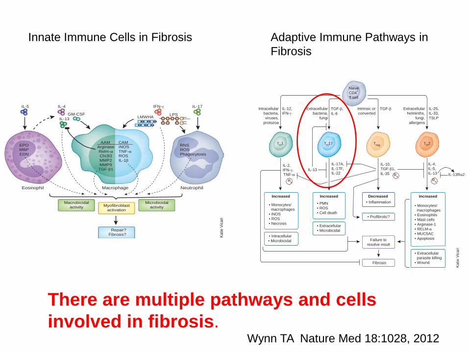
Adaptive Immune Pathways in Fibrosis
Innate Immune Cells in Fibrosis
Wynn TA Nature Med 18:1028, 2012
There are multiple pathways and cells involved in fibrosis.

Role of Tyrosine Kinase Receptors in Peritoneal Fibrosis Wang L, Zhuang S PDI 35:497-505; 2015
Another Part of the Puzzle
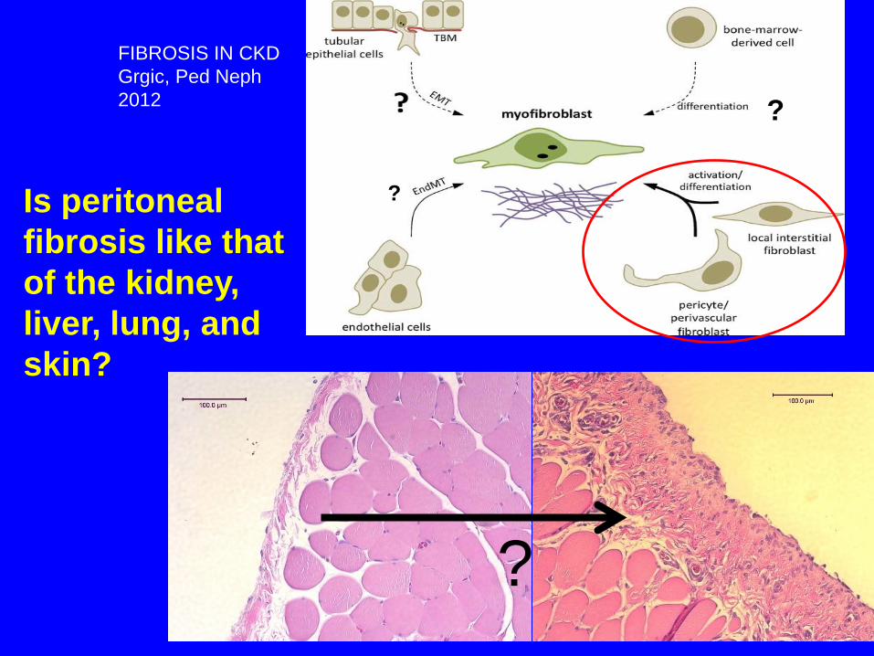
FIBROSIS IN CKD Grgic, Ped Neph 2012
Is peritoneal fibrosis like that of the kidney, liver, lung, and skin?
?
?
?

Lineage Tracing Reveals Distinctive Fates for Mesothelial Cells and Submesothelial Fibroblasts during Peritoneal Injury
JASN 25:2847-2858, 2014 Yi-Ting Chen,*†‡ Yu-Ting Chang,* Szu-Yu Pan,† Yu-Hsiang Chou,*† Fan-Chi Chang,*† Pei-Ying Yeh,* Yuan-Hung Liu,§ Wen-Chih Chiang,† Yung-Ming Chen,† Kwan-Dun Wu,† Tun-Jun Tsai,† Jeremy S. Duffield,| and Shuei-Liong Lin*† National Taiwan University
• Used inducible genetic fate mapping to trace cells after inflammation in 3 transgenic mouse (C57BL6) animal models (hypochlorite, hyperglycemic solutions, TGF-β1)
• Demonstrated mesothelial cells repair the mesothelium and myofibroblasts arise from submesothelial fibroblasts
• Found a possible target (PDGFRβ) for blockade to attenuate fibrosis

Chen YT et al JASN 2014
Model of Peritoneal Inflammation/Fibrosis
80% labeled
65% labeled

In-vitro cultured mesothelial cells do not replicate in vivo results
(Chen et al JASN 2014) • Cultured primary mesothelial from WT1-RFP+ mice for
ability to express αSMA in vitro • No αSMA was seen with culture medium alone. With
TGF-β1, these mesothelial cells expressed αSMA after 48 hours.
• However, there was no αSMA seen in vivo after over-expression of TGF-β1 by injection of Ad TGF-β1 in mesothelial cells in mice.
• This demonstrates the problems with planar cell culture models of a single type of cell.

More basic research is needed using lineage tracing and other
molecular manipulations in order to sort out mechanisms and develop new targets for
combating peritoneal fibrosis.

Topics • Fibrosis: How does it occur? • Causes of Fibrosis • Potential Methods to decrease peritoneal
fibrosis: – New catheter materials – New solutions – Potential pharmacologic additives to current
dialysis solutions


PD Catheter = Foreign Body

Progressive inflammatory response over 1-7 days from sterile polyethylene
catheters in rats E Gomez-Sanchez-Flessner ASN 2010
Trichrome
control 1 day 3 days 7 days

Mouse Adherent Cell Layer at 2 weeks on Polyethylene Catheter

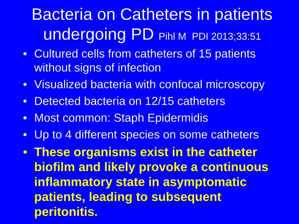
Bacteria on Catheters in patients undergoing PD Pihl M PDI 2013;33:51
• Cultured cells from catheters of 15 patients without signs of infection
• Visualized bacteria with confocal microscopy • Detected bacteria on 12/15 catheters • Most common: Staph Epidermidis • Up to 4 different species on some catheters • These organisms exist in the catheter
biofilm and likely provoke a continuous inflammatory state in asymptomatic patients, leading to subsequent peritonitis.
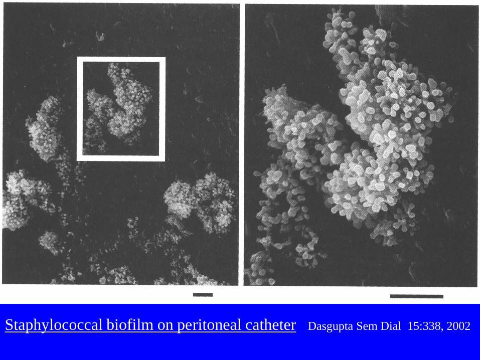
Staphylococcal biofilm on peritoneal catheter Dasgupta Sem Dial 15:338, 2002


Normal Plasma Glucose Concentration = 100 mg/dL
Glucose concentrations in the
dialysis solution:
1.5% = 15 g/L = 1500 mg/dL 2.5% = 25 g/L = 2500 mg/dL
4.25% = 42.5 g/L = 4250 mg/dL


Entire Cohort Low Glucose
High Glucose
Icodextrin
No Icodextrin No peritonitis
1 or more episodes
Davies et al. KI 67:1609, 2005

Topics • Fibrosis: How does it occur? • Causes of Fibrosis • Potential Methods to decrease
peritoneal fibrosis: – New catheter materials – New solutions – Potential pharmacologic additives to current
dialysis solutions


Can we decrease or eliminate inflammation induced by the
catheter? Can we decrease or eliminate
bacterial biofilm?

Molecular Determinants of Biocompatibility
Tang, Hu Expert Rev. Med. Devices 2005, 2:493
• Host cells interact predominantly with adsorbed plasma proteins, which have altered conformations and present sites for cell binding.
• Exposure of the P1/P2 epitopes of fibrinogen bound to biopolymers may be critical for binding of inflammatory cells.
• Different biopolymers have different affinities for protein-cell interactions.
• Blockade of protein sites alters cell binding.

PET=dacron; PE=polyethylene; PVC=polyvinyl chloride; PEU = polyurethane; PDMS = polydimethyl siloxane
Hu, Eatorn, Tang Blood 98:1231, 2001.
Quantitative binding site measurement versus material
Phagocyte accumulation on various biomaterials.
Biocompatible?

Organs-on-a-Chip (large US-NIH Consortium that is entitled Microphysiological Systems): Has demonstrated that cells grown directly on PDMS (polydimethyl siloxane) had properties that were not consistent with in vivo conditions.

Surface Modification of Silicone for Biomedical Applications Requiring Long-Term Antibacterial, Antifouling, and
Hemocompatible Properties Li M et al: Langmuir 28:16408; 2012
• Covalently grafted poly(poly(ethylene glycol) dimethacrylate) – P(PEGDMA)) to medical silicone
• Added polysulfobetaine polymer (P(DMAPS)) to enhance anti-fouling

4 hr Exposure of polymers to S. aureus ( ) Li et al; Langmuir 28:16408; 2012 810 /cells ml
10 μm

3T3 Fibroblast adherence to polymers (24 hr) Li et al Langmuir 28:16408; 2012
200 μm
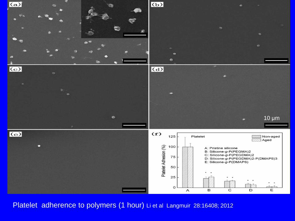
Platelet adherence to polymers (1 hour) Li et al Langmuir 28:16408; 2012
10 μm

Kocuran-functionalized silver glyconanoparticles as antibiofilm coatings
Kumar CG et al Nanotechnology 25:: 32510; 2014
• Synthesized silver 12 nm spherical glyconanoparticles (AgNPs) using Kocuran (exopolysaccharide produced by Kocuria rosea strain BS-1)
SA 48 h E. coli SA 72 h E.coli SA 96 h E.col
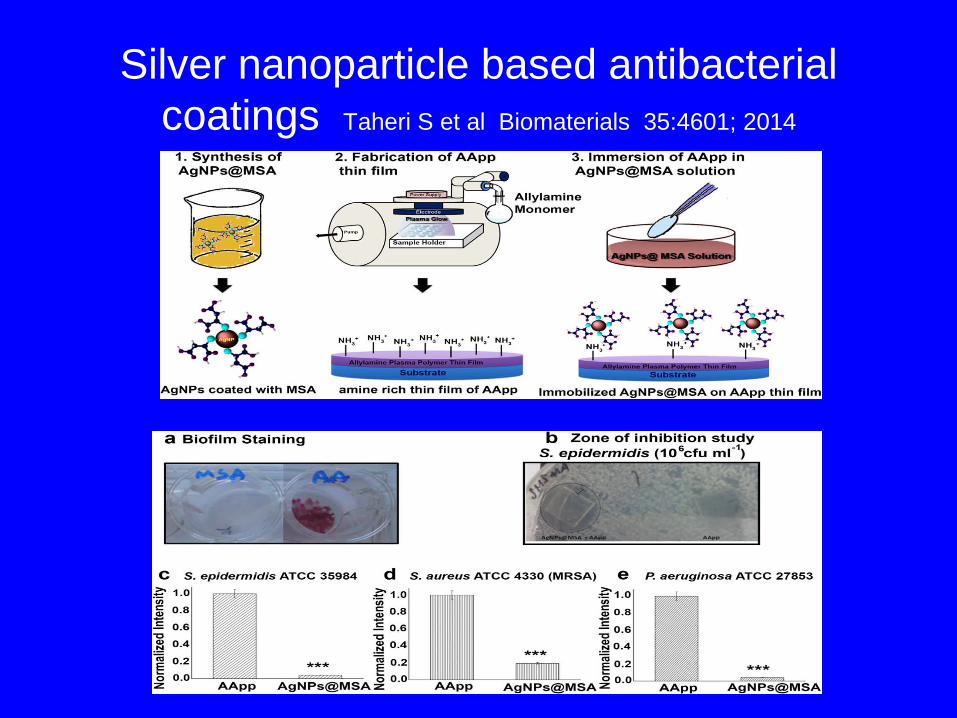
Silver nanoparticle based antibacterial coatings Taheri S et al Biomaterials 35:4601; 2014

Anti-Biofilm Activity of Electrical Current in a Catheter Model Voegele P et al Antimicrob Agents Chemother 2015
• Examined effect of low-amperage DC current exposure on established bacterial and fungal biofilms
• Time and dose (0-500 μA)-dependent bacterial killing observed with DC delivered via an intraluminal platinum electrode
• After 24 hours, 500 μA sterilized the catheter of all bacterial species tested
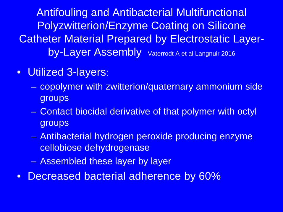
Antifouling and Antibacterial Multifunctional Polyzwitterion/Enzyme Coating on Silicone
Catheter Material Prepared by Electrostatic Layer-by-Layer Assembly Vaterrodt A et al Langnuir 2016
• Utilized 3-layers: – copolymer with zwitterion/quaternary ammonium side
groups – Contact biocidal derivative of that polymer with octyl
groups – Antibacterial hydrogen peroxide producing enzyme
cellobiose dehydrogenase – Assembled these layer by layer
• Decreased bacterial adherence by 60%

Catheter Inflammation and Biofilm
• Remains a major problem for IP, IA, IV, and urethral applications
• Biomaterials are needed to decrease protein adsorption and cell adhesion
• US NIDDK Small Business Initiative: PA-13-050 and PA-13-051: Improved Biomaterials for Urinary and Hemodialysis and Peritoneal Catheters


“Biocompatibility” and lower glucose are reasonable goals, but they have not proven 100% effective in animal experiments or in
randomized human trials.
How about a new osmotic solute?
Impact of low-glucose PD regimen on fibrosis and inflammation markers Yung S et al PDI 35:147; 2015
Studied 43 patients on Physioneal, Extraneal, and Nutrineal (PEN group) vs Controls on Dianeal for 12 months
At 12 months: dialysate levels of CA125, decorin, HepGF, IL-6, adiponectin, adhesion molecules were significantly greater in PEN group compared to controls

Needed: Alternative Osmotic Agent to Glucose • Does not produce GDP or other harmful
substances • Does not produce metabolic changes • Does not increase uremia • Does not inflame or alter the peritoneal
barrier • Exerts effective osmotic pressure over 4-6
hours • Is relatively inexpensive
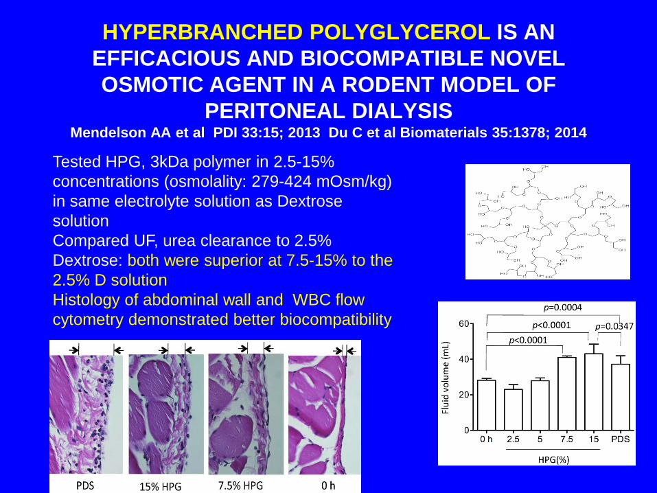
HYPERBRANCHED POLYGLYCEROL IS AN EFFICACIOUS AND BIOCOMPATIBLE NOVEL OSMOTIC AGENT IN A RODENT MODEL OF
PERITONEAL DIALYSIS Mendelson AA et al PDI 33:15; 2013 Du C et al Biomaterials 35:1378; 2014
Tested HPG, 3kDa polymer in 2.5-15% concentrations (osmolality: 279-424 mOsm/kg) in same electrolyte solution as Dextrose solution Compared UF, urea clearance to 2.5% Dextrose: both were superior at 7.5-15% to the 2.5% D solution Histology of abdominal wall and WBC flow cytometry demonstrated better biocompatibility

HPG: Questions/Concerns prior to Human Testing
• Metabolism of hyperbranched polyglycerol?
• Use in uremic animals? • Multiple exchanges: efficacy and
biocompatibility? • Toxicity? Deposition in cells/organs? • Rodents vs humans?

Topics • Fibrosis: How does it occur? • Causes of Fibrosis • Potential Methods to decrease
peritoneal fibrosis: – New catheter materials – New solutions – Potential pharmacologic additives to
current dialysis solutions
New catheter materials and a new osmotic agent in PD are desirable but require years of development
and safety testing.
Can we use additives to current solutions to decrease inflammation and damage to
the peritoneum?

Agent
EMT
Fibrosis
Histology Angiogenesis
AGEs
Small-solute transport rate
Inflammation
Disodium cromoglycate
—
↓
↓
—
—
↓
PPARG agonists ↓ ↓ ↓ ↓ ↓ ↓
Aminoguanidines — ↓ ↓ ↓ ↓ —
Alagebrium — — — ↓ ↓ —
Zopolrestat — ↓ ↓ — — —
Pyridoxamine — — ↓ ↓ ↓ —
Benfotiamine — — ↓ ↓ ↓ ↓
ACE inhibitor or ARB — ↓b ↓ — ↓b ↓
Statins — ↓ — — ↓ —
COX2 inhibitors — ↓ ↓ — ↓ ↓
Vitamin D or VDR activators — — — — ↓b —
Bone morphogenic protein 7 ↓ ↓ ↓ — — —
Sunitinib — — ↓ — = —
Pharmacologic Targets and Peritoneal Remodeling (Farhat et al PDI 2014; 34:114)

Novel Additives to Current PD Solutions
• Decorin (proteoglycan inactivates TGF-β) administered via gold nanoparticles or adenovirus decreases peritoneal fibrosis in an in vivo rodent model. Chaudhary K AJP 307:F777; 2014
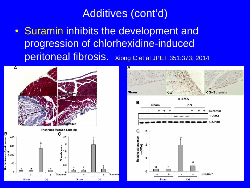
Additives (cont’d) • Suramin inhibits the development and
progression of chlorhexidine-induced peritoneal fibrosis. Xiong C et al JPET 351:373; 2014
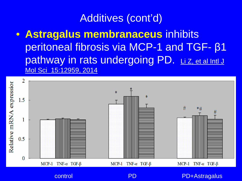
Additives (cont’d) • Astragalus membranaceus inhibits
peritoneal fibrosis via MCP-1 and TGF- β1 pathway in rats undergoing PD. Li Z, et al Intl J Mol Sci 15:12959, 2014
control PD PD+Astragalus

Additives (cont’d)
• MicroRNA-29b inhibits peritoneal fibrosis in mouse model of PD Yu JW et al Lab Invest 94:978;2014
UT = untreated EV = empty vector miR-29b-vector
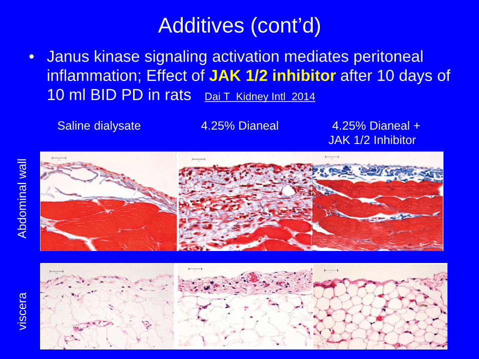
Additives (cont’d) • Janus kinase signaling activation mediates peritoneal
inflammation; Effect of JAK 1/2 inhibitor after 10 days of 10 ml BID PD in rats Dai T Kidney Intl 2014
Abdo
min
al w
all
visc
era
Saline dialysate 4.25% Dianeal 4.25% Dianeal + JAK 1/2 Inhibitor

Oral Paricalcitol decreases Peritoneal Remodeling during PD
Stavenuiter AWD et al BioMed Res Intl 2015
Control 5 wks – PD Fluid 5 wks- PDF +Paricacitol
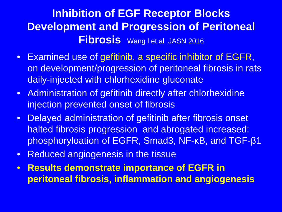
Inhibition of EGF Receptor Blocks Development and Progression of Peritoneal
Fibrosis Wang l et al JASN 2016
• Examined use of gefitinib, a specific inhibitor of EGFR, on development/progression of peritoneal fibrosis in rats daily-injected with chlorhexidine gluconate
• Administration of gefitinib directly after chlorhexidine injection prevented onset of fibrosis
• Delayed administration of gefitinib after fibrosis onset halted fibrosis progression and abrogated increased: phosphoryloation of EGFR, Smad3, NF-κB, and TGF-β1
• Reduced angiogenesis in the tissue • Results demonstrate importance of EGFR in
peritoneal fibrosis, inflammation and angiogenesis

Human Umbilical Mesenchymal Stem Cells Treat Rat Peritoneal Dialysis Induced Fibrosis
Fan Y-P et al Stem Cells Translational Medicine 2016:1-13.
Additives (cont’d) • Endothelin-1 may be a target for prevention of
peritoneal dialysis-associated fibrosis. Busnadiego O et al JASN 26; 2014.
• SMAD2 and SMAD3 play opposing roles in peritoneal fibrosis and are potential targets. Duan W-J et al Am J Pathol 184:2275; 2014.
• Oral Astaxanthin supplementation prevents peritoneal fibrosis in rats. Wakabayashi K et al PDI 2013
• Paracalcitol reduces peritoneal fibrosis in mice through the activation of regulatory T cells and reduction in IL-17. Gonzalez-Mateo GT et al. PLoS One
9:108477; 2014

Additives (cont’d)
• Telmisartan attenuates peritoneal fibrosis via peroxisome proliferator-activated receptor-ɣ activation in uremic rats Su X et al Clin Exp Pharmacol Physiol 42:671; 2015
• The Kampo Medicine Daikenchuto inhibits peritoneal fibrosis (induced with chlorhexidine) in mice by inhibiting inflammation and HSP47 expression. Kitamura M et al Biol Pharm Bull 38:193; 2015

While these pre-clinical studies are promising, each of these
agents require clinical confirmation in government
qualified trials.

Take-Home Points • After an insult, fibrosis occurs in all organs due to the formation
and proliferation of myofibroblasts. Understanding the complex pathways may lead to methods of prevention of the process.
• Studies with cell lineage tracing demonstrate that epithelial de-differentiation along with fibroblast to myofibroblast transition account for repair mechanisms. Other theories include stem cell migration from the bone marrow, transformation of endothelial or mesothelial cell transition to myofibroblasts.
• Major insults to the peritoneum are the PD catheter (foreign body that forms biofilm), high glucose concentrations in dialysate, and overt bacterial infection.
• Needed: better biomaterials that preclude bacterial biofilm formation.
• Needed: improved solutions: Are there alternatives to glucose? • Needed: development and clinical evaluation of pharmacologic
measures to decrease inflammation and to preserve the peritoneum.

Thank you for your attention!
Questions?