pathology of glomerular disease ii
DESCRIPTION
Pathology of Glomerular Disease II. Dr. Álvaro Barboza Quintana. Return to Renal Learning Module. Clinico-pathologic Classification in Renal Syndromes. H = High frequency among patients with the syndrome L = Low frequency among patients with the syndrome 1. Nephrotic Syndrome (NS) - PowerPoint PPT PresentationTRANSCRIPT

Pathology of Pathology of Glomerular Disease Glomerular Disease
IIII
Dr. Álvaro Barboza Quintana.Dr. Álvaro Barboza Quintana.
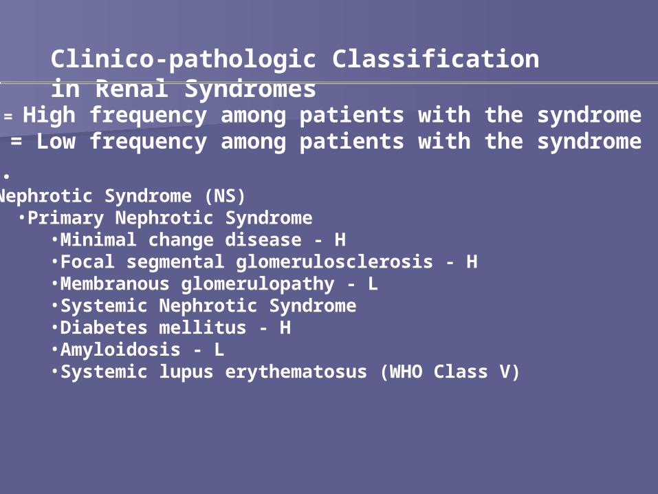
Clinico-pathologic Classification in Renal Syndromes
H = High frequency among patients with the syndrome L = Low frequency among patients with the syndrome1.•Nephrotic Syndrome (NS)
•Primary Nephrotic Syndrome•Minimal change disease - H •Focal segmental glomerulosclerosis - H •Membranous glomerulopathy - L •Systemic Nephrotic Syndrome•Diabetes mellitus - H •Amyloidosis - L •Systemic lupus erythematosus (WHO Class V)

2A. Nephritic Syndrome - Low Serum
Complement
Primary Nephritic Syndrome•Post-infectious glomerulonephritis (GN)
•Membranoproliferative GN - L
Systemic Nephritic Syndrome•Systemic lupus erythematosus (WHO Class III, WHO
Class IV) - H •Infectious endocarditis •HCV-associated cryoglobulinemia

2B. Nephritic Syndrome - Normal Serum Complement
Primary Nephritic Syndrome•IgA Nephropathy
•Hereditary Nephritis (Alport syndrome) - L •Rapidly progressive GN (RPGN), ANCA associated, pauci-immune
Systemic Nephritic Syndrome•Lupus nephritis (WHO Class II) - H •Anti-basement membrane disease (Goodpasture's Syndrome) - L •Systemic vasculitis:
•Polyarteritis nodosa •microscopic polyarteritis •Wegener's granulomatosis •Henoch Schoenlein Purpura
•Thrombotic thrombocytopenic purpura / Hemolytic uremic syndrome
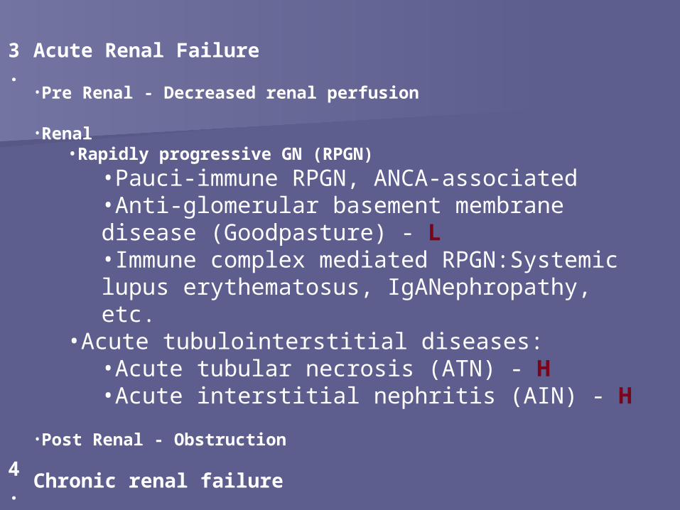
3.
Acute Renal Failure
•Pre Renal - Decreased renal perfusion
•Renal•Rapidly progressive GN (RPGN)
•Pauci-immune RPGN, ANCA-associated •Anti-glomerular basement membrane disease (Goodpasture) - L •Immune complex mediated RPGN:Systemic lupus erythematosus, IgANephropathy, etc.
•Acute tubulointerstitial diseases: •Acute tubular necrosis (ATN) - H •Acute interstitial nephritis (AIN) - H
•Post Renal - Obstruction
4.
Chronic renal failure

Glomerular Glomerular diseases that run diseases that run
with nephrotic with nephrotic syndromesyndrome..

Minimal Change DiseaseMinimal Change Disease(Lipoid Nephrosis)(Lipoid Nephrosis)
Most frequent cause of nephrotic Most frequent cause of nephrotic syndrome in children (2 – 6 years of age).syndrome in children (2 – 6 years of age).
Follows a respiratory infection or routine Follows a respiratory infection or routine prophylactic immunization.prophylactic immunization.
““Its most characteristic feature is its Its most characteristic feature is its usually dramatic response to usually dramatic response to corticosteroid therapy”corticosteroid therapy”
Etiology:Etiology: It’s not known, in most cases is It’s not known, in most cases is idophathic.idophathic.– Interstitial nephritis by medical treatmentInterstitial nephritis by medical treatment– HIV, heroinHIV, heroin– Hodgkin diseaseHodgkin disease
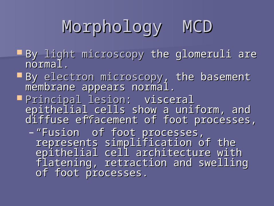
Morphology MCDMorphology MCD By By light microscopylight microscopy the glomeruli are the glomeruli are
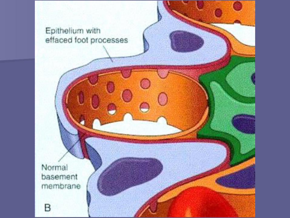
normal.normal. By By electron microscopyelectron microscopy, the basement , the basement
membrane appears normal.membrane appears normal. Principal lesionPrincipal lesion: visceral epithelial cells : visceral epithelial cells
show a uniform, and diffuse effacement show a uniform, and diffuse effacement of foot processes, of foot processes, – ““Fusion” of foot processes, represents Fusion” of foot processes, represents
simplification of the epithelial cell simplification of the epithelial cell architecture with flatening, retraction architecture with flatening, retraction and swelling of foot processes.and swelling of foot processes.
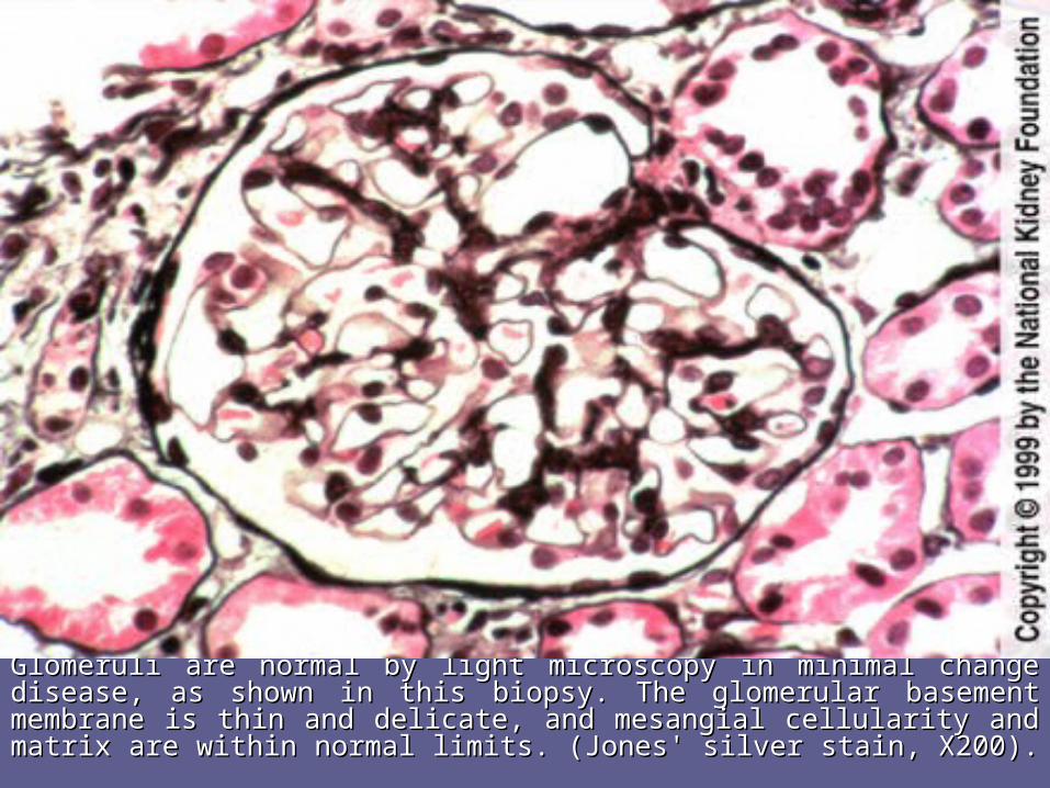
Glomeruli are normal by light microscopy in minimal changeGlomeruli are normal by light microscopy in minimal change disease, disease, as shown in thisas shown in this biopsy. The glomerular basement membrane is thin biopsy. The glomerular basement membrane is thin and delicate, and mesangial cellularity andand delicate, and mesangial cellularity and matrix are within normal matrix are within normal limits. (Jones' silver stain, X200).limits. (Jones' silver stain, X200).

In this electron micrograph, overlying epithelial cell foot processes are In this electron micrograph, overlying epithelial cell foot processes are effaced (giving the appearance of fusion) and run together. effaced (giving the appearance of fusion) and run together.

Morphology MCDMorphology MCD This changes are reversible This changes are reversible
after corticosteroid therapy after corticosteroid therapy and remission of the and remission of the proteinuria.proteinuria.
The cells of the proximal The cells of the proximal tubules are often laden with tubules are often laden with lipid, reflecting tubular lipid, reflecting tubular reabsorotion of lipoproteins reabsorotion of lipoproteins passing through diseased passing through diseased glomeruli: glomeruli: Lipoid nephrosisLipoid nephrosis..
ImmunofluorescenceImmunofluorescence studies studies show no immunoglobulin or show no immunoglobulin or complement deposits.complement deposits.

Clinical CourseClinical Course
Nephrotic syndrome:Nephrotic syndrome:– Proteinuria Proteinuria (albumin) > 3.5 g/day.(albumin) > 3.5 g/day.– HipoalbuminemiaHipoalbuminemia– EdemaEdema– HyperlipidemiaHyperlipidemia– LipiduriaLipiduria– Thromboembolic eventsThromboembolic events– Slow decrease of the glomerular filtrateSlow decrease of the glomerular filtrate
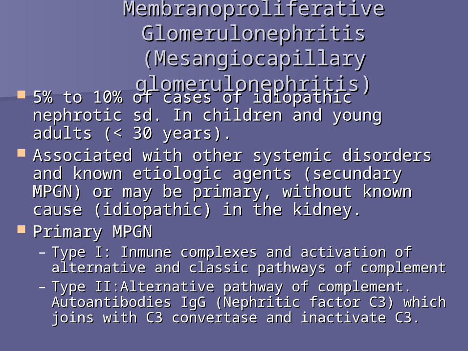
Membranoproliferative GlomerulonephritisMembranoproliferative Glomerulonephritis(Mesangiocapillary glomerulonephritis(Mesangiocapillary glomerulonephritis))
5% to 10% of cases of idiopathic nephrotic sd. 5% to 10% of cases of idiopathic nephrotic sd. In children and young adults (< 30 years).In children and young adults (< 30 years).
Associated with other systemic disorders and Associated with other systemic disorders and known etiologic agents (secundary MPGN) or known etiologic agents (secundary MPGN) or may be primary, without known cause may be primary, without known cause (idiopathic) in the kidney.(idiopathic) in the kidney.
Primary MPGNPrimary MPGN– Type I: Inmune complexes and activation of Type I: Inmune complexes and activation of
alternative and classic pathways of complementalternative and classic pathways of complement– Type II:Alternative pathway of complement. Type II:Alternative pathway of complement.
Autoantibodies IgG (Nephritic factor C3) which Autoantibodies IgG (Nephritic factor C3) which joins with C3 convertase and inactivate C3.joins with C3 convertase and inactivate C3.

Morphology MPGNMorphology MPGN By By light microscopylight microscopy, both types are , both types are
similar.similar.– The glomeruli are large and hipercelular (by The glomeruli are large and hipercelular (by
proliferation of cells in the mesangium).proliferation of cells in the mesangium).– The glomeruli have an “hyperlobular” The glomeruli have an “hyperlobular”
appearance accentuated by the appearance accentuated by the proliferating mesangial cells and increased proliferating mesangial cells and increased mesangial matrix.mesangial matrix.
– The GBM is thickened in the peripheral The GBM is thickened in the peripheral capillary loops.capillary loops.
– The glomerular capillary wall aften shows a The glomerular capillary wall aften shows a “double-contour” or “tram-track” “double-contour” or “tram-track” appearance (appearance (silver or PASsilver or PAS stains). stains).

As seen here, the glomerulus has increased As seen here, the glomerulus has increased overall cellularity, mainly mesangial. overall cellularity, mainly mesangial.
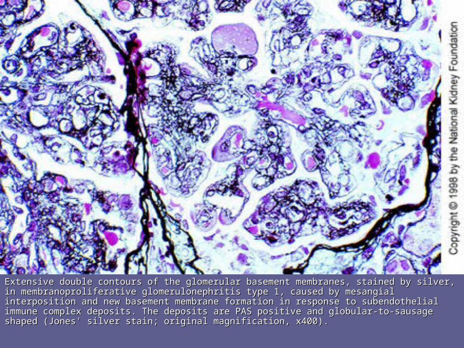
Extensive double contours of the glomerularExtensive double contours of the glomerular basement membranes, stained by silver, inbasement membranes, stained by silver, in membranoproliferative glomerulonephritis type 1, causedmembranoproliferative glomerulonephritis type 1, caused by mesangial interposition and new by mesangial interposition and new basement membranebasement membrane formation in response to subendothelial immune complexformation in response to subendothelial immune complex deposits. The deposits. The deposits are PAS positive anddeposits are PAS positive and globular-to-sausage shaped (Jones' silver stain; originalglobular-to-sausage shaped (Jones' silver stain; original magnification, x400).magnification, x400).
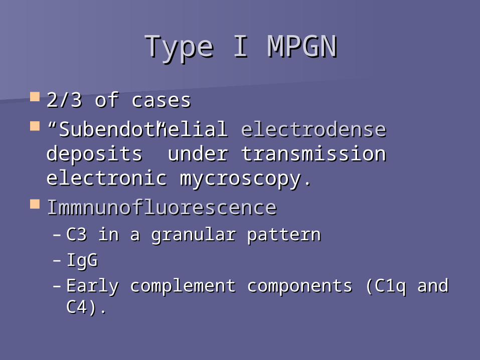
Type I MPGNType I MPGN
2/3 of cases2/3 of cases ““Subendothelial Subendothelial electrodenseelectrodense
deposits” under transmission deposits” under transmission electronic mycroscopy.electronic mycroscopy.
ImmnunofluorescenceImmnunofluorescence– C3 in a granular patternC3 in a granular pattern– IgG IgG – Early complement components (C1q and Early complement components (C1q and
C4).C4).

Membranoproliferative glomerulonephritis type 1.Membranoproliferative glomerulonephritis type 1. The marked endocapillary The marked endocapillary proliferation (proliferatingproliferation (proliferating endothelial and mesangial cells) appears to endothelial and mesangial cells) appears to occlude theocclude the capillary lumen. Numerous large subendothelial andcapillary lumen. Numerous large subendothelial and occasional occasional mesangial-dense immune complex-typemesangial-dense immune complex-type deposits (bottom middle) are present deposits (bottom middle) are present (transmission(transmission electron microscopy; original magnification, x4,700electron microscopy; original magnification, x4,700

Segmental, coarsely granular-to-globular or elongated capillary wall IgG deposits in membranoproliferative glomerulonephritis type 1 (immunofluorescence with anti-IgG; origina magnification, x200).

This electron micrograph demonstrates the dense deposits This electron micrograph demonstrates the dense deposits in the basement membrane of MPGN type II. There are dark in the basement membrane of MPGN type II. There are dark electron dense deposits within the basement membrane electron dense deposits within the basement membrane that often coalesce to form a ribbon-like mass of deposits.that often coalesce to form a ribbon-like mass of deposits.

Type IType I Clinical Course: Clinical Course: Massive proteinuria,Massive proteinuria, Nephrotic sd.Nephrotic sd. Treatment: Treatment: Elimination of the infection.Elimination of the infection. PrognosisPrognosis: Good, 70 – 85% without clinical : Good, 70 – 85% without clinical
alterations.alterations.
Type IIType II• Clinical Course: Nephrotic and Nephritic sd.• Prognosis: Most patients progress to end-
stage renal disease within 10 years.
In both types: Hipocomplementemia, cause of the comsumption in the glomeruli.

Membranous Membranous GlomerulonephritisGlomerulonephritis
Membranous NephropathyMembranous Nephropathy
Epimembranous GNEpimembranous GN
Spikes GNSpikes GN

Membranous GNMembranous GN
Most common cause of nephrotic Most common cause of nephrotic syndrome in adults. syndrome in adults.
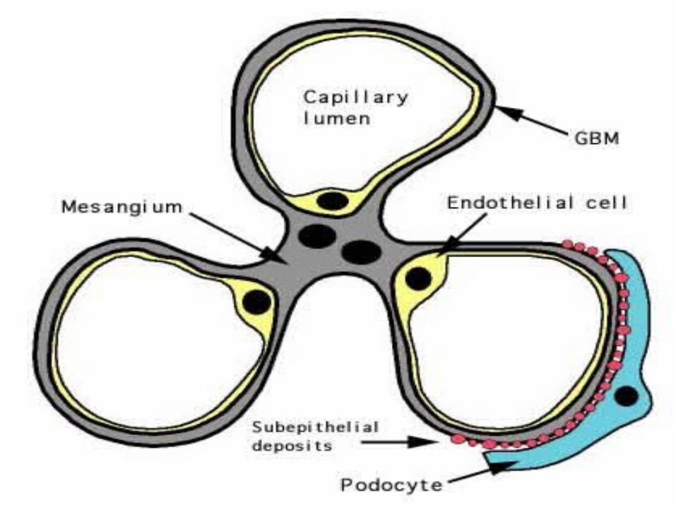
Characterized by:Characterized by:– Diffuse thickening of glomerular Diffuse thickening of glomerular
capillary wallcapillary wall– Accumulation of electron-dense, Accumulation of electron-dense,
deposits of immunoglobulin. deposits of immunoglobulin. – Along the subepithelial side of the Along the subepithelial side of the
basement membranebasement membrane

Membranous GNMembranous GN Idiopathic – 85% of casesIdiopathic – 85% of cases Secondary to: Secondary to:
– Drugs: penicillamine, captopril, gold, Drugs: penicillamine, captopril, gold, NSAIDsNSAIDs
– Underlying malignant tumors: carcinoma Underlying malignant tumors: carcinoma of lung, colon and melanoma. of lung, colon and melanoma.
– Systemic Lupus: Most common type.Systemic Lupus: Most common type.– Infections: chronic hepatitis B/C, syphilis, Infections: chronic hepatitis B/C, syphilis,
schistosomiasis, malaria. schistosomiasis, malaria. – DM, thyroiditis. DM, thyroiditis.
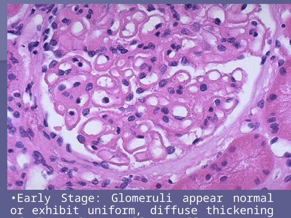
•Early Stage: Glomeruli appear normal or exhibit uniform, diffuse thickening of glomerular capillary wall.

•BM material is laid down between the deposits, appearing as irregular spikes protruding from the GBM (silver stain is the best)


IMMUNOFLUORESCENCE: Deposits of IMMUNOFLUORESCENCE: Deposits of immunoglobulines (G or M) and immunoglobulines (G or M) and complement. complement.

Electron microscopy: thickening by irregular dense deposits between BM and podocites (subepithelial). Podocites have lost their foot processes.
Clinical CourseClinical Course
Nephrotic syndrome or non-selective Nephrotic syndrome or non-selective proteinuria. proteinuria.
Common symptoms: hematuria, Common symptoms: hematuria, hypertension, and symptoms of hypertension, and symptoms of secondary causes. secondary causes.
Course: irregular, indolent. Course: irregular, indolent. As the glomeruli sclerosis progresses: As the glomeruli sclerosis progresses:
BUN elevated, hypertension and BUN elevated, hypertension and reduction in severity of proteinuria. reduction in severity of proteinuria.

Prognosis and TreatmentPrognosis and Treatment
Prognosis: Prognosis: – 60% recovers with persistent proteinuria60% recovers with persistent proteinuria– 10% die or progress to renal 10% die or progress to renal
insufficiency. insufficiency. – Spontaneous remission and better Spontaneous remission and better
prognosis in women with non-nephrotic prognosis in women with non-nephrotic proteinuria. proteinuria.
Treatment:Treatment:– NONNON

Renal AmyloidosisRenal Amyloidosis

AmyloidosisAmyloidosis A systemic immune disease characterized A systemic immune disease characterized
by deposition of amyloid (may be localized)by deposition of amyloid (may be localized) Amyloid is a pathologic proteinaceous Amyloid is a pathologic proteinaceous
substance, deposited between cells in substance, deposited between cells in various organs and tissues with a wide various organs and tissues with a wide variety of clinical settings. variety of clinical settings.
Tipically involves: Tipically involves: – Kidneys, spleen, liver, myocardium, adrenals, Kidneys, spleen, liver, myocardium, adrenals,
thyroid, pituitary and tongue. thyroid, pituitary and tongue. Associated with:multiple myeloma, chronic Associated with:multiple myeloma, chronic
inflammatory conditions, chronic renal inflammatory conditions, chronic renal failure, Alzheimer’s disease, type 2 failure, Alzheimer’s disease, type 2 diabetes.diabetes.
AmyloidosisAmyloidosis Amyloid is formed by fibril proteins in Amyloid is formed by fibril proteins in
95% and by glycloproteins (P component) 95% and by glycloproteins (P component) in 5%.in 5%.
There are 15 biochemically distinct forms There are 15 biochemically distinct forms of amyloid proteins: of amyloid proteins: – Amyloid Light ChainAmyloid Light Chain– Amyloid –associated proteinAmyloid –associated protein– AA amyloid in Alzheimer’s disease amyloid in Alzheimer’s disease– All produce the same consequences and give All produce the same consequences and give
the same pattern in microscopy. the same pattern in microscopy.

Renal amyloidosis is the most common and Renal amyloidosis is the most common and potentially the most serious form of potentially the most serious form of organ involvevement. organ involvevement.
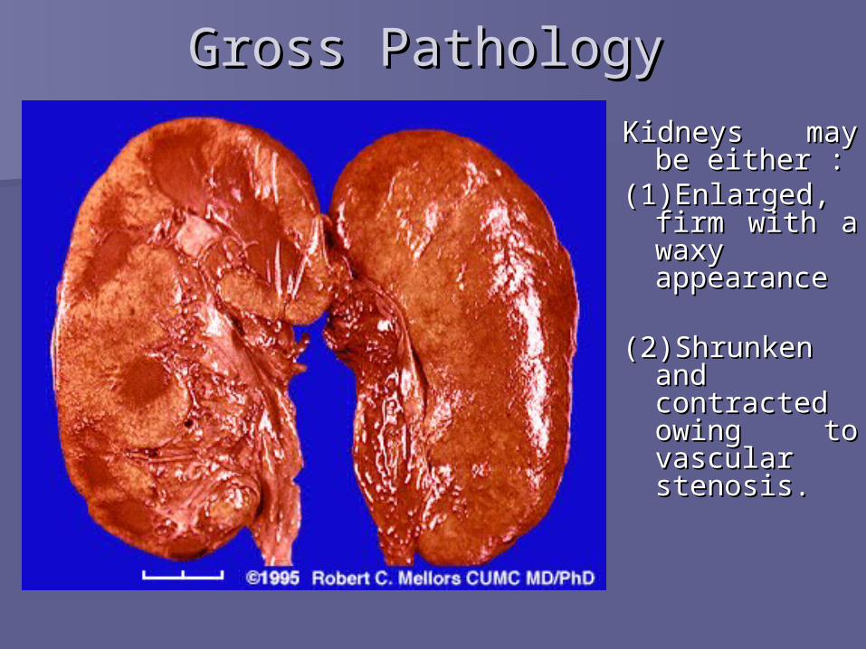
Gross PathologyGross Pathology
Kidneys may be Kidneys may be either :either :
(1)Enlarged, (1)Enlarged, firm with a firm with a waxy waxy appearance appearance
(2)Shrunken (2)Shrunken and and contracted contracted owing to owing to vascular vascular stenosis.stenosis.
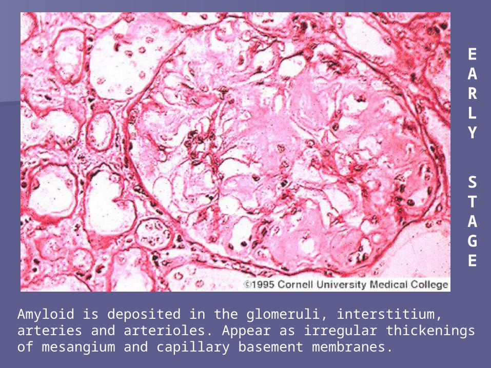
Amyloid is deposited in the glomeruli, interstitium, arteries and arterioles. Appear as irregular thickenings of mesangium and capillary basement membranes.
E A R L Y
S T AG E

Congo Red Stain - Polarizing microscopyShow diffuse amyloid deposition (green birefringence) in glomerular tufts and mesangial regions.
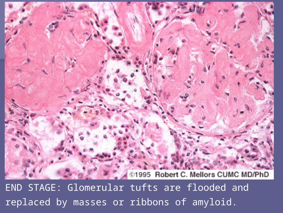
END STAGE: Glomerular tufts are flooded and replaced by masses or
ribbons of amyloid.

Glomerular diseases Glomerular diseases that run with that run with
Nephrytitc SyndromeNephrytitc Syndrome
Poststreptococcal Poststreptococcal GlomerulonephritisGlomerulonephritis(Postinfectious Acute (Postinfectious Acute
Glomerulonephritis)Glomerulonephritis)

Pathogenesis Pathogenesis Secondary to a pharyngeal infección with Secondary to a pharyngeal infección with
varying latent periodvarying latent period
Nephritic syndromeNephritic syndrome– Group A (1,2,3,4,12,18,25,49,55,57,60)Group A (1,2,3,4,12,18,25,49,55,57,60)
Low complement levels, and high titles of Low complement levels, and high titles of streptococcal products.streptococcal products.
GlomeruliGlomeruli– Granular immune depositsGranular immune deposits– Endostreptsin and cationic antigens in afected Endostreptsin and cationic antigens in afected
áreasáreas

MacroMacro
Macroscopic hematuria with a rusty or Macroscopic hematuria with a rusty or smokey hue.smokey hue.
MicroMicro
Glomeruli: bloodless, hypercelular and Glomeruli: bloodless, hypercelular and enlarged.enlarged.
Proliferating mesangial and endothelial Proliferating mesangial and endothelial cells oclude the capillary luminacells oclude the capillary lumina– PMN and monocyte infiltration.PMN and monocyte infiltration.
Exudative and difuse (will affect all the lobules)Exudative and difuse (will affect all the lobules) Interstitium: edemaInterstitium: edema

Electron MicroscopyElectron Microscopy
Dome-shaped deposits projecting Dome-shaped deposits projecting outward from epithelial side of outward from epithelial side of basement membreane. basement membreane. – Epithelial cell slit poresEpithelial cell slit pores– Separated from the basement Separated from the basement
membrane by cearl zone continuos membrane by cearl zone continuos with the lamina rara externa.with the lamina rara externa.
PMN and monocytesPMN and monocytes

Clinical FeaturesClinical Features Spontaneous nefritic syndromeSpontaneous nefritic syndrome
– Fever, nausea, gross hematuria, oliguria after Fever, nausea, gross hematuria, oliguria after recovery from pharyngitis.recovery from pharyngitis.
Note: adults have a less spontaneous start Note: adults have a less spontaneous start with HTA.with HTA.
During epidemics, symptoms may be rare.During epidemics, symptoms may be rare.
1% of children develop intense oliguria and 1% of children develop intense oliguria and progresive glomerulonephritis. progresive glomerulonephritis.
Outcome in adults is less favorable.Outcome in adults is less favorable. During sporadic cases, 60% have an early During sporadic cases, 60% have an early
recovery.recovery.
1 or more weeks.1 or more weeks.

Glomeruli show diffuse hypercellularity due to mesangial and endothelial cell increase and a large number of polymorphonuclear neutrophils (PMNs). H&E

Diffuse proliferative acute postinfectious glomerulonephritis with numerous PMNs with PAS-positive cytoplasm and endocapillary proliferation. PAS

The garland pattern of immune complexes due to large subepithelial deposits in acute postinfectious glomerulonephritis is shown (immunofluorescence

Hump-shaped deposits in acute postinfectious glomerulonephritis with extensive foot process effacement and endocapillary proliferation

Rapidly Progressive Rapidly Progressive GlomerulonephritisGlomerulonephritis
Very uncommonVery uncommon– 2 % of all cases presenting with GN2 % of all cases presenting with GN
Predominates in men (2:1) Predominates in men (2:1) – Young to middle aged Young to middle aged
Rapidly Progressive Rapidly Progressive GlomerulonephritisGlomerulonephritis
General InfoGeneral Info

1.- Post-infectious (Post-1.- Post-infectious (Post-Streptococcal)Streptococcal)
2.- Associated to Systemic Diseases 2.- Associated to Systemic Diseases Lupus, Goodpasture syndrome, vasculitis, Lupus, Goodpasture syndrome, vasculitis,
WegenerWegener
3.- Primary or idiopathic 3.- Primary or idiopathic
Rapidly Progressive Rapidly Progressive GlomerulonephritisGlomerulonephritis
ClassificationClassification
In all cases the basic In all cases the basic pathogenic mechanism is pathogenic mechanism is
immunogenicimmunogenic

The presence of fibrin in Bowman’s The presence of fibrin in Bowman’s space promotes the epithelial space promotes the epithelial proliferation and formation of crescentsproliferation and formation of crescents
Rapidly Progressive Rapidly Progressive GlomerulonephritisGlomerulonephritis
PathogenesisPathogenesis
Fibrin
Fibrin
GLOMERULUSExtravasation due to capillary damage
Production stimulated by factors liberated by Monocytes
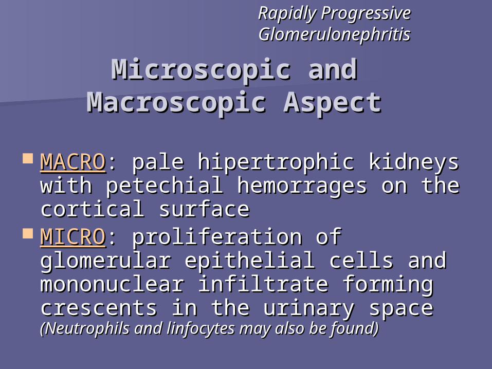
MACROMACRO: pale hipertrophic kidneys with : pale hipertrophic kidneys with petechial hemorrages on the cortical petechial hemorrages on the cortical surfacesurface
MICROMICRO: proliferation of glomerular : proliferation of glomerular epithelial cells and mononuclear epithelial cells and mononuclear infiltrate forming crescents in the infiltrate forming crescents in the urinary space urinary space (Neutrophils and linfocytes may (Neutrophils and linfocytes may also be found)also be found)
Rapidly Progressive Rapidly Progressive GlomerulonephritisGlomerulonephritis
Microscopic and Microscopic and Macroscopic AspectMacroscopic Aspect

CrescentsCrescents: complex mixture of proliferating epithelial : complex mixture of proliferating epithelial cells and infiltrating monocytes forming concentric layers cells and infiltrating monocytes forming concentric layers around the capillary tufts (which are compressed)around the capillary tufts (which are compressed)
This is a case of RPGN due to LupusThis is a case of RPGN due to Lupus
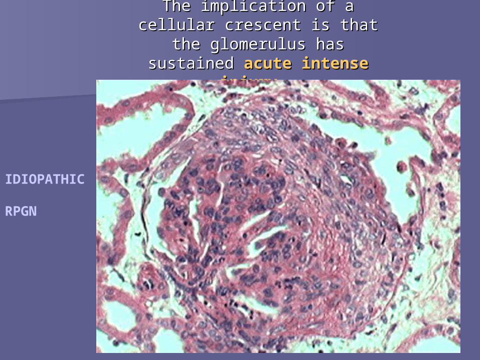
The implication of a cellular The implication of a cellular crescent is that the glomerulus crescent is that the glomerulus has sustained has sustained acute intense acute intense
injuryinjury. .
IDIOPATHIC RPGN

Pathogenesis of symptomsPathogenesis of symptoms
Rapidly Progressive Rapidly Progressive GlomerulonephritisGlomerulonephritis
Glomerular tufts adhere to Bowman’s capsule OLIGURIA
Cicatrization of capillaries POST-GLOMERULARISCHEMIA
TUBULAR ATROPHYAND INTERSTITIALFIBROSIS
Renal Failure
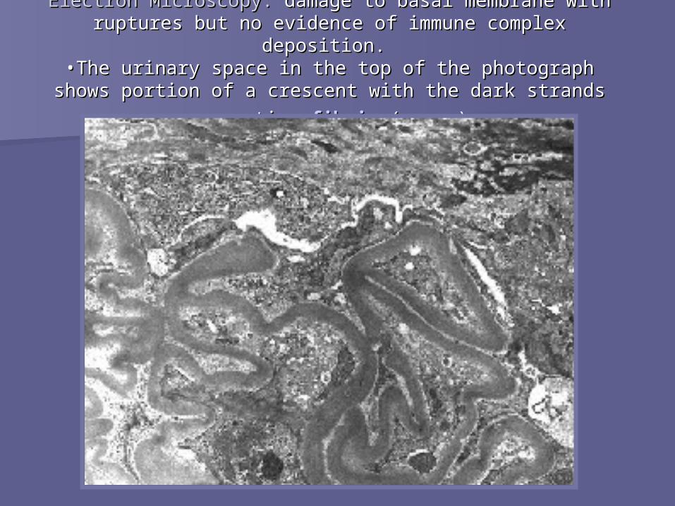
Electron Microscopy: Electron Microscopy: damage to basal membrane with damage to basal membrane with ruptures but no evidence of immune complex deposition. ruptures but no evidence of immune complex deposition.
•The urinary space in the top of the photograph shows •The urinary space in the top of the photograph shows portion of a crescent with the dark strands representing portion of a crescent with the dark strands representing
fibrinfibrin (arrow). (arrow).

ImmunofluorescenceImmunofluorescence: positivity with antibody to : positivity with antibody to fibrinogenfibrinogen. With a rapidly progressive GN, the . With a rapidly progressive GN, the glomerular damage is so severe that fibrinogen glomerular damage is so severe that fibrinogen
leaks into Bowman's space, leading to proliferation leaks into Bowman's space, leading to proliferation of the epithelial cells and formation of a crescent.of the epithelial cells and formation of a crescent.
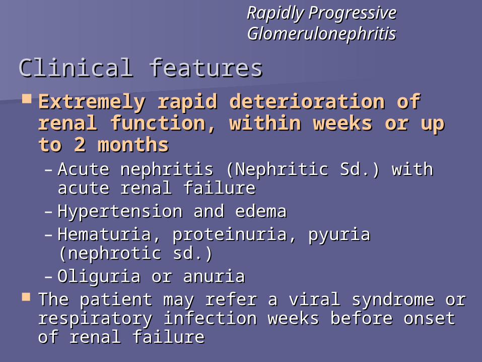
Clinical featuresClinical features Extremely rapid deterioration of Extremely rapid deterioration of
renal function, within weeks or up to renal function, within weeks or up to 2 months2 months– Acute nephritis (Nephritic Sd.) with acute Acute nephritis (Nephritic Sd.) with acute
renal failurerenal failure– Hypertension and edemaHypertension and edema– Hematuria, proteinuria, pyuria (nephrotic sd.)Hematuria, proteinuria, pyuria (nephrotic sd.)– Oliguria or anuriaOliguria or anuria
The patient may refer a viral syndrome or The patient may refer a viral syndrome or respiratory infection weeks before onset of respiratory infection weeks before onset of renal failurerenal failure
Rapidly Progressive Rapidly Progressive GlomerulonephritisGlomerulonephritis

Goodpasture’s Goodpasture’s SyndromeSyndrome
(Anti-Basement Membrane (Anti-Basement Membrane Disease)Disease)

Acute and necrotizing RPGN associated to Acute and necrotizing RPGN associated to pulmonary hemorraging and hemoptysispulmonary hemorraging and hemoptysis
Uncommon disease affecting primarily men Uncommon disease affecting primarily men (4:1) between 20-30 years of age. (4:1) between 20-30 years of age.
Autoimmune disease in which there is Autoimmune disease in which there is production of Anti-Basement Membrane production of Anti-Basement Membrane Antibodies (ABM) that deposit on alveoli and Antibodies (ABM) that deposit on alveoli and glomeruliglomeruli
Goodpasture’s SyndromeGoodpasture’s Syndrome
General InfoGeneral Info

The Goodpasture antigen resides in the non-The Goodpasture antigen resides in the non-collagen portion of the 3-alpha chain of collagen portion of the 3-alpha chain of type IV collagentype IV collagen
Precipitation factor is unknown, may be:Precipitation factor is unknown, may be:– VirusVirus– Hydrocarbonated solventsHydrocarbonated solvents– Tobacco smoke (permissive role)Tobacco smoke (permissive role)– DrugsDrugs– CancerCancer
Genetic predisposition: DRW15/DQW6Genetic predisposition: DRW15/DQW6 The lesion consists of deposits of ABM The lesion consists of deposits of ABM
antibody and complement on the basement antibody and complement on the basement membrane of glomeruli and alveoli causing membrane of glomeruli and alveoli causing their destructiontheir destruction
Goodpasture’s SyndromeGoodpasture’s Syndrome
PathogenesisPathogenesis

MACROMACRO: same aspect as RPGN: same aspect as RPGN MICROMICRO: crescents composed of epithelial cells : crescents composed of epithelial cells
and monocytes surrounding capillary tufts in and monocytes surrounding capillary tufts in the urinary spacethe urinary space– Methenamine silver stainMethenamine silver stain– Electron MicroscopyElectron Microscopy: does not show deposits, only : does not show deposits, only
architectural damage and epithelial proliferationarchitectural damage and epithelial proliferation– ImmunofluorescenceImmunofluorescence
The lungs show alveolar hemorrage, The lungs show alveolar hemorrage, hemosiderin-filled macrophages and thickening hemosiderin-filled macrophages and thickening of alveolar septi. The BM shows immune of alveolar septi. The BM shows immune depositsdeposits
Goodpasture’s SyndromeGoodpasture’s SyndromeMacroscopic and Macroscopic and Microscopic Microscopic
AspectAspect

Silver stainSilver stain

ImmunofluorescenceImmunofluorescence: shows positivity with : shows positivity with antibody to antibody to IgGIgG has a has a smooth, diffuse, linear smooth, diffuse, linear
patternpattern that is PATOGNOMONIC that is PATOGNOMONIC

Goodpasture’s SyndromeGoodpasture’s Syndrome
Clinical featuresClinical features Typically begins as flu-like illness with evidence of Typically begins as flu-like illness with evidence of
pulmonary compromisepulmonary compromise Pulmonary hemorrages HEMOPTYSISPulmonary hemorrages HEMOPTYSIS Progressive dyspneaProgressive dyspnea Nephritis (Nephritic Sd.) and acute renal failureNephritis (Nephritic Sd.) and acute renal failure The disease progresses rapidly with renal failure The disease progresses rapidly with renal failure
ocurring within weeks or monthsocurring within weeks or months
Treatment and PrognosisTreatment and Prognosis High doses of steroids, with or without cytotoxic agents. High doses of steroids, with or without cytotoxic agents. Plasmapheresis removes ABM-antibodies Plasmapheresis removes ABM-antibodies Better prognosis than other diseases causing RPGNBetter prognosis than other diseases causing RPGN

Glomerulonephritis Glomerulonephritis IgAIgAoror
Of Berger Of Berger
ClassificationClassification
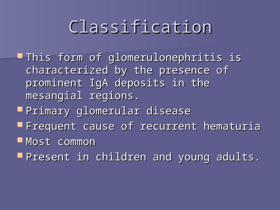
This form of glomerulonephritis is This form of glomerulonephritis is characterized by the presence of characterized by the presence of prominent IgA deposits in the prominent IgA deposits in the mesangial regions.mesangial regions.
Primary glomerular diseasePrimary glomerular disease Frequent cause of recurrent hematuriaFrequent cause of recurrent hematuria Most commonMost common Present in children and young adults.Present in children and young adults.

PathogenesisPathogenesis
Genetic or acquired abnormality of Genetic or acquired abnormality of inmune regulation leading to increased inmune regulation leading to increased mucosal IgA synthesis in response to mucosal IgA synthesis in response to respiratory or GI exposure to respiratory or GI exposure to environmental agents. environmental agents.
IgA1 and IgA1complexes are entrapped in IgA1 and IgA1complexes are entrapped in the mesanguim.the mesanguim.
They activate the alternative complement They activate the alternative complement pathway and initiate glomerular injury.pathway and initiate glomerular injury.

MICRO/ H&EMICRO/ H&E
Mesangial widening Mesangial widening or proliferation or proliferation (arrow)(arrow)
Segmental Segmental proliferationproliferation
Overt crescentic Overt crescentic glomerulonephritis glomerulonephritis (rare)(rare)
Sclerosis (healing of Sclerosis (healing of focal proliferative focal proliferative lesion).lesion).

MICRO/ Electron Mic.MICRO/ Electron Mic.
Mesangial electron Mesangial electron dense deposits and dense deposits and increased mesangial increased mesangial matrix and matrix and cellularity in IgA cellularity in IgA nephropathy nephropathy (transmission (transmission electron electron microscopy, original microscopy, original magnification magnification x8,500). x8,500).
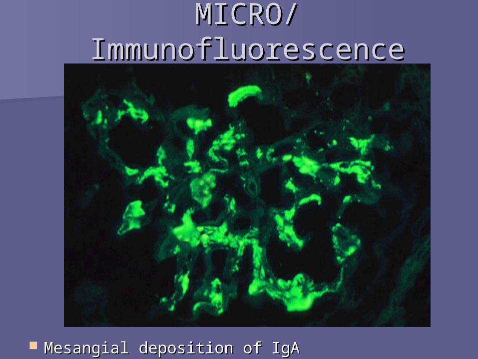
MICRO/ MICRO/ ImmunofluorescenceImmunofluorescence
Mesangial deposition of IgAMesangial deposition of IgA
Clinical CourseClinical Course
Gross hematuria after GI or Gross hematuria after GI or respiratory infection.respiratory infection.
5-10% develop a typical acute 5-10% develop a typical acute nephritic syndrome.nephritic syndrome.
Hematuria lasts several days and Hematuria lasts several days and then subsides, only to return every then subsides, only to return every few months.few months.

Sistemic Lupus Sistemic Lupus ErithematousErithematous
Diffuse Proliverative Diffuse Proliverative GlomerulonephritisGlomerulonephritis

IntroductionIntroduction
SLE- SLE-
Classic prototype of the multisystem disease Classic prototype of the multisystem disease of autoimmune origin, characterized by a of autoimmune origin, characterized by a bewildering array of autoantibodies, bewildering array of autoantibodies, particulary antinuclear antibodies (ANAs). particulary antinuclear antibodies (ANAs). Acute or insidious in its onset, it is a Acute or insidious in its onset, it is a chronic, remitting and relapsing, often chronic, remitting and relapsing, often febrile illness characterized principally by febrile illness characterized principally by injury to the skin, joints, kidney, and serosal injury to the skin, joints, kidney, and serosal membranes.membranes.

ClassificationClassificationLupus GlomerulonephritisLupus Glomerulonephritis
According to the WHO (morphologic class.)According to the WHO (morphologic class.) Class I: Normal by light, electron, and Class I: Normal by light, electron, and
imunofluorescent microscopy imunofluorescent microscopy Class II: Mesangial lupus Class II: Mesangial lupus
glomerulonephritisglomerulonephritis Class III: focal proliferative Class III: focal proliferative
glomerulonephritisglomerulonephritis Class IV: Diffuse proliferative Class IV: Diffuse proliferative
glomerulonephritisglomerulonephritis Class V: Membranous glomerulonephritisClass V: Membranous glomerulonephritis
Kidney appears to be involved in 60 to 70% of cases.


MICRO/ H&EMICRO/ H&E Proliferation of mesangial cells and Proliferation of mesangial cells and
endothelial cells, along with infiltrating endothelial cells, along with infiltrating mononuclear and polymorphonuclear mononuclear and polymorphonuclear leukocytes afecting more than 50% of the leukocytes afecting more than 50% of the glomerular area.glomerular area.
Extensive peripheral capillary wall Extensive peripheral capillary wall subendothelial immune deposition subendothelial immune deposition (wire (wire loop)loop), and extracapillary proliferation in the , and extracapillary proliferation in the form of crescents.form of crescents.
Fibrinoid necrosis, leukocyte infiltration, Fibrinoid necrosis, leukocyte infiltration, wire-loop deposits, hyaline thrombi, and wire-loop deposits, hyaline thrombi, and hematoxylin bodieshematoxylin bodies

MICRO/ OthersMICRO/ Others Half of the tuft is Half of the tuft is
distorted by marked distorted by marked endocapillary endocapillary proliferation with proliferation with occasional infiltrating occasional infiltrating cells. Segmental areas of cells. Segmental areas of basement membrane basement membrane splitting and splitting and eosinophilic eosinophilic subendothelial deposits subendothelial deposits and mesangial and mesangial eosinophilic deposits are eosinophilic deposits are visualized (Jones' silver visualized (Jones' silver stain; original stain; original magnification x400).magnification x400).

Clinical CourseClinical Course Typically have:Typically have:
– high anti-DNA antibody titershigh anti-DNA antibody titers
– low serum complement levelslow serum complement levels
– Very active urinary sediment with:Very active urinary sediment with: erythrocytes erythrocytes
other casts present on urinalysisother casts present on urinalysis
– ProteinuriaProteinuria
– Half of the patients will have nephrotic syndromeHalf of the patients will have nephrotic syndrome
– HypertensionHypertension