pa ge 2/ 20
TRANSCRIPT

Page 1/20
Maraviroc, An Inhibitor of Chemokine Receptor Type5, Alleviates Neuroin�ammatory Response afterCerebral Ischemia/Reperfusion Injury via RegulatingJNK SignalingBeilei Chen
Yangzhou UniversityPingping Cao
Dalian Medical College: Dalian Medical UniversityXin Guo
Dalian Medical College: Dalian Medical UniversityXiaobo Li
Yangzhou UniversityLi Jiang
Yangzhou UniversityXiongfeng Lian
Yangzhou UniversityXin Chen
Yangzhou UniversityChao Jiang
Yangzhou UniversityLuhang Tao
Yangzhou UniversityHailong Yu ( [email protected] )
Northern Jiangsu People's Hospital https://orcid.org/0000-0002-1557-7465
Research Article
Keywords: cerebral ischemia/reperfusion, Maraviroc, in�ammation, microglia, JNK signal pathway.
Posted Date: September 27th, 2021
DOI: https://doi.org/10.21203/rs.3.rs-914711/v1
License: This work is licensed under a Creative Commons Attribution 4.0 International License. Read Full License

Page 2/20

Page 3/20
AbstractNeuroin�ammation is a key factor that contributes to the secondary injury after cerebralischemia/reperfusion (CI/R) injury. Chemokine receptor type 5(CCR5) has shown its pro-in�ammatoryeffects during central nervous system (CNS) diseases. However, the role of CCR5 in CI/R injury is stillunclear. In this study, we administered maraviroc (MVC,APEXBIO,UK-427857), a CCR5 antagonist, to themiddle cerebral artery occlusion(MCAO) mice. In vivo studies showed that MVC was successivelyintraperitoneally (i.p.) with different doses (5, 20, or 50 mg/kg body weight) for 3 days after mice MCAO.MVC showed its neuroprotective effects in alleviating neurological de�cits and infarct volumes afterMCAO. The level of apoptosis and in�ammation were remarkably decreased by MVC treatment after CI/Rinjury. Subsequently, primary microglia were stimulated with different doses of MVC (0.2, 2, 20 or 200nM)for 12h after oxygen-glucose deprivation/reoxygenation model (OGD/R) in vitro. MVC signi�cantlyincreased the viability of primary microglia after (OGD/R). The expression of pro-in�ammatory cytokines(IL-1β and IL-6) in microglia were down-regulated by MVC treatment. Mechanistically, MVC also inhibitedthe secretion of IL-1β and IL-6 by microglia after OGD stimulation. Furthermore, the key components ofNF-κB pathway were measured in vivo and in vitro after MCAO and OGD. MVC signi�cantly inhibited theactivity of NF-κB pathway in the above pathological environments. Finally, our data indicated that MVCtreatment decreased the activation of JNK signaling pathway after CI/R injury in vivo and in vitro. TheJNK activator anisomycin (AN Beyotime SC0132-5mg) reversed the neuroprotective effects of MVC,indicating that the JNK pathway is involved in the anti-in�ammatory and anti-apoptotic mechanisms ofMVC in CI/R injury. Our data demonstrated that CCR5 inhibition exhibits neuroprotective effects after CI/Rinjury. MVC, which is widely used for HIV treatment by its anti-virus effect, is a potential drug for thetreatment of ischemic stroke in the future clinical trials.
IntrodutionAcute ischemic stroke is the most common cerebrovascular event with high morbidity and mortality[1]..Based on its complex pathophysiological mechanisms, the interaction of various factors, such asin�ammatory damage[2, 3]. and blood-brain barrier destruction, [4]. leads to the apoptosis and necrosis ofcerebral tissue and cells. However, there is a lack of effective treatments for ischemic stroke. CCchemokine receptor 5 (CCR5), a G protein-coupled receptor, has the availability to regulate cell activationand migration in response to chemokines[5].. Based on a previously conducted research, CCR5 has beendetermined to play a vital role in the immune and in�ammatory responses by mediating the chemotacticactivity in leukocytes[6].. A previous study showed that CCR5 plays a critical role in intestinalischemia/reperfusion (I/R) injury[7]. and endotoxin-induced lung injury[8].. Nevertheless, the effects ofCCR5 antagonist in CI/R remain unknown. This evidence has led to the successful search for CCR5antagonists as antiretroviral drugs and the approval of maraviroc for both treatment-experienced andtreatment CI/R. However, whether MVC possesses neuroprotective effects in cerebral I/R injury remains tobe understood. Based on its anti-in�ammatory capacity[2, 3]., we hypothesized that MVC may have aprotective effect in cerebral I/R injury. In this study, we employed a mice middle cerebral artery occlusion
Page 4/20
(MCAO) model to investigate the role of MVC in the treatment of cerebral I/R injury and its potentialmechanisms in mice.
Materials And Mmethods
AnimalsAll adult male C57BL/6J (B6) mice (8–10 weeks old, approximately 25–30 g) were purchased from theComparative Medical Center of Nanjing Medical University (Jiangsu Province, China). As previouslydescribed, the mice were housed in a suitable environment.
Grouping and Drug AdministrationMice were randomized into four groups as follows: I) control vehicle-treated group (Sham group); II) MVC-treated group (MVC group); III) vehicle-treated CI/R group (CI/R group); and IV) MVC-treated CI/R group(CI/R + MVC group). After the induction of MCAO, mice received intraperitoneal injections with either MVC(5, 20, or 50mg/kg body weight) or the vehicle(10% dimethyl sulfoxide ,10%DMSO,Amresco, Solon, OH,USA) daily for 3 consecutive days.
The Middle Cerebral Artery Occlusion(MCAO) modelThe animals were anaesthetized with pentobarbital sodium. The MCAO mouse model was established aspreviously described[9].. Brie�y, a 6/0 mono�lament nylon suture (Doccol Corporation, MA, USA) with aheat-rounded tip was inserted into the beginning of the MCA through the internal carotid artery until theipsilateral blood �ow decreased to less than 30% of the baseline value, as monitored using laser Doppler�owmetry (Perimed Corporation, Stockholm, Sweden). After 60 min of occlusion, the �lament waswithdrawn to allow blood reperfusion. In the sham-operated group, the aforementioned procedure wasperformed, but a �lament was not inserted into the MCA. After the operation, we fed the mice jelly orperformed an intraperitoneal (i.p.) injection of normal saline to rehydrate. Pre-dissolved MVC or the samevolume of saline was administered to each animal by an i.p. injection at 30 min, 24 h and 48 h afterMCAO in a double-blind manner.
Behavioral analysis of neurological de�cit scoreNeuro-behavioural function was assessed using the modi�ed neurological severity score (mNSS) andLonga score on day 3 following MCAO by a blinded observer[10].. The mNSS evaluates neurologicalde�cits through motor, re�ex, sensory, and balance testing. The score ranges from 0, indicating noneurologic de�cit, to 18 for animals with the most severe impairment. The Longa score is determined asfollows: 0 point, no neurological de�cit; 1point, cannot fully extend the contralateral forelimb; 2 points,tail-catching phenomenon while walking (circling to the contralateral side); 3 points, unsteady in thestanding position, falling to the contralateral side; and 4 points, no spontaneous walking and decreasedconsciousness. Scores of 0‐2 are classi�edas mild neurological impairment, while scores of 3‐4 are
Page 5/20
classi�ed as severe neurological impairment. The evaluators were blinded to the treatment group of miceduring the neurobehavioral tests.
Cerebral Infarction Volume MeasurementAfter neurological evaluation, mice were sacri�ced and histological evaluation of brain tissue wasperformed. The brain was sectioned into six slices and incubated with 0.2%(w/v) 2,3,5-triphenyltetrazolium chloride (TTC, Sigma-Aldrich)at 37°C for 15 min to determine the infarct volume.Infarcted tissue was pale grey in color, compared to the dark red color of normal brain tissue. Imageswere obtained using a digital camera and analyzed using ImageJ software. The percentage ofhemispheric infarction volume was calculated using the following formula: Infarct size = (contralateralarea—ipsilateral non-infarct area)/contralateral area x 100%
Quantitative Real-Time-Polymerase Chain Reaction (qPCR)Total RNA was extracted from the ischemic cerebral cortex using a commercial TRIzol kit, and RNA wasthen reverse-transcribed into cDNA using a PrimeScript RT reagent Kit(Vazyme,Nanjing,China).Quantitative measurements were performed on an ABI 7500 PCR instrument (Applied Biosystems, USA)using a SYBR green kit(Applied Biosystems). Relative gene expression levels were normalized to GAPDH.The sequences of the primers used are as follows: TNF-a: (forward) TCGGTCCCAACAAGGAGGAG and(reverse) GGGTTGTCACTCGAGTTTTG; IL-1b: (forward) GCAACTGTTCCTGAACTCAACT and (reverse)ATCTTTTGGGGTCCGTCAACT; IL-6:(forward) GGCGGATCGGATGTTGTGAT and (reverse)GGACCCCAGACAATCGGTTG; GAPDH (forward) GCCAAGGCTGTGGGCAAGGT and (reverse)TCTCCAGGCGGCACGTCAGA.
Western Blot AnalysisWestern blot analysis was then performed on the cerebral tissue within the ischemic areas. Proteins werehomogenized in RIPA lysis buffer (Millipore, Billerica, MA, USA) containing protease inhibitors. Thesupernatant was assayed using a BCA Protein Assay Kit (Beyotime Biotech). The protein concentrationwas adjusted to the same concentration with protein buffer. The protein samples were then loaded,electrophoresis was performed, and the separated proteins were electrically transferred to polyvinylidenedi�uoride (PVDF) membrane. The membrane was blocked in PBS containing 0.1% Tween-20 and 5% non-fat milk for 2 h at room temperature. The membrane was then incubated with the primary antibodiesovernight at 4°C. anti-GAPDH(Bioworld), anti-Bax (Cell Signaling Technology), Anti-Bcl-2 (Bioworld),anti-Phospho-IκBα(Cell Signaling Technology),anti-IκBα(Cell Signaling Technology) anti-NF-κB (p-p65) (CellSignaling Technology) anti-NF-κB (p65) (Cell Signaling Technology) anti-Phospho-p38 MAPK(CellSignaling Technology) anti-p38 MAPK(Cell Signaling Technology) anti-Phospho-p44/42 MAPK (Erk1/2)(Cell Signaling Technology) anti-p44/42 MAPK (Erk1/2) (Cell Signaling Technology) anti-SAPK/JNK (CellSignaling Technology), and anti-phospho-SAPK/JNK (Cell Signaling Technology)
After washing with PBST, the blots were incubated with HRP conjugated secondary antibody in blockingsolution for 2 hand developed with the ECL chemiluminescence system(Thermo Company, West Chester,

Page 6/20
PA, USA).secondary antibody: Rabbit Anti-Goat IgG (H + L)-HRP(Bioworld),Mouse Anti-Goat IgG (H + L)AP(Bioworld).
TUNEL StainingTUNEL staining was used to detect the apoptosis of neurons in the ischemic penumbra.TUNEL stainingaccording to the manufacturer’s instructions (Vazyme,Nanjing, China) .As previously described, the frozensections were dried at 37°C for 2 h, washed in PBS 3 times for 5 min each time, soaked in PBS containing0.25%Triton X-100 for 15 min to permeabilize the cells, and washed with PBS 3 times. The cells werecovered with 100 ml of equilibrium buffer and incubated at room temperature for 5‐10min. 50 ul ofterminal deoxynucleotidyl transferase recombinant(RTdT) incubation buffer was then added to eachtissue slice, and the slices were then incubated for 60 min at 37°C in a dark humidi�ed atmosphere. Theslides were soaked in 2X SSC solution, and the reaction was terminated after 15 min. The cells werestained with DAPI for 15 min and then exposed to the quenchant. Laser scanning confocal microscopywas performed with a �uorescence microscope (Olympus X73). Positive cells were quanti�ed via Image-Pro Plus6.0 by blinded observers.
Cytokine measurementsPrimary microglia with MVC (20nM) for 12h after OGD/R. The supernatants were collected, and theconcentrations of the cytokines TNF-α, IL-1β and IL-6 were measured using enzyme-linkedimmunosorbent assays (ELISAs) according to the manufacturer’s instructions (Cusabio Biotech, Wuhan,China)
StatisticsRelevant statistical analyses and graphing were performed using ImageJ and GraphPad Prism 8.0.2 Alldata are expressed as the mean ± SEM. One-way analysis of variance (ANOVA) was used for multiplecomparisons among groups, and Student’s t-test was used for comparisons between two groups. P < 0.05was considered signi�cant.
Results
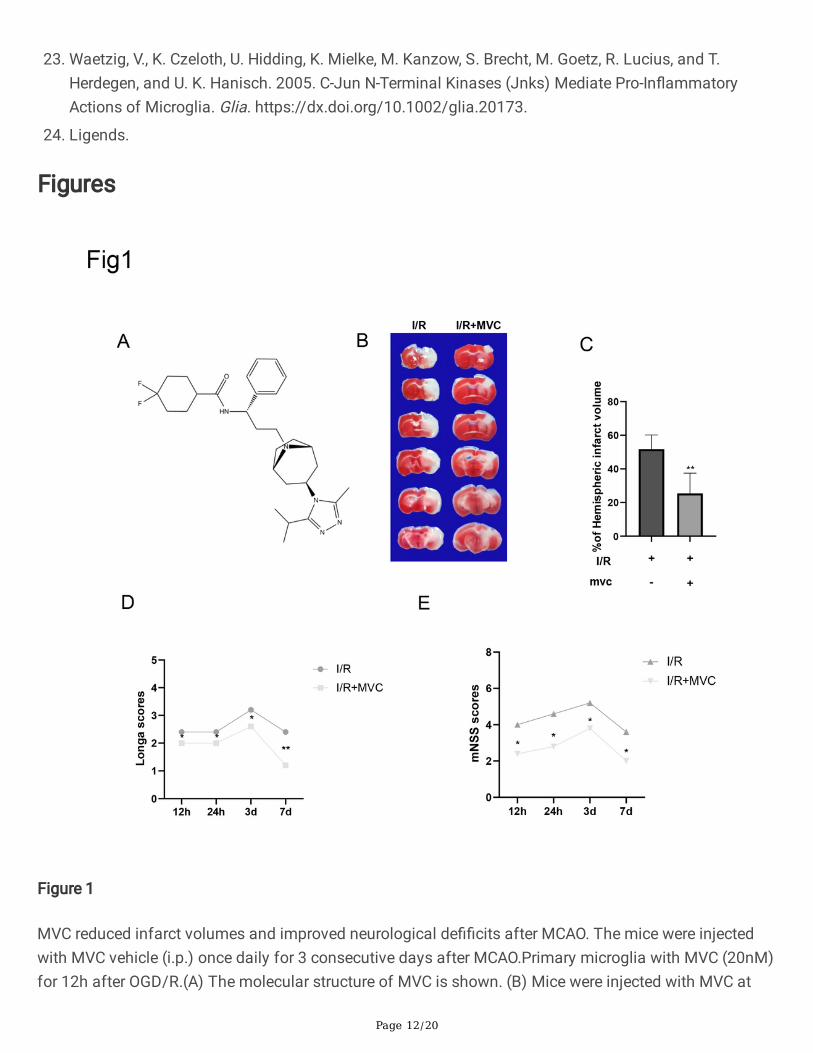
MVC Reduced Infarct Volume and Improved NeurologicalDe�cits After MCAOThe molecular structure of MVC, 4,4-Di�uoro-N-[(1S)-3-[(1R,5S)-3-(3-methyl-5-propan-2-yl-1,2,4-triazol-4-yl)-8-azabicyclo[3.2.1]octan-8-yl]-1-phenylpropyl]cyclohexane-1-carboxamide, is shown in Fig. 1A., MVCtreatment decreased the infarct volume (P < 0.05, Fig. 1B). MVC treatment at higher doses (20 umol/kg)provided signi�cant neuroprotection after CI/R injury (P < 0.05, Fig. 1C). Because the 20umol/kg doseproduced the most signi�cant effects, this dose was selected for future experiments. While the vehiclegroup exhibited poor neurological function as shown by the Longa and mNSS scores, MVC treatment(20umol/kg, i.p.) signi�cantly alleviated the neurological de�cits. Compared to the vehicle group, Longa

Page 7/20
and mNSS scores were signi�cantly lower in the MVC treatment group (P < 0.05, Fig. 1D, E), indicatingthat MVC treatment provided protective effects against CI/R injury in mice.
MVC downregulates apoptosis-associated cytokinesinduced by CI/R injury and OGD/RInhibiting the apoptotic effects of neurons in the ischemic penumbra is considered as a key target toprotect against CI/R injury. Previous studies have shown that the change in Bcl-2/Bax ratio re�ects thelevel of apoptosis[11]. .In this study, Western blot analysis of Bcl-2 and Bax was performed on braintissue in mice at 3 days after MCAO. Consistent with the previous results, the expression ratio of Bcl-2/Bax was decreased in the CI/R + vehicle group compared with those in the sham and MVC groups.However, MVC treatment resulted in an increase in Bcl2/Bax ratio compared to the CI/R + vehicle group (P < 0.05, Fig. 2A, B) with increased Bcl-2 expression and decreased Bax expression. At 12 h after OGD/R, thewestern blots of microglia showed that the expression ratio of Bcl-2/Bax was decreased in the OGD/Rgroup compared with those in the control and MVC groups (P < 0.05, Fig. 2C, D). TUNEL staining has alsobeen used as an apoptotic marker. The number of TUNEL positive cells in the brain tissue wassigni�cantly decreased after MVC treatment compared to the CI/R + vehicle group (P < 0.05, Fig. 2E, F).These results further support the anti-apoptotic effects of MVC in CI/R.
MVC Alleviated the In�ammatory Response After MCAOand OGD/RIn�ammation has been demonstrated to be involved in all stages of CI/R. To explore the potential anti-in�ammatory effects of MVC[12]., we detected the mRNA expression levels of pro-in�ammatorycytokines, including TNFa, IL-1b and IL-6, by quantitative real-time polymerase chain reaction (qPCR). ThemRNA levels of TNFa, IL-1b, and IL-6 were all decreased after MVC treatment compared with the CI/R + vehicle group (P < 0.05, Fig. 3A–C). At 12 h after OGD/R, we evaluated the levels of the IL-1β, IL-6 andTNF-α mRNAs and proteins using real-time PCR and ELISAs, respectively. Consistent with our hypothesis,MVC attenuated OGD-induced IL-1β, IL-6 and TNF-α expression in microglia (Fig. 3D-I). These resultsindicate that MVC signi�cantly alleviated the in�ammatory response after CI/R injury and OGD/R.
MVC Inhibited the NF-kB p65 SignallingAfter CI/R injury, nuclear factor-kappa B (NF-kB) pathway plays a central role in the post-ischemicproin�ammatory response [13].. MVC has been reported to mitigate CI/R injury by inhibiting the NF-kBsignlling pathway. In our study, the core proteins of the NF-kB signlling pathway were detected by Westernblot. CI/R injury signi�cantly increased the levels of phospho-IKβ/a and phospho-p65 compared with thesham and MVC groups. However, MVC treatment downregulated phospho-IkBa and phospho-p65 levels,while upregulated IkBa levels compared with the CI/R + vehicle group (P < 0.05, Fig. 4A-D). At 12 h afterOGD/R, signi�cantly increased the levels of phospho-IKβ/a and phospho-p65 compared with the sham
Page 8/20
and MVC groups. MVC treatment downregulated phospho-IkBa and phospho-p65 levels, whileupregulated IkBa levels compared with the OGD/R group (P < 0.05, Fig. 4E-H). Taken together, these dataindicated that MVC inhibits the NF-kB signaling pathway during CI/R injury and OGD/R.
MVC suppressed the phosphorylation of ERK1/2, JNK andP38 in CI/R and OGD/RThe MAPK signaling pathway is a well-known pathway involved in the in�ammatory response thatmainly consists of three MAPK subfamilies, including the ERK1/2, JNK and p38 families[14, 15]..Therefore, we aimed to investigate the molecular mechanism underlying the anti-in�ammatory effect ofMVC in the CI/R injury. As shown in Fig. 5A-D, CI/R injury increased the levels of the phosphorylatedERK1/2, JNK, and p38 proteins, which was consistent with the results of our previous study. After MVCtreatment, the phosphorylation of p38and JNK, but not ERK1/2, was suppressed (Fig. 5A-D). In addition,consistent with our previous �ndings, OGD/R increased the levels of the phosphorylated ERK1/2, JNK,and p38 proteins. After MVC treatment, the phosphorylation of p38 and JNK, but not ERK1/2, wassuppressed (Fig. 5E-I). According to these results, we concluded that MVC suppresses in�ammation byinhhibiting the p38 and JNK MAPK signaling pathways without altering the activation of ERK1/2 MAPK.
The JNK signaling pathway activator AN reverses the neuroprotective effects of MVC on cerebral I/Rinjury and OGD/R.
To further investigate whether JNK pathway inhibition mediates the neuroprotective effects of MVC oncerebral I/R injury, we treated mice with the JNK signaling pathway activator AN (0.1 mg/kg, i.p.) 30 minbefore ischemia. Western blot analysis showed that AN reversed the change in p-JNK levels induced byMVC treatment (P < 0.05, Fig. 6A-B). For further veri�cation, we evaluated the effects of differentintervention methods on infarct volume in MCAO mice. TTC staining showed that compared with I/R + MVC, AN treatment almost eliminated the protective effect of MVC after cerebral I/R injury (Fig. 7A-B).Based on the results above, the JNK pathway may contribute to the neuroprotective effects of MVC aftercerebral I/R injury.
DiscussionIschemic stroke is a serious vascular disease that signi�cantly threats public health. Due to progressiveimprovements in recent years regarding our understanding of its pathogenesis, it is now recognized thatcerebral ischemia-reperfusion injury is a complex pathophysiological process involving in�ammation,cellular damage by oxygen-derived free radicals, energy depletion in the brain, intracellular calcium onoverload, acidosis, toxicity of excitatory amino acids, nerve cell apoptosis, and the breakdown of theblood-brain barrier[16].. MVC has been reported to have many pharmacological effects, including anti-in�ammatory, and antiapoptotic effects[2].. Pharmacological research has demonstrated that MVC iscapable of penetrating the blood brain barrier (BBB)[17].. MVC is a potent and selective antagonist ofCCR5, which is localized on the surface of a variety of cells [18].. In the central nervous system, CCR5 is
Page 9/20
expressed by neurons, astrocytes and microglia[19].. In the present study, we observed theneuroprotective effect of MVC on the CI/R injury. Our results showed that MVC administration at 30 min,24 h and 48 h after MCAO obviously attenuated the brain infarct size and neurological de�cits on day 3after CI/R injury. In the subsequent experiments, pre-treatment with MVC effectively inhibited the CI/Rinjury-induced proin�ammatory mediators in mice and primary microglia by regulating the activation ofthe MAPK and NF-κB signaling pathways. According to our in vivo data, the i.p. injection of MVCattenuated the brain infarct volume and improved neurological de�cits after transient focal ischemia.Furthermore, MVC signi�cantly suppressed the release of in�ammatory cytokines, suggesting that theprotective effects of MVC on experimentally-induced stroke were at least partially mediated by theattenuation of microglia-mediated neuroin�ammation. Taken together, MVC is a potential neuroprotectiveagent to treat ischemic brain injury. IL-1β, IL-6 and TNF-α are likely the most extensively studied pro-in�ammatory cytokines involved in stroke. The levels of IL-1β, IL-6 and TNF-α are signi�cantly increasedin the brain within the �rst 24h after the induction of focal cerebral ischemia in mice[20].. These threecytokines are mainly synthesized by microglia in the stroke-lesioned rodent brain [21].. In the presentstudy, we con�rmed that MVC not only suppresses the OGD-induced expression of the IL-1β, IL-6 andTNF-α mRNA and proteins in microglia, but also inhibits the production of these pro-in�ammatorycytokines in the mouse brain after CI/R injury. In addition to MAPK pathways, NF-κB activation is alsocritically required for microglia-mediated CNS in�ammation. CI/R injury signi�cantly increased the levelsof phospho-IKβ/a and phospho-p65 compared with the sham and MVC groups. However, MVC treatmentdownregulated phospho-IkBa and phospho-p65 levels, while upregulated IkBa levels compared with theCI/R group. As shown in a recent study by Ganbold et al., NF-κB p65 silencing decreases the expressionof pro-in�ammatory cytokines and facilitates the anti-in�ammatory polarization of microglia[22]..Consistent with these results, we unequivocally demonstrated that MVC pre-treatment signi�cantlyreduced phospho-IkBa and phospho-p65 levels, while upregulated IkBa levels compared with the OGD/Rgroup. Subsequently, MVC suppressed NF-κB p65 activation and its nuclear translocation, suggestingthat the IκB/NF-κB p65 signaling pathway is involved in the anti-in�ammatory effects of MVC in OGD/Rmicroglia. Several studies have reported that the MAPK signaling pathways, including the ERK1/2, JNKand p38 pathways, play vital roles in microglial activation and in�ammatory response[14].. JNKs areessential mediators of microglia-mediated pro-in�ammatory response[23]. .Thus, treatments that inhibitMAPKs may be a promising intervention for in�ammatory responses induced by CI/R injury. In our currentstudy, we con�rmed that these three MAPK signaling pathways that were involved in thatphosphorylation of p38 and JNK were markedly diminished in CI/R injury after MVC pre-treatment,suggesting that p38 and JNK are the two important molecular targets of MVC. Furthermore, MVC tendedto decrease the levels of phospho-JNK. In this study, we provided robust evidence that the inhibition ofJNK activation can mediate the neuroprotective effects of MVC in a cerebral I/R injury model. To con�rmthe above conclusion, a JNK signaling pathway agonist (AN) was employed in the MCAO model. ANsigni�cantly increased the level of p-JNK and reversed the protective effects of MVC on cerebral I/R injury.AN-induced excessive activation of JNK may explain the �nding that AN almost completely blocked theneuroprotective effects of MVC. Nevertheless, to reduce the use of experimental animals, the number ofmice in each group was relatively small, which may cause potential bias. Further experiments will be

Page 10/20
conducted to validate the effect of MVC on mice models of ischemic stroke to make our research moremeaningful and intriguing.
DeclarationsEthics Approval and Consent to Participate All animal testing procedures were approved by the localexperimental ethics committee and were performed in accordance with the guidelines of the NationalInstitutes of Health Guide for the Care and Use of Laboratory Animals. All experimental procedures wereapproved by the Animal Ethics Committee of Yangzhou University (YZUNSFC2020-LCYXY-136).
Con�ict of Interest The authors declare no competing interests.
AcknowledgmentsThe authors would like to acknowledge Xiang Cao, A�liated of Drum Tower Hospital,Medical School of Nanjing University, China.
References1. Hurford, R., A. Sekhar, T. A. T. Hughes, and K. W. Muir. 2020. Diagnosis and Management of Acute
Ischaemic Stroke. Pract Neurol. https://dx.doi.org/10.1136/practneurol-2020-002557.
2. Cipriani, S., D. Francisci, A. Mencarelli, B. Renga, E. Schiaroli, C. D'Amore, F. Baldelli, and S. Fiorucci.2013. E�cacy of the Ccr5 Antagonist Maraviroc in Reducing Early, Ritonavir-Induced Atherogenesisand Advanced Plaque Progression in Mice. Circulation.https://dx.doi.org/10.1161/circulationaha.113.001278.
3. Liu, F. C., C. W. Zheng, and H. P. Yu. 2016. Maraviroc-Mediated Lung Protection Following Trauma-Hemorrhagic Shock. Biomed Res Int. https://dx.doi.org/10.1155/2016/5302069.
4. Kraft-Terry, S. D., A. R. Stothert, S. Buch, and H. E. Gendelman. 2010. Hiv-1 Neuroimmunity in the Eraof Antiretroviral Therapy. Neurobiol Dis. https://dx.doi.org/10.1016/j.nbd.2009.12.015.
5. Escola, J. M., G. Kuenzi, H. Gaertner, M. Foti, and O. Hartley. 2010. Cc Chemokine Receptor 5 (Ccr5)Desensitization: Cycling Receptors Accumulate in the Trans-Golgi Network. J Biol Chem.https://dx.doi.org/10.1074/jbc.M110.153460.
�. Venuti, A., C. Pastori, and L. Lopalco. 2017. The Role of Natural Antibodies to Cc Chemokine Receptor5 in Hiv Infection. Front Immunol. https://dx.doi.org/10.3389/�mmu.2017.01358.
7. Akahori, T., M. Sho, H. Kashizuka, T. Nomi, H. Kanehiro, and Y. Nakajima. 2006. A Novel Ccr5/Cxcr3Antagonist Protects Intestinal Ischemia/Reperfusion Injury. Transplant Proc.https://dx.doi.org/10.1016/j.transproceed.2006.10.115.
�. Grommes, J., M. Drechsler, and O. Soehnlein. 2014. Ccr5 and Fpr1 Mediate Neutrophil Recruitment inEndotoxin-Induced Lung Injury. J Innate Immun. https://dx.doi.org/10.1159/000353229.
9. Chiang, T., R. O. Messing, and W. H. Chou. 2011. Mouse Model of Middle Cerebral Artery Occlusion. JVis Exp. https://dx.doi.org/10.3791/2761.
Page 11/20
10. Yu, H., L. Song, X. Cao, W. Li, Y. Zhao, J. Chen, J. Li, Y. Chen, W. Yu, and Y. Xu. 2020. HederageninAttenuates Cerebral Ischaemia/Reperfusion Injury by Regulating Mlk3 Signalling. Front Pharmacol.https://dx.doi.org/10.3389/fphar.2020.01173.
11. Cong, C., L. Kluwe, S. Li, X. Liu, Y. Liu, H. Liu, W. Gui, T. Liu, and L. Xu. 2019. Paeoni�orin InhibitsTributyltin Chloride-Induced Apoptosis in Hypothalamic Neurons Via Inhibition of Mkk4-JnkSignaling Pathway. J Ethnopharmacol. https://dx.doi.org/10.1016/j.jep.2019.03.030.
12. Karampoor, S., H. Zahednasab, R. Amini, M. Esghaei, M. Sholeh, and H. Keyvani. 2020. MaravirocAttenuates the Pathogenesis of Experimental Autoimmune Encephalitis. Int Immunopharmacol.https://dx.doi.org/10.1016/j.intimp.2019.106138.
13. Zhang, D. D., M. J. Zou, Y. T. Zhang, W. L. Fu, T. Xu, J. X. Wang, W. R. Xia, Z. G. Huang, X. D. Gan, X. M.Zhu, and D. G. Xu. 2017. A Novel Il-1ra-Pep Fusion Protein with Enhanced Brain PenetrationAmeliorates Cerebral Ischemia-Reperfusion Injury by Inhibition of Oxidative Stress andNeuroin�ammation. Exp Neurol. https://dx.doi.org/10.1016/j.expneurol.2017.06.012.
14. Ji, R. R., R. W. th Gereau, M. Malcangio, and G. R. Strichartz. 2009. Map Kinase and Pain. Brain ResRev. https://dx.doi.org/10.1016/j.brainresrev.2008.12.011.
15. Piotrowska, A., K. Kwiatkowski, E. Rojewska, W. Makuch, and J. Mika. 2016. Maraviroc ReducesNeuropathic Pain through Polarization of Microglia and Astroglia - Evidence from In vivo and In vitroStudies. Neuropharmacology. https://dx.doi.org/10.1016/j.neuropharm.2016.04.024.
1�. Jin, R., G. Yang, and G. Li. 2010. In�ammatory Mechanisms in Ischemic Stroke: Role of In�ammatoryCells. J Leukoc Biol. https://dx.doi.org/10.1189/jlb.1109766.
17. Haruwaka, K., A. Ikegami, Y. Tachibana, N. Ohno, H. Konishi, A. Hashimoto, M. Matsumoto, D. Kato, R.Ono, H. Kiyama, A. J. Moorhouse, J. Nabekura, and H. Wake. 2019. Dual Microglia Effects on BloodBrain Barrier Permeability Induced by Systemic In�ammation. Nat Commun.https://dx.doi.org/10.1038/s41467-019-13812-z.
1�. Levy, J. A. 2009. Hiv Pathogenesis: 25 Years of Progress and Persistent Challenges. Aids.https://dx.doi.org/10.1097/QAD.0b013e3283217f9f.
19. Kaul, M., Q. Ma, K. E. Medders, M. K. Desai, and S. A. Lipton. 2007. Hiv-1 Coreceptors Ccr5 and Cxcr4Both Mediate Neuronal Cell Death but Ccr5 Paradoxically Can Also Contribute to Protection. CellDeath Differ. https://dx.doi.org/10.1038/sj.cdd.4402006.
20. Hill, J. K., L. Gunion-Rinker, D. Kulhanek, N. Lessov, S. Kim, W. M. Clark, M. P. Dixon, R. Nishi, M. P.Stenzel-Poore, and F. P. Eckenstein. 1999. Temporal Modulation of Cytokine Expression FollowingFocal Cerebral Ischemia in Mice. Brain Res. https://dx.doi.org/10.1016/s0006-8993(98)01140-8.
21. Lambertsen, K. L., K. Biber, and B. Finsen. 2012. In�ammatory Cytokines in Experimental and HumanStroke. J Cereb Blood Flow Metab. https://dx.doi.org/10.1038/jcbfm.2012.88.
22. Ganbold, T., Q. Bao, J. Zandan, A. Hasi, and H. Baigude. 2020. Modulation of Microglia Polarizationthrough Silencing of Nf-Κb P65 by Functionalized Curdlan Nanoparticle-Mediated Rnai. ACS ApplMater Interfaces. https://dx.doi.org/10.1021/acsami.9b23004.
Page 12/20
23. Waetzig, V., K. Czeloth, U. Hidding, K. Mielke, M. Kanzow, S. Brecht, M. Goetz, R. Lucius, and T.Herdegen, and U. K. Hanisch. 2005. C-Jun N-Terminal Kinases (Jnks) Mediate Pro-In�ammatoryActions of Microglia. Glia. https://dx.doi.org/10.1002/glia.20173.
24. Ligends.
Figures
Figure 1
MVC reduced infarct volumes and improved neurological de��cits after MCAO. The mice were injectedwith MVC vehicle (i.p.) once daily for 3 consecutive days after MCAO.Primary microglia with MVC (20nM)for 12h after OGD/R.(A) The molecular structure of MVC is shown. (B) Mice were injected with MVC at

Page 13/20
doses of 20mg/kg (i.p.) after MCAO. The representative TTC-stained coronal sections in vehicle- andMVC-treated mice are shown. (C) MVC signi��cantly reduced infarct volumes after MCAO. MVCtreatments signi��cantly reduced Longa (D) and mNSS (E) scores after MCAO. The bar graphs representthe mean ± SEM of 6 brains in each group. *P < 0.05 versus the MCAO group
Figure 2

Page 14/20
MVC downregulates apoptosis-associated cytokines induced by CI/R injury and OGD/R. Mice weretreated with MVC (20 mg/kg) or vehicle (i.p.) once daily for 3 consecutive days after MCAO. Primarymicroglia with MVC (20nM) for 12h after OGD/R.Western blot was used to detect the expression levels ofapoptosis-related proteins, and TUNEL staining was used to measure the apoptotic levels after CI/Rinjury. A At 3 days after MCAO, Representative images show the expression levels of Bcl-2, Bax byWestern Blot analysis with GAPDH as a loading control. B Quantitative analysis of the Bcl-2 to Bax ratio.C At 12 h after OGD/R,Representative images show the expression levels of Bcl-2, Bax by Western Blotanalysis with GAPDH as a loading control. D Quantitative analysis of the Bcl-2 to Bax ratio. E TUNEL-positive cells showed green �uorescence, and DAPI-positive cells showed blue �uorescence. FQuanti��cation of the number of TUNEL-positive neural cells in each group. MVC treatment signi��cantlydecreased the number of TUNEL-positive neural cells after CI/R injury. Scale bar, 50 µm; magni��cation,20x. The bar graphs represent the mean ± SEM of three brains in each group. *P < 0.05 versus the CI/Rgroup.

Page 15/20
Figure 3
MVC Alleviated the In�ammatory Response After MCAO and OGD/R.The mice were treated with MVC (20mg/kg, i.p.) or vehicle once daily for 3 consecutive days after CI/R injury. Primary microglia with MVC(20nM) for 12h after OGD/R.Pro-in��ammatory cytokine levels were determined by qRT-PCR, ELISA. A–CThe mRNA levels of TNF-a, IL-6, and IL-1b in the ischaemic penumbra tissues. MVC signi��cantlydecreased the mRNA levels of TNF-a, IL-6, and IL-1b after CI/R injury. D-F The mRNA levels of TNF-a, IL-6,

Page 16/20
and IL-1b in microglia.MVC signi��cantly decreased the mRNA levels of TNF-a, IL-6, and IL-1b afterOGD/R. G-I the protein levels of TNF-a, IL-6 and IL-1b were evaluated by ELISA.MVC signi��cantlydecreased the protein levels of TNF-a, IL-6, and IL-1b after OGD/R.
Figure 4
MVC Inhibited the NF-kB p65 Signalling.The mice were treated with MVC (20 mmol/kg, i.p.) or vehicleonce daily for 3 consecutive days after CI/R. Primary microglia with MVC (20nM) for 12h afterOGD/R.The protein levels were detected by Western blot. A The expression of p-IκBα, IκBα, p65 and p-p65in the ischaemic penumbra tissues was analyzed by Western blotting. GAPDH was used as a loadingcontrol. B-D The greyscale value of each band was evaluated by ImageJ software.Quantitative analysis

Page 17/20
showed that MVC decreased the expression levels of p-IκBα, IκBα and p-p65 after CI/R injury. E Theexpression of p-IκBα, IκBα, p65 and p-p65 in microglia was analyzed by Western blotting. GAPDH wasused as a loading control. G-H Quantitative analysis showed that MVC decreased the expression levels ofp-IκBα, IκBα and p-p65 after OGD/R.The bar graphs represent the mean ± SEM of three brains in eachgroup. *P < 0.05 versus the CI/R group.
Figure 5

Page 18/20
MVC suppressed the phosphorylation of ERK1/2, JNK and P38 in CI/R and OGD/R.Mice were treated withMVC (20 mg/kg, i.p.) or vehicle once daily for 3 consecutive days after CI/R.Primary microglia with MVC(20nM) for 12h after OGD/R. After 3 days of reperfusion, cerebral tissue was collected, and protein levelswere detected by Western blot. A The representative images show the levels of p-p38, p38, p-ERK, ERK, p-JNK, JNK, and GAPDH. GAPDH was used as a loading control. B Quantitative analysis showed that MVCdecreased the expression level of p-p38 and p-JNK, but not that of p-ERK , after CI/R injury. C Therepresentative images show the levels of p-p38, p38, p-ERK, ERK, p-JNK, JNK, and GAPDH. GAPDH wasused as a loading control. D Quantitative analysis showed that MVC decreased the expression level of p-p38 and p-JNK, but not that of p-ERK , after OGD/R. The bar graphs represent the mean ± SEM of threebrains in each group. *P < 0.05 versus the CI/R group and OGD/R group.
Figure 6

Page 19/20
The JNK signaling pathway activator AN reverses the neuroprotective effects of MVC on cerebral I/Rinjury and OGD/R.Mice were treated with MVC (20 mg/kg, i.p.) or vehicle once daily for 3 consecutivedays after CI/R.Primary microglia with MVC (20nM) for 12h after OGD/R. Protein levels were determinedby Western blot. A, B Mice were treated with MVC (20 mg/kg, i.p.) or vehicle 1 h after ischemia and AN(0.1 mg/kg, i.p.) was given 30 min before ischemia. Western blot and quantitative analysis showed thatAN treatment reversed the inhibitory effect of MVC on JNK phosphorylation.C, D Primary microglia withMVC (20nM) after OGD/R and AN (0.1nM) was given 30 min before OGD.Western blot and quantitativeanalysis showed that AN treatment reversed the inhibitory effect of MVC on JNK phosphorylation.
Figure 7
A The volume of cerebral infarction was measured by TTC staining. B Statistical analysis ofrepresentative TTC-stained coronal sections indicated that the JNK agonist AN almost blocked theprotective effect of MVC. The bar graph showed the mean ± S.E.M. of three brains in each group, *,

Page 20/20
indicating P<0.05 compared with the I/R group. C-F MVC treatments signi��cantly reduced Longascores,mNSS scores,Grip test and Time on Rotarod after CI/R injury, but the JNK agonist AN almostblocked the protective effect of MVC.