osteology of the middle triassic archosaur lewisuchus ...054) bittencourt et... · osteology of the...
TRANSCRIPT

Osteology of the Middle Triassic archosaur Lewisuchus admixtus Romer(Cha~nares Formation, Argentina), its inclusivity, and relationships amongst
early dinosauromorphs
Jonathas S. Bittencourta*, Andrea B. Arcuccib, Claudia A. Marsicanoc and Max C. Langerd
aDepartamento de Geologia, Universidade Federal de Minas Gerais, Av. Antonio Carlos 6670, 31270901, Belo Horizonte, Brazil; bAreade Zoologia, Universidad Nacional de San Luis, Chacabuco 917, 5700, San Luis, Argentina; cDepartamento de Ciencias Geol�ogicas,Universidad de Buenos Aires, IDEAN-CONICET, Ciudad Universitaria, Pab. II, C1428 DHE, Ciudad Aut�onoma de Buenos Aires,
Argentina; dLaborat�orio de Paleontologia, Departamento de Biologia, Universidade de S~ao Paulo, Av. Bandeirantes 3900, 1404901,Ribeir~ao Preto, Brazil
(Received 6 March 2013; accepted 13 October 2013)
Lewisuchus admixtus is an enigmatic early dinosauriform from the Cha~nares Formation, Ladinian of Argentina, which hasbeen recently considered a member of Silesauridae. Yet, it differs markedly from Late Triassic silesaurids in dental andvertebral anatomy. Indeed, a detailed redescription of its holotype allowed the identification of several features of theskeleton previously unrecognized amongst silesaurids. These include pterygoid teeth, a dorsomedial posttemporal openingon the otoccipital, foramina associated with cranial nerves X–XII on the caudal region of the prootic–otoccipital, andpostaxial neck/trunk vertebrae with craniocaudally expanded neural spines. The presence of a single row of presacralscutes was also confirmed. Some elements previously referred to, or found associated with, the holotype, includinga lower jaw, pedal elements and an astragalus, more probably correspond to proterochampsid remains. Theanatomical information available for the holotype of L. admixtus was rescored into a new phylogenetic dataset fordinosauromorphs, mostly based on previous works. Lewisuchus admixtus and Pseudolagosuchus major are treated asdistinct OTUs because their preserved skeletons mostly lack overlapping parts. The parsimony analysis supports the basalposition of L. admixtus within dinosauriforms, prior to the silesaurid–dinosaur split, rather than at the base of Silesauridae.This suggests that a higher number of early dinosauriform clades branched in the Middle and Late Triassic than previouslysuggested.
Keywords: Lewisuchus; Pseudolagosuchus; basal dinosauromorphs; Middle Triassic; silesaurids
Introduction
Archosaur remains discovered from the Middle to Late
Triassic of Europe (Dzik 2003; Dzik & Sulej 2007), North
America (Ezcurra 2006; Nesbitt et al. 2007; Nesbitt et al.
2010), Brazil (Ferigolo & Langer 2007) and Africa
(Nesbitt et al. 2010; Kammerer et al. 2012; Peecook et al.
2013) have improved the record of lightly built dinosaur
precursors (Bakker & Galton 1974; Bonaparte 1978;
Sereno 1991a; Novas 1996), until recently known only
from the Ladinian of the Cha~nares Formation, in Argen-
tina (Romer 1971, 1972c; Bonaparte 1975; Arcucci
1987). Various studies positioned these taxa as successive
outgroups to Dinosauria, including Lagerpetidae,Marasu-
chus and Silesauridae (see Nesbitt 2011, for review).
Lagerpetids comprise three species (i.e. Lagerpeton cha-
narensis, Dromomeron romeri and D. gregorii), which
are currently known from Ladinian and Norian deposits of
Argentina and USA (Arcucci 1986; Sereno & Arcucci
1994a; Irmis et al. 2007a; Nesbitt et al. 2009a). Marasu-
chus is monoespecific (M. lilloensis), and restricted to the
Cha~nares Formation (Sereno & Arcucci 1994b). Silesaur-
ids are minimally known from Anisian to Norian strata in
Poland, Brazil, Argentina, United States, Tanzania,
Morocco and Zambia (Dzik 2003; Ferigolo & Langer
2007; Nesbitt et al. 2010; Kammerer et al. 2012; Peecook
et al. 2013). Some authors support the position of silesaur-
ids as basal ornithischians (Ferigolo & Langer 2007;
Langer et al. 2007b; Langer & Ferigolo 2013), but this
hypothesis has been disputed (Irmis et al. 2007b; Nesbitt
et al. 2010). Marasuchus, silesaurids and dinosaurs com-
prise together the Dinosauriformes (Novas 1996; Langer
& Benton 2006; Nesbitt et al. 2010), into which the poorly
known Saltopus from the Late Triassic of Scotland has
been recently included (Benton & Walker 2011).
An enigmatic specimen collected by the La Plata-Har-
vard expedition during 1964–1965 in the Cha~nares For-
mation (Romer 1966; Romer & Jensen 1966; Rogers et al.
*Corresponding author. Email: [email protected]
� The Trustees of the Natural History Museum, London 2014. All Rights Reserved.
Journal of Systematic Palaeontology, 2014
Vol. 0, No. 0, 1–31, http://dx.doi.org/10.1080/14772019.2013.878758

2001), La Rioja, Argentina, became the holotype of Lewi-
suchus admixtus Romer, 1972c. The specimen comprises
a partial skull and post-cranial remains, including most of
the pre-sacral column, pectoral and hind limb elements.
As it was recovered from a concretion that also included
remains of cynodonts and proterochampsids, the associa-
tion of the non-articulated parts to the preserved skeleton
is problematic (Arcucci 1998).
Although the taxonomic validity of L. admixtus has
never been disputed, its phylogenetic position is contro-
versial. Romer (1972c, p. 13) allied the taxon with the tra-
ditional ‘pseudosuchians’ (Cruickshank 1979), supporting
a possible bearing on the origin of ‘coelurosaurs’. Other
authors have suggested affinities with gracilisuchids
(Carroll 1988; Sues 1997), or basal crocodylomorphs
(Sereno 1991a), more specifically with sphenosuchians
(Bonaparte 1981; Olshevsky 1991). Parrish (1993) per-
formed a phylogenetic study in which L. admixtus resulted
as a basal suchian, but this pioneer analysis employed low
sampling of characters and taxa. Alternatively, Paul
(1988) and Arcucci (1997, 1998) suggested its placement
closer to dinosaurs than to other archosaurs, a hypothesis
that has gained support due to its positioning within Sile-
sauridae (Brusatte et al. 2010; Nesbitt et al. 2010; Nesbitt
2011). Data gathered from the femoral anatomy of the
coeval Pseudolagosuchus major Arcucci, 1987, often
regarded as a junior synonym of L. admixtus (Arcucci
1997, 1998; Nesbitt et al. 2010), have added support to
that relationship. However, the synonymization between
these taxa is problematic due to the scarcity of overlap-
ping elements in their preserved skeletons.
In order to improve the available information about L.
admixtus to phylogenetic studies, we performed a detailed
description of its holotype. Both the assignment of the pre-
served elements to the type specimen and the synonymy
between L. admixtus and P. major are scrutinized via the
phylogenetic signal of the preserved bones. The revised
anatomical information on L. admixtus is rescored in a
new character–taxon matrix for basal dinosauromorphs, in
order to discuss its phylogenetic position within that
clade.
Institutional abbreviationsAMNH: American Museum of Natural History, New
York, USA; NHMUK: The Natural History Museum,
London, UK; GPIT: Institut f€ur Geologie und
Pal€aontologie, T€ubingen, Germany; GR: Ghost Ranch
Ruth Hall Museum of Palaeontology, Abiquiu, NM, USA;
MACN: Museo Argentino de Ciencias Naturales, Buenos
Aires, Argentina; MCN: Museu de Ciencias Naturais/
Fundac~ao Zoobotanica, Porto Alegre, Brazil; MCP:
Museu de Ciencias e Tecnologia/PUC, Porto Alegre, Bra-
zil; MNA: Museum of Northern Arizona, Flagstaff, AZ,
USA; NMMNHS: New Mexico Museum of Natural
History and Science, Albuquerque, NM, USA; NMT:
National Museum of Tanzania, Dar es Salaam, Tanzania;
PULR: Universidad Nacional de La Rioja, La Rioja,
Argentina; PVL: Fundaci�on Miguel Lillo, San Miguel de
Tucum�an, Argentina; PVSJ: Universidad Nacional de
San Juan, San Juan, Argentina; SAM: South African
Museum, Cape Town, South Africa; SMNS: Staatliches
Museum f€ur Naturkunde, Stuttgart, Germany; UCMP:
University of California Museum of Paleontology,
Berkeley, CA, USA; UFRGS: Universidade Federal do
Rio Grande do Sul, Porto Alegre, Brazil; ULBRA:
Universidade Luterana do Brasil, Canoas, Brazil; ZPAL:
Institute of Paleobiology of the Polish Academy of
Science, Warsaw, Poland.
Systematic palaeontology
Archosauria Cope, 1869 sensu Nesbitt, 2011
Ornithodira Gauthier, 1986 sensu Sereno, 1991a
Dinosauromorpha Benton, 1985 sensu Nesbitt,
2011
Dinosauriformes Novas, 1992 sensu Nesbitt,
2011
Genus Lewisuchus Romer, 1972c
Revised diagnosis. As for type and only species.
Type species. Lewisuchus admixtus Romer, 1972c.
Lewisuchus admixtus Romer, 1972c
(Figs 1–11)
1972c Lewisuchus admixtus Romer: figs 1 (except
mandible), 2–4, 6–7, 8a.
1978 Lewisuchus admixtus Romer; Bonaparte: fig. 134
(except mandible).
?2011 Lewisuchus admixtus Romer; Nesbitt: figs 24G,
28E, 30F.
Holotype. PULR 01: incomplete left and right maxillae
with teeth; caudal portion of the skull, including the left
laterotemporal region: jugal, postorbital, quadratojugal,
squamosal, and quadrate; braincase, comprising supraoc-
cipital, basioccipital, otoccipital, laterosphenoid, parabasi-
sphenoid and prootic; articulated left pterygoid and
ectopterygoid; cervical vertebrae from atlas to the seventh
vertebra, 11 dorsal vertebrae, nine proximal to mid-caudal
vertebrae; both scapulocoracoids and humeri; incomplete
tibiae. Bones previously referred to the holotype, includ-
ing an isolated dentary and pedal elements (Romer 1972c)
are now assigned to indeterminate proterochampsids.
Locality and horizon. Los Cha~nares Locality, Talam-
paya National Park, La Roja Province, north-western
Argentina, Cha~nares Formation (Romer & Jensen 1966;
Rogers et al. 2001). Several authors assigned a Middle
2 J. S. Bittencourt et al.

Triassic age to the Cha~nares Formation (Bonaparte 1982;
Arcucci 1990), including Anisian (Romer & Jensen 1966;
Arcucci 1990; Bonaparte 1997), Anisian–Ladinian
(Arcucci & Marsicano 1998) or Ladinian (Mancuso &
Marsicano 2008; Desojo & Arcucci 2009). Desojo et al.
(2011) suggested a possible earliest Carnian age for this
stratigraphical unit. New data are required to corroborate
this hypothesis.
Revised diagnosis. Dinosauromorph that can be distin-
guished from its kin by the following combination of fea-
tures (asterisks indicate potential autapomorphies):
extremely elongated skull, supraoccipital nearly horizon-
tal, three foramina caudal to the metotic strut�, craniocau-dally extending rugose ridge on the mid-height of the
lateral surface of the axial neural spine�, postzygodiapo-physeal lamina of the caudal cervical vertebrae projecting
caudally to the tip of the postzygapophysis, and the pres-
ence of a single row of scutes associated to the distal tip
of the cervical and dorsal neural spines.
Description and comparisonsWe conducted a comparative description of the holotype
of Lewisuchus admixtus based on the first-hand analysis
of several archosaur specimens and on the literature
(Supplemental Table 1). Because L. admixtus has been
variously related to coeval archosauriforms such as proter-
ochampsids, Gracilisuchus and Sphenosuchia, compari-
sons with those taxa were also performed, albeit no
evidence supports a close relationship of this species with
any of them.
Maxilla. The isolated right maxilla referred to Lewisu-
chus admixtus is elongated, low and transversely com-
pressed (Fig. 1A–F). Its dimensions suggest that the skull
was low and elongated, similar to that of basal crocodylo-
morphs (Walker 1990; Clark et al. 2000), and longer than
that of any known basal dinosauriform. It differs from the
lower and medially inclined maxillae of the protero-
champsids, and from the dorsoventrally enlarged maxil-
lary body of the robust pseudosuchians (Sill 1974;
Barberena 1978). There is no definitive evidence that the
maxilla belongs to the holotype, because the rostral por-
tion of the jugal is damaged, and they cannot be unambig-
uously articulated. However, they match in anatomy and
size, so we tentatively associate the maxilla to the holo-
type until further evidence is available.
The rostrally rounded rostral ramus (measured from the
rostral edge of the external antorbital fenestra) encom-
passes approximately one-fifth of the total length of the
preserved maxilla (Fig. 1A, B). The ascending ramus
tapers dorsocaudally as in most archosauriforms (Gower
1999; Nesbitt 2011), and its rostral margin forms an angle
of 30� with the alveolar margin of the bone. The base of
the ascending ramus bears a shallow notch, which sets it
apart from the rostral ramus of the maxilla.
On its rostrolateral surface, between the antorbital
fenestra and the alveolar margin, the maxilla is pierced by
a small foramen. Other Late Triassic dinosauriforms, such
as Silesaurus (ZPAL AbIII/361/26), Sacisaurus (MCN
PV10050), Pampadromaeus (ULBRA-PVT016) and Coe-
lophysis (e.g. MNA V3315), possess multiple foramina on
the lateral maxilla. The external antorbital fenestra is cra-
niocaudally elongated, and is marked by a faint rim bor-
dering the caudolateral margin of the ascending ramus,
and the dorsolateral surface of the maxillary caudal pro-
cess. An enlarged version of this rim is seen in sauri-
schians, including Eoraptor (PVSJ 512), and theropods
(Rowe 1989; Rauhut 2003). Ventral to the rim, the lateral
surface of the maxilla bears a shallow and rostrocaudally
elongated sulcus, which ends rostral to the caudal tip of
the caudal ramus, similar to that seen in Sacisaurus (MCN
PV10050).
The antorbital fossa is shallower than the lateral surface
of the caudal maxillary ramus. This morphology is com-
parable to that of Sacisaurus (MCN PV10050), and early
dinosaurs (Langer & Benton 2006). The area of the medial
wall of the antorbital fossa compares to that of early dino-
sauriforms, such as Sacisaurus (MCN PV10050),
Agnosphitys (Fraser et al. 2002), and most basal dinosaurs
(Langer & Benton 2006), because it is transversely wider
than the maxillary ascending ramus. Due to poor preserva-
tion, the existence of an excavation or aperture on the lat-
eral surface of the antorbital fossa, as widely recognized
in dinosauriforms (Sereno 1999; Rauhut 2003; Langer &
Benton 2006) cannot be evaluated.
As in most archosaurs (Brusatte et al. 2010; Nesbitt
2011), the caudal maxillary ramus is elongated (77% of
the total preserved length), tapers caudally, and bears den-
tal alveoli along its entire preserved ventral margin.
Medially, the maxilla is convex along its whole exten-
sion. A faint crest extends along the dorsal portion of the
caudal ramus, internally to the medial wall of the antorbi-
tal fossa. An enlarged version of this crest is present in
one maxilla of Silesaurus (ZPAL AbIII/361/26). Ros-
trally, two additional crests extend on the medial surface
of the maxilla. The upper one is a rostrocaudally bulged
area restricted to the base of the ascending ramus, which
bears a peculiar foramen that probably connects the antor-
bital fossa with the palatal portion of the maxilla. The
lower crest is the palatal process, which forms a steep
margin above the three rostralmost dental alveoli. The
palatal process, like that of Agnosphitys (Fraser et al.
2002), is short and rod-like, but it is unclear if it contacted
its antimere. The dorsal surface of the palatal process
forms a horizontal shelf that probably contacted the cau-
dal portion of the premaxilla.
An additional fragment of a left maxilla with seven in
situ teeth plus two empty alveoli (Fig. 1G) was referred to
L. admixtus by Romer (1972c). The maxillary rim on the
lateral margin of the external antorbital fenestra is similar
Osteology of the Middle Triassic archosaur Lewisuchus admixtus 3

Figure 1. Lewisuchus admixtus, PULR 01. Right maxilla in A, B, lateral, C, D, medial, E, dorsal and F, ventral views. G, left maxilla inlateral view, with a detail of the tooth attachment. H, Sixth and I, seventh preserved maxillary teeth, respectively, in lingual view and incross section. Abbreviations: anfo, antorbital fossa; cr, crest; enl, enamel layer; de, denticles; fo, foramen; mcrp, maxillary cranial pro-cess; no, notch; pap, palatal process; puc, pulp channel. Scale bars: A–F ¼ 20 mm; G–I ¼ 10 mm.
4 J. S. Bittencourt et al.

to that described above for the right maxilla, supporting a
tentative assignment of this bone to PULR 01.
Dentition. The right maxilla possesses 20 preserved
tooth positions with remains of at least 13 teeth in various
states of preservation (Fig. 1A–D). Although the maxilla
is not complete, there is no evidence of additional alveoli
caudal to the last preserved one, thus the maxilla of Lewi-
suchus admixtus bears 20 teeth. Overall, the teeth display
the anatomical pattern of the carnivorous archosauriforms,
being similar to those described for Tropidosuchus (PVL
4601) and Marasuchus (PVL 3870). The crowns are
mostly elongated, labiolingually compressed, caudally
curved, with convex mesial margin and concave distal
margin (Fig. 1A–I). They do not expand from the base
(i.e. basal constriction absent), nor possess a basal cingu-
lum or wear facets, as in herbivorous/omnivorous dino-
sauriforms (Yates 2003; Butler et al. 2008; Nesbitt et al.
2010). The tooth crowns are elliptical in cross section due
to their labiolingual compression.
Although the rostralmost teeth are not complete, the
evidence available from the alveoli and the preserved
base of some crowns suggests that the largest maxillary
teeth are located within the rostral third of the bone. Those
elements are both apicobasally higher and mesiodistally
wider than more caudal teeth. In fact, the overall dimen-
sions of the maxillary teeth gradually diminish towards
the caudal end of the bone. Based on the preserved basal
portion of the fourth tooth, this is probably the largest
maxillary tooth, suggesting the presence of an enlarged
fang-like element close to the rostral portion of the max-
illa. Because the maxilla/premaxilla contact is not pre-
served, the existence of a rostral diastema cannot be
evaluated. Likewise, because the caudal portion of the
maxilla is missing and the contact with the jugal and/or
lacrimal is elusive, the extension of the maxillary tooth
row below the orbit cannot be determined.
The 11th maxillary tooth bears conspicuous evidence of
serration (Fig. 1H). Its crown is shorter than those of more
caudal teeth, suggesting that it was not completely
erupted. It bears five serrations along 0.60 mm in the api-
cal portion of the mesial carina. The serrations are small
knob-like protuberances, distally rounded (i.e. spatulate in
lateral view), expanding with an angle of 90� to the tooth
margin, similar to that observed in carnivorous archo-
saurs. Shallow grooves separate each serration, but these
do not mark the tooth crown beyond the base of the serra-
tion. This differs from the typical chisel-like structure of
the herrerasaurids (Bittencourt & Kellner 2009) and neo-
theropods (Abler 1997). They also differ from the coarser
serration of silesaurids and ornithischians (Dzik 2003;
Norman et al. 2004a; Ferigolo & Langer 2007), and the
sharply pointed denticles of ‘prosauropods’ (Barrett 2000;
Galton & Upchurch 2004).
The serrations observed in the basal portion of the distal
carina of the tooth occupying the 15th maxillary tooth are
somewhat distinct. They are not distally rounded as the
mesial serrations of the 11th tooth, but bear a somewhat
pointed edge in their ‘apical’ portion. This difference may
reflect either a preservation artefact or an individual poly-
morphism given the relative state of development of the
tooth. The tenth maxillary tooth has only faint evidence of
serration, which may result from poor preservation or
wearing. The differences in the serration morphology
between the 11th and 15th maxillary teeth are not due to
wearing.
The incomplete isolated left maxilla bears seven teeth
(Fig. 1G), the two rostralmost of which are the largest
ones (basally longer and apicobasally deeper). Compared
to the more complete right maxilla, the dimensions of the
preserved teeth suggest that they belong to the caudal
third of the bone. Yet, the mode of preservation of the
bone and teeth is rather distinct. The colour of the teeth
varies from red-brown in the more complete maxilla, to
strong carmine in the less complete one. The preserved
teeth are bulged on the labial surface. Differently from the
right maxilla, the more distal teeth are less caudally
curved and apparently not as pointed at the crown tip.
However, these differences may be related to
preservation.
The internal structure of the second preserved tooth of
the left maxilla is exposed (Fig. 1G, detail). The root is
slightly longer than the crown, and is formed by a broad
hollow pulp cavity, encased by a two-layered wall. The
external cementum layer is very thin and covers the thick
dentine layer, which is composed of a palisade of incre-
mental lines. It does not show evidence of ankylosis, as
has been assumed for some silesaurids (Nesbitt et al.
2010; Nesbitt 2011). From the proximal third of the tooth,
a thin layer of enamel fulfils the space between the dentine
and the cementum. The thickness of the enamel layer
increases towards the tip of the tooth, but never achieves
more than twice the width of the dentine stratum. The ser-
rations in the distal carina are spatulate, with a density of
nine serrations per mm.
Postorbital. The low postorbital body expands medially
(Fig. 2A, B), and its dorsal border is marked by a rostro-
caudally oriented crest, similar to those of Tropidosuchus
(PVL 4601) and Gracilisuchus (Romer 1972b; Brinkman
1981). The participation of the postorbital in the dorso-
temporal (¼ supratemporal) fenestra cannot be assured in
Lewisuchus admixtus due to poor preservation. The basal
portion of the incomplete rostral ramus is dorsoventrally
wider than that of the caudal ramus. Despite this incom-
pleteness, the postorbital body was probably closer to
the dorsal edge of the orbit than that of Gracilisuchus
(Brinkman 1981). The dorsal crest roofs a broad lateral
excavation that extends caudally along the preserved
Osteology of the Middle Triassic archosaur Lewisuchus admixtus 5

rostral portion of the caudal process. The excavation spans
ventrally as an elongated sulcus halfway the length of the
ventral ramus, as also seen in other archosauriforms, such
as Chanaresuchus (PULR 07), Gracilisuchus (Brinkman
1981) and Pampadromaeus (ULBRA-PVT016).
The elongated ventral ramus is slender, and tapers
smoothly ventrally. By contrast, the ventral ramus of the
postorbital of Silesaurus (Dzik & Sulej 2007) possesses a
caudal kink. In this respect, the postorbital of L. admixtus
is comparable to that of Gracilisuchus (Brinkman 1981)
and some early dinosaurs (e.g. Pampadromaeus, ULBRA-
PVT016). The dorsal ramus of the jugal is mostly over-
lapped by the ventral ramus of the postorbital, along an
extensive suture between both bones. As seen in most arch-
osauriforms (Nesbitt 2011), the ventral ramus curves ros-
trally, and the rostral border has a concave outline. Its distal
third also bends medially, reaching the floor of the orbit.
Ventrally, the postorbital contacts both the lateral mar-
gin of the ectopterygoid and a medial portion of the jugal.
The medial surface of the postorbital is mostly concave
and bears a faint ridge extending along the proximal third
of the caudomedial border. The transverse width of the
postorbital at its broadest preserved portion (the dorsal
region) encompasses approximately one-fifth of its height.
Jugal. The preserved jugal is interpreted as aligned sub-
horizontally (Fig. 2A, B). Contrasting with Sphenosuchus
(SAM-PK-3014), its rostral ramus is transversely wider
than dorsoventrally high, the medial part of which articu-
lates with the pterygoid–ectopterygoid complex, forming
the floor of the orbit. In this portion, the rostral ramus is
laterally bordered by a low sharp crest, which extends
caudally, disappearing rostral to the caudal ramus of the
bone. A sharp crest on the jugal is recorded amongst arch-
osauriforms, including proterochampsids and dinosaurs
(Nesbitt 2011).
The dorsal ramus of the jugal is similar to that of most
archosaurs (Brusatte et al. 2010), its caudal margin form-
ing an angle of 70� to the rostrocaudal axis of the bone.
The caudal ramus is bifurcated and forms a slot that
receives the quadratojugal. The ventral tine of the caudal
ramus is caudally pointed, bears a faint ridge along its dor-
sal surface, and expands farther caudally than the dorsal
tine, as also seen in basal theropods (Rauhut 2003). The
dorsal tine is laterally bulged, with a rounded caudoven-
tral border and pointed dorsocaudal edge.
We were not able to identify the mandibular bones
associated with the jugal and quadratojugal as figured by
Romer (1972c, fig. 1).
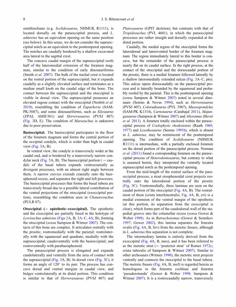
Figure 2. Lewisuchus admixtus, PULR 01. A, B, caudal portion of the skull in lateral view. Abbreviations: cr, crest; dpr, dorsal prooticrecess; cpr, cultriform process; ectp, ectopterygoid; ind, indeterminate bone; j, jugal; ltfe, laterotemporal fenestra; par, parietal; po, post-orbital; popr, paraoccipital process (otoccipital); q, quadrate; qj, quadratojugal; ri, ridge; soc, supraoccipital; su, sulcus. Scale bar ¼20 mm.
6 J. S. Bittencourt et al.

Quadratojugal. The quadratojugal has the common
L-shape morphology seen in several archosauriforms
(Nesbitt 2011), and forms the caudoventral margin of the
laterotemporal ( ¼ infratemporal) fenestra (Fig. 2A, B).
The rostral ramus is aligned with the caudal ramus of the
jugal, and its visible portion ends before the mid-length of
the laterotemporal fenestra, as is common amongst dino-
sauriforms (Nesbitt 2011).
The dorsal ramus of the quadratojugal is straight, ros-
trocaudally slender, and forms a near right angle to the
rostral ramus. As in Gracilisuchus (Brinkman 1981) and
some Late Triassic saurischians (e.g. Herrerasaurus,
PVSJ 407), the dorsal ramus is considerably longer than
the rostral one. Romer (1972c) inferred that the dorsal
ramus probably contacted the ventral tip of the squamosal,
but this cannot be ascertained. Along most of its exten-
sion, the dorsal ramus is appressed to the rostroventral
half of the lateral ala of the quadrate, but there is no evi-
dence of co-ossification to each other. The base of the cau-
dal margin of the dorsal ramus, i.e. the region that merges
with the caudal ramus, overlaps part of the craniolateral
edge of the ventral portion of the quadrate. Both the ros-
tral and the dorsal rami converge into a plate-like body
that continues caudally in a subtriangular process. Unlike
some basal dinosaurs (Yates 2003), the caudal ramus does
not form a waisted process or a distinct heel, although it is
somewhat elongated and slightly projected medially. Cau-
dally, the quadratojugal bears a faint ridge extending onto
the ventral end of its dorsal ramus. The latter condition
differs from that of Chanaresuchus (PVL 4575; PULR
07), in which the lateral surface of the quadratojugal
shows a conspicuous ridge, bordering the caudoventral
margin of the laterotemporal fenestra (Nesbitt 2011).
Squamosal. The left squamosal is preserved as an incom-
plete, slightly displaced subtriangular bone, articulating
with both the quadrate and the paraoccipital process of the
opisthotic (Fig. 2A, B). Both the rostral and ventral rami
are incomplete, so comparisons are hampered. The squa-
mosal of Lewisuchus admixtus contrasts with the plate-
like, rostrally and caudally rounded squamosal body of
Gracilisuchus (Brinkman 1981).
The preserved portion of the ventral ramus of the squa-
mosal of L. admixtus is short, and contacts the rostrodorsal
half of the lateral ala of the quadrate. The reconstruction
of Romer (1972c) suggests that it formed half the height
of the laterotemporal fenestra. However, the squamosal is
displaced, and its contact with the quadratojugal is elu-
sive. Accordingly, its participation in the laterotemporal
fenestra can only be estimated as equivalent to the quadra-
tojugal contribution. The ventral ramus of the squamosal
is connected to the caudal ramus of the same bone by a
thin and caudally projected lamina, rendering a concave
outline to the caudal portion of the squamosal. The caudal
process is short, sharply pointed, and caps part of the
dorsal portion of the quadrate, a feature already noticed in
basal archosauriforms and ctenosauriscids (Nesbitt 2011).
A small piece of an elongated bone, which is expanded at
one extremity, is attached to the lateral body of the squa-
mosal, but does not fit the size and shape of its missing
parts. As pointed out by Romer (1972c), this is probably a
skull element, but we also consider it as indeterminate.
Laterotemporal fenestra. The laterotemporal fenestra is
subrectangular, and its ventral margin is slightly longer
than the dorsal one (Figs 2A, B). The cranial margin of
the fenestra is bordered by the ventral ramus of the postor-
bital and the dorsal ramus of the jugal. The relative partic-
ipation of each of these bones in the cranial margin of the
fenestra is equivalent. The ventral margin possesses con-
cave cranial and caudal corners. The dorsal tine of the
caudal portion of the jugal contributes with 60% of the
floor of the fenestra, the remainder of which is formed by
the cranial ramus of the quadratojugal. Considering that
the ventral ramus of the squamosal is longer than pre-
served, this ramus and the dorsal ramus of the quadratoju-
gal probably had subequal participation in the caudal rim
of the laterotemporal fenestra. The dorsal margin of the
fenestra is not preserved. Nevertheless, there is no evi-
dence of a strong narrowing of the laterotemporal fenestra
either at the mid-height or at the dorsal portion of the
external opening.
Parietal. The caudomedial portion of the parietal is pre-
served in contact with the rostrolateral margin of the otoc-
cipital (Figs 2A, B, 3A–C). It displays a small rostral
excavation, and is laterally bordered by a sharp crest,
which probably composed the medial margin of the dorso-
temporal fenestra. The parietal extends cranially, roofing
the laterodorsal sinus of the endocranial cavity.
Supraoccipital. The supraoccipital is nearly horizontal
to the presumed rostrocaudal axis of the skull (Figs. 2A, B,
3A–C). It is a flattened bone, except for its small sagittal
nuchal crest. The convex dorsal portion of this crest corre-
sponds ventrally to a canal in the roof of the endocranial
cavity (Fig. 6A, B). The laterodorsal contact of the supra-
occipital with the preserved caudal portion of the parietals
is formed by a notch on each side of the bone, rendering a
rectangular appearance to the rostral portion of the supra-
occipital. This configuration is similar to that observed in
Chanaresuchus (PULR 07). Some dinosaurs (e.g. Masso-
spondylus, SAM-PK-K1314) have a foramen associated
with the supraoccipital notch. In Silesaurus (ZPAL AbIII/
361), the notches are elliptical and deeply incised in the
supraoccipital body, for which they are presumed to be
homologous to the posttemporal opening of dinosaurs
(Langer & Benton 2006; Nesbitt 2011). However, the
posttemporal aperture in basal saurischians (Sereno &
Novas 1994), sauropodomorphs (contra Sues et al. 2004),
neotheropods (Sampson & Witmer 2007), and some
Osteology of the Middle Triassic archosaur Lewisuchus admixtus 7
ornithischians (e.g. Scelidosaurus, NHMUK R1111), is
located dorsally on the paraoccipital process, and L.
admixtus has an equivalent opening on the same position
(see below). In this sense, we do not consider the supraoc-
cipital notch as an equivalent to the posttemporal opening.
The notches are caudally bordered by a shallow excavated
area lateral to the sagittal crest.
The concave caudal margin of the supraoccipital roofs
half of the lateromedial extension of the foramen mag-
num, similar to the condition of early dinosauriforms
(Smith et al. 2007). The bulk of the nuchal crest is located
on the rostral portion of the supraoccipital, but it expands
caudally as a slightly elevated surface and terminates as a
median small knob on the caudal edge of the bone. The
contact between the supraoccipital and the otoccipital is
visible in dorsal view. The supraoccipital possesses an
elevated rugose contact with the otoccipital (Nesbitt et al.
2010), resembling the condition of Euparkeria (SAM-
PK-5687), and some dinosauriforms, such as Silesaurus
(ZPAL AbIII/361) and Herrerasaurus (PVSJ 407)
(Fig. 3D, E). The condition of Marasuchus is unknown
due to poor preservation.
Basioccipital. The basioccipital participates in the floor
of the foramen magnum and forms the central portion of
the occipital condyle, which is wider than high in caudal
view (Fig. 3A, B).
In ventral view, the condyle is transversely wider at the
caudal end, and is bordered by a transversely narrow con-
dylar neck (Fig. 5A, B). The basioccipital portion ( ¼ cau-
dal) of the basal tubera expands ventrocranially as
divergent processes, with an almost right angle between
them. A narrow crevice extends cranially onto the basi-
sphenoid recess, and separates the right and left processes.
The basioccipital processes that forms the basal tubera are
transversely broad due to a possible lateral contribution of
the ventral projections of the otoccipital (exoccipital por-
tion), resembling the condition seen in Chanaresuchus
(PULR 07).
Otoccipital (¼ opisthotic–exoccipital). The opisthotic
and the exoccipital are partially fused in the holotype of
Lewisuchus admixtus (Figs 2A, B, 3A–C, 4A, B), forming
the otoccipital (sensu Sampson &Witmer 2007). The con-
tacts of this bone are complex. It articulates rostrally with
the prootic; rostromedially with the parietal; rostrolater-
ally with the squamosal and quadrate; medially with the
supraoccipital; caudoventrally with the basioccipital; and
rostroventrally with parabasisphenoid.
The paraoccipital process is elongated and expands
caudolaterally and ventrally from the area of contact with
the supraoccipital (Fig. 3A, B). In dorsal view (Fig. 3C), it
forms an angle of 120� to its pair. The process has con-
cave dorsal and ventral margins in caudal view, and
bulges ventrolaterally at its distal portion. This condition
is similar to that of Herrerasaurus (PVSJ 407) and
Plateosaurus (GPIT skeleton), but contrasts with that of
Tropidosuchus (PVL 4601), in which the paraoccipital
processes are rather straight and dorsally expanded at the
distal portion.
Caudally, the medial region of the otoccipital forms the
laterodorsal and lateroventral border of the foramen mag-
num. The region immediately lateral to this border is con-
cave, but the remainder of the paraoccipital process is
nearly flat on its caudal surface. In the right process, at the
contact of the otoccipital and the dorsocaudal portion of
the prootic, there is a medial foramen followed laterally by
a shallow lateromedially extended sulcus (Fig. 3A–C, pto).
This sulcus opens dorsocaudally on the paraoccipital pro-
cess and is laterally bounded by the squamosal and proba-
bly roofed by the parietal. This is the posttemporal opening
(sensu Sampson & Witmer 2007) noted in several dino-
saurs (Sereno & Novas 1994), such as Herrerasaurus
(PVSJ 407), Coloradisaurus (PVL 3967), Massospondylus
(SAM-PK K1314), Carnotaurus (Carabajal 2011), Majun-
gasaurus (Sampson & Witmer 2007) and Alioramus (Bever
et al. 2011). A foramen totally enclosed within the paraoc-
cipital process of Coelophysis rhodesiensis (Raath 1969,
1977) and Lesothosaurus (Sereno 1991b), which is absent
in L. admixtus, may be reminiscent of the posttemporal
opening. The condition of Scelidosaurus (NHMUK
R1111) is intermediate, with a partially enclosed foramen
on the dorsal portion of the paraoccipital process. Norman
et al. (2011) found a corresponding foramen on the paraoc-
cipital process of Heterodontosaurus, but contrary to what
is assumed herein, they interpreted the ventrally located
supraoccipital notch as the posttemporal opening.
From the mid-length of the rostral surface of the para-
occipital process, a stout otosphenoidal crest projects ros-
trally onto the laterodorsal portion of the prootic
(Fig. 3C). Ventromedially, three laminae are seen on the
caudal portion of the otoccipital (Fig. 4A, B). The rostral-
most of them (crista interfenestralis; Fig. 4A, B, cri) is a
medial extension of the ventral margin of the opisthotic
(at this portion, its separation from the exoccipital is
clear), which forms part of the caudodorsal wall of the sta-
pedial groove into the columellar recess (sensu Gower &
Weber 1998). As in Batrachotomus (Gower & Sennikov
1997; Gower 2002), this lamina separates the fenestra
ovalis (Fig. 4A, B, fov) from the metotic fissure, although
in L. admixtus this separation is not complete.
The intermediary lamina is entirely derived from the
exoccipital (Fig. 4A, B, mes), and it has been referred to
as the metotic strut (¼ ‘posterior strut’ of Romer 1972c;
crista tuberalis of Sampson & Witmer 2007). Similar to
other archosaurs (Witmer 1990), the metotic strut projects
ventrally and connects the otoccipital to the basal tubera.
The metotic fissure (Fig. 4A, B, mef) is regarded herein as
homologous to the fenestra cochleae and fenestra
‘pseudorotunda’ (Gower & Weber 1998; Sampson &
Witmer 2007). It is a rostrocaudally narrow, transversely
8 J. S. Bittencourt et al.

wide, and laterally opened chamber, which is also inserted
into the columellar recess. A narrower furrow (Fig. 4B)
diverges from this groove and leads to a foramen caudally
opened just caudal to the metotic strut, and possibly serves
as passage for the cranial nerves X and/or XI. This sug-
gests a diversion of the vagal canal, as described for thero-
pod dinosaurs (Rauhut 2003; Sampson & Witmer 2007),
and this condition contrasts with the undivided metotic
fissure of basal archosauriforms (Gower & Sennikov
1996) and rauisuchians (e.g. Batrachotomus, Gower
2002).
The caudal lamina (Fig. 4A, B, cal), which is limited
by a short and transverse crest at the mid-height of the
occipital condyle, is also restricted to the exoccipital. It
forms the laterodorsal knobs of the basioccipital condyle,
differing from the bulged condylar surface of basal arch-
osauriforms (Desojo et al. 2011; Trotteyn & Haro 2011).
The space between the caudal lamina and the metotic
Figure 3. Lewisuchus admixtus, PULR 01. Caudal portion of the skull in A, B, caudal and C, dorsal views. D, Silesaurus. E, Herrera-saurus. Abbreviations: boc, basioccipital condyle; btu, basal tubera; fm, foramen magnum; ind, indeterminate bone; ncr, nuchal crest;otcr, otosphenoidal crest; par, parietal; popr, paraoccipital process (otoccipital); pto, posttemporal opening; soc, supraoccipital. Scalebars: A–D ¼ 10 mm; E ¼ 30 mm.
Osteology of the Middle Triassic archosaur Lewisuchus admixtus 9

strut harbours three subvertically aligned conspicuous
foramina. The upper two were regarded by Romer
(1972c, figs 3, 4) as the openings for the cranial nerve
XII, giving that in several reptiles (Romer 1956; Gower
& Sennikov 1997; Gower & Weber 1998) the hypoglos-
sal nerve has a dichotomous exit in the braincase. How-
ever, as mentioned above, the dorsal foramen is
interpreted herein as the passage for the vagal and/or
accessory nerves. The intermediary and the ventral
foramina are the exit for the hypoglossal nerve (XII),
which is congruent with its position in some early dino-
saurs (Sereno 1991b; Rauhut 2003). The position of the
hypoglossal nerve foramina caudal to the metotic strut is
not exclusive of L. admixtus and silesaurids (Nesbitt
et al. 2010; Nesbitt 2011). A partial braincase attributed
to Marasuchus (PVL 3872) shows a similar configuration
(Sereno & Arcucci 1994b), suggesting that the exit of the
cranial nerve XII occupied a similar position amongst
non-dinosaur dinosauriforms.
Prootic. The prootic forms most of the lateral portion of
the braincase (Fig. 4A, B). It is tightly attached to the dor-
sal surface of the parabasisphenoid and their separation is
elusive. Indeed, several prootic structures described in
this section are probably also composed by parts of the
parabasisphenoid. The prootic is largely exposed in lateral
view, and is also seen in dorsolateral (the otosphenoidal
crest, Fig. 3C) and ventral view (the recesses on the lateral
surface, Fig. 4A, B). It contacts the otoccipital caudally,
the parietal dorsally, and probably the laterosphenoid
cranially. The dorsolateral portion of the prootic is marked
by a dorsal recess close to the supraoccipital-parietal con-
tact, which receives the rostral ramus of the squamosal
(Fig. 2A, B).
The otosphenoidal crest extends from the caudal mar-
gin of the otoccipital, spanning rostrally onto the lateral
surface of the prootic, similar to Marasuchus (PVL 3872)
and Silesaurus (ZPAL AbIII/361). The rostral margin of
this crest folds down and forms part of the ‘middle strut’
of Romer (1972c), which is also composed by the ascend-
ing ramus of the parabasisphenoid (Fig. 4A, B, ‘mids’).
The dorsocaudal portion of the prootic forms the rostral
border of the columellar recess, which houses the stape-
dial groove dorsally (Fig. 4A, B, stg), the fenestra ovalis
(¼ fenestra vestibuli, Sampson & Witmer 2007) cranially
(fov), and the metotic fissure caudally (mef). The latter
two structures are opened into the endocranial space. A
fragmentary right stapes (Fig. 4A, B, stp) is associated to
the corresponding stapedial groove.
A thin lamina projects rostroventrally from the region
of the ‘middle strut’, forming the roof of a cavity here
referred to as the prootic–parabasisphenoid recess. In raui-
suchians (Wu & Russell 2001; Gower 2002), the thin lam-
ina has been referred to as the crista prootica (Fig. 4A, B,
crpro), homologous to that described in dinosaurs (Galton
& Upchurch 2004, fig. 12.2). The ‘anterior strut’ of Romer
(1972c, pp. 5–6) is formed by both the crista prootica and
the caudal wall of the clinoid process of the parabasisphe-
noid. The prootic–parabasisphenoid recess, which is topo-
logically equivalent to the ‘anterior tympanic recess’ of
Figure 4. Lewisuchus admixtus, PULR 01. A, B, caudal portion of the skull in lateroventral view. Abbreviations: cal, caudal lamina; cri,crista interfenestralis; crpro, crista prootica; fov, fenestra ovalis; icaf, internal carotid artery foramen; mef, metotic fissure; mes, metoticstrut; ‘mids’, middle strut; pbas, parabasisphenoid; proo, prootic; pror, prootic–parabasisphenoid recess; ptg, pterygoid; q, quadrate; stg,stapedial groove; stp, stapes; VII, foramen for the facial nerve; XI, foramen for vagal nerve; XII, foramen for hypoglossal nerve. Scalebar ¼ 20 mm.
10 J. S. Bittencourt et al.

theropods (Rauhut 2003), harbours at least two internal
openings. Contrary to the interpretation of Romer
(1972c), the dorsal opening is the foramen for the facial
nerve (cranial nerve VII), which occupies an equivalent
position in several archosauriforms (Gower & Sennikov
1997; Gower & Weber 1998; Sampson & Witmer 2007).
The rostroventral opening is larger and is floored by the
parabasisphenoid. We tentatively interpret it as the pas-
sage for the internal carotid artery and/or the palatine
branch of the facial nerve (Fig. 4A, B, icaf), because a
similar configuration is described for Euparkeria (Gower
& Weber 1998), Batrachotomus (Gower 2002) and Mara-
suchus (PVL 3872).
The prootic–parabasisphenoid recess also houses two
pneumatic spaces: a small excavation below the foramen
for the facial nerve that does not pierce into the endocra-
nial cavity, and a dorsoventral wedge-like concavity that
leads into the internal opening of the carotid artery. Both
the foramen for the facial nerve and the rostral concavity
are at least partially covered by the lateral flange of the
‘anterior strut’. The rostral lamina that roofs the prootic–
parabasisphenoid recess is rostrally covered by a bony
shield (Fig. 6A, B), which can be interpreted as an ossifi-
cation of the laterosphenoid (Romer 1972c). Dorsome-
dially to the prootic–parabasisphenoid recess and
dorsocaudally to the shield of the ‘anterior strut’, there is
an oval opening of the endocranial cavity (the “? fenestra
epiotica” of Romer 1972c, p. 6). This is better interpreted
as the trigeminal foramen, or the exit for the cranial nerve
V, which is in a similar position to the corresponding fora-
men of several archosauriforms (Gower & Weber 1998),
including dinosaurs (Rauhut 2003).
The caudal portion of the endocranial cavity is exposed.
Its wall above the columellar recess expands laterally
forming the cavity for the auricular lobe (flocculus) of the
cerebellum (Larsell 1932; ten Donkelaar 1998), which is
also well developed in Marasuchus (PVL 3872), Silesau-
rus (ZPAL AbIII/361) and dinosaurs (Franzosa & Rowe
2005). At the entrance of the sinus, the dorsal border of
the internal crest bears a blind excavation associated with
a small cranial concavity (not shown), which may be the
passage for the vena cerebralis media (Galton 2001; Gal-
ton & Upchurch 2004).
Laterosphenoid. A partial laterosphenoid is preserved
rostrally to the trigeminal foramen (Fig. 6), but its con-
tacts with other cranial bones are not clear. Apparently, it
lies rostrally to the prootic and caudodorsally to the cli-
noid process of the parabasisphenoid. This position is con-
sistent with that observed in other archosaurs (Gower &
Sennikov 1996), in which the laterosphenoid lies rostral
to the prootic, and caudal to the sphenethmoid. The posi-
tion of the latter bone can be inferred by the dorsal sulcus
of the cultriform process (parabasisphenoid), which
probably received the “cartilaginous sphenethmoidal
braincase” (Romer 1972c, p. 6).
Parabasisphenoid. As commonly observed in archo-
sauriforms (Ewer 1965; Walker 1990; Parrish 1994; Yates
2003), both parasphenoid and basisphenoid are co-ossi-
fied, thus they are referred to here as the parabasisphe-
noid. This is tightly attached to the ventral margin of the
prootic, and forms most of the floor of the braincase
(Figs 4A, B, 5). The basisphenoid component of the basal
tubera is closely attached rostral to its basioccipital por-
tion (Fig. 5). From the rostral margin of each ramus of the
basal tubera, a shallow flange expands dorsomedially and
rostrally, forming the body of the parabasisphenoid.
The basisphenoid recess is shallow if compared to that
of early theropods (Rauhut 2003; Nesbitt et al. 2009b),
and extends from the basal tubera to the proximal portion
of the cultriform process. The foramina for the internal
carotid artery pass through the lateroventral portion of the
prootic–parabasisphenoid, as also described for Marasu-
chus (Sereno & Arcucci 1994b). By contrast, in Chanare-
suchus (PULR 07) and Silesaurus (ZPAL AbIII/361),
these foramina are located on the ventral surface of the
parabasisphenoid.
The lateral borders of the parabasisphenoid are rather
thickened. In ventral view, this bone is constricted at
the base of the basipterygoid process, achieving half of
the width of the basal tubera. Each basipterygoid pro-
cess projects ventrolaterally, forming an angle of 45�
to the transverse axis of the skull, and a right angle to
its pair. As in various dinosauriforms (Dzik 2003;
Yates 2007), the basipterygoid process is as long as the
basal tuber, distally rounded, and there is no web of
bone spanning towards its pair. The distal end of the
basipterygoid process contacts the medioventral surface
of the pterygoid, forming a loose articulation. The para-
basisphenoid is excavated dorsolaterally to the base of
the basipterygoid process, where the well-developed cli-
noid process projects laterocaudally (Fig. 6). The sella
turcica is deep and excavates the rostrodorsal body of
the parabasisphenoid, caudal to the cultriform process.
This excavation is caudally delimited by the prootic
and rostrolaterally by the medial margin of the clinoid
process.
The cultriform process is relatively deep, stripe-shaped,
and at least as long as the remainder of the parabasisphe-
noid (Fig. 5). Its ventral and dorsal surfaces are respec-
tively marked by a sharp edge and a shallow longitudinal
sulcus. The latter is bordered in both sides by the crests
that project rostroventrally from the medial margin of the
clinoid processes. Unlike some early archosauriforms
(Gower & Sennikov 1996, 1997), the basal tubera and
basipterygoid processes of Lewisuchus admixtus are hori-
zontally aligned to each other, whereas the cultriform pro-
cess and the occipital condyle are placed more dorsally,
Osteology of the Middle Triassic archosaur Lewisuchus admixtus 11

although also aligned to one another. This configuration is
also described for basal saurischians (Yates 2003), but dif-
fers from the condition of Silesaurus (ZPAL AbIII/361),
in which the basipterygoid processes are ventrally offset
relative to the basal tubera.
As also seen in Silesaurus (Dzik 2003), the dorsocaudal
portion of the parabasisphenoid is occupied by an ascend-
ing process (¼ the “middle strut” of Romer 1972c, p. 5),
which fuses dorsally with the lateral surface of the prootic,
and possesses a shallow sulcus on its caudoventral portion.
Figure 5. Lewisuchus admixtus, PULR 01. A, B, caudal portion of the skull in ventral view with a detail of the pterygoid teeth. Abbrevi-ations: con, condylar neck; cpr, cultriform process; elr, ectopterygoid lateral ramus; emex, ectopterygoid medial excavation; emr, ectop-terygoid medial ramus; knob, knob-like ventral projection; pbas, parabasisphenoid; pbr, pterygoid basisphenoid ramus; pcpr, pterygoidectopterygoid ramus; pqr, pterygoid quadrate ramus; ptg, pterygoid; ptpr, pterygoid process (parabasisphenoid); pvpr, pterygoid vomero-palatine ramus; q, quadrate; stp, stapes; vex, ventral excavation on the pterygoid. Scale bar ¼ 20 mm.
12 J. S. Bittencourt et al.

Quadrate. The left quadrate is partially preserved, miss-
ing portions of which include the dorsocranial region of
the medial (¼ pterygoid) ala (Figs 2A, B, 5A, B). Simi-
larly to most archosauriforms (Nesbitt 2011), the bone is
subvertical, and the caudal edge of the head is roughly
aligned with the cranio-mandibular articulation. As in
Gracilisuchus (Brinkman 1981), the quadrate head is
capped by the caudal ramus of the squamosal and the ros-
troventral surface of the lateral tip of the paraoccipital
process. The caudal margin of the quadrate shaft is dorso-
ventrally concave.
The lateral (¼ quadratojugal) and the medial alae are
set in right angles to each other. The lateral ala is broad
and bends rostrolaterally, forming a dorsoventral concave
area facing caudolaterally. The quadrate foramen is
located in this region. It is associated with a rounded fossa
that expands onto the medial margin of the quadratojugal.
Most of the ventral portion of the quadrate is concealed
by the quadratojugal, and the point of maximal lateral
expansion of the bone is the region of the possible squa-
mosal/quadratojugal contact (Romer 1972c). The con-
dyles for cranio-mandibular articulation flush ventrally,
but the medial condyle is craniocaudally narrower than
the lateral one.
The subtriangular medial ala of the quadrate projects
further rostrally than the lateral ala. The ventral margin of
the lateral ala possesses a horizontal shelf, in which the
dorsal portion receives the caudal ramus of the pterygoid
(Fig. 5A, B). Although slightly displaced from its original
position, the rostrodorsal margin of the medial ala did not
contact the lateral surface of the braincase.
Pterygoid. The preserved right pterygoid has a trans-
versely waisted body, with a caudally opened excavation
and a cranial knob-like process on its ventral surface
(Fig. 5A, B). Four rami project from the pterygoid body
across the caudal portion of the palate. Similarly to
Chanaresuchus (PULR 07) and basal rauisuchians
(Sereno 1991a), the basisphenoid ramus is short, caudo-
medially projected, and contacts the basipterygoid process
of the parabasisphenoid and the medial ala of the quad-
rate. The latter contact occurs via a caudally directed
spur-like process of this ramus.
Similarly to most archosaurs (Walker 1964; Sereno
1991a), the quadrate ramus is more slender than the basi-
sphenoid ramus, and extends caudolaterally. By contrast,
the quadrate ramus of Chanaresuchus (PULR 07) is
dorsoventrally expanded, forming a plate-like structure.
The quadrate ramus fits into the medial excavation of the
quadrate medial ala (Fig. 5A, B).
The vomeropalatine and the ectopterygoid rami
(Fig. 5A, B) are connected by a thin lamina projecting
mediolaterally from the thickened medial rim of the for-
mer, and rostrally from the caudal border of the latter.
This lamina bears an L-shape excavated area on the
rostroventral portion of the pterygoid. The concave cra-
niolateral margin of the fan-shaped lamina is tightly
appressed to the medial margin of the ectopterygoid.
The vomeropalatine ramus projects rostromedially
from the pterygoid body and forms a right angle to the
ectopterygoid ramus. Its ventral rim bears small alveoli
encased by cortical bone, elliptical in cross section, with a
hint of a central canal. These are interpreted as palatal
teeth (Fig. 5A). They are not as conspicuous as those of
basal archosauriforms (Sereno 1991a; Welman 1998; Wu
& Russell 2001) or dinosaurs (e.g. Eoraptor, PVSJ 512;
Eodromaeus, PVSJ 562; Pampadromaeus, ULBRA-
PVT016), but this may be result of poor preservation.
Ectopterygoid. The preserved left ectopterygoid is com-
posed of a rostrocaudally elongate shaft and a dichotomous
rostral portion (Fig. 5A, B). The shaft is dorsoventrally
deep, slightly curved laterally, and with a thickened ventral
crest extending along its rostrocaudal extension. At the ros-
tral tip, it splits in a lateral process, which bends caudodor-
sally, forming a hook-like jugal ramus; and a medial
process, which arcs rostromedially and contacts the cranio-
lateral portion of the vomeropalatine ramus of the ptery-
goid. The bone is positioned dorsal to the pterygoid, as in
other dinosauriforms (Sereno 1999; Nesbitt 2011).
The jugal ramus is short and sharply pointed at its cau-
dodorsally bowed tip (Fig. 4). It also possesses a trans-
verse crest on its dorsal surface, which forms part of the
orbital floor. The lateral surface of the hook-like process
articulates with the medial surface of the jugal, and also
contacts the ventral tip of the jugal ramus of the
postorbital.
The medial flange of the ectopterygoid is rostrally con-
cave, and bordered by a distinct medial rim connected to
the vomeropalatine ramus of the pterygoid. Similarly to
theropods (Gauthier 1986; Sereno 1999), an excavated
area (Fig. 5A, B) is formed by the ventral concavity of the
medial flange, achieving its maximal depth in the conflu-
ence of the rostral rim of the flange and the ventral margin
of the ectopterygoid shaft. No foramen pierces the rostro-
ventral surface of the ectopterygoid.
Vertebral column. The holotype of Lewisuchus admix-
tus has 16 presacral vertebrae preserved in articulation,
including the atlantal intercentrum and neural arch, the
axis complex (axial intercentrum, centrum and neural
arch, plus the odontoid), and 14 postaxial vertebrae
(Figs 7, 8). Similarly to Marasuchus (PVL 3870) and
Silesaurus (Piechowsky & Dzik 2010), there is a conspic-
uous morphological transition between the seventh and
eighth preserved vertebrae (Fig. 8A), which includes a
reduction in the centrum length (approximately 20%), and
a lesser ventral projection of the caudal articular facet.
Although the preserved ribs articulate with both the neural
arch and the ventrocranial margin of the centrum up to the
10th presacral vertebra, the ribs attached to the eighth to
Osteology of the Middle Triassic archosaur Lewisuchus admixtus 13

Figure 7. Lewisuchus admixtus, PULR 01. Atlas–axis complex in A, B, right and C, D, left lateral, E, F, cranial, G, dorsal and H, ven-tral views. Abbreviations: ati, atlantal intercentrum; atn, atlantal neural arch; atr, atlantal rib; axr, axial rib; ce3, cervical 3; cn, neuralcanal; dex, dorsal excavation; dri, dorsal ridge; ns, neural spine; od, odontoid; prz, prezygapophysis; riat, rib attachment area; vec, verte-bral centrum; vex, ventral excavation; vri, ventral ridge. Scale bar ¼ 10 mm.
Figure 6. Lewisuchus admixtus, PULR 01. A, B, caudal portion of the skull in frontal view. Abbreviations: fl, floccular lobe; fm, fora-men magnum; clpr, clinoid process; cpr, cultriform process; j, jugal; ncr, nuchal crest; ltsp, laterosphenoid; ptpr, pterygoid process(parabasisphenoid); po, postorbital; su, sulcus; V, foramen for the trigeminal nerve. Scale bar ¼ 20 mm.
14 J. S. Bittencourt et al.

10th presacral vertebrae are more robust, thus the
presacral vertebrae from eighth to 16th are considered as
belonging to the dorsal series (Piechowsky & Dzik 2010).
An isolated vertebra embedded in the slab close to the last
articulated dorsal may correspond to a more caudal ele-
ment within the dorsal series (Fig. 8H).
Three semi-articulated vertebrae plus the fragment of a
fourth element lie close to the left scapula, on the left side
of the slab (Fig. 9A). These are probably proximal caudal
vertebrae. Another set of five semi-articulated vertebrae,
also on the left side of the slab, but closer to the cranial
cervicals (Fig. 9B), probably belong to the middle portion
of the tail.
Cervical series. The atlantal intercentrum is preserved
in articulation with the axial intercentrum and the odon-
toid process (Fig. 7A–D). The atlantal intercentrum is U-
shaped in cranial view, in which the lateral tips curve
upwards and bulge craniocaudally (Fig. 7E, F). The con-
cave dorsal basin fits well with the occipital condyle of
Figure 8. Lewisuchus admixtus, PULR 01. Cervical and dorsal vertebrae in A, right lateral view, with B, a detail of the osteoderms. C,trunk vertebrae in right lateral view found with the holotype of the proterochampsid Tropidosuchus (PVL 4601). D, third cervical incross section. E, second dorsal in left lateral view. F, G, seventh and eighth dorsal vertebrae in left lateral view.H, isolated caudal dorsalvertebra in right lateral view. Abbreviations: c5–7, cervical vertebrae; cc, cervical centrum; crcdl, cranial centrodiapophyseal lamina;d1–3, dorsal vertebrae; idf, infradiapophyseal fossa; ns, neural spine; os, osteoderm; poz, postzygapophysis; r, rib; tp, transverse process.Scale bars: A, E ¼ 20 mm; C ¼ 5 mm; D ¼ 3 mm; F, G ¼ 10 mm; H ¼ 2 mm.
Osteology of the Middle Triassic archosaur Lewisuchus admixtus 15

the described skull. This concavity is shallower at the cra-
nial margin, and deeper at the caudal border, where it
receives the cranioventral margin of the odontoid process.
Ventrally, the atlantal intercentrum is transversely wider
than the axis (Fig. 7H). It also harbours a mediolaterally
oriented shallow sulcus that is not seen in Marasuchus
(PVL 3870) or Silesaurus (Piechowsky & Dzik 2010). An
incomplete right rib is tightly articulated with the caudo-
lateral and ventral margin of the atlantal intercentrum
(Fig. 7A, B). The rod-like narrow shaft is parallel to the
cervical column.
The left atlantal neural arch is similar to those of Tropi-
dosuchus (PVL 4601), Marasuchus (PVL 3870) and Sile-
saurus (Piechowsky & Dzik 2010) in the presence of a
robust pedicel that articulates with both the dorsolateral
portion of the atlantal intercentrum and the craniolateral
margin of the odontoid process. Its base is slightly wider
transverselly than craniocaudally, and the ventral region
is facing medially (Fig. 7A–D). As seen in Silesaurus
(Piechowsky & Dzik 2010), the dorsal portion of the
atlantal neural arch is craniocaudally elongated, with the
cranial ramus projecting medially as an arched plate, roof-
ing the laterodorsal border of the neural canal (Fig. 7E, F).
The caudal ramus corresponds to the postzygapophysis
plus the epipophysis, which are indiscernible from each
other. The caudal ramus is a rod-like, elongated and
slightly caudally flattened structure. It articulates with the
dorsal margin of the reduced axial prezygapophysis, and
its caudal tip does not reach the cranial edge of the axial
postzygapophysis, as occurs in most archosaurs (Yates
2003).
The odontoid process (atlantal centrum, Romer 1956) is
a stout piece of bone not completely co-ossified to the axial
centrum (Fig. 7A, B). In right lateral view, its caudoventral
margin is separated from the axial intercentrum. The dorsal
margin of the odontoid process is slightly concave, and
composes the rostral floor of the neural canal. As in the
atlantal intercentrum, the lateral tips of the odontoid pro-
cess are dorsally projected, and receive part of the ventral
surface of the pedicel of the atlantal neural arch.
The axial intercentrum is completely fused with the
axial centrum (Fig. 7A–D), but its caudal margin is
marked by a shallow sulcus, cranioventral to which the
intercentrum is bulged. The craniodorsal portion of the
intercentrum is in close contact with the ventrocaudal sur-
face of the odontoid process. By contrast, its cranioventral
portion receives part of the atlantal intercentrum.
The axial centrum is transversely compressed (Fig. 7G,
H). If combined with the axial intercentrum, it is cranio-
caudally as long as the other cervical centra, and is longer
than the dorsal ones. The lateral surface of the axial cen-
trum bears a dorsal excavation and a ventral concavity
(Fig. 7C, D). The upper excavation is the deepest and
extends along most of the lateral length of the centrum. It
is dorsally bordered by a rugose horizontal crest, which
also demarcates the ventral edge of the neural arch. Ven-
trally, the excavation is bordered by another ridge that
also delimits the lower concave area. The latter projects
onto the ventral margin of the centrum, which possesses a
sharp median keel. No lateral foramina are seen. The con-
figuration of the lateral surface of the axial centrum
described above also matches those of Tropidosuchus
(PVL 4601), Marasuchus (PVL 3870) and Silesaurus
(Piechowsky & Dzik 2010).
The parapophysis occupies a cranioventral position on
the lateral surface of the centrum. Similarly to Marasu-
chus (PVL 3870), it is a bud-like area fused to the caudal
margin of the axial intercentrum (Fig. 7A–D). In the right
side, a rod-like rib is attached to the parapophysis. The rib
is slightly expanded proximally, projects caudally as a
narrower shaft, and does not reach the caudal edge of the
axial centrum.
The cranial articular facet of the axis is not visible due
to attachment of the odontoid process. The caudal articu-
lar facet is concave, significantly higher than wide, and
with thickened borders.
Measured from the floor of the neural canal to the top of
the postzygapophysis, the axial neural arch is slightly
higher than the centrum (Fig. 7A–D). At mid-height, its
lateral surface is laterally bulged, and bears a small crest-
like prezygapophysis in the cranial portion. The postzyga-
pophysis spans caudodorsally from the neural arch as a
thin lamina, and develops a dorsomedial flange that fuses
with the caudoventral margin of the neural spine. The cau-
dodorsal surface of the postzygapophysis is slightly
bulged, but there is no evidence of an epipophysis.
The transversely compressed neural spine is craniocau-
dally longer than the centrum. Differently from Silesaurus
(Piechowsky & Dzik 2010), Herrerasaurus (PVSJ 407),
Lesothosaurus (Sereno 1991b) and Heterodontosaurus
(SAM-PK-1332), which possess a rather straight dorsal
rim of the axial neural spine, in Lewisuchus admixtus this
margin is strongly convex, resulting in an axe-like lateral
outline. Its cranial edge is connected to the prezygapophy-
sis by a thin lamina, and projects beyond the cranial mar-
gin of the centrum, resembling the condition of Silesaurus
(Piechowsky & Dzik 2010). The cranial edge of the neural
spine is lower than the caudal one. Two rugose ridges are
seen on the lateral surface of the axial neural spine. The
dorsal ridge extends along the convexity of the neural
spine dorsal border. The ventral ridge extends craniocau-
dally along the mid-height of the neural spine. The upper
ridge is present in other dinosauriforms, such as Silesau-
rus (Piechowsky & Dzik 2010) and Heterodontosaurus
(SAM-PK-1332), but the lower one is restricted to
L. admixtus.
The post-axial cervical centra are longer than the pre-
served dorsal centra (Fig. 8A, F). This is typical for basal
dinosauriforms (Nesbitt et al. 2010), but also occurs in the
enigmatic Spondylosoma (GPIT 479/30) and some
16 J. S. Bittencourt et al.

rauisuchians (Lautenschlager & Desojo 2011). The cervi-
cal centra are also transversely compressed and laterally
concave. Contrasting with the neck vertebrae of Tropido-
suchus (PVL 4601), rauisuchians (Benton & Walker
2002; Nesbitt 2005) and other dinosauriforms (Yates
2003; Nesbitt et al. 2009b), there is not a conspicuous
‘keel’ extending along the ventral surfaces of the cervical
vertebrae of L. admixtus.
The transverse section of the third cervical vertebra
reveals that its centrum (Fig. 8D), which is separated from
the neural arch by a lateral constriction (¼ the upper exca-
vation of the axis), is very low (4.2 mm high) and narrow
(2.7 mm wide) if compared to the portion encasing the
neural canal (6.5 mm high; 5.3 mm wide). In addition,
the wall around the neural canal is broader (1.3 mm) than
the outer wall of the centrum (0.7 mm). The crest that dor-
sally borders the upper excavation on the lateral surface of
the centrum is very faint in the third cervical vertebra. It
becomes more pronounced in the fourth and fifth cervical
vertebrae, and is replaced by a true transverse process from
the sixth cervical vertebra backwards (Fig. 8A). The thin
ridge that dorsally borders the lower lateral excavation of
the axial centrum is visible in all the remaining cervical
vertebrae, but the lower excavation itself disappears in the
fourth cervical vertebra backwards.
The caudal facet of the centrum is situated more ven-
trally than the cranial one, resulting in a parallelogram
centrum in lateral view. As a consequence, it is ventrally
offset with regard to the cranial margin of the centrum,
similar to Tropidosuchus (PVL 4601), some crurotarsans
(Nesbitt 2005), and dinosauriforms (Gauthier 1986;
Sereno & Arcucci 1994b; Novas 1996).
The centrum of the third cervical vertebra bears two
low knob-like processes on its cranioventral portion, the
ventral of which is topologically equivalent to the para-
pophysis (Fig. 7C). The dorsal knob is part of the dorsolat-
eral crest that limits the neural arch ventrally and,
apparently, it serves as attachment site for the tuberculum
in the third and fourth cervical vertebrae, which do not
have transverse processes. In the sixth to seventh cervical
vertebrae, which bear a true transverse process, the rib
attaches both in the parapophyseal area and in the distal
portion of the transverse process. Despite this change in
the rib attachment, the dorsal knob-like process on the cra-
nial portion of the centrum is maintained at least until the
first dorsal vertebra.
The sixth cervical vertebra bears a hint of the tetraradi-
ated pattern of lamination associated with the transverse
process, a feature that becomes more conspicuous from
the seventh presacral vertebra backwards (Fig. 8A). The
third and fourth cervical vertebrae possess a lamina topo-
logically equivalent to the postzygodiapophyseal lamina
(sensu Wilson 1999) of other archosauriforms, regardless
of the presence of a transverse process. This condition is
similar to that of Marasuchus (PVL 3870), but differs
from Silesaurus (Piechowsky & Dzik 2010), the fourth
cervical vertebra of which already shows robust laminae
expanding from the transverse process. The sixth cervical
vertebra of L. admixtus bears faint cranial and caudal cen-
trodiapophyseal laminae. This suggests that these struc-
tures are serially homologous to the crest that dorsally
borders the upper excavation of more cranial cervical ver-
tebrae, as it is the case inMarasuchus (PVL 3870).
The infradiapophyseal fossa (sensu Harris 2006) is well
developed in the sixth cervical vertebra. The cranial mar-
gin of its transverse process projects craniodorsally, but
does not reach the lateral surface of the prezygapophysis.
On the other hand, it forms the caudal margin of an incipi-
ent cranial infradiapophyseal fossa. The lateral area cau-
dal to the transverse process is slightly depressed, and
dorsally bordered by a robust postzygodiapophyseal lam-
ina, which expands caudally reaching the caudal edge of
the postzygapophysis.
The seventh cervical vertebra possesses a more con-
spicuous pattern of pneumatization. In addition to the
intermediary infradiapophyseal fossa, both cranial and
caudal infradiapophyseal fossae associated with the later-
oventrally directed transverse process are also well devel-
oped. However, contrasting with the dorsal vertebrae, the
cranial margin of the transverse process forms a prezygo-
diapophyseal lamina that is restricted to the base of the
prezygapophysis. In the dorsal vertebrae, with the more
dorsal position of the rib attachment, the cranial margin of
the transverse process is confluent with the lateral margin
of the prezygapophysis.
As observed in Marasuchus (PVL 3870) and Silesaurus
(Piechowsky & Dzik 2010), the neural arch of the post-
axial cervical vertebrae of Lewisuchus admixtus is signifi-
cantly higher than the centrum (Fig. 8A), which partially
results from the strong dorsal projection of the zygapoph-
yses. The prezygapophysis is elongated, dorsoventrally
broad at its base, and distally rounded. Its laterodorsal
border is marked by a ridge that forms the prezygodiapo-
physeal lamina in the dorsal vertebrae. At least in the sixth
and seventh cervical vertebrae, this ridge projects caudally
and reaches the postzygodiapophyseal lamina. The post-
zygapophysis is also elongated, but less dorsally oriented
than the prezygapophysis. Unlike dinosaurs (Sereno 1999;
Langer & Benton 2006), there is no evidence of epipophy-
ses in the cervical vertebrae. Instead, each postzygapoph-
ysis has a dorsal ridge that projects from the caudal
margin of the neural spine.
Unfortunately, in none of the post-axial cervical verte-
brae is the neural spine completely preserved. The seventh
cervical vertebra preserves the basal portion of the neural
spine and its distal tip may correspond to the fragment of
bone situated immediately above it (Fig. 8A, B). The row
of elongated pieces of bone above the cervical vertebrae
was identified as dorsal ‘scutes’ (i.e. osteoderms) by
Romer (1972c, p. 8), which is endorsed herein. These are
Osteology of the Middle Triassic archosaur Lewisuchus admixtus 17

imbricate, with the cranial ends underlying the caudal por-
tion of the preceding element. A conspicuous lateral fur-
row delimits each osteoderm. The ventral surface of these
elements is flat, and their dorsal margin is bulged. A simi-
lar morphology is seen in the 15th preserved vertebra (¼ninth dorsal vertebra), in which the strongly cranially
expanded distal end of the neural spine is capped by an
osteoderm, as in the cervical vertebrae (Fig. 8F, G).
Accordingly, the distal tip of the neural spine is cranially
expanded both in the cervical and dorsal series. As a
result, the cranio- and caudodorsal edges of the neural
spine tips in adjacent vertebrae contact one another.
Within dinosauriforms, the presence of cervical osteo-
derms is unambiguous in the theropod Ceratosaurus
(Gilmore 1920) and ornithischians (Norman et al. 2004b).
In the latter, the elements are disposed in parasagittal
rows, rather than along the median line of the vertebral
column. The morphology of the osteoderms in L. admixtus
is comparable to that of Tropidosuchus (Fig. 8C) and Gra-
cilisuchus (Brinkman 1981), in which one ‘scute’ is
attached to each neural spine. In Chanaresuchus (PVL
4575), there is also a single row of osteoderms, but at least
two elements associate to each cervical neural spine. The
cervical vertebrae of Marasuchus (PVL 3870) are more
similar to those of Silesaurus (Piechowsky & Dzik 2010),
with low and apparently unroofed neural spines.
A rib is attached to the last cervical vertebra (Fig. 8A).
It follows the general pattern of the archosaur cervical
ribs (Romer 1956), with two cranially directed processes,
the medial of which is the longest, and corresponds to the
rib head (capitulum). It is cranially bulged, and contacts
the cranioventral portion of the centrum. The dorsal pro-
cess corresponds to the rib tubercle (tuberculum). It is
shorter than the head, and attaches to the distal portion of
the transverse process. The processes are orthogonal to
each other. Similarly to basal dinosauriforms (Sereno
1999; Dzik 2003), the whole rib lies subparallel to the cer-
vical vertebrae and is almost twice (1.8 �) as long as the
last cervical centrum. The lateral surface of the rib is flat
on the tubercle and the cranial portion of the shaft. More
caudally, the shaft is rod-like, and dorsolaterally and ven-
trally marked by faint longitudinal ridges.
Dorsal series. Nine dorsal vertebrae are preserved in
articulation, and an isolated vertebra probably belongs to
the caudal portion of the trunk. They are similar to the
dorsal vertebrae of most dinosauriforms (Novas 1994;
Sereno & Arcucci 1994b; Dzik 2003), and differences are
concentrated in the neural spine.
The preserved dorsal centra are approximately of the
same length, and considerably shorter than the cervical
centra. As in the neck vertebrae, each centrum has a lat-
eral excavation dorsally bordered by a thick crest, which
marks the ventral limit of the neural arch (Fig. 8D–H).
The caudal articular facet is slightly offset below the
cranial facet, and at least the third and seventh dorsal cen-
tra have the caudal portion ventrally projected, similar to
the last cervical. No keel or sulcus is present on the ventral
surface of the dorsal centra, and no foramina pierce their
lateral surface.
As in the cervical vertebrae, the neural arch is higher
than the respective centrum (Fig. 8F, G), and buttressed
by deep pedicels. At least until the third dorsal vertebra,
the rib attaches both to the cranial portion of the centrum
and to the distal tip of the transverse process. In the fourth
dorsal vertebra (not shown), the rib head attaches to the
limit between the centrum and the neural arch, but from
the fifth element (the 12th presacral vertebra) backwards,
the rib head clearly attaches on the cranial portion of the
cranial centrodiapophyseal lamina, very close to the distal
portion of the transverse process (which is better seen in
the sixth dorsal vertebra). Accordingly, from this vertebra
backwards, the rib attachment has completely shifted to
the neural arch, as also described for basal saurischians
(Yates 2007). Even in the three cranialmost dorsal verte-
brae, the parapophyses gradually get a more dorsal posi-
tion on the centra.
The transverse process is dorsoventrally deeper than
those of the cervical vertebrae. They are quadrangular in
dorsal view, and its craniodistal tip is more laterally pro-
jected than the caudodistal edge. As in most dinosauri-
forms (Welles 1984; Wilson 1999; Yates 2003;
Piechowsky & Dzik 2010), the laminae associated with
the transverse process are well developed. The cranial
centrodiapophyseal lamina projects from the ventral sur-
face of the transverse process to the parapophysis, i.e. the
cranial margin of the centrum. With the shift of the rib
head attachment to a point closer to the distal portion of
the transverse process, only the cranial ramus of the lam-
ina remains, and is equivalent to the cranial centroparapo-
physeal lamina (Wilson 1999).
In the first to third dorsal vertebrae, the prezygodiapo-
physeal lamina projects craniodorsally, but from the
fourth dorsal vertebra backwards, this lamina acquires a
horizontal orientation. This probably results from the less
conspicuous dorsal inclination of the prezygapophysis.
The three fossae ventral to the transverse process and
their laminae are well developed and deeper in the dorsal
vertebrae than in the seventh cervical vertebrae. The cau-
dal infradiapophyseal fossa is medially walled by the neu-
ral arch pedicel, and unlike some dinosauriforms such as
Silesaurus (Piechowsky & Dzik 2010), Guaibasaurus
(MCN-PV 2355, UFRGS PV-0725-T) and Plateosaurus
(GPIT mounted skeletons), there is no evidence of a verti-
cal lamina within it (Langer et al. 2010). In Lewisuchus
admixtus the postzygapophysis articular facet is caudal to
the caudal edge of neural arch pedicel. Similarly to the
prezygapophysis, the postzygapophysis of the three cra-
nialmost dorsal vertebrae is more inclined dorsally than in
the more caudal vertebrae. No hyposphene–hypanthrum
18 J. S. Bittencourt et al.

articulation was recognized, but this may be an artefact as
the region is not well preserved or exposed in any dorsal
vertebra.
The neural spine of the dorsal vertebrae is only slightly
higher than the neural arch (measured from the floor of
the arch to the top of the postzygapophysis). Its cranial
margin is straight and inclined forward due to a strong
cranial projection of its craniodorsal edge, forming a
wedge-like structure in lateral view. In the eighth dorsal
vertebra, the cranial margin of the neural spine is strongly
notched and its craniodorsal tip contacts the caudodorsal
edge of the preceding neural spine (Fig. 8F, G), similar to
the dorsal vertebrae of Marasuchus (PVL 3870). A modi-
fied version of this configuration is also seen in Lagerpe-
ton (Arcucci 1986; Sereno & Arcucci 1994a). In
Silesaurus (Piechowsky & Dzik 2010), the neural spine of
some dorsal vertebrae also contacts adjacent neural
spines, but these structures are rectangular (Dzik 2003;
Piechowsky & Dzik 2010) rather than subtriangular as in
L. admixtus andMarasuchus (PVL 3870).
The distal portion of all preserved neural spines is lat-
erally bulged, probably for articulation with the osteo-
derms. Although this is possibly homologous to the spine
tables of other archosauriforms (Walker 1961; Nesbitt
2011), it is less expanded than in crurotarsans (Gower &
Schoch 2009) and some dinosauriforms (Novas 1994). As
previously mentioned, the neural spine of the eighth dor-
sal vertebra is capped by an elongated bone, which is very
similar to the cervical osteoderms mentioned by Romer
(1972c). This is separated from the neural spine by a ven-
tral furrow, suggesting that these are independent
ossifications.
The isolated possible dorsal vertebra is similar to the
remaining elements of the vertebral column. The centrum
is elongated; the lateral margin of the postzygapophysis is
crested, and the distal end of the neural spine is bulged
(Fig. 8H). Yet, conspicuous differences with more cranial
dorsal vertebrae include: reduction of the lateral excava-
tion of the centrum; transverse process (although incom-
plete) continuous with a cranial lamina with no evidence
of a rib attachment; less conspicuous set of laminae asso-
ciated with the transverse process; elongated neural spine,
forming a rectangular plate rather than a wedge-like struc-
ture, with its caudodorsal tip slightly projected backwards.
Other features of this vertebra include a wider than high
neural canal, quite different from those of the cranial cer-
vical vertebrae. The cranial articular facet is slightly exca-
vated, as seen in all other preserved vertebrae.
Several dorsal ribs are scattered in the slab, but these are
articulated, even if partially, in the first to third dorsal ver-
tebrae (Fig. 8A). The shaft is more robust than in the cervi-
cal series. The head is cranially bulged and contacts the
cranioventral portion of the centrum at least in the first and
second dorsal vertebrae. As in the last cervical vertebra,
the tubercle is also shorter than the head, and both
converge to the distally elongated shaft. The margin
between the tubercle and the shaft is concave, whereas the
margin between the latter and the head is convex. Indeed,
the proximal portion of the rib is curved. The first dorsal
rib is only slightly angled laterally to the longitudinal axis
of the column. The shaft of trunk ribs second to fourth are
gradually more laterally and ventrally directed. They are
caudally concave at the proximal portion, and the concav-
ity becomes a slim sulcus due to the distal narrowing of
the shaft. No ribs are clearly preserved from the fifth dorsal
vertebra backwards, but an element embedded within the
slab belongs to the middle portion of the dorsal series. It
provides an estimate of the rib length, which is at least five
times the length of the longest preserved dorsal vertebra.
Caudal series. Three possible proximal caudal verte-
brae are preserved close to the left scapula. The centra are
about as long as the dorsal centra (Fig. 9A), and both the
proximal and distal articular facets are more excavated
than in other vertebrae. The ventral margin of the distal
facet has flat lateroventral surfaces for the articulation
with haemal arch. The neural arch is as high as in the pre-
viously described vertebrae. The prezygapophysis is
strongly inclined dorsally, and the postzygapophysis is
similar to those of the dorsal vertebrae, i.e. horizontally
directed, with lateroventrally facing articular surface, and
lateral surface marked by a strong ridge. In none of the
caudal vertebrae is the transverse process well preserved,
but they are transversely deep and rounded at the proximal
end. Both the prezygodiapophyseal and cranial centrodia-
pophyseal laminae are present. The cranial, intermediary
and caudal infradiapophyseal fossae are present, but quite
smaller in comparison to those of the dorsal vertebrae.
The condition is similar to the proximal caudal vertebrae
described for Silesaurus (Piechowsky & Dzik 2010).
The neural spine of the caudal vertebrae is distinct from
those of the presacral vertebrae. It does not possess a proxi-
mal expansion at the dorsal tip, nor has osteoderms associ-
ated to it. The neural spine as a whole is restricted to the
caudal portion of the neural arch, as also seen in the middle
caudal of Silesaurus (Piechowsky & Dzik 2010). In the
third preserved caudal vertebra, the proximal portion and
the dorsodistal edge of the neural spine are missing. Its
apex is lateromedially bulged, as in the presacral vertebrae.
The caudal margin of the neural spine is concave, and
merges with the dorsomedial surface of the postzygapophy-
ses, forming a spinopostzygapophyseal lamina.
On the right side of the slab, close to the cranial cervical
vertebrae, a sequence of five incomplete vertebrae is seen
(Fig. 9B). The centra are as long and as high as the dorsal
centra; the incomplete transverse process is deep, and the
base of the neural spine appears axially elongated, with a
hint of a dorsal projection in the distal portion. Although a
distinct ventrodistal facet for haemal arch attachment is
not conspicuous, given the overall resemblances with the
Osteology of the Middle Triassic archosaur Lewisuchus admixtus 19

caudal vertebrae previously described, these elements are
tentatively identified as proximal to middle caudal verte-
brae of Lewisuchus admixtus. One of these vertebrae bears
a foramen on the lateral surface of the centrum. Due to
poor preservation, this occurrence cannot be confirmed in
the remaining vertebrae.
Scapular girdle and forelimb. Romer (1972c) described
two scapulocoracoids and humeri associated with the
holotype of Lewisuchus admixtus (Fig. 10A–D). The right
scapulocoracoid, now isolated, was attached to the fourth
and fifth cervical vertebrae, with its medial area facing lat-
erally (Romer 1972c, fig. 6). A portion of the humeral
head was close to it, but not articulated to the glenoid.
Both are rather different from the left elements, which are
articulated to each other and in the expected position rela-
tive to the vertebral column. Therefore, the description of
the scapular girdle and limb will be based on the left ele-
ments, and the differences with the right ones are stressed
along.
Scapulocoracoid. The lateral surface of the left scapu-
locoracoid is exposed (Fig. 10A, B). It is relatively well
preserved, but the ventral margin of the coracoid is
Figure 10. Lewisuchus admixtus, PULR 01. A, B, left and C,right scapulae, respectively, in lateral and medial views; D, leftand E, right humerus, respectively in medial and lateral views.Abbreviations: acr, acromial process; car, caudal ridge; cor, cor-acoid; crr, cranial ridge; dpc, deltopectoral crest; gl, glenoid; h,humerus; Isu, lateral sulcus; mer, medial ridge; met, medialtuberosity; prgf, preglenoid fossa; prgr, preglenoid ridge. Scalebars: A–D ¼ 30 mm; E ¼ 10 mm.
Figure 9. Lewisuchus admixtus, PULR 01. Isolated caudalvertebrae in A, right lateral and B, dorsolateral views. Abbrevia-tions: ns, neural spine; poz, postzygapophysis; prz, prezygapoph-ysis. Scale bars: A ¼ 5 mm; B ¼ 20 mm.
20 J. S. Bittencourt et al.

missing, and the distal scapular blade is concealed by sed-
iment associated with the caudal vertebrae.
In general, the scapula is very similar to that of Silesau-
rus (ZPAL AbIII/362). It is elongated, medially curved,
and its body is ventrally excavated by a deep preglenoid
fossa (sensu Langer et al. 2007a). At the ventral margin of
the glenoid, a short and incomplete ridge marks the limit
between scapula and coracoid, although they are co-ossi-
fied. Contrasting with Chanaresuchus (PVL 4575), in
which the scapula and coracoid are not fused, the co-ossi-
fication of the scapula and coracoid is seen in dinosauri-
forms such as Marasuchus (PVL 3871), Silesaurus
(ZPAL AbIII/362), Heterodontosaurus (SAM-PK-1332),
and partially in Herrerasaurus (PVSJ 53) and Saturnalia
(MCP 3844-PV). A craniocaudal preglenoid ridge (sensu
Langer et al. 2007a) borders the dorsal margin of the pre-
glenoid fossa and projects both cranially and caudally
onto the lateral surface of the scapula.
The acromial process has a pyramid-like shape and is
ventrally bordered by the cranial extension of the pregle-
noid ridge (Fig. 10A, B). Its lateral surface is bulged and
its dorsal margin forms an angle of 120� to the cranial
margin of the scapular blade. This is distinct from the con-
dition seen in basal dinosaurs (e.g. Herrerasaurus, Satur-
nalia; Sereno 1994; Langer et al. 2007a; Remes 2007), in
which that angle is lower. In contrast, this configuration
agrees with that of silesaurids (Dzik 2003; Ferigolo &
Langer 2007). The scapular glenoid is composed of a ven-
trocaudal projection of the scapular body, continuous with
the caudal margin of the blade. Dorsolaterally, it bears a
short ridge, which corresponds to the reduced tubercle for
the triceps tendon, as observed in dinosauriforms (Langer
et al. 2007a; Remes 2007; Yates 2007). In contrast, this
structure is far more expanded in crurotarsans (Chatterjee
1978; Gower & Schoch 2009), probably due to the
enhanced extensor capability of the antebrachium pro-
moted by the triceps-group muscles (Meers 2003). The
articular surface is directed ventrocaudally and displays a
thickened outer rim.
As in Silesaurus (ZPAL AbIII/362), the scapular blade
is concave along the cranial margin and nearly straight on
the caudal border (Fig. 10A, B). Its minimal craniocaudal
width is in the ventral portion. Dorsally, the blade expands
craniocaudally, achieving its maximal breadth at the dor-
sal end. The width of the dorsal expansion accounts for
less than one quarter of the total scapular length, differ-
ently from the strongly enlarged dorsal margin of the
scapula attributed to Marasuchus (Bonaparte 1975;
Sereno & Arcucci 1994b; but see Remes 2007).
The scapular blade is also transversely wider in the ven-
tral portion (Fig. 10A, B). In this region, its caudal margin
is flattened, laterally bound by a craniocaudally wide and
dorsoventrally elongated crest extending along the latero-
caudal portion of the blade. On the cranial portion, a faint
vertical ridge also extends along the scapular blade. Both
ridges border a dorsoventrally elongated and somewhat
depressed area on the central portion of the lateral surface
of the blade. This morphology is similar to that of Herrer-
asaurus (PVSJ 53), although its blade differs from that of
L. admixtus because it is craniocaudally short, and has rel-
atively straight cranial and caudal margins.
The coracoid/scapula contact is nearly indistinguish-
able, but along with the caudal glenoid crest, the ventral
limit of the preglenoid fossa marks more cranially the dor-
sal edge of the coracoid (Fig. 10A, B). The lateral surface
of the coracoid is damaged, and the exact position of the
coracoid foramen is not clear. The preserved portions sug-
gest that the coracoid was elliptical, and as deep and as
long as the scapular body. The articular surface of the gle-
noid is caudodorsally oriented and possesses a faint ridge
bordering its lateral rim. Its subglenoid portion is not pre-
served, and the presence of a caudoventral process cannot
be determined.
The right scapulocoracoid (Fig. 10C) has some marked
differences relative to its counterpart, such as a less pro-
nounced preglenoid fossa and ridge; a deeper glenoid cav-
ity, a narrower and shorter blade; a more pronounced notch
between the caudal margin of the blade and the glenoid; a
craniocaudally wider bulged area on the ventral portion of
the scapular blade rather than a caudal thick crest, and the
absence of the dorsoventral central depression on the blade.
These differences may be taphonomic, as noted for the
maxilla. Conversely, several resemblances with the left
scapulocoracoid suggest that it actually belongs to the holo-
type. These include: a scapular blade with a concave cra-
nial margin, nearly straight caudal margin, and a more
projected caudodorsal edge; a caudolateral ridge above the
glenoid, corresponding to the reduced triceps tubercle, and
a laterally bulged acromial process. The association of the
right pectoral girdle with the holotype close to the neck–
trunk contact reinforces this conclusion.
Humerus. The left humerus of Lewisuchus admixtus is
partially articulated to the scapulocoracoid, but displaced
from its original position (Fig. 10D). The bone is incom-
plete, lacking the proximalmost surface of the head, parts
of the deltopectoral crest and the distal portion. It has a
straight shaft, as seen in Silesaurus (ZPAL AbIII/362) and
Marasuchus (PVL 3871), and the relatively stout medial
tuberosity articulates with the glenoid as in the latter form
and basal dinosaurs (Sereno 1994).
The proximal margin of the humerus is not entirely pre-
served, but its transverse section is cranially concave
(Fig. 10D). This cranial excavation is deeper and closer to
the lateral portion of the bone than to the medial, extend-
ing ventrally until the level of the ventral edge of the del-
topectoral crest.
The deltopectoral crest projects craniolaterally from the
proximal end of the humerus and is not as prominent as in
Chanaresuchus (PVL 4575), Marasuchus (PVL 3871), and
Osteology of the Middle Triassic archosaur Lewisuchus admixtus 21

especially basal dinosaurs (Santa Luca 1980; Sereno et al.
1993; Langer et al. 2007a; Nesbitt et al. 2009b). It forms
an angle of 160� to the transverse axis of the proximal end
of the humerus in proximal view. Its caudal surface is
excavated by a longitudinal sulcus along most of its exten-
sion, and its distal projection reaches between 45 and 50%
of the total preserved length of the humerus (4.7 cm), but
the stouter portion is located within the 25% of that length,
as also seen in Silesaurus (ZPAL AbIII/362). Although
both the proximal and distal portions of the humerus are
not preserved, these proportions are reasonably accurate.
The humeral shaft is proximodistally well elongated if
compared to that of some early dinosaurs (e.g. Saturnalia),
but resembles the condition of Marasuchus (PVL 3871),
Agnosphitys (Fraser et al. 2002) and Silesaurus (ZPAL
AbIII/362). In transverse section, the shaft is nearly
rounded at mid-length, but becomes transversely expanded
distally. The incomplete distal part of the left humerus
bears a slight rotation with regard to the proximal portion
(Remes 2007).
The proximal fragment of the right humerus associated
with the holotype (Fig. 10E) is more robust than the artic-
ulated left bone, and the deltopectoral crest appears to be
less distally projected.
Hindlimb. Romer (1972c) referred a femur, a tibia, and
metapodial elements to the holotype of Lewisuchus admix-
tus. However, the described structures of the femur, i.e.
‘femoral head’ and the ‘greater trochanter’, are more prop-
erly interpreted as the cnemial crest and the caudoproximal
condyles of a tibia (Fig. 11A–D; Arcucci 1998; Nesbitt
2011). Additional evidence for this interpretation is the
absence of cranial and fourth trochanters, variably observed
in archosaurs. The identification of the other elongated
bone as a tibia (Romer 1972c) is probably correct. The
metapodial remains probably belong to a proterochampsid.
There is no definitive evidence that the preserved tibiae
belong to the holotype of L. admixtus. However, the mor-
phology of the tibia is comparable to that of basal dino-
sauriforms (Fig. 11E), with a stout cnemial crest and a
nearly straight and slender shaft. In addition, they are of
the dimensions expected for the pelvic epipodium of L.
admixtus, if this is scaled with an updated reconstruction
of Silesaurus (Piechowsky & Dzik 2010). In the latter, the
cervical series is about 10% longer than the tibia, and an
equivalent proportion is observed in L. admixtus.
Tibia. The left tibia is more complete than the right
one, and will serve as a reference to the following descrip-
tion (Fig. 11A–D). The proximal portion of the tibia is
crushed, and parts of the proximal and distal regions are
missing. The cnemial crest is stout, craniolaterally pro-
jected, and conspicuously separated from the proximal
portion of the shaft by a lateral sulcus. By contrast, the
Figure 11. Lewisuchus admixtus, PULR 01, and cf. Pseudola-gosuchus, ‘UPLR 53’. Right tibia of L. admixtus in A, lateral, B,medial, C, proximal and D, distal views; E, left tibia assigned toPseudolagosuchus in lateral view. Abbreviations: cnc, cnemialcrest; cvp, caudoventral process; lac, lateral condyle; mec,medial condyle. Scale bars: A, B, E ¼ 30 mm; C, D ¼ 5 mm.
22 J. S. Bittencourt et al.

cnemial crest of typical silesaurids is straight and poorly
expanded (Irmis et al. 2007a; Nesbitt et al. 2010). The lat-
eral sulcus extends proximodistally along the cranial por-
tion of the lateral surface of the tibia. Caudal to this
sulcus, there is a thick and low distinct area that cranially
borders another proximodistal sulcus, which marks the
cranial limit of the fibular condyle. Both lateral sulci and
the bulged low area lie within a large fossa that occupies
most of the lateral surface of the proximal third of the
tibia. The lateral condyle of the proximal tibia is expanded
and caudally rounded. The tibial condyle is not preserved,
and its position relative to the caudal edge of the fibular
condyle cannot be assessed. The medial surface of the
proximal end of the tibia is convex.
As in several gracile archosauriforms, including Tropi-
dosuchus (PVL 4601), Gracilisuchus (Brinkman 1981),
Lagerpeton (PULR 06, PVL 4619) and Marasuchus (PVL
3870, 3871), the tibial shaft of Lewisuchus admixtus is
elongated, straight and, where not crushed (distal mid-
length), rounded in cross section. A distinct edge extends
proximodistally along the caudal surface of the shaft.
The distal surface bears a shallow concavity for the
reception of the astragalar ascending process. The caudo-
ventral process (sensu Novas 1996) is not expanded lat-
erally as in dinosaurs, but this portion of the distal tibia is
incomplete.
Inclusivity of Lewisuchus admixtus
Archosauriform remains found together
with the holotypeAn incomplete right dentary (Fig. 12A–C) was also origi-
nally referred to L. admixtus (Romer 1972c), based on its
size and morphology of the teeth. Indeed, the dentary
teeth are similar to those of the maxillae in the presence
of labiolingually compressed and caudally curved crowns
with nine serrations per mm (left maxilla). However, no
unambiguous evidence supports its assignment to the
holotype. Besides the fact that this dentary was found iso-
lated from the other cranial remains, it shares a potential
synapomorphy with the dentary of Chanaresuchus (PULR
07): the low and rostrocaudally elongated crest on the lat-
eral surface of the bone. This character cannot be evalu-
ated in other proterochampsids due to the lack of more
complete specimens (Trotteyn et al. 2013). Pseudosu-
chians in general lack this feature (Nesbitt 2011). Within
dinosauriforms, ornithischians possess a pronounced bev-
elled eminence (Norman et al. 2011) on the lateral surface
of the dentary rather than an elongated and low crest.
Some early sauropodomorphs (e.g. Panphagia, PVSJ
874) also have a lateral crest on the dentary, but in this
case it is more pronounced and begins further caudally on
the dentary. Additional characters shared with the dentary
of Chanaresuchus reinforce a possible proterochampsids
affinity for the isolated dentary. These include the rostro-
caudally long and dorsoventrally low profile of the bone
and the foramen-sized rostral excavations aligned close to
its alveolar margin.
The assignment of the pedal elements found within the
slab to L. admixtus (Romer 1972c, fig. 8) is also dubious
(Fig. 12H–J). In fact, they share a unique combination of
characters with the proterochampsid pes (e.g. Chanaresu-
chus, PVL 4575), including the elongated metatarsals and
phalanges, enlarged metatarsal II, and narrow unguals
with straight flexor margin (Romer 1972a; Arcucci 1990).
Thus, the isolated partial pes is referred to an indetermi-
nate proterochampsid until further evidence is available.
A small left astragalus is associated with the type mate-
rial of L. admixtus (Fig. 12E–G). It was not described by
Romer (1972c), and no reference to it was found in the lit-
erature. Its overall morphology suggests archosauriform
affinities (Arcucci 1990; Sereno 1991a; Sereno & Arcucci
1994b), but its association to L. admixtus is uncertain. The
astragalus is transversely wider than craniocaudally long.
The laterocranial portion bears a stout pyramidal ascending
process, which is buttressed by four laminae: two are cau-
dally directed, and two located on the cranial portion. This
pattern is also present in other gracile archosauriforms
from the Cha~nares Formation, including Tropidosuchus
(PVL 4601), Marasuchus (PVL 3870) and Pseudolagosu-
chus (MACN 18954). In contrast, the ascending process of
the astragalus associated with the specimen PVL 3454
(Pseudolagosuchus; Supplemental Table 1; Arcucci 1987),
is strongly craniocaudally elongated.
The craniomedial lamina merges with the cranioproxi-
mal border of the astragalus, and the craniolateral lamina
composes the cranial wall of the articular surface that
receives the distal end of fibula (Fig. 12E, F). The cranial
surface of the ascending process bears a fossa, the depth
and mediolateral breadth of which compare with those of
Tropidosuchus (PVL 4601). In Marasuchus (PVL 3870)
and other basal dinosauriforms (Dzik 2003), this excava-
tion is considerably smaller. Similarly to Tropidosuchus
(PVL 4601), the caudomedial lamina merges with the
thick caudoproximal border of the astragalus body, and
the caudolateral lamina terminates right medial to the cau-
dolateral margin of the bone. Between the caudal laminae,
the caudal surface of the ascending process possesses a
shallow concavity, which in proximal view appears like a
triangular notch, being followed by the vertical caudal
surface of the astragalus. Consequently, a caudal basin for
reception of the tibial caudoventral process is not seen,
suggesting a different type of articulation with the pelvic
epipodium than that described for early dinosauriforms
(Novas 1989, 1996; Dzik 2003).
The proximal surface of the astragalus has two
depressed areas. The lateral one is short and narrow, and
corresponds to the surface for fibular articulation. This
area is caudoventrally limited by a transverse sulcus,
Osteology of the Middle Triassic archosaur Lewisuchus admixtus 23

similar to the condition of Chanaresuchus (Sereno
1991a). The medial area is transversely and craniocau-
dally longer, and receives the distal tibia. In dorsal view,
the medial rim of the astragalus is significantly longer cra-
niocaudally than the lateral rim. The craniomedial corner
bears a small dorsal process, which does not match the
strongly dorsally curved medial portion of the astragalus
of PVL 3454 (Arcucci 1987). A transversely oriented
shallow sulcus divides the cranial portion of the astragalus
in a shallower upper portion and a deeper ventral portion.
The ventrolateral margin of the astragalus is craniocau-
dally deeper than the ventromedial margin (Fig. 12G). This
condition is similar to that of Tropidosuchus (PVL 4601),
reinforcing the proterochampsid affinities for this element.
Comparisons with Pseudolagosuchus majorArcucci (1987) erected P. major based on the holotype
plus three referred specimens (the hypodigm sensu Simp-
son 1940). Novas (1996, p. 727) referred to this species a
non-numbered specimen mislabelled as ‘UPLR 53’,
Figure 12. Archosauriform remains found in Lewisuchus concretion. Dentary in A, B, lateral and C,medial views; mandible of Chanar-esuchus (PULR 07) in D, lateral view; left astragalus in E, cranial, F, dorsal and G, ventral views; pedal elements, including H, J, iso-lated metatarsals and I, distal portion of digit. Abbreviations: aapr, astragalar ascending process; afo, astragalar foramen; ffa, fibularfacet; fo, foramen; ldc, lateral dentary crest; mkc, Meckelian channel; mt metatarsal; tif; tibial facet; ung, ungual. Scale bars: A–D ¼20 mm; E–H ¼ 5 mm; I ¼ 10 mm.
24 J. S. Bittencourt et al.

which includes a complete femur and tibia. This specimen
is probably a silesaurid and may represent another indi-
vidual of P. major. Other authors (Hutchinson 2001;
Carrano 2006) ascribed an additional specimen (PVL
3456) from the Cha~nares Formation to Lewisuchus in the
context of its possible synonym with Pseudolagosuchus
(Arcucci 1987; Arcucci 1998). Another specimen collected
from the Cha~nares Formation (PVL 3455) resembles those
mentioned by Arcucci (1987), but its affinities are unclear.
The aforementioned material is summarized in Supplemen-
tal Table 2, and will be reviewed elsewhere.
The only bone of the holotype of L. admixtus also pre-
served in the holotype of P. major is the tibia. There is a
size difference between these (P. major is about 20%
larger), but both are badly preserved at their proximal and
distal portions, hampering comparisons. Other tibiae
referred to P. major, including those of the specimens
PVL 3454, PULR 53 and ‘UPLR 53’ are also smaller than
that of its holotype (PVL 4269), but their distal portion
are in general similar. Yet, a set of diagnostic characters
that unites all of them in the same taxon cannot be
constructed. Nonetheless, the tibia of ‘UPLR 53’ is the
best-preserved one (Fig. 11E), and differs from that of
L. admixtus in the presence of a conspicuous cranial cur-
vature of the ventral part of the shaft, which is also seen in
Silesaurus (ZPAL AbIII/363).
Apart from the tibia, other bones preserved in the holo-
type of L. admixtus, as well as in the specimens attributed
to either that taxon or P. major include only an incomplete
axis and the articulated third and fourth cervical vertebrae
of PULR 53 (Arcucci 1987). These are superficially simi-
lar to the vertebrae of L. admixtus in their size and the
presence of a broad excavation on the lateral surface of
the centrum. Yet, these characters are variously present in
proterochampsids, as well as in Marasuchus (Sereno &
Arcucci 1994b). In addition, PULR 53 is very poorly pre-
served and its assignment to any small bodied archosaur
from the Cha~nares Formation is not clear. In this sense,
the available information from the material attributed to
any of these taxa is not enough for a formal synonymiza-
tion between them (see discussion of Nesbitt et al. 2010).
For this reason, they are coded as independent taxonomic
operational units in the phylogenetic analyses conduct
herein.
Phylogenetic analysis
Dataset and procedureA new character–taxon data matrix for Dinosauromorpha
was assembled (Supplemental Appendices 1, 2), mostly
based on the comprehensive study of Nesbitt (2011), in an
attempt to infer the position of Lewisuchus admixtus on
the light of the reinterpretation of its anatomical traits pre-
sented above. Its pseudosuchian affinity has been tested,
and rejected, by other authors, and the matrix presented
herein does not include representatives of this clade.
Accordingly, only characters that show variation amongst
dinosauromorphs (i.e. they are phylogenetic informative)
were selected from Nesbitt (2011). Characters from other
datasets dealing with early dinosauromorphs have been
added, including new observations (Supplemental
Appendix 1). We also added recently described new basal
dinosaur taxa (Supplemental Table 1).
The assembled new data matrix is composed of 32 taxa
and 291 characters. The basal archosauriform Euparkeria
capensis was employed for rooting purposes. This is a
fairly complete taxon, which allows the polarization of a
reasonable amount of characters. The scoring of Nesbitt
(2011) has all been double-checked and corrected accord-
ing to our own observation of the specimens. The speci-
mens that were not analysed first-hand were coded based
either on unpublished photographs, recent literature, or
following previous authors with first-hand access to the
material (the case of Diodorus scytobrachion and Dromo-
meron). Pseudolagosuchus major has been coded
based only on its holotype, and treated as distinct of L.
admixtus. Missing entries totalled 46% of the scoring in
the matrix.
The matrix was analysed with the software TNT 1.1
(Goloboff et al. 2008). No characters have been ordered.
Search for the most parsimonious trees (MPTs) were con-
ducted via the ‘Traditional search’ (RAS þ TBR), with
the following options: 10,000 random addition sequences,
random seed ¼ 0 (time), hold ¼ 20 (number of trees saved
per replicate), and tree bisection and reconnection (TBR)
for branch swapping. Trees were collapsed under the
‘rule 1’ of TNT 1.1 (default). We also applied the method
IterPCR (Pol & Escapa 2009) to the MPTs, in order to
identify floating taxa.
ResultsThe numeric analysis resulted in 27 MPTs (Consistency
Index ¼ 0.43; Retention Index ¼ 0.60) of 781 steps
(Fig. 13A). As expected, the strict consensus shows
Lagerpetidae and Marasuchus lilloensis as the earliest
splittings of Dinosauromorpha and Dinosauriformes,
respectively. In all trees, Lewisuchus admixtus is recov-
ered as basal to the dichotomy Silesauridae and Dinosau-
ria (see Discussion for the apomorphies that support this
phylogenetic hypothesis), but it collapses with other
dinosauriforms because of the erratic position of Pseudo-
lagosuchus major (see reduced consensus of Fig. 13B).
Asilisaurus kongwe may be a silesaurid or basal to sile-
saurids plus dinosaurs, although it is always more derived
than L. admixtus. These results contrast with recent
works in which these species were found as unambiguous
silesaurids (Brusatte et al. 2010; Nesbitt et al. 2010; Nes-
bitt 2011). Constraining L. admixtus as the sister taxon to
Osteology of the Middle Triassic archosaur Lewisuchus admixtus 25
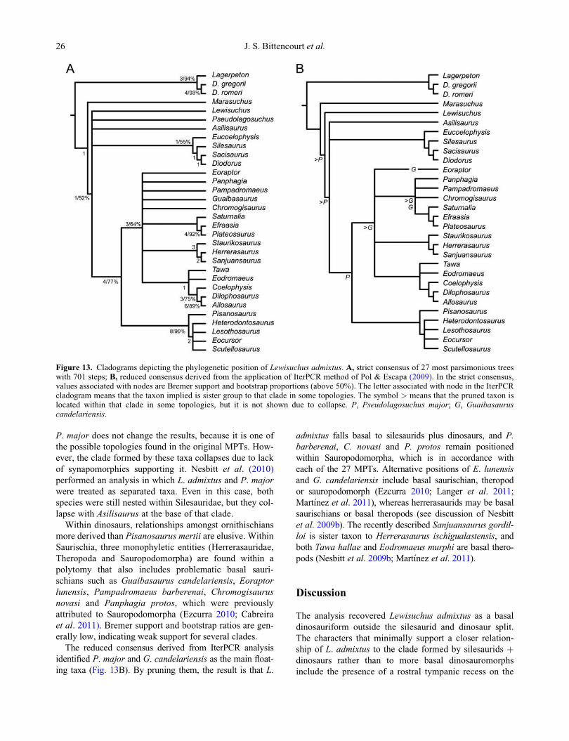
P. major does not change the results, because it is one of
the possible topologies found in the original MPTs. How-
ever, the clade formed by these taxa collapses due to lack
of synapomorphies supporting it. Nesbitt et al. (2010)
performed an analysis in which L. admixtus and P. major
were treated as separated taxa. Even in this case, both
species were still nested within Silesauridae, but they col-
lapse with Asilisaurus at the base of that clade.
Within dinosaurs, relationships amongst ornithischians
more derived than Pisanosaurus mertii are elusive. Within
Saurischia, three monophyletic entities (Herrerasauridae,
Theropoda and Sauropodomorpha) are found within a
polytomy that also includes problematic basal sauri-
schians such as Guaibasaurus candelariensis, Eoraptor
lunensis, Pampadromaeus barberenai, Chromogisaurus
novasi and Panphagia protos, which were previously
attributed to Sauropodomorpha (Ezcurra 2010; Cabreira
et al. 2011). Bremer support and bootstrap ratios are gen-
erally low, indicating weak support for several clades.
The reduced consensus derived from IterPCR analysis
identified P. major and G. candelariensis as the main float-
ing taxa (Fig. 13B). By pruning them, the result is that L.
admixtus falls basal to silesaurids plus dinosaurs, and P.
barberenai, C. novasi and P. protos remain positioned
within Sauropodomorpha, which is in accordance with
each of the 27 MPTs. Alternative positions of E. lunensis
and G. candelariensis include basal saurischian, theropod
or sauropodomorph (Ezcurra 2010; Langer et al. 2011;
Mart�ınez et al. 2011), whereas herrerasaurids may be basal
saurischians or basal theropods (see discussion of Nesbitt
et al. 2009b). The recently described Sanjuansaurus gordil-
loi is sister taxon to Herrerasaurus ischigualastensis, and
both Tawa hallae and Eodromaeus murphi are basal thero-
pods (Nesbitt et al. 2009b; Mart�ınez et al. 2011).
Discussion
The analysis recovered Lewisuchus admixtus as a basal
dinosauriform outside the silesaurid and dinosaur split.
The characters that minimally support a closer relation-
ship of L. admixtus to the clade formed by silesaurids þdinosaurs rather than to more basal dinosauromorphs
include the presence of a rostral tympanic recess on the
Figure 13. Cladograms depicting the phylogenetic position of Lewisuchus admixtus. A, strict consensus of 27 most parsimonious treeswith 701 steps; B, reduced consensus derived from the application of IterPCR method of Pol & Escapa (2009). In the strict consensus,values associated with nodes are Bremer support and bootstrap proportions (above 50%). The letter associated with node in the IterPCRcladogram means that the taxon implied is sister group to that clade in some topologies. The symbol > means that the pruned taxon islocated within that clade in some topologies, but it is not shown due to collapse. P, Pseudolagosuchus major; G, Guaibasauruscandelariensis.
26 J. S. Bittencourt et al.

lateral side of the parabasisphenoid/prootic; a concave
atlantal articulation facet in axial intercentrum, with
upturned lateral borders; and the third cervical vertebra
with centrum longer than those of axis and mid-dorsal
vertebrae (Nesbitt 2011).
Silesaurids plus dinosaurs share apomorphies to the
exclusion of L. admixtus. In the trees which Pseudolagosu-
chus major collapses with L. admixtus outside the dichot-
omy Silesauridae þ Dinosauria, these apomorphies include
minimally the presence of a craniocaudally short distal por-
tion of the dorsal neural spine and the low neural arch of
the dorsal vertebrae. Instead, if P. major is positioned
within silesaurids or closer to dinosaurs than to other dino-
sauriforms, two additional apomorphic characters shared
by silesaurids and dinosaurs, such as the reduction in the
number of maxillary teeth and the loss of pterygoid teeth,
draws L. admixtus to a more basal position. However, it
should be noted that the characters mentioned above are
highly homoplastic within dinosauriforms.
In light of the above anatomical and phylogenetic data,
some characters previously considered to support the posi-
tion of L. admixtus as a basal silesaurid (Nesbitt 2011) have
to be reconsidered. These characters include the subverti-
cally aligned exits of the hypoglossal nerve, which are also
present in M. lilloensis (PVL 3872), and the rugose ridge
on the craniolateral edges of the supraoccipital, also noted
in one specimen of Euparkeria capensis (SAM-PK-5867),
and some dinosaurs (Langer & Ferigolo 2013). Further-
more, the character ‘cervical centra 3–5 longer than mid-
trunk centra’, turned out as a synapomorphy of L. admixtus
plus other dinosauriforms, excluding M. lilloensis.
A taxonomic issue recently created by using Silesauri-
dae, rather than Lewisuchidae, for naming a family-
ranked taxon of ecologically distinct basal dinosauriforms
(Langer et al. 2010; Nesbitt et al. 2010) becomes pointless
with L. admixtus not belonging to that group. Names that
entered the literature including Lewisuchinae and Lewisu-
chidae (Paul 1988; Olshevsky 1991) are redundant by
including only the type species.
Relations within early dinosaurs remain poorly resolved
except for the persistence of the three major clades Thero-
poda, Sauropodomorpha and Ornithischia. The erratic
positions of herrerasaurids, G. candelariensis and E.
lunensis are compatible with recent disputes concerning
the phylogeny of Saurischia (Nesbitt et al. 2009b; Ezcurra
2010; Langer et al. 2011; Mart�ınez et al. 2011), and reflectpresent knowledge of its early evolution.
Conclusions
The Middle Triassic Lewisuchus admixtus is a basal dino-
sauriform, which cannot be synonymized with its coeval
Pseudolagosuchus major due to lack of data. Accordingly,
until further evidence is available, we recommend that
they are treated as a distinct taxa in evolutionary studies.
The holotype of L. admixtus is less complete than pre-
sumed by Romer (1972c), because some attributed bones
(lower jaw and pes) are more consistently referable to pro-
terochampsids. The inclusion of the isolated maxilla in the
holotype is tentative and should be treated with caution. Fea-
tures that are peculiar for basal dinosauromorphs are con-
firmed or reported for the first time in this specimen, such as
osteoderms and palatal teeth. The phylogenetic analyses sug-
gest that L. admixtus is a dinosauriform that branched prior
to the split between silesaurids and dinosaurs.
Acknowledgements
We thank Angela Milner and Sandra Chapman (NHMUK);
Rainer Schoch (SMNS); Ricardo Martinez and Oscar
Alcober (PVSJ); Pat Holroyd and Kevin Padian (UCMP);
Alexander Hohloch (GPIT); Jaime Powell (PVL); Jerzy
Dzik and Tomasz Sulej (ZPAL); Maria Claudia Malabarba
(MCP); Jorge Ferigolo and Ana Maria Ribeiro (MCN);
Cesar Schultz (UFRGS); Sergio Cabreira (ULBRA);
Sheena Kaal (SAM); Spencer Lucas (NMMNHS); Alex
Downs (GR); David Gillette (MNA) and Alejandro Kra-
marz (MACN) for allowing us to examine material under
their care. We also acknowledge the personnel of the
Museo de Ciencias Naturales de la Universidad Nacional
de La Rioja (PULR), especially Silvia Ferraris, EmilioVac-
cari and Lorena Leguizam�on, for their kind hospitality.
The Willi Hennig Society is thanked for making TNT 1.1
freely available. Martin Ezcurra and Randall Irmis are
thanked for reviewing earlier drafts of this manuscript.
This is contribution R-82 of the IDEAN (Instituto de
Estudios Andinos Don Pablo Groeber).
This project was mainly funded by Fundo de Amparo
a Pesquisa do Estado de S~ao Paulo (FAPESP) [Proc.
2010/08891–3, grant to JSB]. Additional funding for this
research was provided by the Universidade Federal de
Minas Gerais [Programa Doutor Rec�em-Contratado
PRPQ (JSB)]; Universidad de Buenos Aires [UBACyT
728 (CM)].
Supplemental material
Supplemental material for this article can be accessed here:http://dx.doi.org/10.1080/14772019.2013.878758
References
Abler, W. L. 1997. Tooth serrations in carnivorous dinosaurs.Pp. 740–743 in P. J. Currie & K. Padian (eds) Encyclopediaof Dinosaurs. Academic Press, San Diego.
Osteology of the Middle Triassic archosaur Lewisuchus admixtus 27

Arcucci, A. 1986. Nuevos materiales y reinterpretacion deLagerpeton chanarensis Romer (Thecodontia, Lagerpetoni-dae nov.) del Triasico Medio de la Rioja, Argentina. Ame-ghiniana, 23, 233–242.
Arcucci, A. 1987. Un nuevo Lagosuchidae (Thecodontia-Pseu-dosuchia) de la fauna de Los Cha~nares (Edad Reptil Cha-~narense, Triasico Medio), La Rioja, Argentina. Ameghiniana,24, 89–94.
Arcucci, A. 1990. Un nuevo Proterochampsidae (Reptilia-Arch-osauriformes) de la fauna local de Los Cha~nares (TriasicoMedio), La Rioja, Argentina. Ameghiniana, 27, 365–378.
Arcucci, A. B. 1997. Dinosauromorpha. Pp. 179–184 in P. J.Currie & K. Padian (eds) Encyclopedia of Dinosaurs. Aca-demic Press, San Diego.
Arcucci, A. B. 1998. New information about dinosaur precursorsfrom the Triassic Los Chanares fauna, La Rioja, Argentina.Journal of African Earth Science, 27, 9–10.
Bakker, R. T. & Galton, P. M. 1974. Dinosaur monophyly anda new class of vertebrates. Nature, 248, 168–172.
Barberena, M. C. 1978. A huge thecodont skull from the Trias-sic of Brazil. Pesquisas, 9, 62–75.
Barrett, P. M. 2000. Prosauropod dinosaurs and iguanas: specu-lations on the diets of extinct reptiles. Pp. 42–78 in H.-D.Sues (ed) Evolution of herbivory in terrestrial vertebrates:perspectives from the fossil record. Cambridge UniversityPress, Cambridge.
Benton, M. J. & Walker, A. D. 2002. Erpetosuchus, a croco-dile-like basal archosaur from the Late Triassic of Elgin,Scotland. Zoological Journal of the Linnean Society, 136,25–47.
Benton, M. J. &Walker, A. D. 2011. Saltopus, a dinosauriformfrom the Upper Triassic of Scotland. Earth and Environmen-tal Science Transactions of the Royal Society of Edinburgh,101, 285–299.
Bever, G. S., Brusatte, S. L., Balanoff, A. M. & Norell, M. A.2011. Variation, variability, and the origin of the avian endo-cranium: Insights from the anatomy of Alioramus altai(Theropoda: Tyrannosauroidea). PlosOne, 6, 1–10.
Bittencourt, J. S. & Kellner, A. W. A. 2009. The anatomy andphylogenetic position of the Triassic dinosaur Staurikosau-rus pricei Colbert, 1970. Zootaxa, 2079, 1–56.
Bonaparte, J. F. 1975. Nuevos materiales de Lagosuchus talam-payensis Romer (Thecodontia - Pseudosuchia) y su significadoen el origen de los Saurischia. Cha~narense inferior, Triasicomedio de Argentina. Acta Geologica Lilloana, 13, 5–90.
Bonaparte, J. F. 1978. El Mesozoico de America del Sur y sustetrapodos. Opera Lilloana, 26, 1–596.
Bonaparte, J. F. 1981. Descripcion de Fasolasuchus tenax y susignificado en la sistematica y evolucion de los Thecodontia.Revista del Museo Argentino de Ciencias Naturales“Bernardino Rivadavia”, 3, 55–101.
Bonaparte, J. F. 1982. Faunal replacement in the Triassic ofSouth America. Journal of Vertebrate Paleontology, 2, 362–371.
Bonaparte, J. F. 1997. El Triasico de San Juan - La RiojaArgentina y sus dinosaurios. Museo Argetino de CienciasNaturales, Buenos Aires, 190 pp.
Brinkman, D. 1981. The origin of the crocodiloid tarsi and theinterrelationships of thecodontian archosaurs Breviora, 464,1–23.
Brusatte, S. L., Benton, M. J., Desojo, J. B. & Langer, M. C.2010. The higher-level phylogeny of Archosauria (Tetra-poda: Diapsida). Journal of Systematic Palaeontology, 8, 3–47.
Butler, R. J., Upchurch, P. & Norman, D. B. 2008. The phy-logeny of the ornithischian dinosaurs. Journal of SystematicPalaeontology, 6, 1–40.
Cabreira, S. F., Schultz, C. L., Bittencourt, J. S., Soares, M.B., Fortier, D. C., Silva, L. R. & Langer, M. C. 2011. Newstem-sauropodomorph (Dinosauria, Saurischia) from theTriassic of Brazil. Naturwissenschaften, 98, 1035–1040.
Carabajal, A. P. 2011. The braincase anatomy of Carnotaurussastrei (Theropoda: Abelisauridae) from the Upper Creta-ceous of Patagonia. Journal of Vertebrate Paleontology, 31,378–386.
Carrano, M. T. 2006. Body-size evolution in the Dinosauria.Pp. 225–268 in M. T. Carrano, T. J. Gaudin, R. W. Blob &J. R. Wible (eds) Amniote paleobiology: perspectives on theevolution of mammals, birds, and reptiles. The University ofChicago Press, Chicago.
Carroll, R. L. 1988. Vertebrate paleontology and evolution. W.H. Freeman and Company, New York, 698 pp.
Chatterjee, S. 1978. A primitive parasuchid phytosaur reptilefrom the Upper Triassic Maleri Formation of India. Palaeon-tology, 21, 83–128.
Clark, J. M., Sues, H.-D. & Berman, D. 2000. A new specimenof Hesperosuchus agilis from the Upper Triassic ofNew Mexico and the Interrelationships of basal crocodylo-morph archosaurs. Journal of Vertebrate Paleontology, 20,683–740.
Cope, E. D. 1869. Synopsis of the extinct Batrachia, Reptiliaand Aves of North America. Transactions of the AmericanPhilosophical Society, 14, 1–252.
Cruickshank, A. R. I. 1979. The ankle joint in some early arch-osaurs. South African Journal of Science, 75, 168–178.
Desojo, J. B. & Arcucci, A. 2009. New material of Luperosu-chus fractus (Archosauria: Crurotarsi) from the Middle Tri-assic of Argentina: the earliest known South American‘rauisuchian’. Journal of Vertebrate Paleontology, 24,1311–1315.
Desojo, J. B., Ezcurra, M. D. & Schultz, C. L. 2011. Anunusual new archosauriform from the Middle-LateTriassic of southern Brazil and the monophyly of Doswellii-dae. Zoological Journal of the Linnean Society, 161, 839–871.
Dzik, J. 2003. A beaked herbivorous archosaur with dinosauraffinities from the early Late Triassic of Poland. Journal ofVertebrate Paleontology, 23, 556–574.
Dzik, J. & Sulej, T. 2007. A review of the early Late TriassicKrasiejow biota from Silesia, Poland. Palaeontologia Polon-ica, 64, 3–27.
Ewer, R. F. 1965. The anatomy of the thecodont reptile Eupar-keria capensis Broom. Philosophical Transactions of theRoyal Society, Series B, 248, 375–435.
Ezcurra, M. D. 2006. A review of the systematic position of thedinosauriform archosaur Eucoelophysis baldwini Sullivan &Lucas, 1999 from the Upper Triassic of New Mexico, USA.Geodiversitas, 28, 649–684.
Ezcurra, M. D. 2010. A new early dinosaur (Saurischia: Sauro-podomorpha) from the Late Triassic of Argentina: a reas-sessment of dinosaur origin and phylogeny. Journal ofSystematic Palaeontology, 8, 371–425.
Ferigolo, J. & Langer, M. C. 2007. A Late Triassic dinosauri-form from south Brazil and the origin of the ornithischianpredentary bone. Historical Biology, 19, 23–33.
Franzosa, J. & Rowe, T. 2005. Cranial endocast of the Creta-ceous theropod dinosaur Acrocanthosaurus atokensis. Jour-nal of Vertebrate Paleontology, 25, 859–864.
28 J. S. Bittencourt et al.

Fraser, N. C., Padian, K., Walkden, G. M. & Davis, A. L. M.2002. Basal dinosauriform remains from Britain and thediagnosis of the Dinosauria. Palaeontology, 45, 79–95.
Galton, P. M. 2001. Endocranial casts of the plated dinosaurStegosaurus (Upper Jurassic, western USA): a completeundistorted cast and the original specimens of OthnielCharles Marsh. Pp. 103–129 in K. Carpenter (ed) TheArmored Dinosaurs. Indiana University Press, Bloomington.
Galton, P. M. & Upchurch, P. 2004. Prosauropoda. Pp. 232–258 in D. B. Weishampel, P. Dodson & H. Osm�olska (eds)The Dinosauria. 2nd edition. University of California Press,Berkeley.
Gauthier, J. 1986. Saurischian monophyly and the origin of birds.Memoirs of the California Academy of Sciences, 8, 1–55.
Gilmore, C. W. 1920. Osteology of the carnivorous Dinosauria inthe United States National Museum, with special reference tothe genera Antrodemus (Allosaurus) and Ceratosaurus. Bulle-tin of the United States National Museum, 110, 1–150.
Goloboff, P. A., Farris, J. S. & Nixon, K. C. 2008. TNT, a freeprogram for phylogenetic analysis. Cladistics, 24, 774–786.
Gower, D. J. 1999. The cranial and mandibular osteology of anew rauisuchian archosaur from the Middle Triassic ofsouthern Germany. Stuttgarter Beitr€age zur NaturkundeSerie B (Geologie und Pal€aontologie), 280, 1–49.
Gower, D. J. 2002. Braincase evolution in suchian archosaurs(Reptilia : Diapsida): evidence from the rauisuchian Batra-chotomus kupferzellensis. Zoological Journal of the LinneanSociety, 136, 49–76.
Gower, D. J. & Schoch, R. R. 2009. Postcranial anatomy of therauisuchian archosaur Batrachotomus kupferzellensis. Jour-nal of Vertebrate Paleontology, 29, 103–122.
Gower, D. J. & Sennikov, A. G. 1996. Morphology and phylo-genetic informativeness of early archosaur braincases.Palaeontology, 39, 883–906.
Gower, D. J. & Sennikov, A. G. 1997. Sarmatosuchus and theearly history of the Archosauria. Journal of Vertebrate Pale-ontology, 17, 60–73.
Gower, D. J. & Weber, E. 1998. The braincase of Euparkeria,and the evolutionary relationships of birds and crocodilians.Biological Reviews of the Cambridge Philosophical Society,73, 367–411.
Harris, J. D. 2006. The axial skeleton of the dinosaur Suuwas-sea emilieae (Sauropoda: Flagellicaudata) from the UpperJurassic Morrison Formation of Montana, USA. Palaeontol-ogy, 49, 1091–1121.
Hutchinson, J. R. 2001. The evolution of femoral osteology andsoft tissues on the line to extant birds (Neornithes). Zoologi-cal Journal of the Linnean Society, 131, 169–197.
Irmis, R. B., Nesbitt, S. J., Padian, K., Smith, N. D., Turner,A. H., Woody, D. & Downs, A. 2007a. A Late Triassicdinosauromorph assemblage from New Mexico and the riseof dinosaurs. Science, 317, 358–361.
Irmis, R. B., Parker, W. G., Nesbitt, S. J. & Jun, L. 2007b.Early ornithischian dinosaurs: the Triassic record. HistoricalBiology, 19, 3–22.
Kammerer, C. F., Nesbitt, S. & Shubin, N. H. 2012. The firstbasal dinosauriform (Silesauridae) from the Late Triassic ofMorocco. Acta Palaeontologica Polonica, 57, 277–284.
Langer, M. C. & Benton, M. J. 2006. Early dinosaurs: A phyloge-netic study. Journal of Systematic Palaeontology, 4, 309–358.
Langer, M. C., Bittencourt, J. S. & Schultz, C. L. 2011. Areassessment of the basal dinosaur Guaibasaurus candelar-iensis, from the Late Triassic Caturrita Formation of south
Brazil. Earth and Environmental Science Transactions ofthe Royal Society of Edinburgh, 101, 301–332.
Langer, M. C., Ezcurra, M. D., Bittencourt, J. S. & Novas, F.E. 2010. The origin and early evolution of dinosaurs. Biolog-ical Reviews, 85, 55–110.
Langer, M. C. & Ferigolo, J. 2013. The Late Triassic dinosaur-omorph Sacisaurus agudoensis (Caturrita Formation; RioGrande do Sul, Brazil): anatomy and affinities. GeologicalSociety, London, Special Publications, 379, doi: 10.1144/sp379.16.
Langer, M. C., Franca, M. A. G. & Gabriel, S. 2007a. Thepectoral girdle and forelimb anatomy of the stem-sauropodo-morph Saturnalia tupiniquin (Upper Triassic, Brazil). Spe-cial Papers in Palaeontology, 77, 113–137.
Langer, M. C., Ribeiro, A. M., Schultz, C. L. & Ferigolo, J.2007b. The continental tetrapod-bearing Triassic of SouthBrazil. Bulletin of the New Mexico Museum of Natural His-tory and Science, 41, 201–218.
Larsell, O. 1932. The cerebellum of reptiles: chelonians andalligator. The Journal of Comparative Neurology, 56,299–345.
Lautenschlager, S. & Desojo, J. B. 2011. Reassessment of theMiddle Triassic rauisuchian archosaurs Ticinosuchus feroxand Stagonosuchus nyassicus. Pal€aontologische Zeitschrift,85, 357–381.
Mancuso, A. C. & Marsicano, C. A. 2008. Paleoenvironmentsand taphonomy of a Triassic lacustrine system (Los RastrosFormation, Central-Western Argentina). Palaios, 23, 535–547.
Mart�ınez, R. N., Sereno, P. C., Alcober, O., Colombi, C. E.,Renne, P. R., Monta~nez, I. P. & Currie, B. S. 2011. Abasal dinosaur from the dawn of the dinosaur era in south-western Pangaea. Science, 331, 206–210.
Meers, M. B. 2003. Crocodylian forelimb musculature and itsrelevance to Archosauria. The Anatomical Record Part A,274A, 891–916.
Nesbitt, S. J. 2005. Osteology of the Middle Triassic pseudosu-chian archosaur Arizonasaurus babbitti. Historical Biology,17, 19–47.
Nesbitt, S. J. 2011. The early evolution of archosaurs: relation-ships and the origin of major clades. Bulletin of the Ameri-can Museum of Natural History, 352, 1–292.
Nesbitt, S. J., Irmis, R. B. & Parker, W. G. 2007. A critical re-evaluation of the Late Triassic dinosaur taxa of North Amer-ica. Journal of Systematic Palaeontology, 5, 209–243.
Nesbitt, S. J., Irmis, R. B., Parker, W. G., Smith, N. D.,Turner, A. H. & Rowe, T. 2009a. Hindlimb osteology anddistribution of basal dinosauromorphs from the Late Triassicof North America. Journal of Vertebrate Paleontology, 29,498–516.
Nesbitt, S. J., Sidor, C. A., Irmis, R. B., Angielczyk, K. D.,Smith, R. M. H. & Tsuji, L. A. 2010. Ecologically distinctdinosaurian sister group shows early diversification of Orni-thodira. Nature, 464, 95–98.
Nesbitt, S. J., Smith, N. D., Irmis, R. B., Turner, A. H.,Downs, A. & Norell, M. A. 2009b. A complete skeleton ofa Late Triassic saurischian and the early evolution of dino-saurs. Science, 326, 1530–1533.
Norman, D. B., Crompton, A. W., Butler, R. J., Porro, L. B.& Charig, A. J. 2011. The Lower Jurassic ornithischiandinosaur Heterodontosaurus tucki Crompton & Charig,1962: cranial anatomy, functional morphology, taxonomy,and relationships. Zoological Journal of the Linnean Society,163, 182–276.
Osteology of the Middle Triassic archosaur Lewisuchus admixtus 29

Norman, D. B., Witmer, L. M. & Weishampel, D. B. 2004a.Basal Ornithischia. Pp. 325–334 in D. B. Weishampel, P.Dodson & H. Osm�olska (eds) The Dinosauria. 2nd edition.University of California Press, Berkeley.
Norman, D. B., Witmer, L. M. & Weishampel, D. B. 2004b.Basal Thyreophora. Pp. 335–342 in D. B. Weishampel, P.Dodson & H. Osm�olska (eds) The Dinosauria. 2nd edition.University of California Press, Berkeley.
Novas, F. E. 1989. The tibia and tarsus in Herrerasauridae(Dinosauria, incertae sedis) and the origin and evolution ofthe dinosaurian tarsus. Journal of Paleontology, 63, 677–690.
Novas, F. E. 1992. Phylogenetic relationships of the basal dino-saurs, the Herrerasauridae. Palaeontology, 35, 51–62.
Novas, F. E. 1994. New information on the systematics andpostcranial skeleton of Herrerasaurus ischigualastensis(Theropoda: Herrerasauridae) from the Ischigualasto Forma-tion (Upper Triassic) of Argentina. Journal of VertebratePaleontology, 13, 400–423.
Novas, F. E. 1996. Dinosaur monophyly. Journal of VertebratePaleontology, 16, 723–741.
Olshevsky, G. 1991. A revision of the parainfraclass Archosau-ria Cope, 1869, excluding the advanced Crocodylia. Meso-zoic Meanderings, 2, 1–196.
Parrish, J. M. 1993. Phylogeny of the Crocodylotarsi, with ref-erence to archosaurian and crurotarsan monophyly. Journalof Vertebrate Paleontology, 13, 287–308.
Parrish, J. M. 1994. Cranial osteology of Longosuchus meadeiand the phylogeny and distribution of the Aetosauria. Jour-nal of Vertebrate Paleontology, 14, 196–209.
Paul, G. S. 1988. Predatory dinosaurs of the world. Touchstone,New York, 464 pp.
Peecook, B., Sidor, C., Nesbitt, S., Smith, R. M. H., Steyer, J.S. & Angielczyk, K. 2013. A new silesaurid from the upperNtawere Formation of Zambia (Middle Triassic) demon-strates the rapid diversification of Silesauridae (Avemetatar-salia, Dinosauriformes). Journal of VertebratePaleontology, 33, 1127–1137.
Piechowsky, R.& Dzik, J. 2010. The axial skleton of Silesaurusopolensis. Journal of Vertebrate Paleontology, 30, 1127–1141.
Pol, D.& Escapa, I. H. 2009. Unstable taxa in cladistic analysis:identification and the assessment of relevant characters. Cla-distics, 25, 515–527.
Raath, M. A. 1969. A new coelurosaurian dinosaur from the for-est sandstone of Rhodesia. Arnoldia, 4, 11–25.
Raath, M. A. 1977. The anatomy of the Triassic theropod Syn-tarsus rhodesiensis (Saurischia: Podokesauridae) and aconsideration of its biology. Unpublished thesis, RhodesUniversity, Salisbury, 233 pp.
Rauhut, O. W. M. 2003. The interrelationships and evolution ofbasal theropod dinosaurs. Special Papers in Palaeontology,69, 1–213.
Remes, K. 2007. Evolution of the pectoral girdle and forelimb inSauropodomorpha (Dinosauria, Saurischia): osteology,myology and function. Unpublished PhD thesis, Universit€atM€unchen, 355 pp.
Rogers, R. R., Arcucci, A., Abdala, F., Sereno, P. C., Forster,C. A. & May, C. L. 2001. Paleoenvironment and taphonomyof the Cha~nares Formation tetrapod assemblage (Middle Tri-assic), Northwestern Argentina: spectacular preservation involcanogenic concretions. Palaios, 16, 461–481.
Romer, A. S. 1956. Osteology of Reptiles. The University ofChicago Press, Chicago, 772 pp.
Romer, A. S. 1966. The Cha~nares (Argentina) Triassic reptilefauna. I. Introduction. Breviora, 247, 1–14.
Romer, A. S. 1971. The Cha~nares (Argentina) Triassic reptilefauna. X. Two new but incompletely known long-limbedpseudosuchians. Breviora, 378, 1–10.
Romer, A. S. 1972a. The Cha~nares (Argentina) Triassic reptilefauna. XII. The postcranial skeleton of the thecodont Cha-naresuchus. Breviora, 385, 1–21.
Romer, A. S. 1972b. The Chanares (Argentina) Triassic reptilefauna. XIII. An early ornithosuchid pseudosuchian, Gracili-suchus stipanicicorum, gen. et sp. nov. Breviora, 389, 1–24.
Romer, A. S. 1972c. The Chanares (Argentina) Triassic reptilefauna. XIV. Lewisuchus admixtus, gen et sp. nov., a furtherthecodont from the Chanares beds. Breviora, 390, 1–13.
Romer, A. S. & Jensen, J. A. 1966. The Chanares (Argentina)Triassic reptile fauna. II. Sketch of the geology of the R�ıoCha~nares-R�ıo Gualo region. Breviora, 252, 1–20.
Rowe, T. 1989. A new species of the theropod dinosaur Syntar-sus from the Early Jurassic Kayenta Formation of Arizona.Journal of Vertebrate Paleontology, 9, 125–136.
Sampson, S. D. & Witmer, L. M. 2007. Craniofacial anatomyof Majungasaurus crenatissimus (Theropoda : Abelisauri-dae) from the Late Cretaceous of Madagascar. Society ofVertebrate Paleontology Memoir, 8, 32–102.
Santa Luca, A. P. 1980. The postcranial skeleton of Heterodon-tosaurus tucki (Reptilia, Ornithischia) from the Stormbergof South Africa. Annals of the South African Museum, 79,159–211.
Sereno, P. C. 1991a. Basal archosaurs: phylogenetic relation-ships and functional implications. Society of VertebratePaleontology Memoir, 2, 1–53.
Sereno, P. C. 1991b. Lesothosaurus, “fabrosaurids,” and theearly evolution of Ornithischia. Journal of Vertebrate Pale-ontology, 11, 168–197.
Sereno, P. C. 1994. The pectoral girdle and forelimb of the basaltheropod Herrerasaurus ischigualastensis. Journal of Verte-brate Paleontology, 13, 425–450.
Sereno, P. C. 1999. The evolution of dinosaurs. Science, 284,2137–2147.
Sereno, P. C. & Arcucci, A. B. 1994a. Dinosaurian precursorsfrom the Middle Triassic of Argentina: Lagerpeton chanar-ensis. Journal of Vertebrate Paleontology, 13, 385–399.
Sereno, P. C. & Arcucci, A. B. 1994b. Dinosaurian precursors fromthe Middle Triassic of Argentina: Marasuchus lilloensis, n. gen.Journal of Vertebrate Paleontology, 14, 53–73.
Sereno, P. C., Forster, C. A., Rogers, R. R. & Monetta, A. M.1993. Primitive dinosaur skeleton from Argentina and theearly evolution of Dinosauria. Nature, 361, 64–66.
Sereno, P. C. & Novas, F. E. 1994. The skull and neck of thebasal theropod Herrerasaurus ischigualastensis. Journal ofVertebrate Paleontology, 13, 451–476.
Sill, W. D. 1974. The anatomy of Saurosuchus galilei and therelationships of the rauisuchid thecodonts. Bulletin of theMuseum of Comparative Zoology, 146, 317–362.
Simpson, G. G. 1940. Types in modern taxonomy. AmericanJournal of Science, 238, 413–431.
Smith, N. D., Makovicky, P. J., Hammer, W. R. & Currie, P.J. 2007. Osteology of Cryolophosaurus ellioti (Dinosauria :Theropoda) from the Early Jurassic of Antarctica and impli-cations for early theropod evolution. Zoological Journal ofthe Linnean Society, 151, 377–421.
Sues, H.-D. 1997. Continental tetrapods of the Early Mesozoic.Pp. 627–643 in J. O. Farlow & M. K. Brett-Surman (eds)The complete dinosaur. Indiana University, Bloomington.
30 J. S. Bittencourt et al.

Sues, H. D., Reisz, R. R., Hinic, S. & Raath, M. A. 2004. Onthe skull of Massospondylus carinatus Owen, 1854 (Dino-sauria : Sauropodomorpha) from the Elliot and Clarens for-mations (Lower Jurassic) of South Africa. Annals ofCarnegie Museum, 73, 239–257.
ten Donkelaar, H. J. 1998. Reptiles. Pp. 1315–1523 in R. Nieu-wenhuys, H. J. ten Donkelaar & C. Nicholson (eds) The Cen-tral Nervous System of Vertebrates. Springer, Berlin.
Trotteyn, M. J., Arcucci, A. B. & Raugust, T. 2013. Protero-champsia: an endemic archosauriform clade from SouthAmerica. Geological Society of London, Special Publica-tions, 379, doi 10.1144/sp379.23.
Trotteyn, M. J. & Haro, J. A. 2011. The braincase of a speci-men of Proterochampsa Reig (Archosauriformes: Protero-champsidae) from the Late Triassic of Argentina.Pal€aontologische Zeitschrift, 85, 1–17.
Walker, A. D. 1961. Triassic reptiles from the Elgin area: Stago-nolepis, Dasygnathus and their allies. Philosophical Trans-actions of the Royal Society, Series B, 244, 103–204.
Walker, A. D. 1964. Triassic reptiles from the Elgin area:Ornithosuchus and the origin of carnosaurs. PhilosophicalTransactions of the Royal Society of London, Series B, 248,53–134.
Walker, A. D. 1990. A revision of Sphenosuchus acutus Haughton,a crocodylomorph reptile from the Elliot Formation (Late
Triassic or Early Jurassic) of South Africa. PhilosophicalTransactions of the Royal Society of London, Series B, 330,1–120.
Welles, S. P. 1984. Dilophosaurus wetherilli (Dinosauria, Ther-opoda). Osteology and comparisons. Palaeontographica,185, 85–180.
Welman, J. 1998. The taxonomy of the South African protero-suchids (Reptilia, Archosauromorpha). Journal of Verte-brate Paleontology, 18, 340–347.
Wilson, J. A. 1999. A nomenclature for vertebral laminae insauropods and other saurischian dinosaurs. Journal of Verte-brate Paleontology, 19, 639–653.
Witmer, L. M. 1990. The craniofacial air sac system of Meso-zoic birds (Aves). Zoological Journal of the Linnean Soci-ety, 100, 327–378.
Wu, X. C. & Russell, A. P. 2001. Redescription of Turfanosu-chus dabanensis (Archosauriformes) and new informationon its phylogenetic relationships. Journal of VertebratePaleontology, 21, 40–50.
Yates, A. M. 2003. A new species of the primitive dinosaur The-codontosaurus (Saurischia: Sauropodomorpha) and itsimplications for the systematics of early dinosaurs. Journalof Systematic Palaeontology, 1, 1–42.
Yates, A. M. 2007. Solving a dinosaurian puzzle: the identity ofAliwalia rex Galton. Historical Biology, 19, 93–123.
Osteology of the Middle Triassic archosaur Lewisuchus admixtus 31