cranial and mandibular osteology of the … · cranial and mandibular osteology of the early...
TRANSCRIPT
CRANIAL AND MANDIBULAR OSTEOLOGYOF THE EARLY TRIASSIC ARCHOSAURIFORM
OSMOLSKINA CZATKOWICENSIS FROM POLAND
MAGDALENA BORSUK−BIAŁYNICKA and SUSAN E. EVANS
Borsuk−Białynicka, M. and Evans, S.E. 2009. Cranial and mandibular osteology of the EarlyTriassic archosauriform Osmolskina czatkowicensis from Poland. Palaeontologia Polonica65, 235–281.
The basal archosauriform Osmolskina czatkowicensis Borsuk−Białynicka et Evans, 2003from the Early Olenekian karst deposits of Czatkowice near Kraków (southern Poland)shares a unique mosaic of skull character states with the African Anisian genus Euparkeria.This is considered, with reservation, as a basis for including them in the family Euparke−riidae Huene, 1920. A provisional diagnosis is given for this family, but no unique derivedcharacter states have been identified to support its monophyly. Osmolskina and Euparkeriadiffer primarily in snout structure. The vertical orientation of the basisphenoid, and thepostero−ventral position of the entry foramina for cerebral branches of the internal carotid ar−tery place both genera crownward of the proterosuchids. Lack of ossification of the medialwall of the otic capsule and the partial retention of pterygoid teeth exclude them fromcrown−group Archosauria, while they also lack erythrosuchid autapomorphies (including ex−tremely short cervical vertebrae).
Key words: Early Triassic, Poland, basal Archosauriformes, euparkeriids.
Magdalena Borsuk−Białynicka[[email protected]], Institut Paleobiologii PAN, Twarda51/55, PL−00−818 Warszawa, Poland.
Susan E. Evans [[email protected]], Research Department of Cell and Developmental Biol−ogy, UCL, University College London, Gower Street, London, WC1E 6BT, UK.
Received 20 April 2005, accepted 29 May 2008
INTRODUCTION
Osmolskina is a small Early Triassic euparkeriid−grade archosauriform, known from the karst fissure de−posits of Czatkowice near Kraków (southern Poland, Czatkowice 1 locality). Osmolskina is the largest andmost common component of a diverse small vertebrate assemblage from these deposits including a smallprolacertiform−grade reptile (Borsuk−Białynicka and Evans 2009), two basal lepidosauromorphs (Evans andBorsuk−Białynicka 2009a), including a basal kuehneosaurid (Evans 2009), procolophonids (Borsuk−Biały−nicka and Lubka 2009) and temnospondyls (Szyszkin and Sulej 2009). A tiny stem−frog, Czatkobatrachuspolonicus (Evans and Borsuk−Białynicka 1998, 2009b; Borsuk−Białynicka and Evans 2002), and some fish,also belong to this fauna.
The Early Triassic archosauriforms are crucial to an understanding of the incompletely known section ofphylogeny preceding the split between the crocodilian and dinosaur/avian lineages. Terrestrial faunas of thisage have been recorded from all over the world, beginning with the most complete assemblages of easternEuropean Russia (Ochev and Shishkin 1985, 1989; Shishkin and Ochev 1993), through the Lower and Mid−dle Germanic Buntsandstein (Induan−Olenekian in age), the North American Wupatki Member (the basalmember of Moenkopi Formation, Morales 1987), the upper part of the Chinese Guodikeng Formation(Jimsarian, Fuguan, and probably Ordosian faunachrons, Lucas 1993), to the Gondwanan formations: theAfrican Karoo (Lystrosaurus and Cynognathus zones), the Antarctic Fremouw, Indian Panchet, and Austra−lian Arcadia formations.
The Early Triassic fauna was not as uniform over Pangaea as was that of the Permian. Therapsids werepresent in the Gondwanan assemblages along with large temnospondyls, basal archosauriforms, and procolo−phonians, but were absent from Laurasian assemblages, except for those of the Induan period (Battail 1993).Small non−archosauriform reptiles and other microvertebrates were distributed more randomly. If theCzatkowice 1 fauna is correctly dated to the earliest Late Olenekian (Szyszkin and Sulej 2009), this is thefirst terrestrial vertebrate assemblage of this age recorded from Euramerica outside the Cis−Urals and EastEuropean Platform.
The objective of the present paper is to give a detailed description of the cranial morphology ofOsmolskina czatkowicensis. The systematic position of Osmolskina, its relationships with Euparkeria, andthe status of the Euparkeriidae, as well as the information value of cranial characters at this stage of evolu−tion, are also discussed.
Institutional acronyms. — BPS, Bayerische Staatssammlung für Paläontologie und Historische Geo−logie, München, Germany; GPIT, Institute und Museum für Geologie und Paläontologie, UniversitatTübingen, Germany; MZ, Museum of the Earth, Polish Academy of Sciences, Warsaw, Poland; PIN, Pale−ontological Institute, Russian Academy of Sciences, Moscow, Russia; SAM, South African Museum,Cape Town, Republic of South Africa; SMNS, Staatliches Museum für Naturkunde, Stuttgart, Germany;ZPAL, Institute of Paleobiology, Polish Academy of Sciences, Warsaw, Poland.
Acknowledgements. — We are indebted to Józef Wieczorek and Mariusz Paszkowski (JagiellonianUniversity, Kraków) who discovered the Czatkowice 1 breccia, and kindly offered it for study. Ourwarmest thanks go to Teresa Maryańska (Museum of the Earth) and the late Halszka Osmólska (Instituteof Paleobiology), both from the Polish Academy of Sciences, who generously offered the Czatkowicematerial to us. MBB expresses her warmest thanks to following persons and institutions that allowed herto study archosauriform material in their collections: Rupert Wild at the Staatliches Museum für Natur−kunde, Stuttgart, Michael Maisch at the Institut and Museum für Geologie und Palaeontologie, Uni−versität Tübingen, and Helmut Mayr at the Bayrische Staatssammlung für Paläontologie und HistorischeGeologie, München. We are grateful to the referees who greatly helped in improving the manuscript.Thanks are also due to the following staff members of the Institute of Paleobiology, Polish Academy ofSciences in Warsaw: Ewa Hara for preparation of the material, Cyprian Kulicki, and Janusz Błaszyk forSEM microphotographs, and Aleksandra Hołda−Michalska for preparing computer illustrations. Thework of MBB was partly supported by the State Committee of Scientific Research, KBN grant No 6PO4D 072 19.
236 MAGDALENA BORSUK−BIAŁYNICKA and SUSAN E. EVANS

GEOLOGICAL SETTING
Czatkowice 1 was the largest of several sediment infilled caves developed in the Early Carboniferous lime−stone quarry. (Paszkowski and Wieczorek 1982). Paszkowski (2009) gives a more detailed account of the geo−logical setting of Czatkowice 1 locality. As a result of economic exploitation of the quarry, the Czatkowice 1fissure no longer exists. The original age determination of the assemblage, as Early Olenekian on the basis ofGnathorhiza (Dipnoi) teeth and procolophonian material (Borsuk−Białynicka et al. 2003), has been revised toearliest Late Olenekian on the basis of temnospondyl material (Shishkin and Sulej 2009). This suggestsCzatkowice 1 karst deposits are slightly younger than the upper part of the Vetlugian Superhorizon of the EastEuropean biozonation (Shishkin et al.1995, 2000; Shishkin and Ochev 1985; Ochev and Shishkin 1989). Ac−cording to Ochev (1993), and Shishkin and Ochev (1993), the locality must have been within the xericcircumequatorial belt occurring at this latitude in Northern Pangaea. The vertebrate assemblage from Czatko−wice 1, including both terrestrial and amphibious animals and some fish (Borsuk−Białynicka et al. 1999), indi−cates that the material was deposited primarily in a freshwater pool, probably developed within an oasis in anotherwise arid Central European Scythian environment. At a higher taxonomic level, the assemblage corre−sponds to others in Laurasia, but it differs dramatically in the size of the animals included (see below) and wasprobably taphonomically biased. The taphonomy of the Czatkowice 1 locality has recently been studied byCook and Trueman (2009). The authors suggest that the diagenetic pathways included reworking of the skeletalremains, originally incorporated into sediments at the bottom of ephemeral lakes and pools, and their rede−position in nearby cave systems after a short fast transport during periods of stormy weather and flooding.
MATERIAL AND METHODS
General comments. — The material is completely disarticulated, and often broken into pieces, both be−fore deposition and in preparation. Chemical breakdown of the matrix by acetic acid is the only method ofpreparation for this material. Freshly dissociated pieces may sometimes be matched with one another andglued together. The bones are mostly white, orange or dark brown, and are generally preserved three−dimensionally. Most of them show relatively little abrasion. To date, about 100 specimens have been cata−logued, but there are hundreds of other specimens. The materials are stored at the Museum of the Earth andthe Institute of Paleobiology, Polish Academy of Sciences, Warsaw.
Attribution of osteological elements. — The primary criteria for the attribution of disarticulated bonesto particular species are size ranges and relative frequency of particular morphotypes in the samples. Ro−bustness of elements, sculpture, articular facet fit, and phylogenetically relevant characters are the next cri−teria. The composition of fossil assemblages is biased by the differential preservational potential of partic−ular elements belonging to the same animal, and those of different animals. Nonetheless, the relative per−centage of elements in a fossil sample seems to be fairly consistent for each member of the assemblage.The largest (20 mm dentary length) and most frequent remains belong to Archosauriformes and are gener−ally considered conspecific. Some less common bones exceeding the normal variability range of the genushave been excluded as representing a separate taxon, their attribution being relegated to future studies.Next in size in the assemblage is a prolacertiform−grade reptile (Borsuk−Białynicka and Evans 2009a), butthe dimensions overlap. The remaining material is a mixture of medium−sized procolophonids, and tinybones (up to a few mm in length) including two rarer lepidosauromorphs (Evans and Borsuk−Białynicka2009a; Evans 2009), tiny temnospondyls (Shishkin and Sulej 2009), and the stem frog Czatkobatrachuspolonicus (Evans and Borsuk−Białynicka 1998, 2009b; Borsuk−Białynicka and Evans 2002). The dimen−sions of these animals do not overlap the range of Osmolskina czatkowicensis.
Reconstruction (Fig. 1A, B). — Based on counts of the best preserved bone fragments, such as frontals,quadrates and articulars, the number of Osmolskina individuals of the Czatkowice 1 taphocoenosis amountsto several hundred. Some more fragile bones are represented by single specimens. The reconstruction of thewhole skull on the basis of such material requires some scaling of elements. The shape of paired premaxillaein the horizontal plane (Fig. 2A1) is conclusive for the horizontal outline of the skull as a whole, while the
OSMOLSKINA CRANIAL AND MANDIBULAR OSTEOLOGY 237

profile of the nasal process of the premaxilla (Fig. 2B2, D1) affects that of the skull in a sagittal plane. Thelength of skull roof bones should correspond to that of the maxilla articulated with the jugal and quadrato−jugal, whereas the gap between the skull roof bones and the ventral profile of the skull should match the sizeof the quadrate, which closes the skull outline from behind. These interdependent dimensions help to correctthe relative sizes of missing parts, especially those of the maxillae. There are no complete specimens of themaxilla. The posterior parts of the maxillae preserved as separate pieces (Figs 5B, 14D, E) are tentativelyconsidered as belonging to the same species if they match the size and thecodont tooth implantation of the an−terior fragments. The length of the maxilla has been deduced on the basis of the gap left between the jugaland the anterior part of the maxilla. Among other problematic elements, the pterygoids barely fit within thespace outlined by the maxillae and jugals (Fig. 1A) unless aligned obliquely with the dentigerous fields de−scending ventro−laterad to produce a V−shaped concave palate open ventrally.
In spite of a poor ossification of the walls of the braincase, the component bones are fairly well preservedand allow a reconstruction of the whole element and of its details (Figs 17, 25).
238 MAGDALENA BORSUK−BIAŁYNICKA and SUSAN E. EVANS
palatineas preserved
palatinereconstructed
surangular
prearticular facet on angular
dentary
splenial prearticular
surangular
retroarticular process
articular
splenial facet
splenial
possible coronoid position
possible coronoid position
alveolar nerveentrance
10 mm
10 mm
10 mm
10 mm
mandibular fenestra
Fig. 1. Osmolskina czatkowicensis Borsuk−Białynicka et Evans, 2003, Early Triassic of Czatkowice 1, Poland. Reconstructions:A. Skull. B. Skull with mandible. E–G. Mandibles, different views of mandibular bones; without splenial and prearticular (E),without prearticular (F), complete (G); out of scale. C. Euparkeria capensis Broom, 1913, skull with mandible (from Ewer 1965,fig. 2). D. Erythrosuchus africanus Broom, 1905, skull with mandible (from Gower 2003, fig. 1). Ventral (A), left lateral (B–D),
and medial (E–G) views.

The braincase structure of the basal archosauriforms Sarmatosuchus, Erythrosuchus, and Euparkeria,and of the basal crurotarsians Batrachotomus and Stagonolepis (Gower and Sennikov 1997, Gower 1997,Gower 2002, Gower and Weber 1998, and Gower and Walker 2002, respectively) has been used as a refer−ence. Nonetheless, the complex nature of this part of the skeleton, and preservation problems, substantiatethe use of extant comparative material. Lizards, although more distantly related to archosauriforms than croc−odiles, have a less specialized skull structure than the latter group. Hence, lizard skulls have been used as thebasis of structural interpretations, and the terminology of Oelrich (1956) has been applied.
Several points contribute to the reconstruction of the structure and the proper life position of the mandi−ble. Most informative is the mandibular symphysis, which determines the alignment of the dentaries and theirinclination to one another.
Individual age and size. — The braincase specimens show poor ossification of the internal surface, withalmost no finished bone (except for the passage of nerve canals) suggesting that they are immature. The rarityof fully attached teeth may further suggest that much of the material is immature. However, cervical verte−brae, that match the size of the skull bones, have neurocentral suture closed which is a criterion of maturity incrocodiles and, most probably, in crocodilian relatives (Brochu 1996). The most abundant vertebrae ofOsmolskina, with neural arches fused, are about the size of those of an adult individual of the lizardHeloderma suspectum (ZPAL RV/26) used for comparison. This specimen has a skull 60 mm in length,which corresponds to the average skull length reconstructed for Osmolskina. The closure of the neurocentralsutures does not necessarily mean a complete stoppage of growth (Brochu 1996).
The term “grade” used herein refers to taxa that share the same combination of primitive and derivedcharacters but no unique synapomorphies.
SYSTEMATIC BACKGROUND
The Archosauriformes (sensu Gauthier 1986) correspond to what Romer (1956) designated as Archosauria.They belong to the diapsid subgroup Archosauromorpha (Gauthier, 1986) along with rhynchosaurs, “pro−lacertiforms” and a number of isolated genera which stand as plesions (Evans 1988). According to Gauthier’sdefinition, Archosauria is restricted to the two extant groups, Aves and Crocodilia, their most recent commonancestor and all its descendants, and thus correspond to the “crown group Archosauria” of e.g., Benton andClark (1988), or to Avesuchia of Benton (1999). Archosauriformes is a more−inclusive taxon encompassing aseries of taxa on the archosaurian stem. Phylogenetic relationships within this assemblage have been studied bymany authors (Benton and Clark 1988; Gauthier 1986; Sereno and Arcucci 1990; Sereno 1991; Parrish 1993;Juul 1994; Gower and Sennikov 1996; Gower 1997; Gower and Walker 2002). Gower and Wilkinson (1996)demonstrated a substantial agreement in the general topology of cladograms resulting from those studies, allsupporting the monophyly of Archosauria sensu Gauthier (1986). The semistrict reduced consensus cladogramof those analyses (Gower and Wilkinson 1996, fig. 4) is a framework (Fig. 31) for discussion in the present pa−per. According to this tree, proterochampsids, euparkeriids, erythrosuchids, and proterosuchids are consecutiveoutgroups of Archosauria. The exclusion of these taxa from the Archosauria is based mainly on the retention ofa palatal dentition (lacking in Archosauria sensu Gauthier 1986) and a virtually transverse construction of thetarsus, rather than the posteriorly deflected calcaneum of archosaurs (Juul 1994, p. 38). It is also supported bybraincase data, most notably by a lateral position of the entrances of the cerebral branches of the internal carotidarteries into the parabasiphenoid (Gower and Weber 1998).
Proterosuchids are the most basal of the non−crown−group archosauriforms, including the medium−sizedArchosaurus from the Late Permian of Russia (Tatarinov 1960; Sennikov 1995; Gower and Sennikov 2000),Proterosuchus from the Early Triassic (Lystrosaurus Zone) of South Africa (Cruickshank 1972), Fugusuchushejiapanensis from the Early Triassic of China (Cheng 1980; Gower and Sennikov 1997), and the Mid TriassicRussian Sarmatosuchus otschevi (Gower and Sennikov 1997).
Erythrosuchids are large archosauriform predators recorded from the Late Olenekian through Ladinian ofRussia, China, Argentina, and South Africa, and reviewed by Parrish (1992; see also Gower 2003). Euparkeriafrom the Anisian of the South African Karoo Formation (Cynognathus Zone), the only adequately known
OSMOLSKINA CRANIAL AND MANDIBULAR OSTEOLOGY 239

euparkeriid genus, was roughly contemporaneous with Erythrosuchus in South Africa (Mid Triassic, earlyAnisian, Cynognathus Zone; Hancox et al. 1995; Shishkin et al. 1995), but is much smaller in size.
Euparkeria capensis Broom, 1913 became the type genus of the family Euparkeriidae Huene, 1920.Browniella africana (Broom, 1913) from exactly the same site as Euparkeria capensis is currently consid−ered conspecific with the latter (Haughton 1922; Ewer 1965). Among four Chinese genera assigned to thefamily, Wangisuchus Young, 1964, from the Upper Ehrmaying Formation (early Mid Triassic), XilousuchusWu, 1981 (Early Triassic, Heshanggou Fm), and Halazhaisuchus Wu, 1982 (Early Triassic, Lower Ermay−ing Fm; Lucas 1993) are of doubtful affinity (Gower and Sennikov 2000). Sennikov (1989) added a new pos−sible euparkeriid, Dorosuchus neoetus, from the Anisian Donguz Formation to this list (Gower and Sennikov2000). Turfanosuchus Young, 1973 from the late Early Triassic Lower Ehrmaying Formation, originally re−garded as an euparkeriid (Young 1973), is more probably a crurotarsian (Parrish 1993 and Gower andSennikov 2000, contra Wu and Russell 2001).
The Mid through Late Triassic Proterochampsidae (Sill 1967; Romer 1971, 1972a) includes medium−sized, lightly built semiaquatic crocodile−like animals from South and North America. Their highly distinc−tive anatomy is poorly documented.
SYSTEMATIC PALEONTOLOGY
Clade Archosauromorpha Huene, 1946Clade Archosauriformes Gauthier, 1986
Family Euparkeriidae Huene, 1920
Provisional diagnosis. — Basal archosauriforms differing from crown−group Archosauria in the lateralorientation of the calcaneal tuber and the unossified medial wall of the otic capsule. They share a verticalorientation of the basisphenoid and the absence of an astragalocalcaneal canal with all archosauriforms ex−cept proterosuchids. They differ from erythrosuchids in the lighter construction of the skeleton, relativelysmaller skull, and generally more elongate cervical vertebrae (centrum length/depth usually around 1.4–1.6instead of 0.4–1.0 in erythrosuchids).
Generic composition. — Euparkeria Broom, 1913, Osmolskina Borsuk−Białynicka et Evans, 2003, mostprobably Dorosuchus Sennikov, 1989.
Occurrence. — Olenekian to Anisian of Pangaea (localities in Europe and South Africa).
Genus Osmolskina Borsuk−Białynicka et Evans, 2003
Diagnosis. — As for the species.
Osmolskina czatkowicensis Borsuk−Białynicka et Evans, 2003
Holotype: The fragmentary maxilla ZPAL RV/77 (Borsuk−Białynicka and Evans 2003, fig. 2A; and Fig. 5 herein).Type horizon: Early Late Olenekian.Type locality: Czatkowice 1, southern Poland.
Occurrences. — Type locality only.Emended diagnosis. — An euparkeriid similar to Euparkeria, but smaller, having a modal skull length of
about 60 mm, modal femur and tibia length about 40 mm and 30 mm, respectively. Differs from Euparkeria inhaving a slightly overhanging premaxilla (but less so than in proterosuchids) that has a deeper body (maximumlength to depth 10:3 in Osmolskina, versus 10:4 in Euparkeria), a more oblique posterolateral process (slopingat an angle of 50� versus almost 90� in Euparkeria). The posterolateral process was weakly attached to themaxilla (with no peg and socket articulation developed), and was probably separated from it by a slit−like addi−tional antorbital space. Osmolskina differs from Euparkeria in having a subquadrangular nasal process of themaxilla, and a barely recessed antorbital fenestra. The preorbital part of the skull is less elongated than inEuparkeria. The maximum maxilla length to depth is 5:1 in Osmolskina czatkowicensis, versus 7:1 inEuparkeria capensis. The estimated tooth count is 13 in both species, but the teeth are less compressed in O.
240 MAGDALENA BORSUK−BIAŁYNICKA and SUSAN E. EVANS
czatkowicensis. In Osmolskina the ventral bordering of the orbit is smoothly concave, and the orbit morerounded while tapering ventrally in Euparkeria. The mandible of Osmolskina does not increase in depth poste−riorly unlike that of Euparkeria. Osmolskina differs from Euparkeria in the shorter humerus; more twisted fe−mur (distal to proximal end angle is about 55� in Osmolskina, 32� in Euparkeria), and the extremely anteriorposition of the coracoid foramen or notch. Compared to Dorosuchus (femur about 90 mm, tibia about 70 mm inlength, femur twist about 40�) Osmolskina is smaller and has the femur more twisted.
Material. — About 100 catalogued skull bones, including isolated braincase and mandibular elements,and several hundred less complete cranial elements.
SKULL ROOF
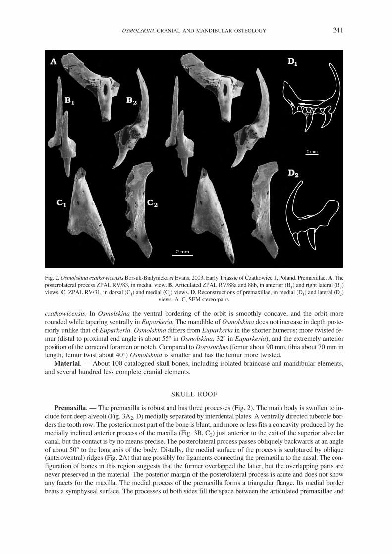
Premaxilla. — The premaxilla is robust and has three processes (Fig. 2). The main body is swollen to in−clude four deep alveoli (Fig. 3A2, D) medially separated by interdental plates. A ventrally directed tubercle bor−ders the tooth row. The posteriormost part of the bone is blunt, and more or less fits a concavity produced by themedially inclined anterior process of the maxilla (Fig. 3B, C2) just anterior to the exit of the superior alveolarcanal, but the contact is by no means precise. The posterolateral process passes obliquely backwards at an angleof about 50� to the long axis of the body. Distally, the medial surface of the process is sculptured by oblique(anteroventral) ridges (Fig. 2A) that are possibly for ligaments connecting the premaxilla to the nasal. The con−figuration of bones in this region suggests that the former overlapped the latter, but the overlapping parts arenever preserved in the material. The posterior margin of the posterolateral process is acute and does not showany facets for the maxilla. The medial process of the premaxilla forms a triangular flange. Its medial borderbears a symphyseal surface. The processes of both sides fill the space between the articulated premaxillae and
OSMOLSKINA CRANIAL AND MANDIBULAR OSTEOLOGY 241
2 mm
2 mm
Fig. 2. Osmolskina czatkowicensis Borsuk−Białynicka et Evans, 2003, Early Triassic of Czatkowice 1, Poland. Premaxillae. A. Theposterolateral process ZPAL RV/83, in medial view. B. Articulated ZPAL RV/88a and 88b, in anterior (B1) and right lateral (B2)views. C. ZPAL RV/31, in dorsal (C1) and medial (C2) views. D. Reconstructions of premaxillae, in medial (D1) and lateral (D2)
views. A–C, SEM stereo−pairs.

protrude posteromedially. They leave no space for the entrance of the vomer (Figs 2C1, 3A1, A2). On each side,a slight dorsal concavity received the underside of the anterior process of the maxilla (Fig. 3C1). The premaxillawas probably held in place mainly by connective tissue.
The anterior (nasal) process is very tall, thin (Fig. 2B), and posteriorly concave. In transverse section, it ismedially flat and laterally evenly convex. It suggests the external nares were fairly large. It tapers distally andbecomes sub−tetrahedric at about 1/4 of its height, with the anterior and lateral surfaces flat, the former beingnarrower than the latter. The lateral surface probably served for the nasal contact. In anterior view, the articu−lated premaxillae are very slender. A longitudinal furrow follows the symphysis line on each side (Fig. 2B1).
The length of the premaxilla body varies from ca. 7 to10 mm. Smaller specimens have more slender pro−portions and the angle of the posterior process to the horizontal is more acute. This variability is tentativelyascribed to allometric growth.
Maxilla. — There is no complete specimen. Usually, the maxilla breaks into four pieces that are difficult tomatch to one another. The central section (Fig. 4A) is usually sub−triangular but it develops dorsally into a tall,narrow sub−quadrangular nasal process (Fig. 6) and posteriorly into a horizontal tooth bearing ramus (ca. 30%the total height of the maxilla Fig. 6A, D–F). The borders of the nasal process are almost vertical, but becomemore oblique ventrally as the process broadens out. The lateral surface of the central portion is flat. Togetherwith the curvature of the premaxilla (see above) and the position of the nasal (see below) it suggests that thesnout was deep. Only the posterodorsal border of the maxilla curves slightly medially. The ventral border of the
242 MAGDALENA BORSUK−BIAŁYNICKA and SUSAN E. EVANS
2 mm
R/31
R/89
R/8
R/77R/482 R/294 mm2 mm
Fig. 3. Osmolskina czatkowicensis Borsuk−Białynicka et Evans, 2003, Early Triassic of Czatkowice 1, Poland. A. Reconstruc−tion of articulated premaxillae, in dorsal (A1) and ventral (A2) views. B. Reconstruction of anterior part of skull, in left lateralview. C. Premaxillae: ZPAL RV/30 (left) and 31 (right) combined with anterior part of maxilla ZPAL RV/29, in dorsal (C1) andleft lateral (C2) views. D. Left premaxilla ZPAL RV/78, in medial view. C, D SEM stereo−pairs. Shortened catalogue numbers
indicate the specimens on which the reconstruction is based.

maxilla is straight (Fig. 6A, C). A large exit foramen for the superior alveolar canal opens on the labial surfaceat the base of the premaxillary process (Figs 4A1, 6A1). The estimated number of alveoli is 13.
In the type specimen, ZPAL RV/77, the anterior border of the nasal process is damaged but the lossesseem to be insignificant. The medial surface of the anterior border bears a longitudinal furrow descending al−most half way down the total height of the maxilla. This groove is the posterior part of the nasal facet. Behindit the medial surface of the nasal process bears a pattern of sub−vertical ridges and furrows (Fig. 4A2) mark−ing the position of the lacrimal. The posterior border of the nasal process crosses the dorsal margin of themaxilla to extend onto the medial surface. The dorsal margin of the horizontal ramus continues over the lat−eral face of the nasal process as an oblique crest ascending anterodorsally. The crest forms the anterior borderof the weak antorbital recess.
OSMOLSKINA CRANIAL AND MANDIBULAR OSTEOLOGY 243
2 mm
nasal
maxilla
Fig. 4. Osmolskina czatkowicensis Borsuk−Białynicka et Evans, 2003, Early Triassic of Czatkowice 1, Poland. Anterior frag−ment of the holotype maxilla ZPAL RV/77 combined with a fragmentary nasal ZPAL RV/8, in lateral (A1) and medial (A2)
views. SEM stereo−pairs.
2 mm
2 mm
Fig. 5. Osmolskina czatkowicensis Borsuk−Białynicka et Evans, 2003, Early Triassic of Czatkowice 1, Poland. A. Middle part ofright maxilla ZPAL RV/81, in lingual (A1) and labial (A2) views. B. Posterior fragment of right maxilla ZPAL RV/160, in lingual
(B1), dorsal (B2), and labial (B3) views. SEM stereo−pairs.

The premaxillary process curves medially. It is an anteromedial pyramidal extension of the alveolar mar−gin and corresponds to what Gow (1970) called an anterior median flange. There is no discrete articular facetfor the premaxilla along the acute anterior border of the maxilla, nor is there any space between the nasal andmaxilla for the posterior process of the premaxilla that probably fitted the anterior surface of the nasal. Thecontact between the processes of the premaxilla and maxilla was probably quite loose, possibly leaving anaccessory antorbital fenestra. There are also no traces of a lateral overlap between the body of the maxilla andthat of premaxilla. The bones probably only touched each other in this region, while the faintly sculpturedventral surface of the premaxillary process of the maxilla probably overlapped the dorsal surface of the me−dial process of the premaxilla in the anteriormost part of the palate (Fig. 3A). Articulated in such a way, thepremaxilla slightly overhangs the maxilla (Fig. 3A2, C), although the posterior premaxillary teeth are levelwith the maxillary tooth−row.
The medial face of the premaxillary process bears a sub−horizontal longitudinal furrow on the dorsal sur−face of the superior alveolar shelf. In articulated bones, this furrow is in line with the vascular foramen pierc−ing the posterior margin of the premaxilla. It probably served for the neurovascular supply of the premaxillafrom the superior alveolar canal. Faint sculpture on the lateral face of the premaxillary process, directly be−hind the anterior margin, may reflect the presence of ligaments that attached the premaxilla. Directly behindthe nasal process, the dorsal border of the horizontal ramus is slightly concave, but the profile of the moreposterior part is poorly known. It clearly sloped posteroventrally at the tip.
244 MAGDALENA BORSUK−BIAŁYNICKA and SUSAN E. EVANS
lacrimal facet
sagittal nasal suture
2 mm
maxillary facet
premaxillaryfacet
sagittal nasal suture
superior alveolarcanal exit
antorbital fossa recess
jugal facet
R/29 R/77 R/81 R/475 R/191 R/160
superior alveolarcanal entrance
palatine facet
maxillary pillar
lacrimal facet
2 mm
jugal facet
nasal facet
jugal
Fig. 6. Osmolskina czatkowicensis Borsuk−Białynicka et Evans, 2003, Early Triassic of Czatkowice 1, Poland. A. Reconstructionof the left maxilla, in lateral (A1) and medial (A2) views. B. Left nasal ZPAL RV/8, in lateral (B1) and medial (B2) views. C. Cen−tral part of maxilla ZPAL RV/81, in medial view. D. Posterior fragment of maxilla ZPAL RV/484 combined with the anterior partof the jugal ZPAL RV/281, in medial view (right side bones reversed). E. Anterior part of maxilla ZPAL RV/29, in medial view.
C–E SEM micrographs. Shortened catalogue numbers indicate the specimens on which the reconstruction is based.

The medial surface of the main body of the maxilla (Figs 4A2, 5A1) displays a very deep, medially swol−len alveolar part, and a sub−vertical pillar inclined anterodorsally and buttressing the fragile nasal process.Both the alveolar part and the pillar are sub−circular in transverse section, but the pillar is much smaller in di−ameter. The alveolar part is separated from the main lamina of the maxilla by a longitudinal furrow. A fewmillimetres posterior to the pillar base, and usually roofed by an oblique blade of bone (Fig. 6A2), is the en−trance of the superior alveolar canal for the maxillary artery and superior alveolar branch of the maxillarynerve passing from the palatine. This opening (posterior alveolar foramen of Oelrich 1956) should corre−spond in position with the infraorbital foramen of the palatine. The palatine facet consists of a sub−perpendic−ular surface extending along the medial border of the alveolar edge between roughly the fifth to ninthalveolus, and tapering both anteriad (Figs 5A1, 6A2) and posteriad (Fig. 14D). The deepest part, facingobliquely dorsomedially at the level of about the sixth to seventh tooth, was probably received into a longitu−dinal concavity on the maxillary process of the palatine (see below). As a result, the anteromedial part of thepalatine slightly overlapped the dorsal surface of the alveolar part of the maxilla.
As in the premaxilla, the maxillary alveoli are bordered by interdental plates situated slightly medial tothe lingual side of the dentigerous margin. Labially the interdental plates pass into interdental septa. The al−veolar part becomes flatter posteriorly (Fig. 6F), as the alveoli become shallower, and its dorsal wall bearsnumerous irregular perforations (possible reason for its poor preservation). The dorsal overlap of the jugal onthe maxilla is fairly long (see e.g., Figs 5B2, 6D).
Nasal. — The nasal is represented by a single damaged right specimen, ZPAL RV/8 (Figs 4, 6B), and somefragments. It is a transversally curved bone blade turning anterolaterally into a long process that descendsobliquely down the lateral face of the snout. The posterior process, sub−triangular shape, allow for reconstruc−tion of the U−shaped incision between joint nasals. The incision probably received the anteriorly protruding partof the frontal (Fig. 8B). An elongated wavy medial facet (Fig. 6B2) extending along approximately posteriorone half of the preserved ventral border of the nasal should have received the lacrimal, but this bone has not yetbeen identified. The sagittal suture is straight and simple. The maxillary facet begins anterior to the lacrimal oneand runs down the posterolateral border of the anterolateral process. As preserved, the process is rounded at thetop and tapers ventrally. The anterior border of the process bears remnants of a premaxillary facet (Fig. 6B1).
Frontal. — The frontals (Fig. 7A–C) are represented by many specimens. They are flat, paired and stronglybuilt, and ca. 3.5 times as long as wide. The sagittal suture is straight along the anterior third of its length, be−coming increasingly sinuous posteriad. The frontonasal suture is broadly U−shaped (Fig. 8B) with anterolateralcorners retracted, and bearing a dorsal nasal facet. Anterolaterally and posterolaterally the frontal joins theprefrontal and postfrontal respectively, so that it borders the orbit for only about one quarter of its length (thesecond quarter from the rear). The prefrontal facet incises the dorsal surface of the frontal obliquely (Fig. 8A1)at the level of the second quarter of the frontal from the front. The facet protrudes slightly laterally from themain body of the frontal. It is convex in transverse section and longitudinally ridged, which suggests a ratherrigid junction permitting no mobility in the transverse plane. The postfrontal facet incises the posterior quarterof the lateral frontal margin, but is usually little exposed in dorsal view. Between the prefrontal and postfrontal,the orbital margin of the frontal is slightly concave. The ventral surface of each frontal bears a strong, laterallyconcave sub−olfactory process (Fig. 8A2). The longitudinal concavity of the olfactory canal deepens at bothends. The posterolateral part of each frontal is slightly dorsally concave (Fig. 7B). It bears posterior and medialparietal facets facing ventrally (Fig. 8C), and a ventrolateral postfrontal facet (Fig. 8A2, D). However, the me−dial border of the process also bears a dorsal facet (Fig. 7B), and was probably overlapped by a thin superficialsheet of the parietal. The posterior border of the combined frontals is U−shaped.
The total length of frontals varies between roughly 15 and 22 mm. The posterior width is 100–118% ofthe anterior width. This variability affects the shape of the lateral border, which varies from almost straight toconcave. Some larger specimens do not fit into the variability range of the majority form. They are relativelyshorter and stouter (in ZPAL RV/96 posterior width attains ca. 155% of the anterior one), while thepostfrontal facet invades the dorsal surface of the frontal. The large type is much less numerous in the assem−blage. Whether it is a variant of the majority form or belongs to another animal cannot be resolved at the pres−ent time. The frontals of the majority variant are usually longer and more slender than those of Euparkeria(Ewer 1965), but the topography of the bones in the frontal region, including the suture structure, is exactlythe same.
OSMOLSKINA CRANIAL AND MANDIBULAR OSTEOLOGY 245

Parietal. — The parietals (Figs 7D, E, 8B) are paired and there is no trace of a parietal foramen. By com−parison with the frontal, the parietal is short (roughly about half the frontal length in sagittal axis). As awhole, the sagittal suture is interdigitating. The anterior half of the suture surface bears V−shaped ridges anddiffers from the posterior one, which is vertically ridged. The posterior part becomes thinner and more sus−ceptible to damage. When the anterior parts are articulated, the posterior parts leave a long incision betweenthem, which combined with the suture morphology is suggestive of the presence of an interparietal. This
246 MAGDALENA BORSUK−BIAŁYNICKA and SUSAN E. EVANS
2 mm
Fig. 7. Osmolskina czatkowicensis Borsuk−Białynicka et Evans, 2003, Early Triassic of Czatkowice 1, Poland. A. Left frontalZPAL RV/91, in dorsal (A1) and ventral (A2) views. B. Right frontal ZPAL RV/1370, in dorsal view. C. Posterior fragment of theright frontal ZPAL RV/250, in ventral view. D. Left parietal ZPAL RV/293, in dorsal view. E. Left parietal ZPAL RV/285,
in dorsal view. SEM stereo−pairs.

OSMOLSKINA CRANIAL AND MANDIBULAR OSTEOLOGY 247
parietal facetfrontal facet
postfrontal facet
subolfactoryprocess
nasalfacet
nasal
frontal
prefrontalfacet
postfrontalfacet
postfrontalfacet
possiblyundamaged margin
postorbitalfacet
supratemporalcrest
lacrimalposition
prefrontalposition
postfrontal
postorbital
postorbitalfacet frontal
facet
parietalfacet
frontalfacet 2 mm
2 mm
2 mm
(E, G)
Fig. 8. Osmolskina czatkowicensis Borsuk−Białynicka et Evans, 2003, Early Triassic of Czatkowice 1, Poland. A. Right frontal,in dorsal (A1) and ventral (A2) views. B. Skull roof, in dorsal view. C. Right parietal, in dorsal (C1) and ventral (C2) views.D. Combined left side skull roof bones of different individuals, in ventral view: frontal ZPAL RV/259, ZPAL RV/291 parietal,ZPAL RV/547 postfrontal, postorbital ZPAL RV/315, and squamosal ZPAL RV/134. E. Right postfrontal ZPAL RV/544,in ventral view. F. Right postfrontal ZPAL RV/543, in dorsal view. G. Left prefrontal ZPAL RV/66, in lateral view. A–C, recon−
structions; E–G, SEM micrographs; D, E, G, stereo−pairs.

would be consistent with the structure of this region in the other basal archosauriforms (Ewer 1965, Gower2003), but is not supported by actual preservation of the interparietal. The anterior part of the parietal bears astrong facetted flange that is wavy in transverse section (Fig. 7D, E). It accommodated the frontal medially,and the postfrontal and postorbital laterally (Fig. 8B1, D). The central part is convex and swollen dorsally,and protrudes anteriad to fit the overlapping frontal. Laterally a longitudinal groove receives the rear of thedescending crest of the frontal. The facet is longitudinally split to prevent lateral dislocation. The lateral wingof the parietal is oriented anterolaterally, and accommodates the combined postorbital−postfrontal. Its dorsalsurface is convex. The ventral surface, correspondingly concave and roughened, may have received the dor−sal part of the laterosphenoid. The lateral margin of the parietal bears an oblique surface for the origin of thetemporal muscles. It faces dorsolaterally, is overhung by the supratemporal crest, and continues onto the lat−eral surface of the squamosal process.
In length, the squamosal processes probably exceeded that of the parietal body (see reconstruction Fig. 8D),but they are usually incomplete distally. Their posteromedial surfaces (sites of neck muscle attachment) are tri−angular, concave in transverse section, and are overhung by crests. They taper medially. The parietal andsupraoccipital must have been connected by connective tissue. The ventral surface of the parietal has three con−cavities (Fig. 8D) matching the convexities of the dorsal surface: anteromedial, anterolateral (possibly receivingthe laterosphenoid, see above), and posterior (extending approximately half the length of the squamosal pro−cess). In the distal half, the ventral surface of the process is more or less flattened, and probably fitted the end ofthe paroccipital process. A slit−like posttemporal fenestra perhaps separated the proximal parts.
The parietals are fairly uniform in overall shape, while differing in size (from about 8 to 13 mm in sagittallength). The supratemporal crests vary in strength.
Prefrontal. — The prefrontal forms a roughly semilunar conch (Fig. 8G) tapering both posteriorly andanteroventrally in the life position, and having a strongly concave ventromedial surface. The flat triangularand superficially ornamented dorsal wall of the bone (oriented to the left in Fig. 8G) overlaps the ridgedprefrontal facet of the frontal (Figs 7A1, 8A1), and contributes to the skull table. Externally, the suture be−tween the two bones is finely sinuous and extends along a parasagittal plane. Anteriorly the prefrontal be−comes thinner and is rarely preserved intact, so that both its anterior extent and its contact with the lacrimalare poorly known. An elongated step−like surface preserved at the anterior border of ZPAL RV/66 (Fig. 8G,right lower angle of the specimen as oriented in the figure) is a fragment of the lacrimal facet.
Postfrontal. — The postfrontal is a small roughly triangular bone (Fig. 8E, F) that is wedged in betweenthe parietal, frontal and postorbital (Fig. 8B, D). Its dorsal surface is a regular smooth triangle, the orbital bor−der forming the longest side. Two ridged surfaces extend along the anterior and posterior sides of the triangleto contact the frontal and postorbital respectively. Both face mainly dorsally. The frontal facet is narrowerand extends slightly onto the ventral side of the bone. The postorbital facet is larger and is not evident in ven−tral aspect. Between the two facets, there is a tiny triangular articular surface that is concave and ridged. Thisfaces ventrally and slightly medially and contributes to the postorbital−postfrontal complex that overlaps thedorsal surface of the parietal. The orbital border of the postfrontal forms a smooth wall that is elongate in theparasagittal plane, but is only slightly concave longitudinally. In life position it faces ventrally and onlyslightly laterally.
Postorbital. — The postorbital is a triradiate bone (Fig. 9A–C). The axes of the posterior and ventral pro−cesses extend approximately at right angles to each other, but the processes themselves are connected by amore or less extensive blade of bone. This is laterally flat and medially concave. The posterior process fitsinto a groove on the squamosal to produce the upper temporal arcade. The medial process ascends slightlyanteromedially (at ca. 130� to the ventral process). Its stout tip bears an ovoid concave parietal facet facingventromedially, and this combines with the adjacent surface of the postfrontal to overlap the parietal (Fig.8D). More anterior and lateral in position is an elongated postfrontal facet. The elongated, tapering ventralprocess is bent anteriorly at about half its length. It is sub−triangular in transverse section with its lateral sur−face flat. The anterior surface contributes to the curved posterior orbital border. It faces directly anteriad andis separated from the posterior surface by a sharp crest. The posterior surface is concave in transverse sectionand faces posteromedially. The slit−like jugal facet extends onto this surface along the distal half of the lateralborder.
248 MAGDALENA BORSUK−BIAŁYNICKA and SUSAN E. EVANS

There is some variability in the postorbitals, in size, thickness, angulation between the anterior and poste−rior processes, and transverse section of the processes (more or less flattened laterally). However, the inter−esting parameters are difficult to measure accurately due to imperfect preservation.
Squamosal. — The squamosal is a quadriradiate bone that consists of a bulky rhomboid body with pro−cesses extending from each of its corners (Figs 9C, 10). The anterior process is stouter in lateral aspect thanthe posterior one, and bears a deep triangular socket on its lateral surface to receive the postorbital. The pos−terior one is twisted medially to overhang the quadrate, which makes its lateral wall more slender and posteri−
OSMOLSKINA CRANIAL AND MANDIBULAR OSTEOLOGY 249
parietal facet
postorbitalfacet
quadratefacet
parietal facet
quadratefacet
quadratefacet
2 mm
2 mm
parietal facet
Fig. 10. Osmolskina czatkowicensis Borsuk−Białynicka et Evans, 2003, Early Triassic of Czatkowice 1, Poland. Right squamo−sals: ZPAL RV/398 (A), ZPAL RV/399 (B), and ZPAL RV/27 (D). C. Left squamosal ZPAL RV/27. Lateral (A1, B1, C, D) and
ventromedial (A2, B2) views. C, D, SEM stereo−pairs.
parietalfacet
jugalfacet
R/275
R/273
R/270
R/270 R/276
R/281
postorbital facet
maxillary facet ectopterygoid facetposterior extentof maxillary facet
R/281
quadratojugalfacet
squamosal facet
2 mm
2 mm
Fig. 9. Osmolskina czatkowicensis Borsuk−Białynicka et Evans, 2003, Early Triassic of Czatkowice 1, Poland. A, B. Rightpostorbitals, two ontogenetic stages: ZPAL RV/318 (A) and ZPAL RV/319 (B). C. Left posterior skull fragment combined fromdifferent individual bones: jugal fragment ZPAL RV/273, postorbital ZPAL RV/319, squamosal ZPAL RV/871, and quadrateZPAL RV/872. D. Reconstruction of the left jugal, mostly on the basis of the specimens illustrated in Fig. 11 (shortened
catalogue numbers around). Medial (A1, B1, D2) and lateral (A2, B2, C, D1) views. C, SEM stereo−pair.

orly tapering. Both processes extend in line with the diagonal of the main body. This diagonal was orientedapproximately horizontally as is the resulting supratemporal arcade. The posterior and medial processes con−tribute to an elongate rough parietal facet which generally faces posteromediad, but becomes more dorsal inorientation on the medial process. Ventrally, the squamosal extends into a long, slender process. Its lateralsurface is flat and tapers to a point. A deep concavity between the posterior and ventral process received thequadrate head, while the lateral quadrate flange lay against the posterior wall of the ventral process.
There is variation in the size and proportions of the body of the squamosal, its shape and curvature, and inthe angle between the postorbital and quadratojugal processes, but this cannot be quantified, partly because ofdifferences in preservation. There seem to be two morphotypes, but they grade into one another. The smallermorphotype (width of the quadratojugal process: 1.0–2.1 mm, n = 7) is rhomboid in shape and the length of thebody greatly exceeds its width (Fig. 10A1, C). The quadratojugal process extends anteroventrad at an anglemuch less than 90� to the postorbital processes. The larger morphotype (Fig. 10B1, D) (width of the quadrato−jugal process: 2–3.5 mm n = 7) is squarer in outline, with a quadratojugal process that is relatively stouter andflatter, as well as almost vertical in orientation, but this may be just a matter of size and individual age.
As illustrated by Ewer (1965, fig. 2), the anterior and ventral processes of the squamosal in Euparkeriaenclose an angle of about 90�. In contrast, the same angle in Sarmatosuchus is 115� (Gower and Sennikov1997, p. 63, fig. 3C).
Quadrate. — The quadrate (Fig. 11) has a pillar−like body arched in the parasagittal plane and posteriorlyconcave. The proximal head is simply a small rounded termination of the central pillar. The medial wing istriangular with its apex situated in the upper one third of the bone, and the base extending down to the lowerone third of the pillar. The less extensive lateral wing is triangular with its apex more dorsally positioned, atabout one quarter the quadrate height from the top. It is separated from the lower extremity of the bone by adeep notch that contributes to the quadratojugal foramen (Fig. 11A1). The wings extend from the body inanteromedial and anterolateral directions, the anterior surface of the bone thus enclosing a widely openV−shaped concavity in transverse section. The lower part of the anterior face, depressed for the articulationwith the pterygoid, is separated from the upper part by an oblique crest (pterygoid crest on Fig. 11A1, B2) ex−tending ventrolaterad along the lower margin of the medial wing toward the quadratojugal articulation. Thiscrest marks an abrupt change in orientation of the anterior surface of the bone. At the distal end of the pillar,there is a heavy medial condyle for the mandibular articulation (Fig. 11A1). The lateral condyle protrudeslaterad from the shaft. It is less developed than the medial one and is separated from the latter by a shallowsmooth concavity. Both extend onto the anterior face of the bone, suggesting a posteroventral inclination ofthe bone in life (Fig. 11A3, A4). Given an approximately horizontal life position of the mandibular articula−tion, the upper concavity of the shaft faced anteriad and the lower one anteroventrad (Fig. 11A3, A4). Thelower border of the medial wing should have been sub−horizontal in life position. The ventrolateral margin ofthe bone bears a narrow oval facet for the quadratojugal (Fig. 11A1, C), while a row of tubercles, perhaps forligaments, parallels this surface on the posterior face of the bone.
The height of the quadrate varies from 11 to 24 mm with a modal value of about 16 mm. One of the largestspecimens is ZPAL RV/37. Allowing for differences in size, the quadrate of Osmolskina czatkowiensis isvery similar to that of Sarmatosuchus otschevi (Gower and Sennikov 1997, p. 63, fig. 5).
Quadratojugal. — The quadratojugal (Fig. 12F) consists of two limbs enclosing an angle of ca. 40�, andcontributing to a sub−triangular posteriorly rounded body. The body is medially concave to fit the ventro−lateral extremity of the quadrate, exactly as in Euparkeria (Ewer 1965). The horizontal limb is a simple bartapering anteriorly to fit the quadratojugal facet on the medial side of the jugal. The vertical limb is compli−cated by the addition of a medial lamina (or ridge). It closes the quadratojugal foramen laterally, and it istwisted just above it.
Jugal. — The jugal is a triradiate bone (Figs 9D, 12A–D). The middle part, including the postorbital pro−cess (Fig. 12C), is usually the best preserved, whereas the anterior and posterior rami are generally brokenoff. A number of anterior fragments may readily be combined with the middle parts, but only three specimenshave been preserved with the base of the posterior ramus that demonstrates the existence of the lower tempo−ral arcade (Fig. 9C).
The main part of bone is straight and sub−horizontal, but ascends anteriad to overlap the maxilla along anoblique line (Fig. 6D). The lateral surface is convex with a smooth sub−orbital part facing slightly dorso−
250 MAGDALENA BORSUK−BIAŁYNICKA and SUSAN E. EVANS

laterally and a strongly pitted ventral part facing ventrolaterally (Fig. 12A). The maxillary facet is complex.The main part of the facet is elongate and faces laterally but is set off from the main lateral surface of the bone(Fig. 12A). It must have been received into the furrow−like posteromedial surface of the maxilla (Fig. 6D).The posterior border of the main maxillary facet is always damaged. The same is true of the anterior extrem−ity of the jugal, and hence the contact with the prefrontal and lacrimal remains unknown. The posterior pro−cess (Figs 9D, 12D, E) is long and narrow. As preserved, it makes up 58% of the reconstructed length of thebone. The quadratojugal facet is a long V−shaped incision in the ventromedial surface of the distal end (Fig.12E). The postorbital process forms a triangle with a large base, and an apex directed posterodorsally withthe slope of its axis at about 52� to 65� to the long axis of the bone.
The postorbital facet is a slit−like furrow extending on the anterior margin of the process, and, more dor−sally, onto its medial side (Figs 12C, D). Medially, the quadratojugal and postorbital processes are separatedfrom each other by a deep concavity extending sub−horizontally below the base of the postorbital process,and bordered ventrally by what is probably the ectopterygoid facet. However, the nature of the jugal−ecto−pterygoid contact is far from clear. Further anteriorly, a sub−triangular concavity, open anteriorly (Figs 9D2,12B), corresponds to the region overlying the maxilla. The concavity is bordered ventrally by a crest that
OSMOLSKINA CRANIAL AND MANDIBULAR OSTEOLOGY 251
2 mm
squamosal facet
quadratojugalfacet
medial condylelateral condyle
medial winglateralwing
pterygoid crest
quadratojugal facetlateral condyle
medial wing
lateral wing
pterygoid crest
medialwing
2 mm
quadratojugalfacet
lateralcondyle
medialcondyle
Fig. 11. Osmolskina czatkowicensis Borsuk−Białynicka et Evans, 2003, Early Triassic of Czatkowice 1, Poland. A. Rightquadrate reconstructed on the basis of the specimens illustrated below, in anterior (A1), posterior (A2), medial (A3), lateral (A4),and distal (A5) views. B. Right quadrate ZPAL RV/38, in posterior (B1) and anterior (B2) views. C. Right quadrate ZPAL RV/39,
in distal view. D. Right quadrate ZPAL RV/39, in posterior (D1 ) and anteromedial (D2) views. B–D, SEM stereo−pairs.

probably contacted the anterior flange of the ectopterygoid, overlying the maxilla medial to the jugal. A ven−tral incision separates the maxillary facet from the posterior part of the jugal (Fig. 12A).
As a rule, the anterior ramus is convex laterally in transverse section. Directly anterior to the postorbital pro−cess, and posterior to this process, the jugal wall is concave. Some specimens differ in having an evenly convexlateral surface with no concavities in the vicinity of the postorbital process. They are otherwise quite similar tothe main type and are provisionally considered as variants. However, some very flat specimens (ZPAL RV/ 279and 280), with a more vertical postorbital process (angle 72–78�), may belong to another animal.
PALATAL COMPLEX
Palatine. — As reconstructed on the basis of specimens ZPAL RV/33 and 34 (Figs 13A, B, 14A, B), thepalatine is an elongated plate roughly rectangular in outline and approximately twice as long as wide (Fig.13D). A V−shaped notch in its anterolateral end marks the posterior limit of the choana. The exact length andmorphology of the anteromedial process are unknown, as are the posterior extent and outline of the bone and
252 MAGDALENA BORSUK−BIAŁYNICKA and SUSAN E. EVANS
2 mm
2 mm
(A–E)
(F)
Fig. 12. Osmolskina czatkowicensis Borsuk−Białynicka et Evans, 2003, Early Triassic of Czatkowice 1, Poland. Jugal fragments.A. Central portion with maxillary facet (left upper angle), and the base of a postorbital process ZPAL RV/274. B. Central portionwith bases of both postorbital and quadratojugal processes ZPAL RV/277. C. Postorbital process with fragment of the main bodyZPAL RV/275. D. Quadratojugal and postorbital processes ZPAL RV/273. E. Posterior part of jugal ZPAL RV/270 combinedwith quadratojugal ZPAL RV/394. F. Quadratojugal ZPAL RV/53. Lateral view (A, C2, D, F) and medial view (B, C1, E). Left
bones, SEM stereo−pairs.

the shape of the sub−orbital fossa. As a whole the dorsal surface is concave because of the upward curvatureof both the choanal border and the medial one, but the main portion is flat. A sharp arcuate, posteriorly con−cave crest borders a sub−transverse slit giving entry perhaps to branches of the medial palatal artery. Anotherforamen or foramina, situated at the lateral border of the bone, probably served for the entry of a passage usedby the inferior orbital artery and a palatine branch of the trigeminal nerve (Fig. 13D1). The general position ofthis passage in the palatine should correspond with that of the posterior opening of the superior alveolar canalof the maxilla (the posterior alveolar foramen of Oelrich 1956; see also Evans 1980, figs 21–25) which deter−mines the mutual position of the bones. The maxillary facet is double and extends from the tip of theanterolateral process of the bone backwards (Fig. 14A, B). Ventrally, an elongated, ridged surface extendsalong the entire length of the bone. Close to the posterior border of the choana, this surface passes dorsallyinto a triangular facet along the medial border of the upturned flange of the palatine. The two parts enclose alongitudinal furrow that more or less matches the palatine facet of the maxilla (Fig. 14D). This places the pal−atine in position within the palate (Fig. 1A).
The ventral surface of the bone is bordered medially by a narrow pterygoid surface tapering forwards andextending medially in its posterior part. An oblique crest extends posteromedially almost in line with the lat−
OSMOLSKINA CRANIAL AND MANDIBULAR OSTEOLOGY 253
teeth
interior orbitalartery and trigeminalnerve branch entry
pterygoidfacet
medialpalatalarteryentry
pterygoidfacet
2 mm
5 mm
pterygoidfacet
5 mm
5 mm
5 mm
Fig. 13. Osmolskina czatkowicensis Borsuk−Białynicka et Evans, 2003, Early Triassic of Czatkowice 1, Poland. A. Left palatinefragment ZPAL RV/34. B. Left palatine fragment ZPAL RV/33. C–F. Partial reconstructions of left palatines. C. Sarmatosuchusotschevi (after Gower and Sennikov 1997). D. Osmolskina czatkowicensis. E. Euparkeria capensis (after Ewer 1965, fig. 27).F. Batrachotomus kupferzellensis (after Gower 1999, fig. 14). All but A2, B2, D1 in ventral view. Shaded areas show the missing
parts. A, B, SEM stereo−pairs.

eral border of the choanal notch. The crest bears traces of a tooth row that was probably continuous with thatof the pterygoid. A triangular section of palatine blade, posterolateral to this crest, ascends obliquely towardthe maxillary border.
The attribution of this type of palatine to Osmolskina is based primarily on its having a size that best fits thelargest and most numerous bones of the assemblage. The possibility of procolophonid provenance has beenconsidered, but seems unlikely because of its elongated and rectangular outline, with the choanal notch occupy−
254 MAGDALENA BORSUK−BIAŁYNICKA and SUSAN E. EVANS
2 mm
Fig. 14. Osmolskina czatkowicensis Borsuk−Białynicka et Evans, 2003, Early Triassic of Czatkowice 1, Poland. Left palatines inlateral view exposing maxillary facet (anterior to the top): ZPAL RV/34 (A) and ZPAL RV/33 (B). C. Jugal fragment ZPALRV/281, in lateral view. D. Posterior fragment of maxilla ZPAL RV/484 combined with the anterior part of the jugal ZPAL
RV/281, in medial view. E. Posterior fragment of maxilla ZPAL RV/1371. SEM stereo−pairs.
crest
crest
5 mm
Fig. 15. Reconstructions of right pterygoids. A, B. Osmolskina czatkowciensis. C, D. Euparkeria capensis (a drawing afterEwer’s 1965, fig. 26; stereo−photographs). Ventral (A, C) and medial (B, D) views.

ing a short margin. This differs from the short, sub−rhomboid palatine of procolophonians that bears articularfacets on all margins, with the choanal notch situated in one corner (Carroll and Lindsay 1985, fig. 6E). As pre−served the bone corresponds to the middle portion of the palates in Sarmatosuchus otschevi (Fig. 13C; Gowerand Sennikov 1997, fig. 4B), Batrachotomus kupferzellensis (Fig. 13F; Gower 1999, fig. 14), and Euparkeriacapensis (Fig. 13E; Ewer 1965, fig. 1), while corresponding to the latter species in detail (Fig. 13C–E).
Pterygoid. — The pterygoid consists of two wings, palatal and quadrate, united by a short neck. In dorsalaspect, the main part of the palatal wing is fan−shaped and convex in transverse section. However, towardsthe medial side, the convexity passes into a radially oriented furrow that, in turn, is bordered by the prominentdorsomedial border. The posterior angle of the palatal wing, between the fairly deep medial wall and thethick laterally directed posterior border, is ca. 80�. The ventral surface of the palatal wing is separated fromthe neck by a sharp crest (Figs 15A, 16B), and similar crest occurs in Euparkeria capensis (Fig. 15C). Thelateral part of the palatal wing is ventrally concave and funnel−shaped. Sharply delimited on both sides, thedentigerous zone is flat, and bears about 4 longitudinal rows of small teeth. The anterior part of the pterygoidis never preserved, and was reconstructed (Fig. 15A) on the basis of the space left by the palatines andmaxillae. The lateral extreme is a fairly stout, laterally thickened sub−quadrangular blade bearing a flat inci−sion for the ectopterygoid (Fig. 16D, E). The ectopterygoid facet is never complete in existing material.
OSMOLSKINA CRANIAL AND MANDIBULAR OSTEOLOGY 255
2 mm
Fig. 16. Osmolskina czatkowicensis Borsuk−Białynicka et Evans, 2003, Early Triassic of Czatkowice 1, Poland. A–C. Centralportions of left pterygoids. A. ZPAL RV/74. B. ZPAL RV/72. C. Basipterygoid facet and base of the quadrate process ZPALRV/73. D. Right pterygoid combined with ectopterygoid ZPAL RV/604. E. Lateral part of the left ptergoid wing withectopterygoid facet ZPAL RV/605. F. Left ectopterygoid ZPAL RV/465. G. Right ectopterygoid ZPAL RV/408. Ventral (A1, B,
D, G), medial (A2), ventromedial (C), and dorsal (F) views. All but B, D, E SEM micrographs; all stereo−pairs.

The pterygoid neck has a ventromedially protruding hooked process (Fig. 16A, C) which contributes tothe basipterygoid articulation. Situated on the posterior face of this process, the articular surface is sub−per−pendicular to the plane of the palatal wing. It continues onto the medial surface of the ventral twig of thequadrate process (Fig. 16A, C). A tuber for insertion of the pterygoideus muscle (Schumacher 1973) or forligament attachment is situated about the middle of the neck. Its size is variable, and the apex is directed later−ally. At the very base of the quadrate ramus is a bone blade that is flat both ventrally and dorsally. Distally, itdivides into two processes and extends obliquely posterolaterally.
Ectopterygoid. — The ectopterygoid is a strongly curved bone with a relatively small hook−shaped lat−eral head and a body that extends strongly to a sub−triangular medial conch. The latter is dorsally convex(Fig. 16F) and ventrally concave (Fig. 16G). The concave surface is probably continuous with the concavityof the ventral surface of the pterygoid, and it is bordered by a V−shaped crest that opens medially. Thepterygoid facets it bears are obscure, and contact must be reconstructed on the basis of the facet on the lateralmargin of the pterygoid (Fig. 16D). The lateral head (turned down in Fig. 16F, G) bears an articular facet hereinterpreted as the jugal facet, but the nature of its contact with neighbouring bones is conjectural.
BRAINCASE
The braincase is represented by all constituent elements, with the exception of a laterosphenoid, the pres−ence of which is conjectural. The braincase material is disarticulated except for the parabasisphenoid, whichis a fully integrated element, and the exoccipitals that are almost always fused with the opisthotic (e.g., ZPALRV/115, 422). A few specimens also have the supraoccipital or prootic, or both (ZPAL RV/419), fused withthe opisthotic, but in existing material, the Osmolskina exoccipitals are never fused with the basioccipital.The base of the cranial cavity is, as a rule, covered by unfinished bone, except for the ossified basal groovesfor the metotic foramen, and occasionally the abducens grooves (ZPAL RV/424), as well as the articular fac−ets for neighbouring bones in a few larger specimens.
Basioccipital. — The basioccipital (Fig. 17A, B) is a thick bone that is cordate in shape, with the apexforming the majority of the occipital condyle. It contacts the basisphenoid by means of two sub−circular sur−faces facing anteriad and separated from each other by a notch (Fig. 17A3, B2). More laterally, an ovoid sur−face probably received the tip of the ventral ramus of the opisthotic that fitted between the basioccipital andthe posterolateral parabasisphenoid wing. The surface is situated on the anterolateral face of each basal tuber.The exoccipital facets are symmetrical, flattened, and posterolaterally inclined. They almost touch one an−other in the midline, but do not completely exclude the basioccipital from the foramen magnum. Anteriorly,the exoccipital facets diverge, and the basioccipital contributes a narrow zone to the braincase floor. How−ever, the contribution of the basioccipital to the braincase floor may have decreased in ontogeny with pro−gressing co−ossification of the bones. An elongated furrow of finished bone, corresponding to the base of theembryonic metotic fissure (nerves IX and X), lies anterior to the exoccipital facets, and posterolateral to theopisthotic facet of both sides. The apices of the basal tubera are situated at about the anterior one third of thebasioccipital length. Medially they meet the crests that border the V−shaped ventral concavity of thebraincase floor posteriorly.
Supraoccipital. — The supraoccipital (Fig. 18) is a hexagonal plate that is thickened ventrally on bothsides. The thickened parts bear oblique articular facets, for the prootic anterolaterally and for the opisthoticposterolaterally. The former is pierced by the entrance of the anterior semicircular canal, the latter by thatof the posterior canal. The canals converge medially as more or less open furrows, and are sometimes sepa−rated from each other by a transverse septum (Fig. 18C1). Their eventual fusion into a crus communis musthave occurred below the ossified part. Between the lateral otic facets, the ventral surface of the supra−occipital provides a concave, fully ossified ceiling to the brain cavity. Its posterior border is laterally in−cised by surfaces for the exoccipitals. Between them, the border of the supraoccipital contributes to the fo−ramen magnum. On the ventral surface of the bone, medial to the semicircular canal regions, the anteriormargin bears symmetrical triangular incisions (Fig. 18A1, C1). The incisions display a surface of finishedbone and extend laterally to the cavities continuous with the space for the dural venous sinus draining theprootic (see below) or for the endolymphatic ducts. The cavities most probably had their exits on the dorsalsurface of the bone (Fig. 25B, C).
256 MAGDALENA BORSUK−BIAŁYNICKA and SUSAN E. EVANS

The supraoccipital displays a fairly consistent structure, but the stage of ossification and the level ofco−ossification with neighbouring bones vary with respect to the details of the posterior margin. The triangu−lar surfaces for the exoccipitals are more or less widely spaced (Fig. 18B, D), sometimes separated by anacute roof above the foramen magnum incised in the sagittal axis, in some others the exoccipital facets areclose together and blend to produce a small finger−shaped process (Fig. 18B). The specimens do not differfrom one another in other aspects.
Exoccipital. — The exoccipitals (Fig. 19) are hour−glass shaped bones with enlarged upper and lowerends contacting the opisthotic, supraoccipital, and basioccipital respectively. The exoccipitals diverge dor−sally and contribute to the lateral borders of the foramen magnum (Fig. 25), but do not usually make contactabove it. Two hypoglossal foramina pierce the exoccipital pillar. The exits lie on the lateral side, with thelarger one postero−dorsal to the smaller one, which almost touches the suture with the basioccipital. Twoacute sub−vertical crests border the posterior foramen from both anterior and posterior sides (Fig. 24A). Theanterior crest forms the posterior border of the metotic fissure while the opisthotic ventral ramus makes upthe anterior wall of the fissure, and the basioccipital the furrow−like floor. The sutures between theexoccipital and the basioccipital close later in ontogeny than those between the exoccipital and the opisthotic,the latter two frequently being preserved as a single element (Fig. 25).
Opisthotic. — The opisthotic (Fig. 19) is a pyramidal bone that makes up the main body of theparoccipital process. The tip is bilaterally flattened and distally bears a dorsal ridge for the parietal contact. In
OSMOLSKINA CRANIAL AND MANDIBULAR OSTEOLOGY 257
occipital condyle
basisphenoid facet
exoccipitalfacet
opisthotic ventralramus facet
space foropisthoticventral ramus
basioccipitalbasal tuber exoccipital
facet
arteria carotisinterna furrow
semilunarfossa
occipital condyle
metotic foramenborder
metoticforamen floor
exoccipitalfacet
2 mm
2 mm
basipterygoidprocess
parasphenoidrostrum
metoticforamenfloor
parabasisphenoidbasal tubera
Fig. 17. Osmolskina czatkowicensis Borsuk−Białynicka et Evans, 2003, Early Triassic of Czatkowice 1, Poland. A, B. Basi−occipital ZPAL RV/114, in ventral (A1, B1), lateral (A2), and dorsal (A3, B2) views. C, D. Parabasisphenoid with basioccipital
ZPAL RV/413 combined from two specimens, in left lateral view. B, SEM micrographs; B, C, stereo−pairs.

life, the paroccipital processes of both sides extend posteriad enclosing an angle of about 55� (Fig. 25B). Theventral surface bears three longitudinal crests (Fig. 19B1). The blunt medial crest is continuous with the pos−terior wall of the exoccipital. Topographically, it roughly corresponds to the tuberal crest sensu Säve−Söderbergh (1947, see also Oelrich 1956) but is medial instead of lateral to the hypoglossal foramina. Thelateral crest extends half way along the paroccipital process and is continuous with the main line of theprootic crest. Intermediate in position, the third, most acute, crest extends along the posterior side of the ven−tral ramus of the opisthotic. It roughly corresponds to the interfenestral crest of lizards (Oelrich 1956) in thatit separates the region of fenestra ovalis from that corresponding to the embryonic metotic fissure. InOsmolskina, the metotic fissure is largely open, and no separate compensatory window is formed exactly asin the case of Euparkeria (Gower and Weber 1998). The “interfenestral crest” borders a stapedial fossa,which leads to the fenestra ovalis, widely open in Osmolskina.
The ventral ramus of the opisthotic has a twofold structure (Fig. 19A). It is composed of two elongatedprocesses, the posterior one is fused along its anterior margin with a second process protruding from the ven−tral border of the posterior vestibular recess. The suture between the two parts remains visible in all speci−mens having the ventral ramus preserved. It produces a blunt ridge cutting the stapedial grove sub−perpendic−ular to its axis. The ridge produces a semicircle partly surrounding the fenestra ovalis. Anterior to it is a pit,possibly for the ligament supporting the stapes. The anterior margin of the ventral ramus is pierced proxi−mally by a tiny canal extending posteriad (Fig. 19A). On the other side of the process it opens into the dorsalpart of the metotic foramen, possibly carrying the glossopharyngeal nerve (IX), or a part of it.
The heavy proximal end of the opisthotic (Fig. 19A) bears articular facets for neighbouring bones:exoccipital, supraoccipital, and prootic, as well as for the basioccipital and possibly for the parabasisphenoidwhich it contacts by means of its ventral ramus. The contact between the opisthotic and exoccipital issubhorizontal, the latter extending ventromedially. Dorsolateral to it, and facing antero−medially, is thesupraoccipital facet. This is slightly convex, and pierced by the circular opening of the posterior semicircularcanal that penetrates the posterolateral margin of the supraoccipital after having left the opisthotic (Fig. 19A).The prootic facet cuts obliquely across the proximal end of the opisthotic. This surface is overlapped laterallyby the posterior process of the prootic and is pierced by the horizontal semicircular canal. Below this, theopisthotic contains a large ventral cavity for the posteroventral part of the vestibule, with the exit of the hori−zontal semicircular canal and a posterior ampullary recess (Fig. 23A) into which the posterior semicircularcanal opens. The lagenar recess must have been located medial to the ventral ramus Fig. 23A).
258 MAGDALENA BORSUK−BIAŁYNICKA and SUSAN E. EVANS
posterior semicircularcanal
venoussinus
anterior semicircularcanal
anterior semicircularcanal
exoccipitalfacets
exoccipitalfacet
posteriorsemicircular
canal
2 mm
exoccipitalfacet
Fig.18. Osmolskina czatkowicensis Borsuk−Białynicka et Evans, 2003, Early Triassic of Czatkowice 1, Poland. Supraoccipital:ZPAL RV/125 (A, C), ZPAL RV/420 (B), and ZPAL RV/417 (D). Ventral (A1, C1), anterior (A2, C2), and posterior (A3, B)
views. B–D, SEM stereo−pairs.

Parabasisphenoid. — Some specimens from Czatkowice 1 (e.g., ZPAL RV/412) display a clear division ofthe parabasisphenoid (Fig. 20C2) into a spongy endochondral, probably basisphenoid part, and a thin sheet offinished dermal bone, probably corresponding to the parasphenoid. The parasphenoid rostrum is roughlyU−shaped in transverse section. It bears a blunt sagittal crest ventrally and a longitudinal dorsal furrow. Al−though the exact life position is unknown, an oblique, anterodorsal orientation of the parasphenoid rostrum(Fig. 17C) is suggested by that of the neighbouring bones. As a whole, the parabasisphenoid is a triangular bonewith the apex extended into an elongated parasphenoid rostrum and the body divided dorsally into anterior andposterior parts by clinoid processes united by a transverse crest (crista sellaris). The parasphenoid sheet extendsposteriad over the ventral surface of the basisphenoid to produce large posterolateral flanges that cover thebasisphenoid–basioccipital contact from below (Fig. 21C1). They protrude well beyond the basisphenoid. Theirtips have been referred to as parabasisphenoid basal tubera (Gower and Weber 1998), but in Osmolskina theyseem to be produced by the parasphenoid. The flanges are separated from each other by a roughly V−shaped in−cision, the margin of which is very thin and usually badly damaged. It is therefore difficult to say how large theparasphenoid overlap was on the surface of the basioccipital. A pitted surface on the ventral aspect of thebasioccipital (Fig. 17B1) suggests that the parasphenoid retained a posteromedial flange similar to that ofProlacerta. There is however no evidence of a transverse thickening homologous to that in erythrosuchids(Parrish 1992, fig. 4) and described as an intertuberal plate by Gower and Sennikov (1996).
The braincase floor of Osmolskina is organised on two levels, the floor of the braincase cavity being situ−ated significantly higher than the parasphenoid rostrum and more or less parallel to the latter. Between themthe basisphenoid body is essentially vertically aligned. The anteriorly directed surface is occupied by thehypophysial fossa below and the dorsum sellae above it. The crista sellaris separates the dorsum sellae from
OSMOLSKINA CRANIAL AND MANDIBULAR OSTEOLOGY 259
hypoglossalforamina
opisthoticventral ramus
lateralsemicircular
canal
posteriorsemicircular
canal
exoccipital
fenestra ovalissite
posteriorvestibularrecess
?IX n. canal
prootic facet
hypoglossalforamina
opisthotic ventral ramus
basioccipitalfacet
exoccipital
incipientfenestra ovalis
posteriorvestibular recess
stapedial groove
prootic facet
lateralsemicircular
canal
R/422
R/115
interfenestral crest
metoticforamen
tuberalcrest
interfenestralcrest
prooticcrest
? IX nerve
2 mm
2 mm
metotic foramenregion
Fig. 19. Osmolskina czatkowicensis Borsuk−Białynicka et Evans, 2003, Early Triassic of Czatkowice 1, Poland. A. Leftopisthotic + exoccipital ZPAL RV/422, in antero−lateral view. White line in A shows the possible course of the IXth nerve canal.B. Left opisthotic + exoccipital ZPAL RV/115, in ventral view. A1, B1, SEM stereo−pairs. Shortened catalogue numbers indicate
the specimens on which the reconstruction is based.

the basisphenoid part of the braincase floor. Some specimens (Fig. 20A, C) preserve paired furrows of fin−ished bone that cut through the crista sellaris and represent the floor of the abducens canals, the roof beingmade by the anterior inferior prootic processes. The hypophysial fossa is a small concavity continuous withthe dorsal furrow of the rostrum (Fig. 20B, C1). It is pierced by paired foramina for the cerebral branches ofthe carotid artery, but some additional vascular foramina produced by superficial bone bridges may also ap−pear. A sagittal crest divides the dorsum sellae into bilateral concavities for eyeball muscles (m. retractorbulbi and m. bursalis) and the venous system (Säve−Söderbergh 1946). It is bordered on each side by an acutelateral crest, and is pierced by tiny vascular foramina. The dorsal surface of the basisphenoid is essentiallyX−shaped. The clinoid processes diverge anteriad. They are separated from the posterolateral parts of thebasisphenoid by deep bilateral incisions. Each incision corresponds to the lateral depression of Gower andWeber (1998), and is a sub−vertical furrow on the lateral face of the parabasisphenoid destined for the palatalbranch of the facial nerve that descended from the prootic. The internal carotid artery probably also used thispassage to reach the entrance foramen on the ventral side of the parabasisphenoid between the basipterygoidprocesses.
Posteriorly, the basisphenoid bears paired ovoid surfaces for basioccipital contact. These are covered by athin layer of finished bone. Lateral to them, small surfaces, one on each side, might have received the
260 MAGDALENA BORSUK−BIAŁYNICKA and SUSAN E. EVANS
basipterygoidprocess
carotid canal exit
clinoid process
abducens canalfloor
posterolateral parasphenoid flange basioccipital facet
cristasellaris
dorsumsellae
hypophysial fossa
basisphenoidpart of braincase
cavity floor
basipterygoid process
abducens canalfloor
posterolateral parasphenoidflange
carotid canalexit
brain cavityfloor
vascularforamen
R/412
vascularforamen
R/412
R/540
carotid canalexit
posterolateralparasphenoid
flange
2 mm
Fig. 20. Osmolskina czatkowicensis Borsuk−Białynicka et Evans, 2003, Early Triassic of Czatkowice 1, Poland. A, B. Parabasi−sphenoid reconstruction based on specimen ZPAL RV/412, in dorsal (A) and anterior (B) views. C. Parabasisphenoid ZPAL RV/412, in anterior (C1) and dorsal (C2) views. D. Stapes ZPAL RV/59. C1, C2, stereo−pairs. Shortened catalogue numbers indicate
the specimens on which the reconstruction is based.

opisthotic ventral rami. The surfaces are bordered ventrally by well−ossified parasphenoid laminae extendingposterolaterad (parasphenoid flanges), and facing dorsally.
The lateral wall of the parabasisphenoid (Fig. 24A) turns anteroventrally into the neck of the basi−pterygoid process. The dorsal border of the lateral wall extends between the clinoid process and the apex ofthe posterolateral parasphenoid flange, its posterior corner bearing a semilunar fossa (Fig. 24A) facing later−ally. The anterior border of the wall is the lateral crest of the dorsum sellae, whereas the posterior one is theventrolateral crest. Posterior to the basipterygoid process, the lateral wall of the parabasisphenoid bears asub−vertical furrow (Figs 17C, D, 24A) for the palatine branch of the facial nerve and the internal carotid ar−tery. At the contact with the prootic, the furrow is deep, and is bordered anteriorly by a distinct prootic crest(Fig. 24A). Both furrow and crest fade out ventrally. The nerve and artery must have wound around the pos−terior angle of the basipterygoid stalk, with the artery then dividing into two parts. As recognized by earlierauthors (Evans 1986; Gower and Sennikov 1998), the cerebral part entered the foramen on the ventral side ofthe braincase (Fig. 21A), while the anterior, palatal branch, along with the palatine branch of the facial nerve,could have followed the sub−horizontal furrow on the ventral side of the parasphenoid rostrum (the palatinebranch is lost in adult crocodiles; personal communication from the referee D. Gower).
Ventrally, the braincase is excavated by a deep fossa (Fig. 21C1), referred to as basioccipital−basi−sphenoid fossa by Gower and Sennikov (1996). The fossa extends posteriorly to the apices of the para−sphenoid flanges and is bordered laterally by the acute ventrolateral crests which fuse with each other poste−rior to the basipterygoid processes.
OSMOLSKINA CRANIAL AND MANDIBULAR OSTEOLOGY 261
parasphenoid rostrum
basioccipital basal tuber
hypoglossal foramen
metotic foramen
stapedia
l fossa
clinoid process
lateral furrow
semilunarfossa
exoccipital facet
metotic foramen floor
space for opisthoticventral ramus
opisthotic ventralramus
prootic
carotid canalexit
carotid canalentrance
basipterygoid process
VI
ventro-lateralcrest
ventro-lateral
crestcarotid canal
entrance
R412
basioccipital-basisphenoid
fossa
parasphenoidbasal tuber
2 mm
2 mm
Fig. 21 Osmolskina czatkowicensis Borsuk−Białynicka et Evans, 2003, Early Triassic of Czatkowice 1, Poland. A. Braincase re−construction, in ventral view. B. Reconstruction of parabasisphenoid combined with basioccipital, in dorsal view. C. Partialbraincase ZPAL RV/413 combined from different individuals, in ventral (C1) and dorsal (C2) views. D. Parabasisphenoid ZPAL
RV/412, in ventral view. C, stereo−pairs.

The basipterygoid processes are thick rounded bodies on short stout stalks that are mainly directed ven−trally. An articular surface of finished bone is preserved in only a few specimens (e.g., ZPAL RV/412), butusually the whole distal part is damaged. The mutual relations of the basipterygoid processes show somevariability. This variability also affects the position of the carotid foramina, but, as a rule, they are situated onthe ventral surface of the parabasisphenoid, posterior to the basipterygoid stalks and in front of the archedcrest produced by fused anterior sections of the ventrolateral crests.
Prootic. — The prootic (Fig. 22) encloses the main part of the osseous labyrinth of the inner ear. Its lateralsurface is dominated by elongated swellings corresponding to the anterior and lateral semicircular canals andampullae (Figs 22, 23A, B). The position of the lateral canal determines the orientation of the long axis of thebone and this appears to be sub−vertical (Fig. 22A, B, D). Posteriorly, the main part of the prootic extends into atriangular process that overlaps the opisthotic, and is usually broken off. Anteriorly, the otic region is separatedfrom the anterior inferior process by an open U−shaped trigeminal notch. The notch is partially divided by aprotruding part of its margin into two parts (Fig. 24A), the upper part for the exit of the medial cerebral vein,and the lower part, the trigeminal notch proper, for the trigeminal nerve. On the lateral side of the bone (Fig.22C, D), the trigeminal notch extends into a large sub−horizontal furrow under the prominence corresponding tothe anterior ampullar recess. The furrow fades out posteriad where it is crossed by a sub−vertical anterior sectionof the prootic crest. The extension of the crest onto the opisthotic borders the stapedial fossa and forms the lat−
262 MAGDALENA BORSUK−BIAŁYNICKA and SUSAN E. EVANS
posterior fossa
supraoccipital facet
posterior ampullary recess
vestibular recess
posterior semicircular canal passage
anterior semicircular canalpassage
anterior ampullaryrecess
metotic foramen
opisthotic ventral ramusbasioccipital facet
VIIVI
VIII
VI
VIII
horizontalsmicircular
canal passage
2 mm
2 mm
subarcuatefossa
Fig. 22. Osmolskina czatkowicensis Borsuk−Białynicka et Evans, 2003, Early Triassic of Czatkowice 1, Poland. A. Reconstruc−tion of the opisthotic and exoccipital combined with the prootic, in medial view. B, D. Prootic ZPAL RV/124, in medial (B, D1)
and lateral (D2) views. C. Prootic ZPAL RV/123, in lateral view. C, D, SEM stereo−pairs.

eral wall of the tympanic cavity. Anteriorly, it provided attachment for the aponeurosis of origin of the protrac−tor pterygoideus muscle (Oelrich 1956). A single large circular foramen for the facial nerve (VII) is situated in adeep concavity posterior to the prootic crest (Fig. 22A) and is overhung by the crest. The groove for the palatinebranch of the facial nerve, and for the carotid artery, extends anteroventrally from the facial foramen region. Itpasses from the prootic onto the basisphenoid, thus determining their mutual contact. The second furrow, forboth the hyomandibular branch of the facial nerve and the carotid artery fades out posteriad. There is no distinctpartition of the furrow into nervous and vascular parts.
The lateral surface of the anterior inferior process is flat, and bordered dorsally by a faint blunt horizontalcrest (possibly for the protractor pterygoidei muscle: Oelrich 1956; Gower and Sennikov 1996; Gower 2002),and bordered posteriorly by the prootic crest. The anterior end of the process bears a slightly concave,anteroventrally directed facet of finished bone (ZPAL RV/532), the only possible trace of the laterosphenoid (ifpresent). The abducens nerve presumably passed through the basisphenoid−prootic suture leaving shallow fur−rows in both elements (Fig. 20A, B, C1). The canals certainly cut through the crista sellaris to open directlyabove the dorsum sellae. The ventral border of the anterior inferior process bears an elongated ventromediallyfacing facet for the clinoid process of the basisphenoid, but this is usually poorly preserved. When oriented, theprootics converge slightly anteroventrad. As preserved, they do not meet each other in the midline, but the lackof contact is probably only a matter of preservation or of poor ossification in immature animals.
In medial aspect (Fig. 22A, B, D1), the prootic is excavated by a complicated system of concavities sepa−rated from each other by blunt ridges of unfinished bone that were probably extended by connective tissue inlife. The supraoccipital and opisthotic facets contribute to the posterodorsal borders of the cavities. They facemedially and bear a circular opening for the anterior semicircular canal near the anterior end, and a trace ofthe lateral canal along the posterior opisthotic process. The largest and most posterior of the cavities enclosedthe lower lateral part of the vestibule. The lateral semicircular canal begins with the anterior ampullary recessin the deepest, most dorsal part of the concavity, and extends into the posterior process. It has its circular exitat the end of this process where it penetrates the lateral face of the opisthotic. The anterior semicircular canalextends directly dorsad to exit through the supraoccipital facet. Towards the posteroventral margin of thebone, the vestibular concavity has a funnel−shaped ventral prolongation that continues into the opisthotic(Fig. 22A) and includes the posterior ampullary recess with the entrance of the posterior semicircular canal.The funnel−shaped space (Fig. 22B, D1, vestibular recess) must have housed the ventralmost part of the laby−rinth that passed into the lagena (Fig. 23A, B; Wever 1978). There is no crest to separate the lagena from themain vestibular part. In contrast, a crest has been described in at least some archosaurs (Gower 2002; crista
OSMOLSKINA CRANIAL AND MANDIBULAR OSTEOLOGY 263
opisthotic ventral ramus
venous sinus
posterior semicircular canal
supraoccipital
opisthotic
vestibulum
supraoccipital venous sinus
lagena
anterior ampullaposteriorampulla
horizontal semicircularcanal
anterior semicircularcanal
vestibulum
anteriorampulla
posteriorampulla
VIII
VIII
VIIVI
metoticforamen
anteriorsemicircular
canal
posteriorsemicircularcanal
lagena
Fig. 23. Osmolskina czatkowicensis Borsuk−Białynicka et Evans, 2003, Early Triassic of Czatkowice 1, Poland. A. Reconstruc−tion of a labyrinth based on the bones illustrated in Fig. 22B, D1. B. Labyrinth in Alligator mississipiensis (according to Wever
1978, fig. 24−15). Not to scale.

vestibuli). The lagena must have lain ventrally within the unossified part of the braincase wall. Ventral to thebase of the posterior process, and posterior to the vestibular concavity, there is a triangular shelf (posteriorfossa; Fig. 22B, D1; see also ZPAL RV/532) facing posteromedially. It contributes a small anterolateral partto the stapedial groove.
Dorsal to the vestibular recess and separated from it by a ridge, and medial to the anterior semicircular ca−nal, there is a shallow concavity corresponding to the subarcuate fossa (Fig. 22B). This is more or less circu−lar in outline but opens into a wide funnel−shaped mouth anteriorly. It probably housed a cerebellar flocculus(concerned with the maintainance of equilibrium), and a dural venous sinus as in Youngina capensis andProlacerta broomi according to the interpretation by Evans (1986, 1987). The venous sinus could have ex−tended into a lateral cavity in the anterior border of the supraoccipital (Fig. 18A1, A2, C2). The furrow perfo−rated by the facial nerve (VII) forms a third anteroventral concavity in the medial face of the prootic. Anteri−orly, this concavity opens towards the trigeminal notch. Posterodorsally a furrow, possibly for the vestibulo−cochlear nerve (VIII), leads into the vestibular cavity and is medially closed by a narrow bridge of bone.
Laterosphenoid. — As yet, the laterosphenoid has not been recognized in Osmolskina, and evidence forits presence is inconclusive. Regions of possible contact can be suggested on the basis of Clark’s et al (1993,fig. 2) description of Proterosuchus fergusi in which the laterosphenoid articulates with the alar process (dor−sal wing) of the prootic and with the tip of the anterior inferior process of the same bone (thus enclosing thetrigeminal foramen). Dorsally, it meets the skull roof, articulating with a facet/recess on the under surface ofthe parietal wing (medial to the postfrontal articulation) as well as with the descending crest of the frontal. InOsmolskina, the dorsal wing of the prootic shows a small descending extension to the supraoccipital facet(Fig. 22) while the tip of the anterior inferior process, sometimes bears a small surface of finished bone thatmay be for the laterosphenoid. It should be stressed, however, that the trigeminal notch is much more openthan it is in Proterosuchus fergusi. On the other hand, the underside of the lateral wing of the parietal bears arugose surface, immediately medial to the postfrontal facet that matches the position of the laterosphenoid at−tachment surface in Proterosuchus (Clark et al. 1993, fig. 2).
In extant crocodiles, the laterosphenoid ossifies early in embryogenesis (Iordansky 1973, p. 252, fig. 13),and was probably ossified in Osmolskina despite the possible immaturity of the preserved bones.
264 MAGDALENA BORSUK−BIAŁYNICKA and SUSAN E. EVANS
basipterygoid process
stapedial fossa
semilunar fossa
lateral semicircularcanal
anterior semicircular canal prootic crest
foramen ovalesite
VII
semilunar ganglionposition
opisthotic ventral ramus
prootic crest
trigeminalnotch
midcerebralvein exit
2 mm
foramen ovale
XII
midcerebral vein exit
VIIstapendial
fossa
subhorizontalridge
interfenestralcrest
tuberalcrest
tuberalcrest
interfenestralcrest
facialnerve
foramen
facial nerve foramen
2 mm
2 mm
Fig. 24. Osmolskina czatkowicensis Borsuk−Białynicka et Evans, 2003, Early Triassic of Czatkowice 1, Poland. A. Reconstruc−tion of the braincase, in lateral view. B. Varanus griseus braincase, Recent, in lateral view. C. Opisthotic associated with frag−
mentary prootic ZPAL RV/540, in ventral view. B2, C, stereo−pairs.

MANDIBLE
Dentary. — As reconstructed, the dentary (Fig. 26A) is approximately six times longer than deep,slightly deeper at the posterior end. The posterior margin is always damaged, and shows neither facets for theposterior mandibular bones, nor the anterior border of the mandibular fenestra. The lateral face is convex intransverse section, uneven and permeated by nutrient foramina that are distributed mainly along the dorsalmargin of the bone and in the mental region. As a rule, one of the foramina, situated at the level of the secondtooth position, is much larger than the others. Some of the posterior foramina extend posteriad into furrows.Lingually, the Meckelian fossa is long and low, expanding slightly in the vertical plane towards the rear. Itbears three foramina that probably carried the inferior alveolar nerve and its branches. The posteriormost fo−ramen is situated slightly behind the mid−length of the bone and may have allowed the alveolar nerve to passfrom the Meckelian fossa into the inferior alveolar canal. The Meckelian fossa is bordered by two ridges,both bearing faint traces of the splenial. The sub−dental ridge is sub−equal in depth to the fossa, and is deeperthan the sub−Meckelian ridge. Anteroventrally, at about the fifth tooth position (Fig. 26C, D), it is obliquelycut by the splenial facet. The shape and anterior limit of the splenial (Fig. 26B) has been reconstructed from
OSMOLSKINA CRANIAL AND MANDIBULAR OSTEOLOGY 265
occipitalcondyle
opisthoticventral ramus
medial part ofexoccipital
facet
basioccipitalbasal tuber
hypoglossalforamina
posteriorvestibular recess
venoussinus
hypoglossal foramen
venous sinusmetotic
foramenfloor
lateral semicircular canal
anteriorsemicircular
canal
occipitalcondyle
hypoglossalforamen
2 mm
posteriorvestibular recess
hypoglossal foramen
lateralsemicircular canal
lateral semicircular canal
lateralsemicircular canal
anteriorsemicircular
canal
Fig. 25. Osmolskina czatkowicensis Borsuk−Białynicka et Evans, 2003, Early Triassic of Czatkowice 1, Poland. Posterior partof the braincase ZPAL RV/424 combined from different individuals, in occipital (A), ventral (B), and anterior (C) views.
A2, B2, C2, stereo−pairs.

these traces. The symphyseal surface is barely delimited, but seems to extend posteriorly to the fourthalveolus. The alveolar border is subdivided by interdental septa into alveoli that are usually preserved empty.Interdental plates border the lingual side of each alveolus at the front and rear (Fig. 30D, E). When the teethare preserved they are fastened by spongy bone of attachment. The longest preserved specimen, ZPALRV/36 (Fig. 26G2), bears 12.5 alveoli. There is variation in the robusticity of the dentaries and in the corre−sponding shape of their anterior ends (Fig. 26C2, F). This is considered to represent individual andontogenetic variation.
Surangular. — The surangular is represented by numerous fragments (Fig. 27A, D, E), of which themost complete are from the posterior region. The bone usually breaks into pieces, particularly in its very frag−ile anteroventral portion. No part of the anteroventral border is ever preserved, and the presence of the man−dibular fenestra may only be deduced from the dorsal border of the angular (see below). The outline of thefenestra (Fig. 1F, G) is conjectural, but the lateral wall of the surangular is quite deep at its contact with thedentary, suggesting a fairly low position for the inferred fenestra.
266 MAGDALENA BORSUK−BIAŁYNICKA and SUSAN E. EVANS
2 mm
R/210
R/304
symphysealfacet
symphysealfacet
splenialfacet
R/35
R/14 R/36
alveolarnerve
entrance
Fig. 26. Osmolskina czatkowicensis Borsuk−Białynicka et Evans, 2003, Early Triassic of Czatkowice 1, Poland. A. Dentary re−construction. B. Right splenial reconstruction based on ZPAL RV/210 and 304. C. Left dentary ZPAL RV/14. D. Anterior part ofright dentary ZPAL RV/562. E. Right splenial ZPAL RV/304 (below) and ZPAL RV/210 (above). F. Right dentary ZPALRV/35. G. Left dentary ZPAL RV/36. Lingual (A, B, C1, D, E, F, G2) and labial (C2, G1) views. C–G2, SEM micrographs; all but
F and G2 stereo−pairs. Shortened catalogue numbers indicate the specimens on which the reconstruction is base.

The bone is a flat elongated plate that is laterally convex along the longitudinal axis. It is overhung, bothlaterally and medially, by a thick dorsal ridge. In the posterior portion of the bone, the dorsal ridge produces ahook−like medial projection that forms an anterior buttress for articulation with the articular bone. This part ispierced by two foramina (Fig. 27D–F) probably corresponding to the entrance (the medial one) and the exit(the lateral one) of the posterior surangular canal (Oelrich 1956). Posterior to the hook−like projection, thesurangular extends into a long pyramidal process, its ventrolateral wall (Fig. 29A1) tapering posteriad. Thelongitudinal convexity of the dorsomedial wall of the pyramid (Fig. 27E) matches the concavity of theventrolateral face of the articular (Fig. 27C). The third wall of the pyramid (Fig. 29A3) is an elongated trian−gular continuation of the thick dorsal ridge of the anterior portion.
OSMOLSKINA CRANIAL AND MANDIBULAR OSTEOLOGY 267
entrance of anteriorsurangular canal
entrance ofposterior surangular
canal
retroarticularprocess
prearticularfacetexit of chorda
tympani
possibledentary
facet
possible coronoidfacet articular facet
entrance ofchordatympani
exit of anteriorsurangular
canal
exit of posteriorsurangular
canal
articularfacet
vascularforamen
exit andgroovefrom
anteriorsurangular
canal
entranceof anteriorsurangular
canal
4 mm
2 mm
2 mm
possibleangularfacet
Fig. 27. Osmolskina czatkowicensis Borsuk−Białynicka et Evans, 2003, Early Triassic of Czatkowice 1, Poland. A. Anterior frag−ment of right surangular ZPAL RV/551, in dorso−lateral (A1, A3)) and medial (A2) views. B. Left articular ZPAL RV/549, in dor−sal view. C. Left articular ZPAL RV/321, in ventral view. D. Posterior end of the left surangular ZPAL RV/550, in ventral view.E. Posterior end of the left surangular ZPAL RV/548, in dorsal view. F, G. Reconstruction of the surangular and articular, in me−
dial (F), and dorsal (G) views. A1, A2, B, C, D1, E1, SEM stereo−pairs.

Anterior to the hook−like projection, the dorsal ridge is bordered by two crests, a lateral one that over−hangs the lateral face of the bone (Fig. 27A1), and a medial one (Fig. 27A2) that passes gently into the con−cave dorsomedial surface of the mandibular fossa. The lateral crest probably corresponds to the structure de−scribed as an external flange of the surangular. This marks the origin of the lamina lateralis of the tendon sup−porting pterygoideus posterior muscle in extant crocodiles (Busbey 1989) and birds (Lakjer 1926). Clark etal. (1993) considered it to be characteristic of basal archosauriforms.
The anterior end of the dorsal ridge is produced into a small dorsal tuber, in front of which is a ridged sur−face (Fig. 27A2) for possible articulation with the coronoid, although it might be, partly or entirely, a trace ofthe attachment of the mandibular adductor tendon. More anterior and slightly medial to this surface is thedentary facet. This part of surangular is pierced by foramina for branches of the mandibular artery and themandibular division of the trigeminal nerve entering the mandibular fossa. This probably corresponds to theanterior surangular foramen (Oelrich 1956). The medial surface of the surangular (Fig. 27A1, F) is a possibleentrance, the lateral a possible exit (Fig. 27A1, A3) for the anterior surangular canal. The main plate of thesurangular forms the lateral wall of the mandibular fossa and the posterodorsal border of a probable lateralmandibular fenestra. Where preserved, the lateral surface of the posteromedial border of the surangular bearsan obliquely ridged facet for the overlapping angular (Fig. 27D1, D2).
Splenial (Fig. 26B, E). — The best−preserved splenials (PAL RV/210, 211, and 304) are too large tomatch the preserved dentaries, and are assigned only tentatively. This bone may be reconstructed from facetson the medial surface of the dentary (Fig. 26C1) and angular (Fig. 29E). Based on the best−preserved speci−men (ZPAL RV/210), the splenial was an elongate sub−triangular bone contacting the dentary ridges border−ing the Meckelian fossa (see above). It must have extended to at least the fourth tooth position, its anteriorborder descending obliquely to a point between the third and second tooth position, before turning downaround the level of the middle inferior alveolar nerve foramen, to the level of the second tooth position. Me−dially, the splenial is flat, except for the ventral border that curves towards the dentary. The lateral (internal)surface is correspondingly concave ventrally. Its lower border is overhung in a step−like manner by a hori−
268 MAGDALENA BORSUK−BIAŁYNICKA and SUSAN E. EVANS
R/207
R/208
R/209
angularfacet
articularfacet
2 mm
articularfacet
angularfacet
angularfacet
2 mm
Fig. 28. Osmolskina czatkowicensis Borsuk−Białynicka et Evans, 2003, Early Triassic of Czatkowice 1, Poland. Prearticular:ZPAL RV/208 (A), ZPAL RV/214 (B), ZPAL RV/ 209 (C), and ZPAL RV/207 (D). A–C. Right bones, in medial view. D. Rightbones, in lateral view. E, F. Reconstructions of the bone, in medial (E) and lateral (F) views. A–D, SEM stereo−pairs. Shortened
catalogue numbers indicate the specimens on which the reconstruction is base.

zontal shelf that must have met the dorsomedial surface of the dentary sub−meckelian ridge (see above),whereas the ventral margin of the splenial contacted the medial surface of the ridge. The lateral (intra−mandibular) concavity of the splenial is subdivided by a longitudinal anteroventral crest.
Articular (Figs 27B, C, F, G, 29A). — In the Czatkowice material, the articular is never co−ossified with thesurangular. This may be a genuine character or a reflection of immaturity. The articular is a fairly massive, ven−trally crested, and dorsally tripartite bone. The anteriormost part of the dorsal face is occupied by articularcotyles for the quadrate condyles, and is divided posteriorly by a prominence (Fig. 27B). It is covered by unfin−ished and usually damaged bone, but was probably completed by a laterally protruding part. The central andposterior parts of the articular contribute to the retroarticular process. The central part displays a saddle−shapeddorsal surface divided into two concavities by a sagittal crest. The posterior part is a heavy, laterally com−pressed process (Fig. 27B) referred to as a “hooked ascending process” in Batrachotomus kupferzellensis(Gower 1999). It was probably for the attachment of the depressor mandibulae muscle tendon, as in extant croc−
OSMOLSKINA CRANIAL AND MANDIBULAR OSTEOLOGY 269
prearticularfacet
dentaryfacet
surangular facet
splenialfacet
R/197
R/212
R/197
R/212
5 mm 2 mm
Fig. 29. Osmolskina czatkowicensis Borsuk−Białynicka et Evans, 2003, Early Triassic of Czatkowice 1, Poland. A. Right mandi−ble ZPAL RV/ 35+35a combined from different individuals, in lateral (A1), ventral (A2), dorsal (A3), and medial (A4) views.B. Anterior portion of right angular ZPAL RV/197, in lateral (B1) and medial (B2) views. C. Posterior portion of angular ZPALRV/212, in lateral (C1) and medial (C2) views. D, E. Reconstructions of right angular, in medial (D) and lateral (E) views.
A–C, SEM stereo−pairs. Shortened catalogue numbers indicate the specimens on which the reconstruction is based.

odiles. When the surangular is oriented in a possible life position, the hooked ascending process of the articularis inclined medially. This inclination is similar to that in Batrachotomus (Gower 1999), but differs from that ofcrocodiles. The ventrolateral aspect of the articular bears a concavity (Fig. 27C) matching the convexity of theposterior process of the surangular to which it is applied (Fig. 27E). The medial surface is overhung by a pro−truding semi−circular shelf, and is pierced by a foramen (Fig. 27B, C, G). A similarly located foramen in extantcrocodiles is an entrance into a blind pneumatic space (foramen aerum Iordansky 1973). In Osmolskina, the fo−ramen leads to a canal that opens on the dorsomedial surface of the articular, exactly as it does in numerousrauisuchians and is similarly interpreted as a chorda tympani canal (Gower 1999). The structure of theretroarticular process is closely comparable to that of an extant crocodile, but is shorter and broader. It is longerthan in Batrachotomus (Gower 1999).
Prearticular. — As reconstructed (Fig. 28E, F) from fragmentary remains (ZPAL RV/207–209, 213–215),the prearticular consists of an elongated body joining two sub−triangular ends. Ventrally, the bone angles later−ally along a straight ridge to form a horizontal step−like shelf extending toward the angular. The dorsal border ofthe bone is arcuate. It bordered the medial entrance to the mandibular fossa. The posterior end probably had awide contact with the articular, and the anterior end ascended slightly towards the splenial and coronoid, but thestructure of these contacts is unknown. The internal surface of the bone that walls the mandibular fossa medi−ally is slightly concave in transverse section, and is richly permeated by neurovascular canals.
Angular. — This element is usually preserved in pieces (Fig. 29B–E). The longest specimen (ZPALRV/212) demonstrates that the posterior end of the angular is slightly larger than the rest, and was covered bytraces of radiating blood vessels on its inner, concave surface. The dorsal part of this surface probably over−lapped the ventromedial margin of the surangular (Fig. 27D2), but the surface does not reach far anteriorly.The more anterior dorsal border of the angular bears no trace of facets for the surangular and, thus, probablybordered the mandibular fenestra.
Along the ventral border of the bone there is an elongated surface that should have matched the one on theprearticular shelf, but the match is not perfect. Specimen ZPAL RV/197 is an anterior fragment of a right bone(Fig. 29B). A long triangular lateral facet (Fig. 29E), open dorsally but ending ventrally in a deep sub−horizon−tal furrow, probably received a posterior process of the dentary that is not preserved in the material, whereas anelongated ventral step in the medial surface probably met the splenial (Figs. 29D, 36E–G). The position of theangular in relation to the other mandible bones, and particularly to the surangular, is far from clear.
Coronoid. — The coronoid has not been identified and the only information on this bone is a possibletrace of this bone (Fig. 27G) at the anterior end of the dorsal ridge of the surangular (see above). Its positionis reconstructed (Fig. 1G) on the basis of crocodile structure.
DENTITION
General remarks. — Osmolskina is well represented by jaw elements and many isolated teeth, but nospecimen has a complete dentition preserved. This causes some difficulty in determining regional variation(see below). Those jaw fragments retaining teeth, firmly held by bone of attachment, are mostly large andperhaps closer to maturity. The teeth are typically worn, lack surface features, and may represent an adultmorphology somewhat different from that of the majority of isolated teeth in the deposit.
Basic tooth morphology. — In describing a typical tooth, we are focusing on isolated, unankylosed, andtherefore immature examples from the mid−part of the tooth series. A typical tooth of this type (e.g., Fig. 30B)has a root and crown of roughly equal proportions. The tooth is widest at the gingival boundary. The root tapersonly slightly towards the tip, but the crown does so sharply. The surface of the root is generally marked by par−allel longitudinal striae reflecting the development of periodontal connective tissue holding the tooth in place;the cross−section remains ovoid (long axis anteroposterior). The crown is labio−lingually compressed and has aposteriorly recurved tip (convex anterior margin, concave distal one). The surface of the crown is shiny andsmooth except for keeled and serrated anterior and posterior edges. The posterior margin bears a row of around25 denticles (independent of tooth size) along the entirety of its edge. The anterior margin usually bearsdenticles on only the apical half (although the row may be longer), but these denticles are borne on a distinct,lingually inflected keel that may continue further than the serrations themselves. A broad furrow separates this
270 MAGDALENA BORSUK−BIAŁYNICKA and SUSAN E. EVANS

anterior keeled margin from the remainder of the tooth surface on the lingual side (the labial side of the toothcurves uniformly) (e.g., Fig. 30F). The denticles themselves are close set with parallel, weakly grooved marginsand square or slightly rounded apices. Together they form a sharp, serrated cutting edge.
Implantation and replacement. — The implantation is fully thecodont. In a typical mid−jaw tooth, theroot and the crown are of roughly equal length. The alveolar margin of each tooth−bearing element is dividedinto a series of rounded alveoli by a combination of lingual interdental plates and interdental septa ( a combi−nation referred to as an interdental unit by Senter 2003) composed mainly of bone of attachment (Fig. 30D,E). The attachment bone forms a thin layer on the internal wall of the alveolus in juveniles or where the teethare not yet fully implanted (Fig. 30D), but fills the alveolus and blends with the surface of the tooth itselfwhere the teeth are mature (Fig. 30E). During replacement, the lingual side of the alveolar bone is eroded firstthen the attachment bone, and finally the root. Once the functional tooth is lost, the replacement tooth fills upa socket, but is only weakly attached. Only once the root is fully formed does bone of attachment become de−posited around it. The high incidence, therefore, of isolated unerupted crowns (Fig. 30A), and complete butnot yet fully attached, teeth, suggests that replacement was rapid. The rarity of fully attached teeth may sug−gest that much of the material is immature.
Two specimens (ZPAL RV/ 562 and 563) provide a good demonstration of the stages of implantation, at−tachment, and replacement. The juvenile jaw ZPAL RV/562 (Fig. 30D) contains no implanted teeth, butthere is a single unerupted tooth crown in tooth position three. The alveoli have only a thin layer of attach−ment bone on the walls. The unerupted tooth crown is pristine and the anterior and posterior serrations areclearly visible. This is a labio−lingually compressed tooth of the type described above as typical. ZPALRV/563 (Fig. 30E) is the central part of an older maxilla, preserving five tooth positions from the level of the
OSMOLSKINA CRANIAL AND MANDIBULAR OSTEOLOGY 271
attachmentbone
interdentalseptum
1 mm
2 mm
Fig. 30. Osmolskina czatkowicensis Borsuk−Białynicka et Evans, 2003, Early Triassic of Czatkowice 1, Poland. A–C, F. Isolatedteeth. A. Juvenile tooth crown ZPAL RV/565. B. ZPAL RV/564. C. ZPAL RV/566. F. ZPAL RV/567. D. Anterior part of rightdentary ZPAL RV/562. E. Middle part of right maxilla ZPAL RV/563. Lingual (A–C, F) and occlusal (D, E) views. SEM micro−
graphs; all but A stereo−pairs.

anterior maxillary pillar backwards. It shows alternate tooth replacement. Positions one, three and five (aspreserved) contain mature teeth (broken tooth bases), while positions two and four are empty and presumablyheld developing teeth. Again, the structure of the interdental septa (interdental plate and bone of attachment)is clearly visible. All three implanted teeth are firmly attached, but all are undergoing replacement from thelingual side, as shown by the cavities that notch both the alveolar margin of the maxilla and the alveolar at−tachment bone. The cavities must have contained very small loose replacement teeth now lost. Presumablyonce the mature teeth had finally been eroded, lost, and replaced, the developing teeth in positions two andfour would have been firmly implanted.
Variation. — As far as can be judged from the jaw elements themselves, the teeth vary in size and, tosome degree, morphology along the jaws, and show some differences between elements.
Each premaxilla contains four teeth housed in large rounded sockets (Fig. 2). The teeth are relatively lon−ger and narrower than those on the dentary and maxilla, and are more rounded in cross−section. None of theteeth preserved in situ bears serrations, but some isolated teeth of otherwise similar morphology bearserrations on a sharp keel along the posterior margin only, the anterior margin of the tooth being rounded. InZPAL RV/88 (Fig. 2B), only the most medial premaxillary tooth is preserved. It is fully rounded in cross−sec−tion with no trace of keels. In contrast, ZPAL RV/83 (Figs 2A, 3D) preserves the last premaxillary tooth, and,although broken, the cross−section shows a clear posterior keel and a slight anterior ridge.
Each maxilla bears an estimated 13 teeth. These vary in size with the largest teeth in positions four toeight, in the region of the nasal process and anterior pillar. This is reflected by an increased depth of the su−pra−alveolar ridge (Fig. 6C–E). Behind this point the teeth decrease in size, with the most posterior teeth be−ing tiny denticles. On the limited evidence available, the most anterior maxillary teeth resemble those of thepremaxilla in being rather more rounded in cross−section.
At least 13 teeth were present in the dentary which is never complete. Overall, the dentary teeth seem tohave been slightly smaller than those on the maxilla. The dentary ZPAL RV/14 demonstrates that teeth in po−sitions four to eleven were subequal in size. Judging by alveolar size and depth, the first two positions wereheld by relatively small teeth and this may reflect the slightly overhanging position of the premaxilla recon−structed for this animal.
DISCUSSION
As revised by Juul (1994), the diagnosis of archosauriforms (Gauthier et al.1988) has been reduced to twowell supported synapomorphies, the possession of an antorbital fenestra and an ossified laterosphenoid (seealso Clark et al. 1993), supplemented by a third, the possession of a mandibular fenestra (Parrish 1992; Juul1994). Of these, only the antorbital fenestra has been confidently reconstructed in Osmolskina, while thepresence of the laterosphenoid was deduced on the basis of the prootic and the skull roof morphology, andthat of the mandibular fenestra was reconstructed from the outline of the angular margin. The presence of allthe above synapomorphies would be expected in Osmolskina, because this genus presents a combination ofmany other archosauriform features such as the very small postfrontal (character 1 of Gauthier 1988, and ofBenton and Clark 1988), the tall columnar quadrate (Romer 1956), and deep rooted (thecodont) teeth (char−acters 8 and 18 Gauthier et al. 1988) showing lateral compression and serrated margins (character 12 ofBenton and Clark 1988, and character 7 of Gauthier et al. 1988 respectively), as well as characters relegatedby Juul (1994) to some less inclusive groups.
The absence of teeth on the transverse flange of the pterygoid (character 21 of Gauthier et al. 1988) isconsistent with a position crownward of proterosuchids, while the retention of toothed zones along the me−dial margin of the pterygoid and on the palatine (the vomer is not identified) tends to exclude Osmolskinafrom the crown−group. The presence of interdental plates tends to locate Osmolskina more crownwords thanthe erythrosuchids. They are perfectly developed also in Euparkeria (Senter 2003). According to Juul (1994),the interdental plates are characteristic of some basal archosaurs (rauisuchians, Herrerasaurus, Ornitho−suchus), while being absent from non−archosauriform Archosauromorpha (rhynchosaurs and Prolacerta),proterosuchids (Cruickshank 1972, Gower and Sennikov 1997), and erythrosuchids (Parrish 1992, Gower2003). However, the relationship between Osmolskina and the Erythrosuchidae is far from clear. Osmolskina
272 MAGDALENA BORSUK−BIAŁYNICKA and SUSAN E. EVANS

differs in its much smaller size and lighter build as well as in having a ventral maxillary margin that is straightinstead of convex. Some characters considered derived within the non−archosaurian archosauriforms (Juul1994, p. 38), such as a fairly large antorbital fenestra with an antorbital fossa around it, and the absence of aparietal foramen cannot discriminate between the above taxa. The antorbital fossa does occur in erythro−suchids (Juul 1994, p. 6; Gower 2003), and both its size and the structure of its anterior border are similar inErythrosuchus (Parrish 1992, fig. 6), Osmolskina, and Euparkeria (Ewer 1965, fig. 22). The parietal foramenis retained only in Garjainia (Parish 1992, p. 96).
The above discussion shows only that Osmolskina is a non−archosaur archosauriform situated morecrownward than the proterosuchids in the phylogeny.
In the Appendix 2, Osmolskina has been added to the data matrix of braincase characters of Gower andSennikov (1996), although this has been restricted to those characters and taxa relevant to the present case.The characters discussed below are numbered and scored according to Gower and Sennikov (1996) unlessotherwise indicated.
Character 1. — Position of the entry foramina for the cerebral branches of the internal carotid artery:ventral or ventrolateral = 0, lateral =1 (according to Gower and Sennikov 1996)). Gower (2002) updated thischaracter, and recognized three states: foramina posterior = 0; posterolateral = 1, and anterolateral = 2. Ourscoring is different: foramina lateral to the ventrolateral crests = 0; directly ventral, anterior to the fusedventrolateral crests = 1; lateral to basipterygoid stalks = 2. Osmolskina is scored 1.
According to Gower (2002), the foramina on the posterior surface of the parabasisphenoid occur innon−crown−group archosaurs, Proterosuchus, Garjainia, and Euparkeria, so this state is probably equal tothe ventral or ventro−lateral position of Gower and Sennikov (1996).
In our opinion, all the genera included in our matrix (Appendix 2) have the carotid foramina situated closeto the ventral surface of the parabasisphenoid, and also posterior to the basipterygoid processes, and so quali−fications such as just ventral or posterior are not informative enough. Our position is that there is a differencebetween Proterosuchus and Euparkeria–Osmolskina that should be expressed in scoring. The illustrations ofbraincases in Prolacerta and Proterosuchus (Gow 1975, figs 35A, 36C; Evans 1986) suggest they are similarin having the foramina located lateral to the ventrolateral crests of the parabasisphenoid, which is probablyalso true of Fugusuchus given the reconstruction of Gower and Sennikov (1996, fig. 4D). The anteriormostparts of the crests converge gradually over a long distance and at a small angle to each other, whereas inOsmolskina they turn abruptly mediad and fuse with each other. This change in the course of the ventrolateralcrests associated, for simple geometrical reasons, with both a sub−vertical position of the basisphenoid (char−acter 7), and a more vertical position of the basipterygoid processes (character 7a), both derived according toGower and Sennikov (1996), is considered derived as well. If it is correct, the directly ventral position of thecarotid foramina, anterior to the fused ventrolateral crests (we score 1 herein) should also be considered de−rived with respect to their primitive lateral position in proterosuchids, Prolacerta, and a prolacertiform gradeanimal from Czatkowice 1 (Borsuk−Białynicka and Evans 2009, fig. 8E1). Osmolskina shares the position“directly ventral, and anterior to the fused ventrolateral crests” with Euparkeria (Gower and Weber 1998, fig.4), Dorosuchus (Sennikov personal communication), Garjainia (Gower and Sennikov 1996, fig. 1), Xilou−suchus (Gower and Sennikov 1996, fig. 5), and probably Turfanosuchus (Wu and Russell 2001, fig. 5). Thedirectly ventral position of the foramina (1) in the rhynchosaurs (Mesosuchus, Dilkes1998; Hyperodapedon,Benton 1983, figs 10, 11), which are among the outgroups of archosauriforms, is probably homoplastic inview of the highly specialized skull.
The lateral position of the carotid foramina has been considered a synapomorphy of proterochampsids +crown−group archosaurs (Parrish 1993), or of the crown−group alone (Gower and Weber 1998). TheCrurotarsi (at least Postosuchus, Batrachotomus, Parasuchus, and Stagonolepis) have these foramina in alateral position (Chatterjee 1978; Gower and Sennikov 1996; Gower 2002), and the same is true of earlyornithodirans (Marasuchus, Sereno and Arcucci 1994; prosauropods, Galton and Upchurch 2004; and basalSaurischia, Langer 2004). However, the Middle Triassic Arizonasaurus babbitti from the United States(Gower and Nesbitt 2006), and the Late Triassic dinosauriform, Silesaurus opolensis, from Poland (Dzik2003) have the carotid foramina directly ventral.
Character 2. — Basisphenoid intertuberal plate: present = 0; absent = 1. Osmolskina is scored 1. Thebasisphenoid intertuberal plate is a transverse septum separating the parabasisphenoid from the basioccipitalpart of the ventral concavity. Parrish (1992) proposed that it was an autapomorphy of erythrosuchids. Ac−
OSMOLSKINA CRANIAL AND MANDIBULAR OSTEOLOGY 273

cording to Gower and Sennikov (1996), it was shared not only by Proterosuchus and Fugusuchus but also byProlacerta, and on this basis has been considered plesiomorphic of archosauriforms. In our opinion, thiswould suggest that what is labelled basisphenoid intertuberal plate might be homologous with the posteriormargin of the parasphenoid, at least in some taxa.
According to Gow (1975, figs 35, 36), the parasphenoid of early diapsids was long and bridged the gap be−tween the overlying basisphenoid and basioccipital. It has subsequently shortened, in parallel and at differentrates, in different diapsid clades, although its posterolateral corners continued to contribute to the basal tubera,and the posteromedial part retained a horizontal flange protruding posteriad to overlap the basioccipital. Thishorizontal flange is variable in size and shape, and its presence is here considered as a primitive character state.It occurs in Prolacerta (Gow 1975, figs 35; Evans 1986, figs 4, 5) and in Osmolskina, (according to impres−sions left on the basioccipital surface Fig. 33B), and probably corresponds to the thickened border of theparasphenoid in Proterosuchus (Gow 1975, fig. 36). The erythrosuchid intertuberal plate unites the basal tuberaof the parabasisphenoid, but extends clear of the posterior border of this bone (as demonstrated by Garjainiaand Xilousuchus, Gower and Sennikov 1996, figs. 2, 5; and Erythrosuchus, Gower 1997, fig. 2). It does notseem to be homologous with the posteromedial parasphenoid flange of other archosauriforms and Prolacerta,but was probably added to it. If this interpretation is correct, its presence would be derived as originally statedby Parrish (1992). Its absence in Prolacerta and non−erythrosuchid archosauriforms, including both Osmols−kina and Euparkeria, would be primitive (contra Gower and Sennikov 1996). However, this problem requiresfurther study and this equivocal character is removed from the matrix (see Appendix 2).
Character 3. — Abducens canal position: between the basisphenoid and prootic = 0, within prootic only= 1. Osmolskina is scored 0. In early amniotes, as exemplified by Captorhinus and Milleretta (Evans 1986),the abducens canals are located within the basisphenoid. A canal situated within the prootic−basisphenoid su−ture is characteristic of the immediate outgroups of the Archosauriformes (Prolacerta, Evans 1986; Hypero−dapedon, Benton 1983), as well as of Proterosuchus (Cruickshank 1972) and Euparkeria (Gower and Weber1998). In some erythrosuchids, at least, the canal has shifted into the prootic (Gower and Sennikov 1996).However, in basal archosaurs it seems to remain within, or return to, the basisphenoid (e.g., the phytosaurianParasuchus, Chatterjee 1978; and the prosauropod Thecodontosaurus, Benton et al. 2000). If properly re−constructed (Figs 20A, 21B), Osmolskina shares the primitive archosauromorph condition, but the polarity ofthis character, and its developmental basis, remain unclear.
Character 5. — Ventral ramus of the opisthotic: prominent = 0; recessed = 1. Osmolskina is scored 0.Character 6. — Ridge on the anterior inferior prootic process below the trigeminal notch: present = 0;
absent = 1. Osmolskina is scored 0.Character 7. — Basisphenoid orientation: horizontal = 0 , sub−vertical = 1. Osmolskina is scored 1.Character 7a. — Basipterygoid processes directed laterally = 0, ventrolaterally =1. Osmolskina is scored 1.
Based on the work of Gower and Sennikov (1996, description and fig. 4C, D) the braincase of Fugusuchus isprimitive not only in having a horizontal basisphenoid, but also in having laterally directed basipterygoid pro−cesses and carotid foramina lying lateral to the ventrolateral crests (though ventrolateral with respect to the wholebraincase). In contrast, erythrosuchids (Erythrosuchus, Garjainia, and Xilousuchus) display a high braincasewith a very short, sub−vertically oriented parabasisphenoid (Gower 1997; Gower and Sennikov 1996). InXilousuchus (Gower and Sennikov 1996, fig. 5), the processes are slightly more lateral, whereas in Garjainia(Gower and Sennikov 1996, fig. 1) and Erythrosuchus (Gower 1997, figs 2, 11) they are directed more ventrally.As a result, the carotid foramina are further apart in Xilousuchus and less so in the illustrated representatives ofGarjainia and Erythrosuchus. Euparkeria has a sub−vertical parabasisphenoid (Ewer 1965, Gower and Weber1998, p. 385). The basioccipital−basisphenoid fossa seems to be wedged slightly between the carotid foramina(Gower and Weber 1998, fig. 4) as it is in Osmolskina, and in both genera the foramina are located close to oneanother posteromedial to the basipterygoid processes on the ventral surface of the parabasisphenoid (Fig. 21A,E). The whole pattern seems to fall within the range of variability manifested by the erythrosuchids, which wasprobably the range of the non−archosaurian archosauriforms crownward of Proterosuchus. However, thebraincase of Sarmatosuchus otschevi from the Anisian of Russia, considered to be a proterosuchid Gower andSennikov 1997, is strikingly derived in having essentially vertical alignment, with a ventrolateral orientation ofthe basipterygoid processes as well as ventral orientation of the internal carotid foramina.
Clark et al. (1993) considered that the verticalization of the parabasisphenoid, which occurred during theearly course of archosauriform evolution, resulted in an increase in the space available for the adductor mus−
274 MAGDALENA BORSUK−BIAŁYNICKA and SUSAN E. EVANS

cles. According to these authors, this was an adaptation towards macropredaceous habits. A reorientation ofthe basipterygoid processes to a more vertical position, closer to each other, may have been a response to thesame selective agent. As hypothesised above, the changes in the basisphenoid configuration influenced theposition of the anteriormost parts of the ventrolateral crests so that they were pushed back and became con−fluent. They eventually fused across the midline to form a sharp border to the basisphenoid fossa. As thebasipterygoid processes were pushed together towards the midline, the entry foramina for the cerebralbranches of the internal carotid artery followed them onto the ventral side of the braincase, into the space an−terior to the basioccipital−basisphenoid fossa. The internal carotid artery extends down the dorsomedial wallof the tympanic fossa in lizards at least (Oelrich 1956) or along the ventrolateral crests (Evans 1986; Gower2002). Its cerebral branch would have passed from the lateral to the ventral face of the parabasisphenoid toreach the entry foramen. The ventral position of the foramina required that the artery wound ventrally aroundthe posterior surface of the basipterygoid process. This probably accounts for the S−shaped path of the carotidartery (Gower and Weber 1998) associated with verticalization of the basisphenoid. The return of the foram−ina to the lateral side of the parabasisphenoid, which occurred in crown−group archosaurs, probably elimi−nated this convolution to make the path of the artery more direct.
Character 9. — Prootic midline contact on the endocranial floor: absent = 0; present = 1. Osmolskina isscored 1.
The lack of a midline contact of the prootics on the endocranial floor might be correlated with an exposureof the basisphenoid in the midline (character 10), but according to Gower and Sennikov (1996), this is not al−ways the case. Apart from Parasuchus (Chatterjee 1978), Osmolskina probably has the prootic contact (lim−ited to the crista sellaris region) and, at the same time, the midline exposure of the basisphenoid in thebraincase floor.
Character 10. — Basisphenoid midline exposure on endocranial cavity floor: present = 0; absent = 1Osmolskina is scored 0.
Character 11. — Semilunar depression: present = 0; absent = 1 Osmolskina is scored 0.
Further character states preserved in Osmolskina (Appendix 2) and scored “0” on the basis of Gower andSennikov (1996) are as follows:
Character 16. — Number of hypoglossal foramina: two = 0; one = 1. Osmolskina is scored 0. The char−acter may be uninformative because it shows some variability e.g., Hyperodapedon can have two or three fo−ramina (Benton 1983) and the number supposedly changes with age in Sphenodon (SEE). If Gower andSennikov’s (1996) scoring is correct, both Euparkeria and Osmolskina and some archosaurs (see Appendix2) show the primitive state, whereas the condition in erythrosuchids and Fugusuchus which have only a sin−gle foramen is derived.
Character 17. — Ventral contact of the exoccipitals in the sagittal plane: absence = 0; presence = 1.Osmolskina is scored 1. This character appears to be quantitative and gradual rather than qualitative. In bothOsmolskina (Figs 17, 25) and Euparkeria (Ewer 1965, p. 391; Gower and Weber 1998, fig. 4) thebasioccipital is almost excluded from the border of the foramen magnum. The state in these taxa is far fromprimitive and close to the derived condition, and so is scored as such (Appendices 1 and 2).
Character 25. — Fusion of opisthotic with exoccipital: absent = 0; present = 1. Osmolskina is scored 1.The elements are suturally distinct in Prolacerta (Evans 1986), but the condition is variable in rhyncho−saurs Benton 1983, p. 632). In Osmolskina the exoccipital is consistently tightly fused with the opisthotic,even in smaller (probably younger) individuals, in contrast to the condition in a prolacertiform−grade rep−tile (Borsuk−Białynicka and Evans 2009) represented in the Czatkowice 1 assemblage.
Character 26. — Medial wall of the vestibule (character 7 of Gower 2002): unossified = 0; ossified = 1.Osmolskina is scored 0. This character discriminates, more or less, between non−crown group archo−sauriforms and archosaurs, although some archosaurs (phytosaurs) display the primitive state, and the char−acter should be scored on adult individuals as it is age dependent.
Character 30. — Supraoccipital contribution to the foramen magnum: no contribution = 0; supra−occipital contributes to foramen magnum = 1. This character may be subject to ontogenetic variation, as ischaracter 17, because fusion involves enlargement of bones. In Osmolskina the exoccipital facets are vari−ably spaced on the supraoccipital margin (Fig. 18). In Euparkeria capensis the exoccipital facets are sepa−rated as shown by Ewer 1965, fig. 2b). Prolacerta is polymorphic in this respect (Evans 1986).
OSMOLSKINA CRANIAL AND MANDIBULAR OSTEOLOGY 275

The following non−braincase characters might be of some phylogenetic value. They are added to those ofGower and Sennikov (1996), and are numbered consecutively here.
Character 33. — Snout position: snout not downturned = 0; snout downturned due to internal geometryof the premaxilla = 1; snout downturned due to angulation of premaxilla−maxilla contact = 2. Osmolskina isscored 1. The downturned appearance of the snout was originally considered as a proterosuchid character(Charig and Sues 1976), but occurs in some other archosauriforms and is not always homologous. Two fac−tors may contribute to this character: the internal geometry of the premaxilla, with the ventrolateral borderdescending anteroventrally so that its anterior end lies below the horizontal palatal process, as in Protero−suchus Cruickshank (1972) and Sarmatosuchus (Gower and Sennikov 1997, p. 62), and the oblique orienta−tion of the premaxilla resulting from its joint with the maxilla. The downturned snout we reconstruct forOsmolskina results from the second factor (Fig. 3B, C2), and this is also the case for some other stem−grouparchosauriforms and early archosaurs, as exemplified by Erythrosuchus (Gower 2003) and Riojasuchus(Parrish 1993, fig. 4A) respectively. It may suggest a degree of kinetism at this joint. In Osmolskina a smalladditional antorbital foramen may have opened within the premaxilla−maxilla suture (Figs 1B, 3B) as aby−product of this loose contact.
Character 34. — Ventral pterygoid crest on the posterior border of the palatal wing of the pterygoid: ab−sent = 0, present = 1. Osmolskina is scored 1 (Fig. 15A). In Euparkeria (Ewer 1965, fig. 1b), Sarmatosuchus(Gower and Sennikov 1997, fig. 4A), Gracilisuchus (Romer 1972b, fig. 3), and seemingly Proterosuchus(Cruickshank 1972, fig. 3), the palatal wings of the pterygoids are also bordered posteriorly by a distinct, an−teriorly concave, crest but this is not the case in Prolacerta (Gow 1975, fig. 17, see also Borsuk−Białynickaand Evans 2009). The crest is a potential synapomorphy of the Archosauriformes, but requires further com−parative studies.
276 MAGDALENA BORSUK−BIAŁYNICKA and SUSAN E. EVANS
TA
TA
RIA
NIN
DU
AN
OL
EN
EK
IAN
AN
ISIA
NL
AD
INIA
NC
AR
NIA
N
Ae
tosa
uria
Pa
rasu
ch
ia
Pte
rosa
uria
Din
osa
uria
Pro
tero
su
ch
ida
e
Ery
thro
su
ch
ida
e
Pro
tero
ch
am
psid
ae
Eu
pa
rke
ria
Cro
co
dylo
mo
rph
a
“ra
uis
uch
ia”
Orn
ith
osu
ch
us
Ma
rasu
ch
us
Osm
ols
kin
a
Fig. 31. Pattern of early archosauriform evolution based on Gower and Wilkinson’s (1996, fig. 4) cladogram plotted againstbiochronology mainly after Gower and Sennikov (2000). Position of Aetosauria modified according to Gower and Walker (2002).

Summing up the results of the above discussion, Osmolskina is identical to Euparkeria in all 12 charactersthat can be scored (Appendix 2). Given this correspondence, a phylogenetic analysis of Osmolskina seems re−dundant at the present time as this genus would take exactly the same position in the archosauriform phylogeny,crownward of proterosuchids and close to the crown group (Fig. 31), as does Euparkeria (Gower andWilkinson 1996). Several of their character states (characters 3, 5, 6, 10, 16) suggest a position stem−ward oferythrosuchids, from which Osmolskina and Euparkeria also differ in overall skeleton and vertebrae propor−tions. This position is firmly supported by three of 13 braincase character states (1, 7. 7a). They are mostly thoseof the braincase verticality complex discussed above. The unique combination of primitive and derived charac−ter states of the whole skeleton (Appendices 1 and 2; Borsuk−Białynicka and Sennikov 2009) shared byOsmolskina and Euparkeria, as well as a general similarity of the body form, leads us, with reservation, to ac−cord them family status within Euparkeriidae Huene, 1920 although as yet no unique shared derived characterstates have been identified to support the monophyly of Euparkeriidae Huene 1920 (Charig and Sues 1976).
Osmolskina (Borsuk−Białynicka and Evans 2003) is differentiated from Euparkeria at a generic levelbased mainly on skull proportions. Osmolskina has the preorbital part of the skull less elongated thanEuparkeria, a difference that is best expressed in maxilla proportion, the maximum length to depth being 5:1and 7:1 respectively, but the maxillary tooth count is estimated as 13 in both genera. According to our recon−struction, the premaxilla is downturned in Osmolskina, and it is probably separated from the maxilla by anadditional antorbital foramen. Its body is shallower (maximum length to depth 10:3) than in Euparkeria(10:4), and its posterolateral process slopes at an angle of about 50� in contrast to the near vertical orientationin Euparkeria. Orbit shape also differs. In Osmolskina, the ventral border of the orbit is smoothly concavedue to the widely divergent processes of the jugal, whereas the same region is more angular in Euparkeria.Finally, unlike the mandible of Euparkeria, that of Osmolskina does not increase in depth posteriorly. We ac−knowledge that many of these differences could be artifacts of reconstruction. However, in the absence of ar−ticulated material, these reconstructions represent the best approximation of the real state.
CONCLUSIONS
Osmolskina czatkowicensis is a non−archosaur archosauriform situated more crownward than the protero−suchids in the phylogeny of Archosauriformes (Fig. 31), in exactly the same position on the cladogram as isusually accorded to Euparkeria capensis (Gower and Wilkinson 1996).
Osmolskina and Euparkeria share a unique combination of primitive and derived archosauriform charac−ter states of the braincase (see above) and postcranium (Borsuk−Białynicka and Sennikov 2009). On this ba−sis, they are tentatively placed as sister taxa within a monophyletic Euparkeriidae Huene, 1920, although nosynapomorphy has been found to support the clade.
The differences between Osmolskina and Euparkeria are here regarded as generic. Among them, onlyone, the localization of the coracoid foramen (Borsuk−Białynicka and Sennikov 2009), is uncontentious.Some differences in skull morphology, and notably skull proportions, are dependent on the accuracy of thereconstructions. Nonetheless, given the geographical (Europe versus South Africa) and stratigraphical (earli−est Late Olenekian versus Anisian) differences, although these do not represent valid criteria in themselves, itseems preferable to retain generic distinction pending the recovery of further material from other localities.
REFERENCES
Battail, B. 1993. On the biostratigraphy of Triassic therapsid−bearing formations. In: S.G. Lucas and M. Morales (eds), TheNonmarine Triassic. New Mexico Museum of Natural History & Science Bulletin 3, 31–35. University of New MexicoPrinting Services. Albuquerque.
Benton, M.J. 1983. The Triassic reptile Hyperodapedon from Elgin: functional morphology and relationships. PhilosophicalTransactions of the Royal Society of London B 302, 605–717.
Benton, M.J. 1985. Classification and phylogeny of the diapsid reptiles. Zoological Journal of the Linnean Society 84, 97–164.
OSMOLSKINA CRANIAL AND MANDIBULAR OSTEOLOGY 277

Benton, M.J. 1999. Scleromochlus taylori and the origin of dinosaurs and pterosaurs. Philosophical Transactions of the RoyalSociety of London B 354, 1423–1446.
Benton, M.J. and Clark, J.M. 1988. Archosaur phylogeny and the relationships of the Crocodilia. In: M.J. Benton (ed.) The Phy−logeny and Classification of the Tetrapods. 1. Amphibians, Reptiles, Birds. Systematic Association Special Volume 35A,295–338. Clarendon Press, Oxford.
Benton, M.J., Shishkin, M.A., Unwin, D., and Kurochkin, E.N. (eds) 2000. The Age of Dinosaurs in Russia and Mongolia. 697 pp.Cambridge University Press, Cambridge.
Borsuk−Białynicka, M. and Evans S.E. 2002. The scapulocoracoid of an Early Triassic stem−frog from Poland. Acta Palaeonto−logica Polonica 47, 79–96.
Borsuk−Białynicka, M. and Evans S.E. 2003. A basal archosauriform from the Early Triassic of Poland. Acta PalaeontologicaPolonica 48, 649–652.
Borsuk−Białynicka, M. and Evans, S.E. 2009. A long−necked archosauromorph from the Early Triassic of Poland. Palaeonto−logia Polonica 65, 203–234.
Borsuk−Białynicka, M. and Lubka, M. 2009. Procolophonids from the Early Triassic of Poland. Palaeontologia Polonica 65,107–144.
Borsuk−Białynicka, M. and Sennikov, A.G. 2009. Archosauriform postcranial remains from the Early Triassic karst deposits ofsouthern Poland. Palaeontologica Polonica 65, 283–328.
Borsuk−Białynicka, M., Cook, E., Evans, S.E., and Maryańska, T. 1999. A microvertebrate assemblage from the Early Triassicof Poland. Acta Palaeontologica Polonica 44, 167–188.
Borsuk−Białynicka, M., Maryańska, T., and Shishkin, M.A. 2003. New data on the age of the bone breccia from the localityCzatkowice 1 (Cracow Upland, Poland). Acta Palaeontologica Polonica 48, 153–155.
Brochu, C.A. 1996. Closure of neurocentral sutures during crocodilian ontogeny: implications for maturity assessment in fossilarchosaurs. Journal of Vertebrate Paleontology 16, 49–62.
Broom, R. 1913. Note on Mesosuchus browni Watson and on a new South African Triassic pseudosuchian (Euparkeriacapensis). Records of the Albany Museum 2, 394–396.
Busbey, A.B. III 1989. Form and function of the feeding apparatus of Alligator mississippiensis. Journal of Morphology 202,99–127.
Charig, A.J. and Sues, H.−D. 1976. Suborder Proterosuchia Broom 1906. In: O. Kuhn (ed.) Handbuch der Palaeoherpetologie,Vol. 13, 11–39. Gustav Fischer Verlag, Stuttgart.
Chatterjee, S. 1978. A primitive parasuchid (phytosaur) reptile from the Upper Triassic Maleri Formation of India. Palaeontol−ogy 21, 83–127.
Clark, M.J., Welman, J., Gauthier, J.A., and Parrish, J.M. 1993 The laterosphenoid bone of early Archosauriformes. Journal ofVertebrate Paleontology 13, 48–57.
Cook, E. and Trueman, C. 2009. Taphonomy and geochemistry of a vertebrate microremains assemblage from the Early Trias−sic karst deposits at Czatkowice 1, southern Poland. Palaeontologia Polonica 65, 17–30.
Cruickshank, A.R.I. 1972. The proterosuchian thecodonts. In: K.A. Joysey and T.S. Kemp (eds) Studies in Vertebrates Evolu−tion, 89–119. Oliver & Boyd, Edinbourgh.
Dzik, J. 2003. A beaked herbivorous archosaur with dinosaur affinities from the early Late Triassic of Poland. Journal of Verte−brate Paleontology 23, 556–574.
Evans, S.E. 1980. The skull of a new eosuchian reptile from the Lower Jurassic of South Wales. Zoological Journal of the Lin−nean Society 70, 203–264.
Evans, S.E. 1986. The braincase of Prolacerta broomi (Reptilia, Triassic). Neues Jahrbuch für Geologie und Paläontologie173, 181–200.
Evans, S.E. 1987. The braincase of Youngina capensis (Reptilia: Diapsida; Permian). Neues Jahrbuch für Geologie undPaläontologie 4, 193–203.
Evans, S.E. 1988. The early history and relationships of the Diapsida. In: M.J. Benton (ed.) The Phylogeny and Classification ofthe Tetrapods. 1. Amphibians, Reptiles, Birds. Systematic Association Special Volume 35A, 221–260. Clarendon Press,Oxford.
Evans, S.E. 2009. An early kuehneosaurid reptile (Reptilia: Diapsida) from the Early Triassic of Poland. PalaeontologiaPolonica 65, 145–178.
Evans, S.E. and Borsuk−Białynicka, M. 1998. A stem−group frog from the Early Triassic of Poland. Acta PalaeontologicaPolonica 43, 573–580.
Evans, S.E. and Borsuk−Białynicka, M. 2009a. A small lepidosauromorph reptile from the Early Triassic of Poland. Palae−ontologia Polonica 65, 179–202.
Evans, S.E. and Borsuk−Białynicka, M. 2009b. The Early Triassic stem−frog Czatkobatrachus from Poland. PalaeontologicaPolonica 65, 79–105.
Ewer, R.F. 1965. The anatomy of the thecodont reptile Euparkeria capensis Boom. Philosophical Transactions of the Royal So−ciety of London B 248, 379–435.
Galton , P.M. and Upchurch, P. 2004. Prosauropoda. In: D.B. Weishampel, P. Dodson, and H. Osmolska (eds, The Dinosauria,232–258. University of California Press, Berkeley.
Gauthier, J. 1986. Saurischian monophyly and the origin of birds. In: K. Padian (ed.) The Origin of Birds and the Evolution ofthe Flight. Memoirs, California Academy of Sciences 8, 1–58.
Gauthier, J., Kluge, A.G., and Rowe, T. 1988 Amniote phylogeny and the importance of fossils. Cladistics 4, 105–209.Gow, C.E. 1970. The anterior of the palate in Euparkeria. Palaeontologia Africana 13, 61–2.
278 MAGDALENA BORSUK−BIAŁYNICKA and SUSAN E. EVANS

Gow, C.E. 1975. The morphology and relationships of Youngina capensis Broom and Prolacerta broomi Parrington. Palae−ontologia Africana 18, 89–131.
Gower, D.J.1997 The braincase of the early archosaurian reptile Erythrosuchus africanus. Journal of Zoology, London 242,557–576.
Gower, D.J. 1999. Cranial osteology of a new rauisuchian archosaur from the Middle Triassic of Southern Germany. StuttgarterBeitrage zur Naturkunde B 280, 1–49.
Gower, D.J. 2000. Rauisuchian archosaurs (Reptilia, Diapsida): An overview. Neues Jahrbuch der Geologie und Paläonto−logie, Abhandlungen 218, 447–488.
Gower, D.J. 2002. Braincase evolution in suchian archosaurs (Reptilia: Diapsida): evidence from the rauisuchian Batra−chotomus kupferzellensis. In: D.B. Norman and D.J. Gower (eds) Archosaurian Anatomy and Palaeontology. Essays inMemory of Alick D. Walker. Zoological Journal of the Linnean Society 136, 49–76.
Gower, D.J. 2003. Osteology of the early archosaurian reptile Erythrosuchus africanus Broom. Annals of the South AfricanMuseum 111, 1–88.
Gower, D.J. and Nesbitt, S.J. 2006 The braincase of Arizonasaurus babbitti — further evidence for the non−monophyly of“rauisuchian” archosaurs. Journal of Vertebrate Paleontology 26 (1), 79–87.
Gower, D.J. and Sennikov, A.G. 1996. Morphology and phylogenetic informativeness of early archosaur braincases. Palaeon−tology 39, 883–906.
Gower, D.J. and Sennikov, A.G.1997. Sarmatosuchus and the early history of the Archosauria. Journal of Vertebrate Paleon−tology 17, 60–73.
Gower, D.J. and Sennikov, A.G. 2000. Early archosaurs from Russia. In: M.J. Benton, M.A. Shishkin, D. Unwin, and E.N.Kurochkin (eds) Dinosaur Age in Russia and Mongolia, 120–139. Cambridge University Press, Cambridge.
Gower, D.J. and Walker, A.D. 2002. New data on the braincase of the aetosaurian archosaur (Reptilia: Diapsida) Stagonolepisrobertsoni Agassiz. In: D.B. Norman and D.J. Gower (eds) Archosaurian Anatomy and Palaeontology. Essays in Memoryof Alick D. Walker. Zoological Journal of the Linnean Society 136, 7–23.
Gower, D.J. and Weber, E. 1998. The braincase of Euparkeria, and the evolutionary relationships of birds and crocodilians. Bi−ological Review 73, 367–411.
Gower D.J. and Wilkinson, M. 1996. Is there any consensus on basal archosaur phylogeny. Proceedings of the Royal Society ofLondon B 263, 1399–1406.
Hancox, P.J.,Shishkin, M.A., Rubbidge, B.S., and Kitching, J.W. 1995. A threefold subdivision of the Cynognathus assemblageZone (Beaufort Group, South Africa) and its palaeogeographical implications. South African Journal of Science 91,143–144.
Haughton, S.H. 1922. On the reptilian genera Euparkeria Broom and Mesosuchus Watson. Transactions of the Royal Society.South Africa 10, 81–88.
Huene, F. v. 1920. Osteologie von Aetosaurus ferratus O. Fraas. Acta Zoologica 1 (3), 465–491.Iordansky, N.N. 1973. The skull of Crocodilia. In: C. Gans and Th.S. Parsons (eds) Biology of the Reptilia, Vol. 4, 201–262. Ac−
ademic Press, London.Juul, L. 1994. The phylogeny of basal archosaurs. Palaeontologia Africana 31, 1–38.Langer, M.C. 2004. Basal Saurischia. In: D.B. Weishampel, P. Dodson, and H. Osmólska (eds) The Dinosauria, 25–46. Univer−
sity of California Press, Berkeley.Lucas, S.L. 1993. Vertebrate biochronology of the Triassic of China. In: S.E. Lucas and M. Morales (eds) The Nonmarine Tri−
assic. New Mexico Museum of Natural History & Science Bulletin 3, 301–306.Ochev V.G. 1993. Early Triassic tetrapod biogeography. In: S.E. Lucas and M. Morales (eds) The Nonmarine Triassic. New
Mexico Museum of natural History & Science Bulletin 3, 375–377.Ochev, V.G. and Shishkin, M.A. 1989. On the principles of global correlation of the continental Triassic on the tetrapods. Acta
Paleontoogica Polonica 34, 149–173.Oelrich, Th.M. 1956. The anatomy of the head of Ctenosaura pectinata (Iguanidae). Miscellaneous Publications. Museum of
Zoology, University of Michigan 94, 1–122.Parrish, J.M. 1992. Phylogeny of the Erythrosuchidae (Reptilia: Archosauriformes). Journal of Vertebrate Palaeontology 12,
93–102.Parrish, J.M. 1993. Phylogeny of the Crocodylotarsi with reference to archosaurian and crurotarsan monophyly. Journal of Ver−
tebrate Paleontology 13, 287–308.Paszkowski, M. 2009. The Early Triassic karst of Czatkowice 1, southern Poland. Palaeontologia Polonica 65, 7–16.Paszkowski, M. and Wieczorek, J. 1982. Fossil karst with Mesosoic bone breccia in Czatkowice (Cracow Upland, Poland).
Kras i Speleologia 3, 32–38.Romer, A.S. 1956. Osteology of the Reptiles. 772 pp. The University Chicago Press, Chicago.Romer, A.R. 1971. The Chanares (Argentina) Triassic Reptile fauna XI. Two long−snouted thecodonts, Chanaresuchus and
Gualosuchus. Breviora of the Museum of Comparative Zoology 379, 1–22.Romer, A.R. 1972a. The Chanares (Argentina) Triassic Reptile fauna XII. The postcranial skeleton of the thecodont Chanare−
suchus. Breviora of the Museum of Comparative Zoology 385, 1–21.Romer, A.R. 1972b. The Chanares (Argentina) Triassic Reptile fauna XIII. An early ornithosuchid pseudosuchian, Gracili−
suchus stipanicorum gen. et sp. nov. Breviora of the Museum of Comparative Zoology 389, 1–24.Säve−Söderbergh, G. 1946. The fossa hypophyseos and the attachment of the retractor bulbi group in Sphenodon, Varanus and
Lacerta. Arkiv for Zoologii 38A (11), 1–24.
OSMOLSKINA CRANIAL AND MANDIBULAR OSTEOLOGY 279

Säve−Söderbergh, G. 1947. Notes on the brain−case in Sphenodon and certain Lacertilia. Zoologiska Bidtrag från Uppsala 25,489–516.
Schumacher, G.−H. 1973. The head and hyolaryngeal skeleton of turtles and crocodiles. In: C. Gans and Th.S. Parsons (eds)Biology of the Reptilia, Vol. 4, 101–199. Academic Press, London.
Sennikov, A.G. [Sennikov, A.G.] 1989. Novyj êuparkeriid (Thecodontia) iz srednego triasa Ó�nogo Priurali¹ [A neweuparkerid (Thecodontia) from the Middle Triassic of the Southern Cis−Urals]. Paleontologiæeskij ¿urnal 2, 71–78.
Sennikov, A.G. [Sennikov, A.G.] 1995. Rannie tekodonty Vosto�noj Evropy [Early thecodonts of Eastern Europe]. Trudy
Paleontologiæeskogo Instituta 263, 1–139.Senter, Ph. 2003. New information on cranial and dental features of the Triassic archosauriform reptile Euparkeria capensis.
Palaeontology 46, 613–621.Sereno, P.C. 1991 Basal archosaurs: phylogenetic relationships and functional implications. Journal of Vertebrate Paleontol−
ogy (Supplement) 11, 1–53.Sereno, P.C. and Arcucci, A.B. 1990.The monophyly of crurotarsi archosaurs and the origin of bird and crocodile ancle joints.
Neues Jahrbuch fur Geologie und Palaeontologie. Abhandlungen 180, 21–52.Sereno, P.C. and Arcucci, A.B. 1994. Dinosaur precursors from the Middle Triassic of Argentina: Marasuchus lilloensis gen.
nov. Journal of Vertebrate Paleontology 14, 53–73.Shishkin, M.A. and Ochev, V.G. [�iœkin, M.A. i O�ev, V.G.] 1985. Zna�enie nazemnyh pozvono�nyh dl¹ stratigrafii
triasa Vosto�no−Evropejskoj platformy [Significance of land vertebrates for stratigraphy of Triassic of East EuropeanPlatform]. In: A.V. Vostri¹kov [A.V. Vostriakov] (ed.) Triasovye otlo¿eni¹ Vostoæno−Evropejskoj platformy
[Triassic Deposits of East European Platform], 28–43. Izdatelqstvo Saratovskogo Universiteta, Saratov.Shishkin, M.A. and Ochev, V.G. 1993. The Permo−Triassic transition and the early Triassic history of the Euramerican tetrapod
fauna. In: S.E. Lucas and M. Morales (eds) The Nonmarine Triassic. New Mexico Museum of Natural History & ScienceBulletin 3, 301–306.
Shishkin, M.A. and Sulej, T. 2009. The Early Triassic temnospondyls of the Czatkowice 1 tetrapod assemblage. PalaeontologiaPolonica 65, 31–77.
Shishkin, M.A., Ochev, V.G., and Tverdohlebov, V.P. [�iœkin, M.A., O�ev, V.G. i Tverdohlebov, V.P.] (eds) 1995.Biostratigrafi¹ kontinentalqnogo ó¿nogo Priurali¹ [Biostratigraphy of the Triassic of the Southern Cis−Urals]. 205 pp. Nauka, Moskva.
Shishkin, M.A., Ochev, V.G, Lozovsky, V.R., and Novikov, I.V. 2000. Tetrapod biostratigraphy of the Triassic of Eastern Eu−rope. In: M. Benton, M.A. Shishkin, D. Unwin, and E.N. Kurochkin (eds) Dinosaur Age in Russia and Mongolia, 120–139.Cambridge University Press, Cambridge.
Sill, W.D. 1967. Proterochampsa barrionuevoi and the early evolution of the Crocodilia. Bulletin of the Museum of Compara−tive Zoology 135, 415–446.
Tatarinov, L.P. [Tatarinov, L.P.] 1960. Materialy psevdozuhij v berhnej permi SSSR [Discovery of pseudosuchians in theUpper Permian of SSSR]. Paleontologiæeskij ¿urnal 4, 74–80.
Wever, E.G. 1978. The Reptile Ear — Its Structure and Function. 1024 pp. Princeton University Press, Princeton.Wu, X.−Ch. 1981 The discovery of a new thecodont form North−East Shensi. Vertebrata PalAsiatica 19, 122–132.Wu, X.−Ch. 1982. Two pseudosuchian reptiles from Sha−Gan−Ning Basin (in Chnese with English summary). Vertebrata
PalAsiatica 20, 289–301.Wu, X.−Ch. and Russell, A.P. 2001. Redescription of Turfanosuchus dabanensis (Archosauriformes) and new information on
its phylogenetic relationships. Journal of Vertebrate Paleontology 21, 40–50.
280 MAGDALENA BORSUK−BIAŁYNICKA and SUSAN E. EVANS

APPENDIX 1
Character list based mainly on Gower and Sennikov (1996). Character numbering according to the sameauthors.
1. Cerebral branches of the internal carotid entrances: lateral to the ventrolateral crests = 0; directly ventral, an−terior to the fused ventrolateral crest = 1; lateral to basipterygoid stalks = 2
3. Abducens canal: between the basisphenoid and prootic = 0; within prootic only = 15. Ventral ramus of the opisthotic: prominent = 0; recessed = 16. Ridge on lateral surface of anterior inferior prootic process below the trigeminal notch: present = 0; absent = 17. Basisphenoid: oriented horizontally = 0; or more vertically = 17a. Basipterygoid processes: directed laterally = 0; ventrolaterally = 19. Prootic midline contact on endocranial cavity floor: absent = 0; present = 1
10. Basisphenoid midline exposure on endocranial cavity floor: present = 0; absent = 111. Semilunar depression: present = 0; absent = 116. Number of hypoglossal foramina: two = 0; one = 117. Midline contact of the exoccipitals in braincase floor: absent = 0; present = 125. Fusion of opisthotic−exoccipital: absent = 0; present = 126. Medial wall of vestibulum: open = 0; ossified = 1
APPENDIX 2
Character state matrix for selected Archosauromorpha with Osmolskina included.
Character no 1 1 3 5 6 7 7a 9 10 11 16 17 25 26Prolacerta 0 0 0 0 0 0 0 0 0 0 0 0 0 0Hyperodapedon 1 0 0 0 0 0 0 0 0 1 0 1 1 0Proterosuchus 0 0 0 0 0 0 0 ? ? 0 ? ? ? 0Erythrosuchus 1 1 1 1 1 1 1 1 1 0 1 1 1 0Euparkeria 1 1 0 0 0 1 1 1 ? 0 0 1 1 0Osmolskina 1 1 0 0 0 1 1 1 0 0 0 1 1 0Dorosuchus 1 ? ? 0 ? 1 1 ? ? ? ? ? 0 ?Turfanosuchus 1 ? ? 0 ? 1 1 ? ? 1 0 ? 1 ?Chanaresuchus 2 2 ? ? ? ? 0 0 ? ? ? ? ? ?
Parasuchia 2 2 0 1 1 ? 1 1 0 1 0 1 ? 0Batrachotomus 2 2 ? 0/1 0 1 1 1 1 1 1 1 1 1Stagonolepis 2 2 ? 0 1 1 ? ? ? 1 0 ? ? 1
Batrachotomus according to Gower (2002)Dorosuchus according to Sennikov (1989, 1995)Euparkeria according to Gower and Weber (1998)Hyperodapedon after Benton (1983)Proterochampsa according to Romer (1971)Prolacerta according to Gow (1975, fig. 35)Stagonolepis according to Gower and Walker (2002)Turfanosuchus according to Wu and Russell (2001, p. 43, figs 3, 5)
OSMOLSKINA CRANIAL AND MANDIBULAR OSTEOLOGY 281
