osteology of russian toads and frogs for paleontological ... bufo... · osteology of russian toads...
TRANSCRIPT
Acta zoologica cracoviensia, 44(1): 1-23, Krakow, 30 March, 2001
Osteology of Russian toads and frogs for paleontologicalresearches
Viatcheslav Yu. RATNIKOV
Received: 24 May, 2000Accepted for publication: 28 Dec., 2000
RATNIKOV V. Yu. 2001. Osteology of Russian toads and frogs for paleontological re-searches. Acta zoologica cracoviensia, 44(1): 1-23.Abstract. Osteological diagnosis of modern genera Bufo and Rana as well as species Bufobufo, B. gargarizans, B. verrucosissimus, B. viridis, B. raddei, B. calamita, Rana tempo-raria, R. chensinensis, R. arvalis, R. asiatica, R. amurensis, R. ridibunda, R. lessonae andR. nigromaculata are given. The genera investigated are distinguished very easily, be-cause practically all the skeletal elements, with the exception of distal elements of thelimbs, have their specific generic features. Bufonids have 9-11 bones used for speciesidentification. The most important of them are ilium, frontoparietale, maxillare, andparasphenoideum. Ranids have 6-7 such bones, the most important of which are ilium,frontoparietale, and scapula.Key words: Osteology, Bufo, Rana, Modern species, Russia.
Viatcheslav Yu. RATNIKOV, Geological Faculty, Voronezh State University, UniversitySq. 1, Voronezh, 394693 Russia.E-mail: [email protected]
I. INTRODUCTION
The geological researches of anhtropogenic deposits of the East-European platform conductedrecently, have shown, that amphibian and reptile remains in them occur not so rarely as had beenpreviously supposed. As a part of fossil fauna, they bear information concerning the geological pastand can be used for the reconstruction of paleogeographical conditions and the determination of thegeological age of bone-bearing deposits (RATNIKOV, 1995, 1996a). Most of the remains in quater-nary sediments belong to modern species, requiring their careful study for comparison with fossilmaterials. Paleontologists should also take into consideration the change of systematic status ofsome forms in connection with the new ideas of the neoherpetologists and finding osteologicaldistinctions in newly described species.
In the present paper the author brings criteria of identification of skeleton elements of somemodern representatives of genera Bufo and Rana, which are often found in anthropogene deposits ofRussia. He found these criteria when comparing the fossil remains with samples of osteological col-lection, which contains (sensu BORKIN, DAREVSKY, 1987) (the digits denote the number of speci-mens):
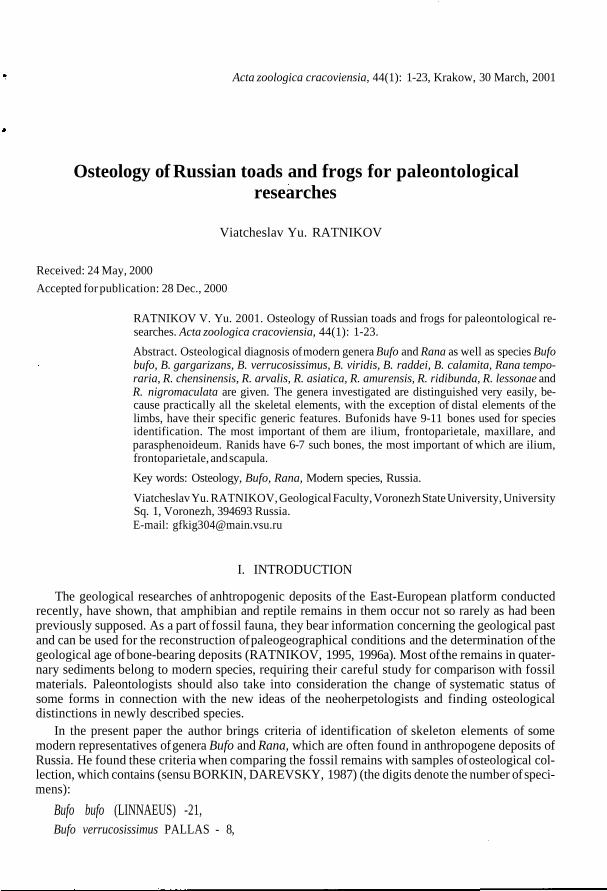
Bufo bufo (LINNAEUS) -21,Bufo verrucosissimus PALLAS - 8,
V. Yu. RATNIKOV2
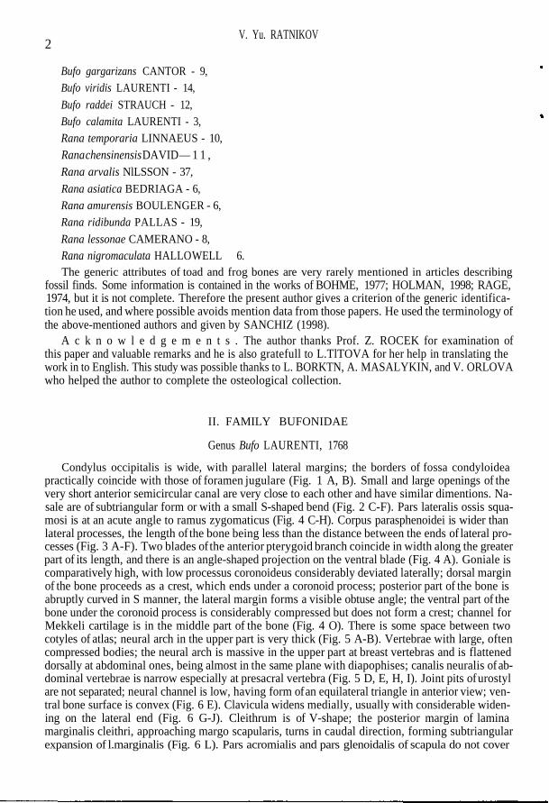
Bufo gargarizans CANTOR - 9,Bufo viridis LAURENTI - 14,Bufo raddei STRAUCH - 12,Bufo calamita LAURENTI - 3,Rana temporaria LINNAEUS - 10,Rana chensinensis DAVID — 1 1 ,Rana arvalis NlLSSON - 37,Rana asiatica BEDRIAGA - 6,Rana amurensis BOULENGER - 6,Rana ridibunda PALLAS - 19,Rana lessonae CAMERANO - 8,Rana nigromaculata HALLO WELL - 6.The generic attributes of toad and frog bones are very rarely mentioned in articles describing
fossil finds. Some information is contained in the works of BOHME, 1977; HOLMAN, 1998; RAGE,1974, but it is not complete. Therefore the present author gives a criterion of the generic identifica-tion he used, and where possible avoids mention data from those papers. He used the terminology ofthe above-mentioned authors and given by SANCHIZ (1998).
A c k n o w l e d g e m e n t s . The author thanks Prof. Z. ROCEK for examination ofthis paper and valuable remarks and he is also gratefull to L.TlTOVA for her help in translating thework in to English. This study was possible thanks to L. BORKTN, A. MASALYKIN, and V. ORLOVAwho helped the author to complete the osteological collection.
II. FAMILY BUFONIDAE
Genus Bufo LAURENTI, 1768
Condylus occipitalis is wide, with parallel lateral margins; the borders of fossa condyloideapractically coincide with those of foramen jugulare (Fig. 1 A, B). Small and large openings of thevery short anterior semicircular canal are very close to each other and have similar dimentions. Na-sale are of subtriangular form or with a small S-shaped bend (Fig. 2 C-F). Pars lateralis ossis squa-mosi is at an acute angle to ramus zygomaticus (Fig. 4 C-H). Corpus parasphenoidei is wider thanlateral processes, the length of the bone being less than the distance between the ends of lateral pro-cesses (Fig. 3 A-F). Two blades of the anterior pterygoid branch coincide in width along the greaterpart of its length, and there is an angle-shaped projection on the ventral blade (Fig. 4 A). Goniale iscomparatively high, with low processus coronoideus considerably deviated laterally; dorsal marginof the bone proceeds as a crest, which ends under a coronoid process; posterior part of the bone isabruptly curved in S manner, the lateral margin forms a visible obtuse angle; the ventral part of thebone under the coronoid process is considerably compressed but does not form a crest; channel forMekkeli cartilage is in the middle part of the bone (Fig. 4 O). There is some space between twocotyles of atlas; neural arch in the upper part is very thick (Fig. 5 A-B). Vertebrae with large, oftencompressed bodies; the neural arch is massive in the upper part at breast vertebras and is flatteneddorsally at abdominal ones, being almost in the same plane with diapophises; canalis neuralis of ab-dominal vertebrae is narrow especially at presacral vertebra (Fig. 5 D, E, H, I). Joint pits of urostylare not separated; neural channel is low, having form of an equilateral triangle in anterior view; ven-tral bone surface is convex (Fig. 6 E). Clavicula widens medially, usually with considerable widen-ing on the lateral end (Fig. 6 G-J). Cleithrum is of V-shape; the posterior margin of laminamarginalis cleithri, approaching margo scapularis, turns in caudal direction, forming subtriangularexpansion of l.marginalis (Fig. 6 L). Pars acromialis and pars glenoidalis of scapula do not cover

Osteology of Russian toads and frogs
4mm
Fig. 1. Cranial bones. A-C - exoocipital, posterior view: A - Bufo bufo, B - Bufo viridis, C- Rana temporaria; D-E - prooti-cum, dorsal and ventral views: D - Rana temporaria, E - Rana ridibunda; F-L - maxillare, F-H, K, L - outer view, I-J - in-ner view: F - Bufo bufo, G - Bufo gargarizans, H - Bufo verrucasissimus, I - Bufo viridis, } - Bufo raddei, K — Bufocalamita, L - Rana arvalis; M-S - sphenethmoideum, dorsal, ventral (J, M, P- and anterior) views: M - Bufo bufo, N -Bufo gargarizans, O - Bufo verrucosissimus, P - Bufo viridis, Q - Bufo raddei, R - Bufo calamita, S - Rana temporaria.
V. Yu. RATNIKOV
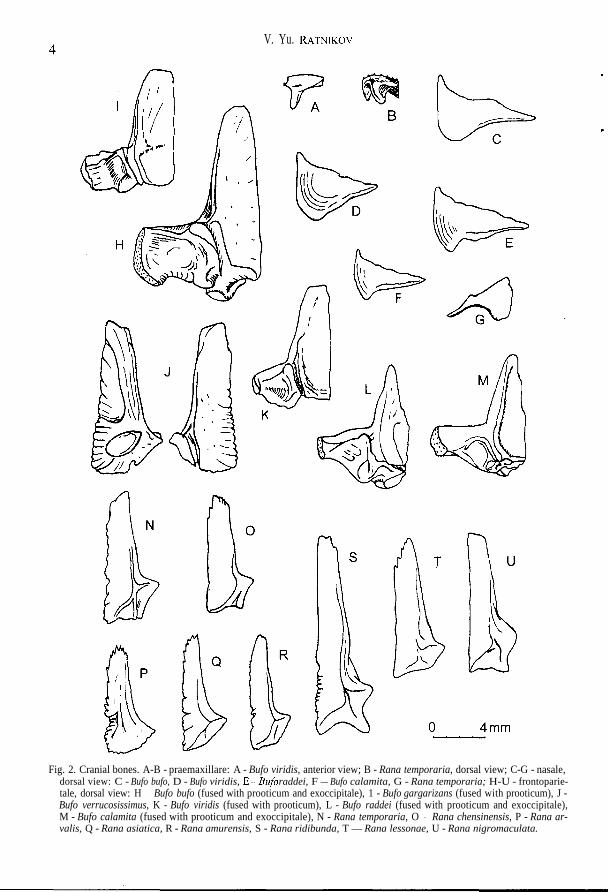
Fig. 2. Cranial bones. A-B - praemaxillare: A - Bufo viridis, anterior view; B - Rana temporaria, dorsal view; C-G - nasale,dorsal view: C - Bufo bufo, D - Bufo viridis, E — B u f o raddei, F — Bufo calamita, G - Rana temporaria; H-U - frontoparie-tale, dorsal view: H - Bufo bufo (fused with prooticum and exoccipitale), 1 - Bufo gargarizans (fused with prooticum), J -Bufo verrucosissimus, K - Bufo viridis (fused with prooticum), L - Bufo raddei (fused with prooticum and exoccipitale),M - Bufo calamita (fused with prooticum and exoccipitale), N - Rana temporaria, O - Rana chensinensis, P - Rana ar-valis, Q - Rana asiatica, R - Rana amurensis, S - Rana ridibunda, T — Rana lessonae, U - Rana nigromaculata.

Osteology of Russian toads and frogs
Fig. 3. Parasphenoideum, ventral view. A - Bufo bufo, B - Bufo gargarizans, C - Bufo verrucosissimus, D - Bufo viridis, E -Bufo calamita, F - Bufo raddei, G - Rana temporaria, H - Rana chensinensis, I - Rana arvalis, J — .Sara asiatica, KXa«a amurensis, L - ffa«a ridibunda, M - Sana lessonae, N - Sana nigromaculata.
V. Yu. RATNIKOV
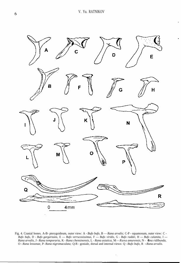
Fig. 4. Cranial bones. A-B- pterygoideum, outer view: A - Bufo bufo, B — Rana arvalis; C-P - squamosum, outer view: C -Bufo bufo, D - Bufo gargarizans, E — Bufo verrucosissimus, F — Bufo viridis, G - Bufo raddei, H — Bufo calamita, I —Rana arvalis, J - Rana temporaria, K - Rana chensinensis, L - Rana asiatica, M—Rana amurensis, N - ^f ana ridibunda,O - Rana lessonae, P- Rana nigromaculata; Q-R - goniale, dorsal and internal views: Q - Bufo bufo, R - Rana arvalis.
Osteology of Russian toads and frogs
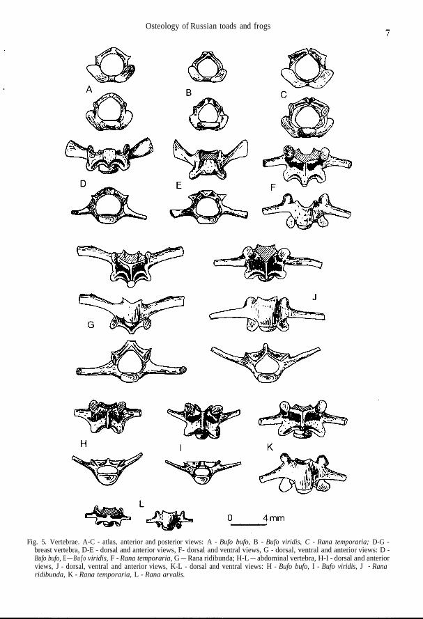
Fig. 5. Vertebrae. A-C - atlas, anterior and posterior views: A - Bufo bufo, B - Bufo viridis, C - Rana temporaria; D-G -breast vertebra, D-E - dorsal and anterior views, F- dorsal and ventral views, G - dorsal, ventral and anterior views: D -Bufo bufo, E — B u f o viridis, F - Rana temporaria, G — Rana ridibunda; H-L — abdominal vertebra, H-I - dorsal and anteriorviews, J - dorsal, ventral and anterior views, K-L - dorsal and ventral views: H - Bufo bufo, I - Bufo viridis, J - Ranaridibunda, K - Rana temporaria, L - Rana arvalis.
V. Yu. RATNIKOV
M
4mm N
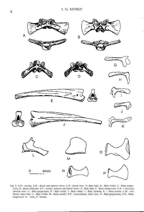
Fig. 6. A-D - sacrum, A-B - dorsal and anterior views, C-D - dorsal view: A- Bufo bufo, B - Bufo viridis, C - Rana tempo-raria, D - Rana ridibunda; E-F - urostyl, anterior and lateral views: E - Bufo bufo, F - Rana temporaria; G-K - clavicula,internal view: G - Bufo gargarizans, H - Bufo viridis, I - Bufo raddei, J - Bufo calamita, K — Rana arvalis; L-M - clei-thrum, inner view: L - Bufo viridis, M - Rana arvalis; N-P - coracoideum, inner view: N - Bufo gargarizans, O-P - Ranatemporaria: O - male, P - female.
Osteology of Russian toads and frogs
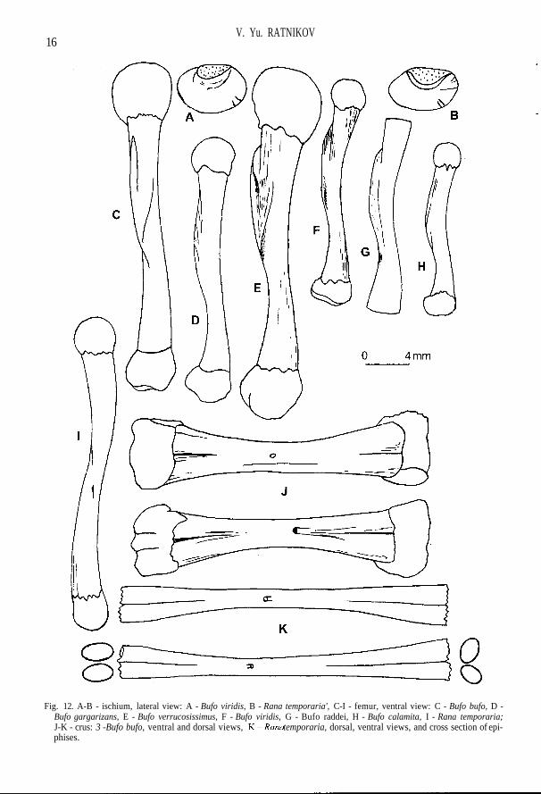
each other; facies lunata is turned laterally; crista longitudinalis is weakly expressed (Fig. 7 A-F).The width of extremitas lateralis coracoidei is approximately equal to that of extremitas medialis;medial margin of the bone is either straight or slightly rounded (Fig. 6 N). The axis of olecranontrace is displaced laterally relatively to long axis of humerus; there may be up to three ventral crestson a proximal half of the bone (Fig. 8). The greatest ischium width is approximately on the middleline; the area for attachment of hind limb muscles is wide (Fig. 12 A). Crus is short, sharply widenedtowards the ends, in the middle part (in the region of foramen nutritium) compressed, often has akiel from tibia side, rarely - from both sides; sections of tibia and fibula at the distal end more or lessrounded, at the proximal end they are oval; the diameter of tibia section on the distal end is muchsmaller than that of the fibula one, whereas on the proximal end they are approximately equal; nearthe foramen nutritium, close to the distal bone end, there is a deep furrow, turning into sulcus inter-medius in distal direction (Fig. 12 J). The additional characteristics of the majority of toad bones isspecific "toad" shading which is absent in other modern Anura of Russia.
A number of works are devoted to the estimation of species features of pleistocene toads(BAILON, 1986; BOHME, 1977; HODROVA, 1980; HOLMAN, 1989,1998; SANCHIZ, 1977). All theseresearches are based on the assumption that the same three modern toad species existed in WesternEurope in the past: Bufo bufo, B. calamita, and B. viridis. This assumption is not acceptable in diag-nostics of the Eastern Europe fossil material. Firstly, the tendency of transference of former subspe-cies to species has made it necessary to search for identification criteria of new species and revisionof old ones, concerning first of all B. bufo. Secondly, the present author has admitted the possibilityof occurence in anthropogene deposits not only forms living these at present, that has later proved tobe true (RATNIKOV, 1996b). Thus, the number of possible levels of fossil material determination hasincreased.
The modern representatives of genus Bufo of Eurasia are united by zoologists in unsystematicgroups (complexes) on the basis of certain attributes. The toads of Russia and adjacent countriesform two complexes: Bufo (bufo) sp. (the grey toad group) and Bufo (viridis) sp.(the green toadgroup). Representatives of each complex have similar features in the structure of some skeletonelements, distinguishing them from representatives of another complex, and similar ecological at-tachment, which is very important for reconstruction of the paleogeographical conditions during theformation of the locality (RATNIKOV, 1996a).
Bufo (bufo) sp.
Condylus occipitalis is very wide; torus terminalis is usually narrow and high (Fig. 1 A). Eleva-tio prootici is well expressed and is disposed along the anterior margin of the bone; ramus lateralisprootici is massive, almost unnarrowed laterally (Fig. 2 H, I). Sphenethmoid is usually with narrowposterior openings of canalis olfactorii and weakly expressed folds in their cavities (Fig. 1 M-O).The anterior end of maxilla pars facialis as a process projects forward further than anterior end ofpars dentalis (Fig. 1 F-H). Nasale has visible S-shaped bend (Fig. 2 C). Frontoparietale graduallynarrows in the anterior part; dorsal surface is flattened and is not limited by crest; lateral margin ofthe bone is abruptly curved downwards (Fig. 2 H-J). Squamosum has considerably widened ramusretrozygomaticus (Fig. 4 C-E). Ventral surface of corpus parasphenoidei forms abrupt bend in re-gion of lateral processes and has more abrupt relief than representatives of Bufo (viridis) sp. do(Fig. 3 A-C). Atlas has wide cotyles and oval condyle (Fig. 5 A). Centra of breast vertebras are ovalin cross section; cotyles at abdominal vertebrae acquire a half-moon form; dorsal plane of neuralarch as a rule is longer than ventral one; diapophises widened at the basis and compressed (Fig. 5 D,H). Sacral neurapophyses are of wide A-shaped form (Fig. 6 A). Humerus has usually more convexdorsal surface on the distal end than do representatives of green toads. Ala ossis ilii is comparativelythick without pre-acetabular pit; subtriangular form is characteristic for the bone body (Fig. 10A-C). Femur with comparatively short crista femoris (Fig. 12 C-E).
It was considered that only one species from the group of grey toads - Bufo bufo (L.) with sev-eral subspecies - existed on the territory of the former USSR (BANNIKOV et al., 1977). Not long ago
V. Yu. RATNIKOV
herpetologists acknowledged the independence of a Far Eastern subspecies, which is now includedinto the structure of Bufo (bufo) sp. under the species name Bufo gargarizans CANTOR (BORKIN,DAREVSKY 1987). The question of systematic position of the Caucasian subspecies has notyetbeenfinally solved. Some herpetologists consider it an independent species (ORLOVA, TUNIEV 1989),whereas others (GUNTHER 1985) are of the opinion that only one species exist in Europe. V. M.CKHIKVADZE (1984) pointed to the existence of differences in osteology and independence of theCaucasian toad. The materials investigated by the present author show that the level of differencesbetween B. bufo and 5. gargarizans is the same as that between B. bufo and B. bufo verrucosissimus.He has therefore identified some fossil bones as B. verrucosissimus or B. cf. verrucosissimus(RATNIKOV 1992). In the last list of the former USSR herpetofauna (ANANJEVA et al. 1998) Bufoverrucosissimus is given as a species. In that work the description of osteological features of threegrey toad species - Bufo bufo (L.), Bufo gargarizans CANTOR and Bufo verrucosissimus (PALL.) - isadduced.
Bufo bufo (LINNAEUS, 1758)
Lateral processes of sphenethmoideum are short, ventral crests on them being weakly expressed(Fig. 1 M). External surface of maxillare without folds; posterior process of pars palatina is un-derdeveloped, not projecting over the upper margin of pars facialis (Fig. 1 F). Frontoparietale gentlynarrowed toward the anterior end; facies cerebralis posterior are of elliptic form with a long axis al-most perpendicular to margo sagittalis of the bone (Fig. 2 H). Ramus zygomaticus ossis squamosi isusually short; margins of ramus retrozygomaticus are parallel or slightly divergent, the upper mar-gin often with wavy bend (Fig. 4 C). Lateral processes and corpus parasphenoidei are of equalwidth; on an axial line of the bone in region of lateral proceses there is a well-defined crest recallinga "tick" in its outlines; straight lateral margins of the bone body that one abruptly bent turn into ante-rior margins of lateral processes; posterior process is usually narrow (Fig. 3 A). Scapula with com-paratively wide head and narrow neck; the anterior margin of pars acromialis may be stretched intenuitas acromialis (Fig. 7 A). Medial crest of humerus usually does not reach the middle of thebone, it is wide, with rounded margin, slightly deviated dorsally; spina medialis dorsalis can be ob-served, whereas spina medialis ventralis is barely noticable (Fig. 8 A). Ilium has often low butsometimes high tuber superior on which as a rule there is one large tubercle often displaced forward(Fig. 10 A). Femur has a single crista femoris; there is sometimes an additional obtuse crest in largespecimens, which begins not far from the distal end of crista femoris (Fig. 12 C).
Bufo gargarizans CANTOR, 1842
The posterior part of ventral sphenethmoid area is wide; lateral processes are rather long withabrupt ventral crests (Fig. 1 N). Smooth fold is visible on external surface of maxillare from the an-terior end; posterior process of pars palatina almost does not project over the upper margin of parsfacialis (Fig. 1 G). Lateral margin of frontoparietale is as a rule weakly bent in an arch-like manner;facies cerebralis posterior is practically round (Fig. 2 I). Ramus zygomaticus osis squamosi is usu-ally comparatively long and thin, and the margins of ramus retrozygomaticus appreciably diverge(Fig. 4 D). Corpus parasphenoidei is wider than the lateral process; convexed lateral margins of cor-pus, being gently bent, turn into anterior margins of lateral processes; "tick" in region of lateral pro-cesses is absent, there is a fairly wide ventral area in its place; the crest passing from one lateralprocess toward another is sometimes visible; the posterior process is usually wide (Fig. 3 B). Scap-ula with a comparatively wide head and narrow neck; anterior margin of pars acromialis may bestretched in tenuitas acromialis (Fig. 7 B). Medial crest of humerus usually reaches the middle of thebone, with rounded margin, greatly deviated dorsally; spina medialis dorsalis may be appreciabllydeveloped, whereas spina medialis ventralis is usually not visible (Fig. 8 B). Ilium has high tuber su-perior either with no small tubercles, or with one tubercle on its lateral surface (Fig. 10 B). Femurwith the only crista femoris; sometimes there is an additional slightly visible obtuse crest in largespecimens, which begins from the middle of crista femoris (Fig. 12 D).
Osteology of Russian toads and frogs11
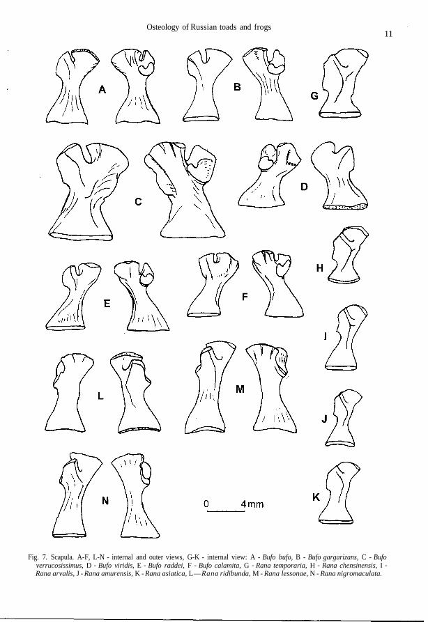
Fig. 7. Scapula. A-F, L-N - internal and outer views, G-K - internal view: A - Bufo bufo, B - Bufo gargarizans, C - Bufoverrucosissimus, D - Bufo viridis, E - Bufo raddei, F - Bufo calamita, G - Rana temporaria, H - Rana chensinensis, I -Rana arvalis, J - Rana amurensis, K - Rana asiatica, L—Rana ridibunda, M - Rana lessonae, N - Rana nigromaculata.
12v. Yu. RATNIK.OV
B
V/1
V//
4mm
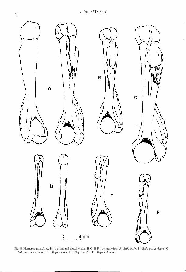
Fig. 8. Humerus (male). A, D - ventral and dorsal views, B-C, E-F - ventral view: A- Bufo bufo, B - Bufo gargarizans, C -Bufo verrucosissimus, D - Bufo viridis, E - Bufo raddei, F - Bufo calamita.
Osteology of Russian toads and frogs 13
4mm
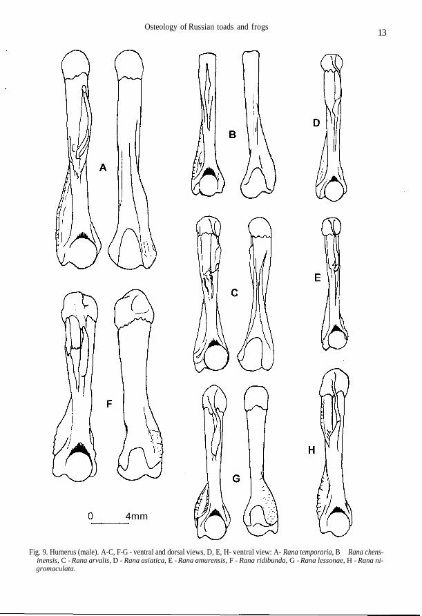
Fig. 9. Humerus (male). A-C, F-G - ventral and dorsal views, D, E, H- ventral view: A- Rana temporaria, B - Rana chens-inensis, C - Rana arvalis, D - Rana asiatica, E - Rana amurensis, F - Rana ridibunda, G - Rana lessonae, H - Rana ni-gromaculata.
14 V. Yu. RATNIKOV
4mm
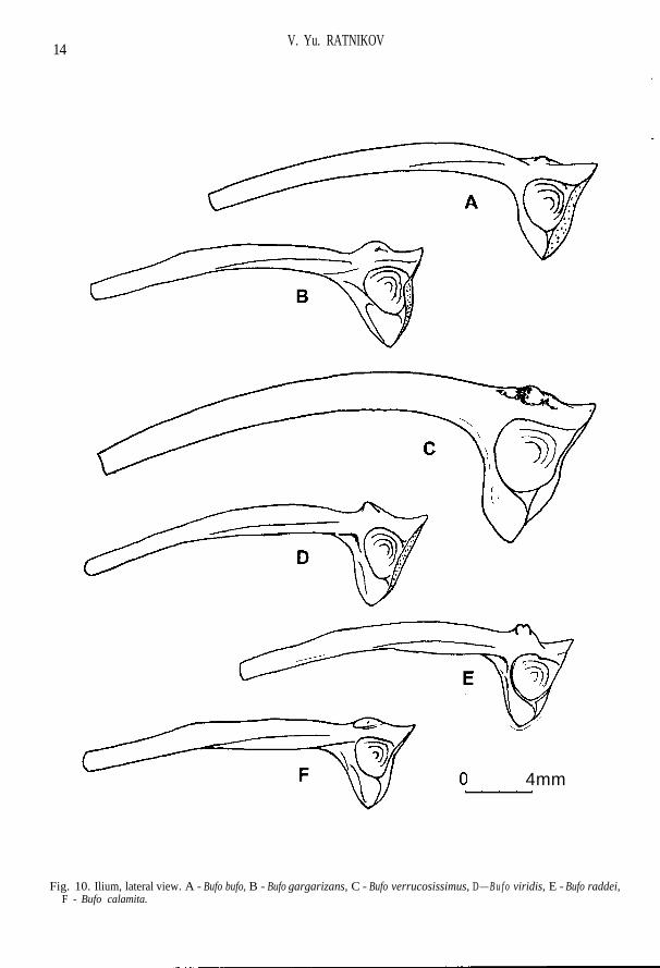
Fig. 10. Ilium, lateral view. A - Bufo bufo, B - Bufo gargarizans, C - Bufo verrucosissimus, D — B u f o viridis, E - Bufo raddei,F - Bufo calamita.
Osteology of Russian toads and frogs15
4mm
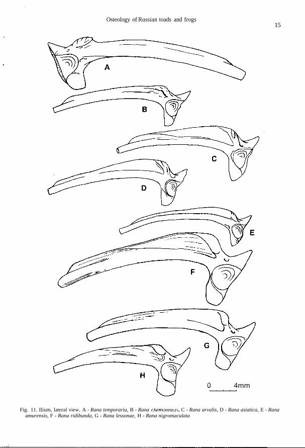
Fig. 11. Ilium, lateral view. A - Rana temporaria, B - Rana chensinensis, C - Rana arvalis, D - Rana asiatica, E - Ranaamurensis, F - Rana ridibunda, G - Rana lessonae, H - Rana nigromaculata
16V. Yu. RATNIKOV
Fig. 12. A-B - ischium, lateral view: A - Bufo viridis, B - Rana temporaria', C-I - femur, ventral view: C - Bufo bufo, D -Bufo gargarizans, E - Bufo verrucosissimus, F - Bufo viridis, G - Bufo raddei, H - Bufo calamita, I - Rana temporaria;J-K - crus: 3 -Bufo bufo, ventral and dorsal views, Y^-Rana temporaria, dorsal, ventral views, and cross section of epi-phises.
Osteology of Russian toads and frogs
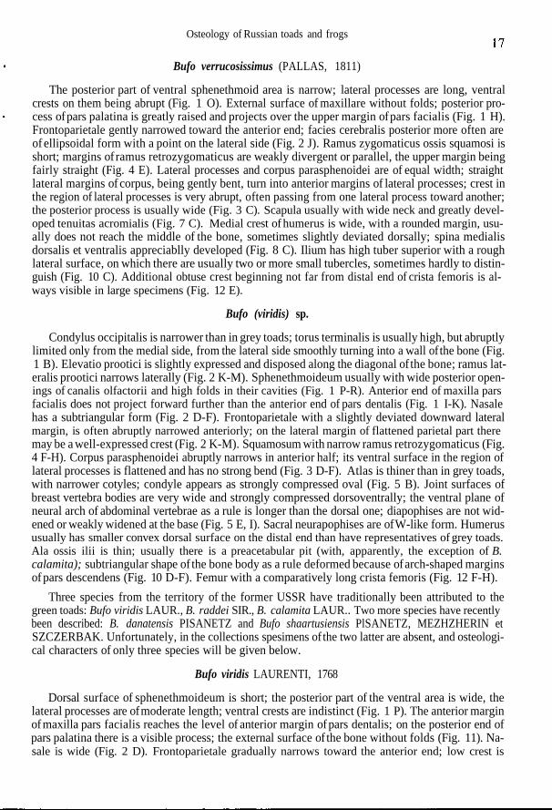
Bufo verrucosissimus (PALLAS, 1811)
The posterior part of ventral sphenethmoid area is narrow; lateral processes are long, ventralcrests on them being abrupt (Fig. 1 O). External surface of maxillare without folds; posterior pro-cess of pars palatina is greatly raised and projects over the upper margin of pars facialis (Fig. 1 H).Frontoparietale gently narrowed toward the anterior end; facies cerebralis posterior more often areof ellipsoidal form with a point on the lateral side (Fig. 2 J). Ramus zygomaticus ossis squamosi isshort; margins of ramus retrozygomaticus are weakly divergent or parallel, the upper margin beingfairly straight (Fig. 4 E). Lateral processes and corpus parasphenoidei are of equal width; straightlateral margins of corpus, being gently bent, turn into anterior margins of lateral processes; crest inthe region of lateral processes is very abrupt, often passing from one lateral process toward another;the posterior process is usually wide (Fig. 3 C). Scapula usually with wide neck and greatly devel-oped tenuitas acromialis (Fig. 7 C). Medial crest of humerus is wide, with a rounded margin, usu-ally does not reach the middle of the bone, sometimes slightly deviated dorsally; spina medialisdorsalis et ventralis appreciablly developed (Fig. 8 C). Ilium has high tuber superior with a roughlateral surface, on which there are usually two or more small tubercles, sometimes hardly to distin-guish (Fig. 10 C). Additional obtuse crest beginning not far from distal end of crista femoris is al-ways visible in large specimens (Fig. 12 E).
Bufo (viridis) sp.
Condylus occipitalis is narrower than in grey toads; torus terminalis is usually high, but abruptlylimited only from the medial side, from the lateral side smoothly turning into a wall of the bone (Fig.1 B). Elevatio prootici is slightly expressed and disposed along the diagonal of the bone; ramus lat-eralis prootici narrows laterally (Fig. 2 K-M). Sphenethmoideum usually with wide posterior open-ings of canalis olfactorii and high folds in their cavities (Fig. 1 P-R). Anterior end of maxilla parsfacialis does not project forward further than the anterior end of pars dentalis (Fig. 1 I-K). Nasalehas a subtriangular form (Fig. 2 D-F). Frontoparietale with a slightly deviated downward lateralmargin, is often abruptly narrowed anteriorly; on the lateral margin of flattened parietal part theremay be a well-expressed crest (Fig. 2 K-M). Squamosum with narrow ramus retrozygomaticus (Fig.4 F-H). Corpus parasphenoidei abruptly narrows in anterior half; its ventral surface in the region oflateral processes is flattened and has no strong bend (Fig. 3 D-F). Atlas is thiner than in grey toads,with narrower cotyles; condyle appears as strongly compressed oval (Fig. 5 B). Joint surfaces ofbreast vertebra bodies are very wide and strongly compressed dorsoventrally; the ventral plane ofneural arch of abdominal vertebrae as a rule is longer than the dorsal one; diapophises are not wid-ened or weakly widened at the base (Fig. 5 E, I). Sacral neurapophises are of W-like form. Humerususually has smaller convex dorsal surface on the distal end than have representatives of grey toads.Ala ossis ilii is thin; usually there is a preacetabular pit (with, apparently, the exception of B.calamita); subtriangular shape of the bone body as a rule deformed because of arch-shaped marginsof pars descendens (Fig. 10 D-F). Femur with a comparatively long crista femoris (Fig. 12 F-H).
Three species from the territory of the former USSR have traditionally been attributed to thegreen toads: Bufo viridis LAUR., B. raddei SIR., B. calamita LAUR.. Two more species have recentlybeen described: B. danatensis PISANETZ and Bufo shaartusiensis PlSANETZ, MEZHZHERIN etSZCZERBAK. Unfortunately, in the collections spesimens of the two latter are absent, and osteologi-cal characters of only three species will be given below.
Bufo viridis LAURENTI, 1768
Dorsal surface of sphenethmoideum is short; the posterior part of the ventral area is wide, thelateral processes are of moderate length; ventral crests are indistinct (Fig. 1 P). The anterior marginof maxilla pars facialis reaches the level of anterior margin of pars dentalis; on the posterior end ofpars palatina there is a visible process; the external surface of the bone without folds (Fig. 11). Na-sale is wide (Fig. 2 D). Frontoparietale gradually narrows toward the anterior end; low crest is
V. Yu. RATNIKOV18
sometimes present on the lateral margin of flattened parietal part (Fig. 2 K). Pars horizontalis ossissquamosi is mostly widened in the middle part (Fig. 4 F). Corpus parasphenoidei is much wider thanthe lateral processes; the flattened part of the bone body is wide (Fig. 3 D). Clavicula is compara-tively narrow and long (Fig. 6 H). Scapula is fairly short, with very wide collum scapulae and widepars acromialis, whose anterior margin is stretched in tenuitas acromialis (Fig. 7 D). Humerus islong; crista medialis is narrow, with a straight margin, slightly deviated dorsally; spinae medialisdorsalis et ventralis are absent (Fig. 8 D). Ilium with high, long, asimmetrical tuber superior (the an-terior end steeper than the posterior one), bearing variable amounts of tubercles (Fig. 10 D). Femuris comparatively thin, having the shortest crista femoris among the green toads (which is, however,longer than in the representatives ofBufo (bufo) sp. (Fig. 12 F).
Bufo raddei STRAUCH, 1876
Dorsal sphenethmoid surface is short; the posterior part of ventral area is narrow; the lateral pro-cesses long; ventral crests abrupt (Fig. 1 Q). The anterior margin of maxilla pars facialis does notreach the level of that of pars dentalis; processes on posterior end of pars palatina are weakly de-veloped; the external surface of the bone without folds (Fig. 1 J). Nasale is of moderate width (Fig. 2E). Frontoparietale considerably narrows in the anterior part; on the lateral margin of parietal part inadult specimens there may be a low crest (Fig. 2 L). Pars horizontalis ossis squamosi is not widenedin the middle part; ramus retrozygomaticus has an arch-shaped upper margin (Fig. 4 G). The flat-tened part of corpus parasphenoidei is narrow; the lateral processes are mostly widened at aboutone-third of their length from the distal end where they are as wide as the bone body (Fig. 3 E).Clavicula is short and wide (Fig. 6 I). Scapula is long, with narrow collum scapulae; the anteriormargin of pars acromialis may be stretched in tenuitas acromialis (Fig. 7 E). (Investigated materialshows variations in the form of this bone: the scapulas of the toads from the Far East have compara-tively wider collum scapulae than Mongolian specimens and are very similar to scapulas of B.calamita). Humerus (Fig. 8 E) is short and curved; the crista medialis is wide, with rounded margin,deviated dorsally; spinae medialis dorsalis et ventralis are well expressed. Ilium has a very high,simmetrically or almost simmetrically rising, tuber superior with large tubercle in the central part, inmost cases split into two peaks or with a pit above (Fig. 10 E). Crista femoris is long, high, and thin(Fig. 12 G).
Bufo calamita LAURENTI, 1768
Dorsal sphenethmoid surface is long; the ventral crests abrupt; posterior part of ventral area ofmoderate width; lateral processes are long (Fig. 1 R). The anterior margin of maxilla pars facialisdoes not reach the level of the anterior margin of pars dentalis; process on posterior end of pars pa-latina is weakly expressed; there is a visible fold in the anterior part of the bone (Fig. 1 K). Nasale isnarrow (Fig. 2 F). Frontoparietale is strongly narrowed in the anterior part; there is a clearly ex-pressed crest along the lateral margin of the parietal part of the bone, extending on the frontal part inlarger specimens (Fig. 2 M). Pars horizontalis of squamosum is widened in the middle part; ramusretrozygomaticus has somewhat abrupt bend of the upper margin at an obtuse angle (Fig. 4 H). Flat-tened part of corpus parasphenoidei is narrow; the width of the lateral processes is approximatelythe same almost along their extent and is appreciablly less than corpus width (Fig. 3 E). Clavicula isshort, of moderate width (Fig. 6 J). Scapula is long, its neck being wider than that of B. raddei; theanterior margin of pars acromialis can be stretched in tenuitas acromialis (Fig. 7 F). Crista medialisossis humeri is of moderate width, with straight margin on the greater part of its length, slightly de-viated dorsally; spina medialis dorsalis et ventralis are well expressed (Fig. 8 F). Ilium without prea-cetabular pit; tuber superior is high, short, narrow, sometimes with a single weakly developedtubercle in central part (Fig. 10 F). Crista femoris is long and thick (Fig. 12 H).
Osteology of Russian toads and frogs
IV. FAMILY RANIDAE
Genus Rana LINNAEUS, 1758
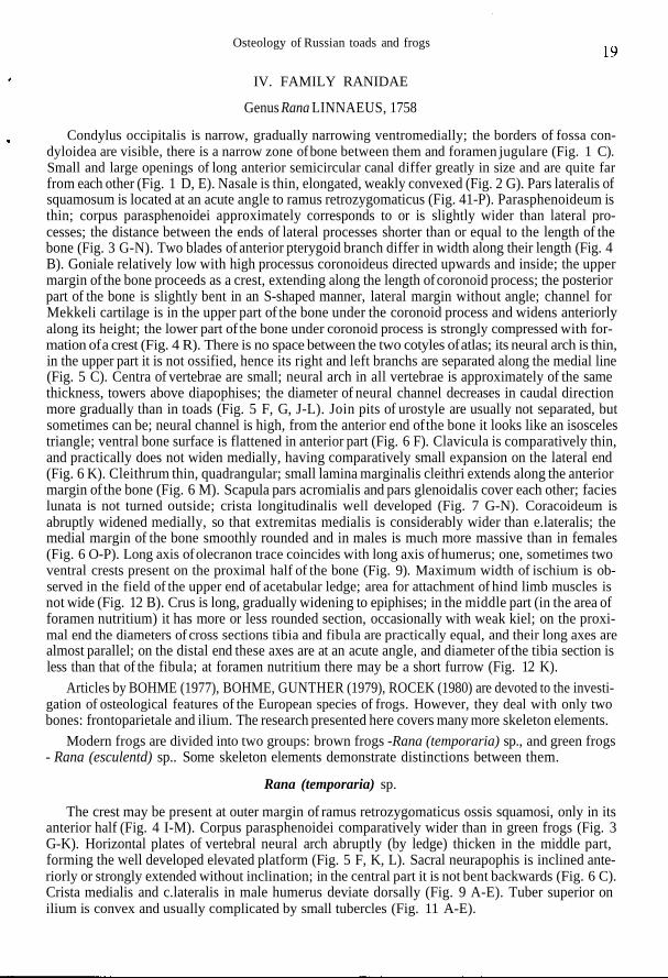
Condylus occipitalis is narrow, gradually narrowing ventromedially; the borders of fossa con-dyloidea are visible, there is a narrow zone of bone between them and foramen jugulare (Fig. 1 C).Small and large openings of long anterior semicircular canal differ greatly in size and are quite farfrom each other (Fig. 1 D, E). Nasale is thin, elongated, weakly convexed (Fig. 2 G). Pars lateralis ofsquamosum is located at an acute angle to ramus retrozygomaticus (Fig. 41-P). Parasphenoideum isthin; corpus parasphenoidei approximately corresponds to or is slightly wider than lateral pro-cesses; the distance between the ends of lateral processes shorter than or equal to the length of thebone (Fig. 3 G-N). Two blades of anterior pterygoid branch differ in width along their length (Fig. 4B). Goniale relatively low with high processus coronoideus directed upwards and inside; the uppermargin of the bone proceeds as a crest, extending along the length of coronoid process; the posteriorpart of the bone is slightly bent in an S-shaped manner, lateral margin without angle; channel forMekkeli cartilage is in the upper part of the bone under the coronoid process and widens anteriorlyalong its height; the lower part of the bone under coronoid process is strongly compressed with for-mation of a crest (Fig. 4 R). There is no space between the two cotyles of atlas; its neural arch is thin,in the upper part it is not ossified, hence its right and left branchs are separated along the medial line(Fig. 5 C). Centra of vertebrae are small; neural arch in all vertebrae is approximately of the samethickness, towers above diapophises; the diameter of neural channel decreases in caudal directionmore gradually than in toads (Fig. 5 F, G, J-L). Join pits of urostyle are usually not separated, butsometimes can be; neural channel is high, from the anterior end of the bone it looks like an isoscelestriangle; ventral bone surface is flattened in anterior part (Fig. 6 F). Clavicula is comparatively thin,and practically does not widen medially, having comparatively small expansion on the lateral end(Fig. 6 K). Cleithrum thin, quadrangular; small lamina marginalis cleithri extends along the anteriormargin of the bone (Fig. 6 M). Scapula pars acromialis and pars glenoidalis cover each other; facieslunata is not turned outside; crista longitudinalis well developed (Fig. 7 G-N). Coracoideum isabruptly widened medially, so that extremitas medialis is considerably wider than e.lateralis; themedial margin of the bone smoothly rounded and in males is much more massive than in females(Fig. 6 O-P). Long axis of olecranon trace coincides with long axis of humerus; one, sometimes twoventral crests present on the proximal half of the bone (Fig. 9). Maximum width of ischium is ob-served in the field of the upper end of acetabular ledge; area for attachment of hind limb muscles isnot wide (Fig. 12 B). Crus is long, gradually widening to epiphises; in the middle part (in the area offoramen nutritium) it has more or less rounded section, occasionally with weak kiel; on the proxi-mal end the diameters of cross sections tibia and fibula are practically equal, and their long axes arealmost parallel; on the distal end these axes are at an acute angle, and diameter of the tibia section isless than that of the fibula; at foramen nutritium there may be a short furrow (Fig. 12 K).
Articles by BOHME (1977), BOHME, GUNTHER (1979), ROCEK (1980) are devoted to the investi-gation of osteological features of the European species of frogs. However, they deal with only twobones: frontoparietale and ilium. The research presented here covers many more skeleton elements.
Modern frogs are divided into two groups: brown frogs -Rana (temporaria) sp., and green frogs- Rana (esculentd) sp.. Some skeleton elements demonstrate distinctions between them.
Rana (temporaria) sp.
The crest may be present at outer margin of ramus retrozygomaticus ossis squamosi, only in itsanterior half (Fig. 4 I-M). Corpus parasphenoidei comparatively wider than in green frogs (Fig. 3G-K). Horizontal plates of vertebral neural arch abruptly (by ledge) thicken in the middle part,forming the well developed elevated platform (Fig. 5 F, K, L). Sacral neurapophis is inclined ante-riorly or strongly extended without inclination; in the central part it is not bent backwards (Fig. 6 C).Crista medialis and c.lateralis in male humerus deviate dorsally (Fig. 9 A-E). Tuber superior onilium is convex and usually complicated by small tubercles (Fig. 11 A-E).
V. Yu. RATNIKOV
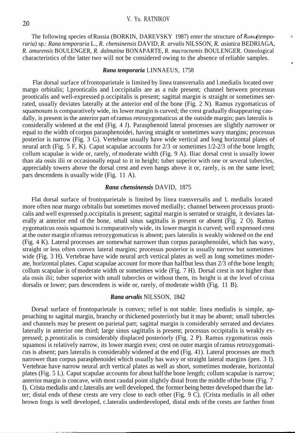
The following species of Russia (BORKIN, DAREVSKY 1987) enter the structure ofRana (tempo-raria) sp.: Rana temporaria L., R. chensinensis DAVID, R. arvalis NlLSSON, R. asiatica BEDRIAGA,R. amurensis BOULENGER, R. dalmatina BONAPARTE, R. macrocnemis BOULENGER. Osteologicalcharacteristics of the latter two will not be considered owing to the absence of reliable samples.
Rana temporaria LINNAEUS, 1758
Flat dorsal surface of frontoparietale is limited by linea transversalis and l.medialis located overmargo orbitalis; l.prooticalis and l.occipitalis are as a rule present; channel between processusprooticalis and well-expressed p.occipitalis is present; sagittal margin is straight or sometimes ser-rated, usually deviates laterally at the anterior end of the bone (Fig. 2 N). Ramus zygomaticus ofsquamosum is comparatively wide, its lower margin is curved; the crest gradually disappearing cau-dally, is present in the anterior part of ramus retrozygomaticus at the outside margin; pars lateralis isconsiderally widened at the end (Fig. 4 J). Parasphenoid lateral processes are slightly narrower orequal to the width of corpus parasphenoidei, having straight or sometimes wavy margins; processusposterior is narrow (Fig. 3 G). Vertebrae usually have wide vertical and long horizontal plates ofneural arch (Fig. 5 F, K). Caput scapulae accounts for 2/3 or sometimes 1/2-2/3 of the bone length;collum scapulae is wide or, rarely, of moderate width (Fig. 9 A). Iliac dorsal crest is usually lowerthan ala ossis ilii or occasionally equal to it in height; tuber superior with one or several tubercles,appreciably towers above the dorsal crest and even hangs above it or, rarely, is on the same level;pars descendens is usually wide (Fig. 11 A).
Rana chensinensis DAVID, 1875
Flat dorsal surface of frontoparietale is limited by linea transversalis and 1. medialis locatedmore often near margo orbitalis but sometimes moved medially; channel between processus prooti-calis and well expressed p.occipitalis is present; sagittal margin is serrated or straight, it deviates lat-erally at anterior end of the bone, small sinus sagittalis is present or absent (Fig. 2 O). Ramuszygomaticus ossis squamosi is comparatively wide, its lower margin is curved; well expressed crestat the outer margin of ramus retrozygomaticus is absent; pars lateralis is weakly widened on the end(Fig. 4 K). Lateral processes are somewhat narrower than corpus parasphenoidei, which has wavy,straight or less often convex lateral margins; processus posterior is usually narrow but sometimeswide (Fig. 3 H). Vertebrae have wide neural arch vertical plates as well as long sometimes moder-ate, horizontal plates. Caput scapulae account for more than half but less than 2/3 of the bone length;collum scapulae is of moderate width or sometimes wide (Fig. 7 H). Dorsal crest is not higher thanala ossis ilii; tuber superior with small tubercles or without them, its height is at the level of cristadorsalis or lower; pars descendens is wide or, rarely, of moderate width (Fig. 11 B).
Rana arvalis NlLSSON, 1842
Dorsal surface of frontoparietale is convex; relief is not stable: linea medialis is simple, ap-proaching to sagittal margin, branchy or thickened posteriorly but it may be absent; small tuberclesand channels may be present on parietal part; sagittal margin is considerably serrated and deviateslaterally in anterior one third; large sinus sagittalis is present; processus occipitalis is weakly ex-pressed; p.prooticalis is considerably displaced posteriorly (Fig. 2 P). Ramus zygomaticus ossissquamosi is relatively narrow, its lower margin even; crest on outer margin of ramus retrozygomati-cus is absent; pars lateralis is considerably widened at the end (Fig. 41). Lateral processes are muchnarrower than corpus parasphenoidei which usually has wavy or straight lateral margins (pen. 3 I).Vertebrae have narrow neural arch vertical plates as well as short, sometimes moderate, horizontalplates (Fig. 5 L). Caput scapulae accounts for about half the bone length; collum scapulae is narrow;anterior margin is concave, with most caudal point slightly distal from the middle of the bone (Fig. 7I). Crista medialis and c.lateralis are well developed, the former being better developed than the lat-ter; distal ends of these crests are very close to each other (Fig. 9 C). (Crista medialis in all otherbrown frogs is well developed, c.lateralis underdeveloped, distal ends of the crests are farther from
Osteology of Russian toads and frogs
each other (Fig. 9 A, B, D, E). Usually dorsal crest is much higher than ala ossis ilii, its highest pointbeing over tuber superior; pars descendens is narrow; tuber superior with small tubercles sometimessmooth (Fig. 11 N).
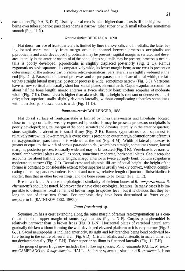
Rana asiatica BEDRIAGA, 1898
Flat dorsal surface of frontoparietale is limited by linea transversalis and l.medialis, the latter be-ing located more medially from margo orbitalis; channel between processus occipitalis andp.prooticalis and underdeveloped l.prooticalis may be present; sagittal margin is serrated and devi-ates laterally in the anterior one third of the bone; sinus sagittalis may be present; processus occipi-talis is poorly developed; p.prooticalis is slightly displaced posteriorly (Fig. 2 O). Ramuszygomaticus ossis squamosi is comparatively wide, its lower margin bent; acute crest is present onouter margin of the anterior part of ramus retrozygomaticus; pars lateralis is slightly widened at theend (Fig. 4 L). Parasphenoid lateral processes and corpus parasphenoidei are of equal width, the lat-ter has straight lateral margins; posterior process is wide, sometimes narrow (Fig. 3 J). Vertebraehave narrow vertical and usually short horizontal plates of neural arch. Caput scapulae accounts forabout half the bone length; margo anterior is twice abruptly bent; collum scapulae of moderatewidth (Fig. 7 K). Dorsal crest is higher than ala ossis ilii; its height is constant or decreases anteri-orly; tuber superior usually slightly deviates laterally, without complicating tubercles sometimeswith tubercles; pars descendens is wide (Fig. 11 D).
Rana amurensis BOULENGER, 1886
Flat dorsal surface of frontoparietale is limited by linea transversalis and l.medialis, locatedclose to margo orbitalis; weakly expressed l.prooticalis may be present; processus occipitalis ispoorly developed; sagittal margin of the bone serrated and deviates laterally in the anterior quarter;sinus sagittalis is absent or is small if any (Fig. 2 R). Ramus zygomaticus ossis squamosi isrelatively narrow, its lower margin is even; crest is present on outer margin of anterior part of ramusretrozygomaticus; pars lateralis is widened at the end (Fig. 4 M). Width of lateral processes isgreater or equal to the width of corpus parasphenoidei, which has straight, sometimes wavy, lateralmargins; posterior process is usually wide and may be bifurcated (Fig. 3 K). Vertebrae have narrowneural arch vertical plates as well as short, sometimes moderate, horizontal plates. Caput scapulaeaccounts for about half the bone length; margo anterior is twice abruptly bent; collum scapulae ismoderate to narrow (Fig. 7 J). Dorsal crest and ala ossis ilii are of equal height; the height of theformer is constant to considerable extent; tuber superior is usually weakly convex, without compli-cating tubercles; pars descendens is short and narrow; relative length of junctura ilioischiadica isshorter, than that in other brown frogs, and the bone seems to be longer (Fig. 11 E).
R e m a r k s . A close morphological similarity of skeleton bones of R. temporaries and R.chensinensis should be noted. Moreover they have close ecological features. In many cases it is im-possible to determine fossil remains of brown frogs to species level, but it is obvious that they be-long to one of these two forms. For emphasis they have been determined as Rana ex gr.temporaria L. (RATN1KOV 1992, 1996b).
Rana (esculenta) sp.
Squamosum has a crest extending along the outer margin of ramus retrozygomaticus as a con-tinuation of the upper margin of ramus zygomaticus (Fig. 4 N-P). Corpus parasphenoidei isrelatively narrower than in brown frogs (Fig. 3 L-N). Horizontal plates of vertebral neural archgradually thicken without forming the well-developed elevated platform or it is very narrow (Fig. 5G, J). Sacral neurapophis is inclined anteriorly, its right and left branches being bend backward be-fore fusing in the centre of neural arch (Fig. 6 D). Crista medialis and c.lateralis in male humeri arenot deviated dorsally (Fig. 9 F-H). Tuber superior on ilium is flattened laterally (Fig. 11 F-H).
The group of green frogs now includes the following species: Rana ridibunda PALL., R. lesso-nae CAMERANO and R.nigromaculata HALL. . So far the systematic situation of R. esculenta L. is not
V. Yu. RATNIKOV
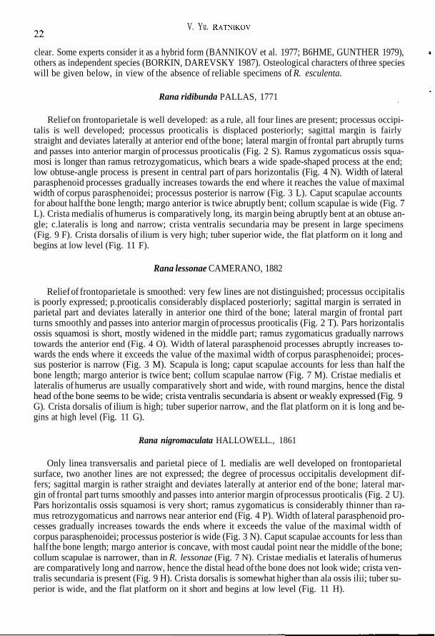
clear. Some experts consider it as a hybrid form (BANNIKOV et al. 1977; B6HME, GUNTHER 1979),others as independent species (BORKIN, DAREVSKY 1987). Osteological characters of three specieswill be given below, in view of the absence of reliable specimens of R. esculenta.
Rana ridibunda PALLAS, 1771
Relief on frontoparietale is well developed: as a rule, all four lines are present; processus occipi-talis is well developed; processus prooticalis is displaced posteriorly; sagittal margin is fairlystraight and deviates laterally at anterior end of the bone; lateral margin of frontal part abruptly turnsand passes into anterior margin of processus prooticalis (Fig. 2 S). Ramus zygomaticus ossis squa-mosi is longer than ramus retrozygomaticus, which bears a wide spade-shaped process at the end;low obtuse-angle process is present in central part of pars horizontalis (Fig. 4 N). Width of lateralparasphenoid processes gradually increases towards the end where it reaches the value of maximalwidth of corpus parasphenoidei; processus posterior is narrow (Fig. 3 L). Caput scapulae accountsfor about half the bone length; margo anterior is twice abruptly bent; collum scapulae is wide (Fig. 7L). Crista medialis of humerus is comparatively long, its margin being abruptly bent at an obtuse an-gle; c.lateralis is long and narrow; crista ventralis secundaria may be present in large specimens(Fig. 9 F). Crista dorsalis of ilium is very high; tuber superior wide, the flat platform on it long andbegins at low level (Fig. 11 F).
Rana lessonae CAMERANO, 1882
Relief of frontoparietale is smoothed: very few lines are not distinguished; processus occipitalisis poorly expressed; p.prooticalis considerably displaced posteriorly; sagittal margin is serrated inparietal part and deviates laterally in anterior one third of the bone; lateral margin of frontal partturns smoothly and passes into anterior margin of processus prooticalis (Fig. 2 T). Pars horizontalisossis squamosi is short, mostly widened in the middle part; ramus zygomaticus gradually narrowstowards the anterior end (Fig. 4 O). Width of lateral parasphenoid processes abruptly increases to-wards the ends where it exceeds the value of the maximal width of corpus parasphenoidei; proces-sus posterior is narrow (Fig. 3 M). Scapula is long; caput scapulae accounts for less than half thebone length; margo anterior is twice bent; collum scapulae narrow (Fig. 7 M). Cristae medialis etlateralis of humerus are usually comparatively short and wide, with round margins, hence the distalhead of the bone seems to be wide; crista ventralis secundaria is absent or weakly expressed (Fig. 9G). Crista dorsalis of ilium is high; tuber superior narrow, and the flat platform on it is long and be-gins at high level (Fig. 11 G).
Rana nigromaculata HALLO WELL., 1861
Only linea transversalis and parietal piece of 1. medialis are well developed on frontoparietalsurface, two another lines are not expressed; the degree of processus occipitalis development dif-fers; sagittal margin is rather straight and deviates laterally at anterior end of the bone; lateral mar-gin of frontal part turns smoothly and passes into anterior margin of processus prooticalis (Fig. 2 U).Pars horizontalis ossis squamosi is very short; ramus zygomaticus is considerably thinner than ra-mus retrozygomaticus and narrows near anterior end (Fig. 4 P). Width of lateral parasphenoid pro-cesses gradually increases towards the ends where it exceeds the value of the maximal width ofcorpus parasphenoidei; processus posterior is wide (Fig. 3 N). Caput scapulae accounts for less thanhalf the bone length; margo anterior is concave, with most caudal point near the middle of the bone;collum scapulae is narrower, than in R. lessonae (Fig. 7 N). Cristae medialis et lateralis of humerusare comparatively long and narrow, hence the distal head of the bone does not look wide; crista ven-tralis secundaria is present (Fig. 9 H). Crista dorsalis is somewhat higher than ala ossis ilii; tuber su-perior is wide, and the flat platform on it short and begins at low level (Fig. 11 H).
Osteology of Russian toads and frogs
IV. CONCLUSION
Bone shape of toads and frogs permit their identification up to the level of species, complex, andgenus. It is hardly possible to consider the above diagnostic criteria exhaustive. Further researchesof fossil materials will probably make it possible to find additional features of the bone morphologyof various anuran groups. Some observations carried out on a small number of specimens undoubt-edly need control and more accurate definition.
In the skeletons of toads are to be found the greatest number of elements (9-11) which make itpossible to define species. The most important of them are ilia, frontoparietalia, maxillaria, paras-phenoidea, and male humeri.
Only 6-7 bones are used for specific identification of frogs. Most diagnostic elements are ilia,frontoparietalia, and scapulae. Other bones do not always give an unequivocal definition.
Genera are distinguished very easily: almost all skeletal elements, with the exception of distalelements of limbs, have their generic signs.
REFERENCES
ANANJEVA N. B., BORKIN L. Y., DAREVSKY I. S, ORLOV N. L. 1998. Zemnovodnye i presmykayuschiesya.Encyklopedia prirody Rossii. 576 pp. ABF, Moscow. [In Russian].
BAILON S. 1986. Los anfibios y los reptiles del yacimiento de Cueva Hora (Darro, Granada). AntropologiayPaleontologia Humana, 4: 131-155.
BANNIKOV A. G., DAREVSKY I. S., ISCHENKO V. G., RUSTAMOV A. K., SZCZERBAK N. N. 1977. OpredelitePzemnovodnykh i presmykayuschikhsya fauny SSSR. 415 pp. Prosveschenie, Moscow. [In Russian].
BORKIN L. J., DAREVSKY I. S. 1987. List of amphibian and reptiles of USSR fauna. [In:] I. S. DAREVSKY (ed.)- Amphibii i reptylii zapovednych territory. 128-141. Moscow. [In Russian].
BOHME G. 1977. Zur Bestimmung quartarer Anuren Europas an Hand von Skelettelementen. Wissens-chaftliche Zeitschrift der Humboldt-Universit&t zu, Mathematisch-Naturwissenschaftliche Reihe, 26(3): 283-300.
BOHME G., GUNTHER R. 1979. Osteological stadies in the European Water frogs Rana ridibunda, R. lessonaeand R. "esculenta" (Anura, Ranidae). Mitteilungen aus dem Zoologischen Museum in Berlin, 55(1):203-215.
CKHIKVADZE V. M. 1984. Survey of fossil tail and tailless amphibians of the USSR, hvestiya Akademii NaukGSSR, 10(1): 5-13. [In Russian].
ENGELMANN W.-E., FRITZSCHE J., GUNTHER R., OBST F. J. 1985. Lurche und Kriechtiere Europas. 420 ss.Neumann Verlag, Leipzig.
HODROVA M. 1980. A toad from the Middle Miocene at Devinska Nova Ves near Bratislava. Vestnik Ustred-niho ustavu geologicekho, 55(5): 311-316.
HOLMAN J. A. 1989. Identification of Bufo calamita andBitfo bufo on the basis of skeletal elements. BritishHerpetological Society Bulletin, 29: 54-55.
HOLMAN J. A. 1998. Pleistocene Amphibians and Reptiles in Britain and Europe. 254 pp. Oxford UniversityPress, New York-Oxford.
ORLOVA V. F., TUNIEV B. S. 1989. To systematic of Caucasian grey toads of Bufo bufo verrucosissimus(PALLAS) group (Amphibia, Anura, Bufonidae) - Byulleten' Moskovskogo Obshchestva Ispytatelei Prirody.Otdel Biologicheskii, 94(3): 13-24. [In Russian].
RAGE J. 1974. Les batraciens des gisements des quaternaires europeens; Determination osteologique. Bulletinmensuel de la Societe Linneenne de Lyon, 43(8): 276-289.
RATNIKOV V. Yu. 1992. Eopleistocene and Pleistocene anuran faunas of the East European Platform. Paleon-tologicalJournal, 26(1): 112-126. [In Russian]
RATNIKOV V. Yu. 1995. Late Cenozoic evolution of batrachofauna of the East-European Platform. Byulleten'Moskovskogo Obshchestva Ispytatelei Prirody. Otdel Geologicheskii, 70(5): 98-103. [In Russian].
RATNIKOV V. Yu. 1996a. Methods of paleogeographic reconstructions based upon fossil remains of amphibi-ans and reptiles of the Late Cenozoic of the East European Platform. PalaeontologicalJournal, 30(1): 77-83.
RATNIKOV V. Yu. 1996b. On the finds of Green Toads (Bufo viridis complex) in the late Cenozoic of the EastEuropean Platform. PalaeontologicalJournal, 30(2): 225-231.
ROCEK Z. 1980. A contribution to the systematics of European ranid frogs (Amphibia, Ranidae) on the basis ofthe incrassatio frontoparietalis. V&stnik ceskoslovenske spolecnosti zoologicki, 44(3): 219-229.
SANCHIZ B. 1977. La familia Bufonidae (Amphibia, Anura) en el Terciario europeo. Trabajos sobreNeogeno-Cuaternario CSIC, 8: 75-111.
SANCHIZ B. 1998. Salientia. Encyclopedia of Paleoherpetology. Part 4. 276 pp. Verlag Dr. Friedrich Pfeil,Munchen.